Case Report

Improved Myocardial Function in Autoimmune-Mediated Fetal Complete Atrioventricular Block Following Dexamethasone and Intravenous Immunoglobulin: A Case Report

by
Maria Elisa Martini Albrecht
1,
Milena Giuberti Bathomarco
1,
Gustavo Yano Callado
2,
Nathalie Jeanne Bravo-Valenzuela
3 and
Edward Araujo Júnior
1,2,4,*
1
Department of Obstetrics, Paulista School of Medicine, Federal University of São Paulo (EPM-UNIFESP), São Paulo 04023-062, SP, Brazil
2
Albert Einstein Israelite College of Health Sciences, Albert Einstein Israelite Hospital, São Paulo 05652-900, SP, Brazil
3
Department of Pediatrics, Pediatric Cardiology, Federal University of Rio de Janeiro (UFRJ), Rio de Janeiro 21941-901, RJ, Brazil
4
Discipline of Woman Health, Municipal University of São Caetano do Sul (USCS), São Caetano do Sul 09521-160, SP, Brazil
*
Author to whom correspondence should be addressed.
Women 2025, 5(2), 20; https://doi.org/10.3390/women5020020
Submission received: 14 May 2025 / Revised: 24 May 2025 / Accepted: 27 May 2025 / Published: 6 June 2025

Abstract

:
This case report describes a fetus diagnosed with complete atrioventricular block (CAVB) associated with positive maternal anti-Ro and anti-La antibodies, referred to our fetal cardiology unit at 25 weeks of gestation. The diagnosis of systemic lupus erythematosus (SLE) was established during the investigation of the fetal condition. Oral dexamethasone was initiated and well tolerated, with no adverse effects reported throughout the remainder of the pregnancy. The fetal heart rate (HR) remained above 50 bpm, and, therefore, no beta-sympathomimetic agents were administered. Due to progressive reduction in myocardial contractility and the appearance of early signs of endocardial fibroelastosis, intravenous immunoglobulin (IVIG) therapy was initiated. The patient was hospitalized for the infusion, which was well tolerated without complications, and a second IVIG cycle was administered four weeks later. Significant improvement in ventricular contractility and reduction in fibroelastosis were observed. As reported in the literature, no chronotropic effect was noted, and fetal HR remained stable after treatment. Weekly monitoring of cardiovascular profile score and fetal HR was maintained, with the score consistently remaining at 8 throughout gestation, supporting continued outpatient management. Delivery occurred at 36 weeks and 3 days due to spontaneous preterm labor. A male neonate weighing 3025 g was delivered with Apgar scores of 8 and 9, and an initial heart rate of 84 bpm. Neonatal electrocardiography confirmed persistent CAVB, and the newborn was monitored in the neonatal intensive care unit. At follow-up, the infant remains clinically stable and has not required permanent pacemaker implantation.

1. Introduction

Fetal complete atrioventricular block (CAVB) is a condition characterized by the disruption of electrical conduction between the atria and ventricles in the fetal heart, resulting in a complete heart block. This condition is rare, with an incidence of approximately 1 in 15,000 to 1 in 20,000 births [1]. CAVB can occur in isolation or in association with structural heart defects. The clinical implications of CAVB are significant, as it can lead to fetal hydrops, heart failure, and even intrauterine or neonatal death. If the fetus survives, lifelong cardiac pacing is often required due to the irreversible nature of the block [2,3].
Autoimmune-mediated CAVB is primarily caused by the transplacental passage of maternal autoantibodies, specifically anti-Ro/SSA and anti-La/SSB antibodies. These antibodies can lead to immune-mediated injury of the fetal atrioventricular node, resulting in inflammation and fibrosis, which disrupts the conduction system [4]. The highest risk period for the development of CAVB is between 16 and 28 weeks of gestation [4]. Many mothers carrying these antibodies are asymptomatic, and less than one-third have a preexisting diagnosis of a rheumatological disorder [1].
The pathogenesis involves a two-stage process: initially, maternal autoantibodies bind to fetal cardiomyocytes, causing dysregulation of calcium homeostasis and apoptosis. This can lead to first-degree heart block, which may be reversible [5]. However, in genetically predisposed fetuses, this process can progress to inflammation, fibrosis, and complete heart block [6]. Despite various treatment attempts, such as the use of fluorinated corticosteroids, the efficacy in reversing complete heart block remains controversial and largely ineffective [7,8]. Regular monitoring and early detection are crucial for managing pregnancies at risk for autoimmune-mediated CAVB [8].
In this context, we report the case of a fetus diagnosed with autoimmune-mediated CAVB at 25 weeks of gestation in a mother with previously diagnosed rheumatoid arthritis and high titers of anti-SSA and anti-SSB antibodies. The case is notable for the early initiation of oral dexamethasone followed by two cycles of intravenous immunoglobulin (IVIG), leading to progressive improvement in myocardial function and stabilization of the clinical course. Despite the persistence of complete heart block, the newborn remained hemodynamically stable and did not require pacemaker implantation during the first six months of life. This case contributes to the growing body of evidence on the potential role of combined immunomodulatory therapy in altering the natural history of antibody-mediated fetal cardiac injury.

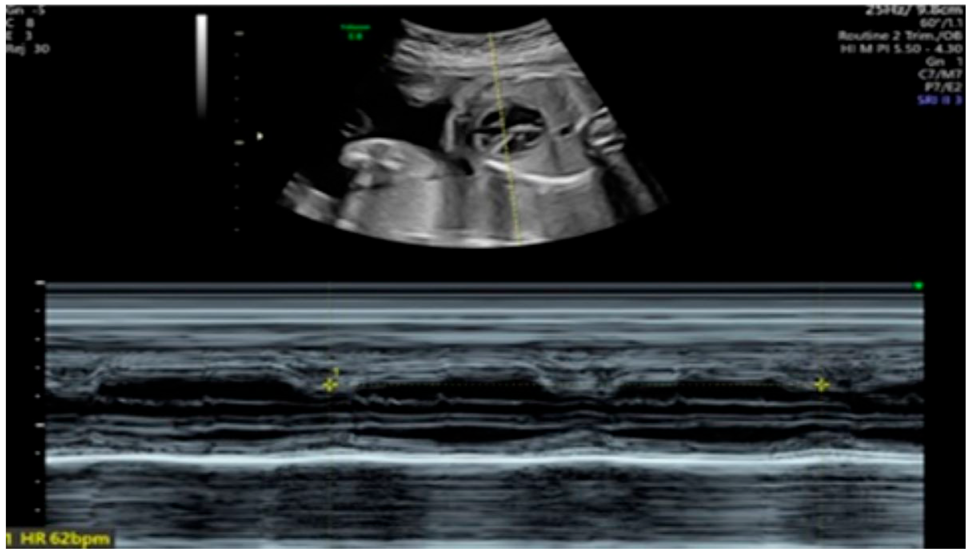
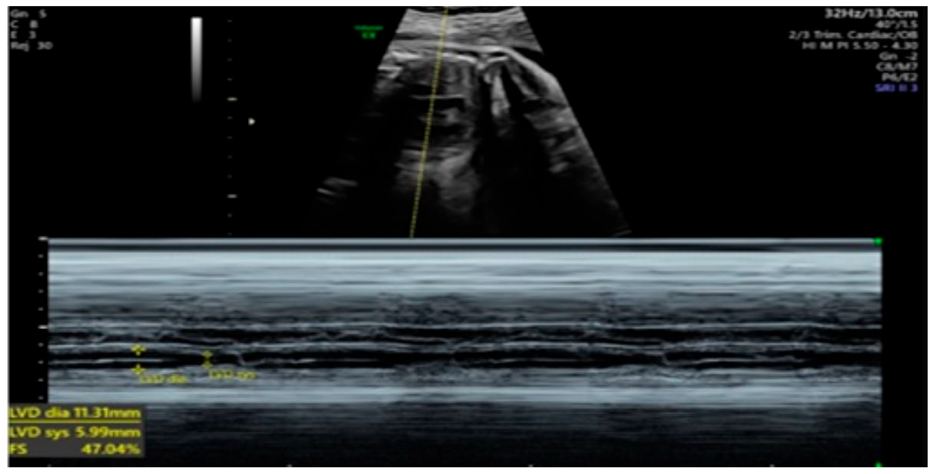
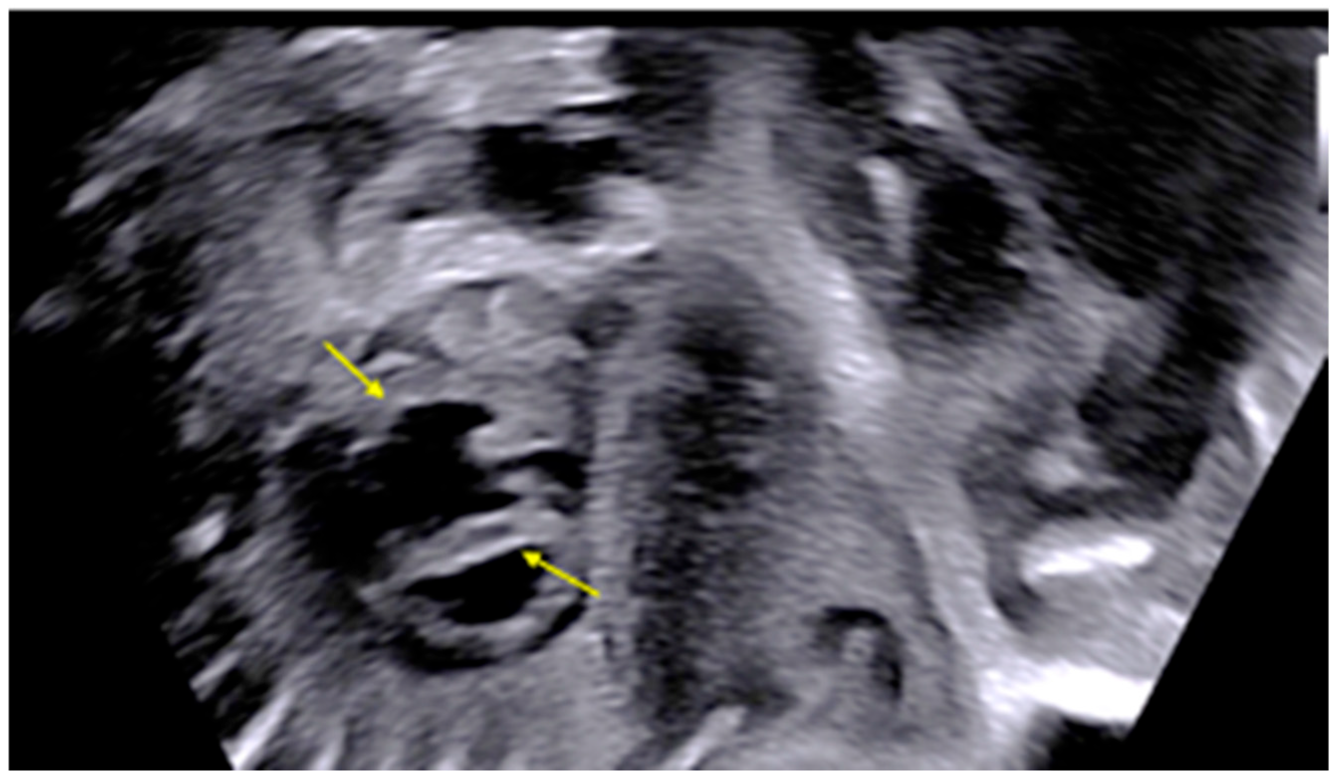
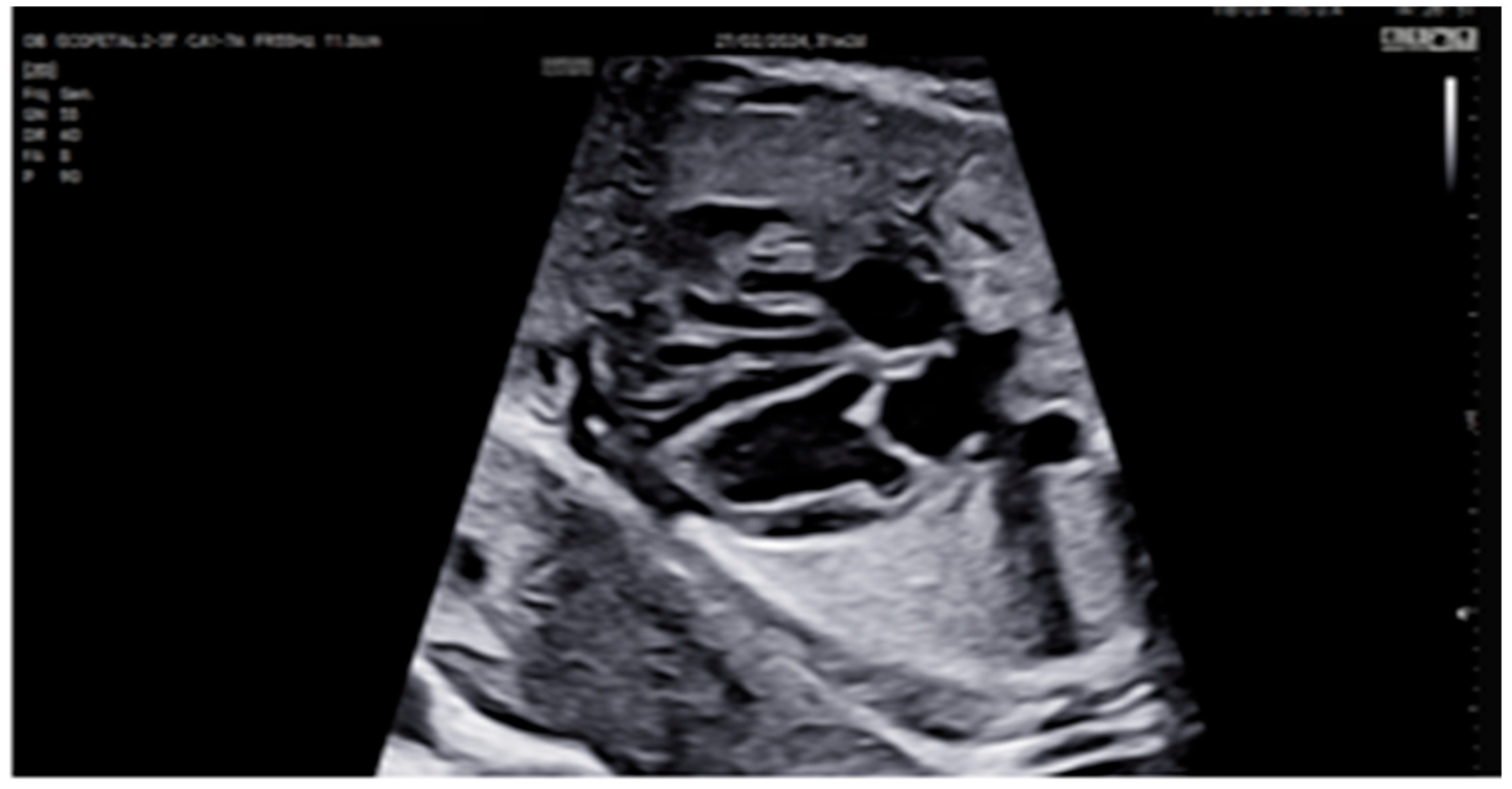
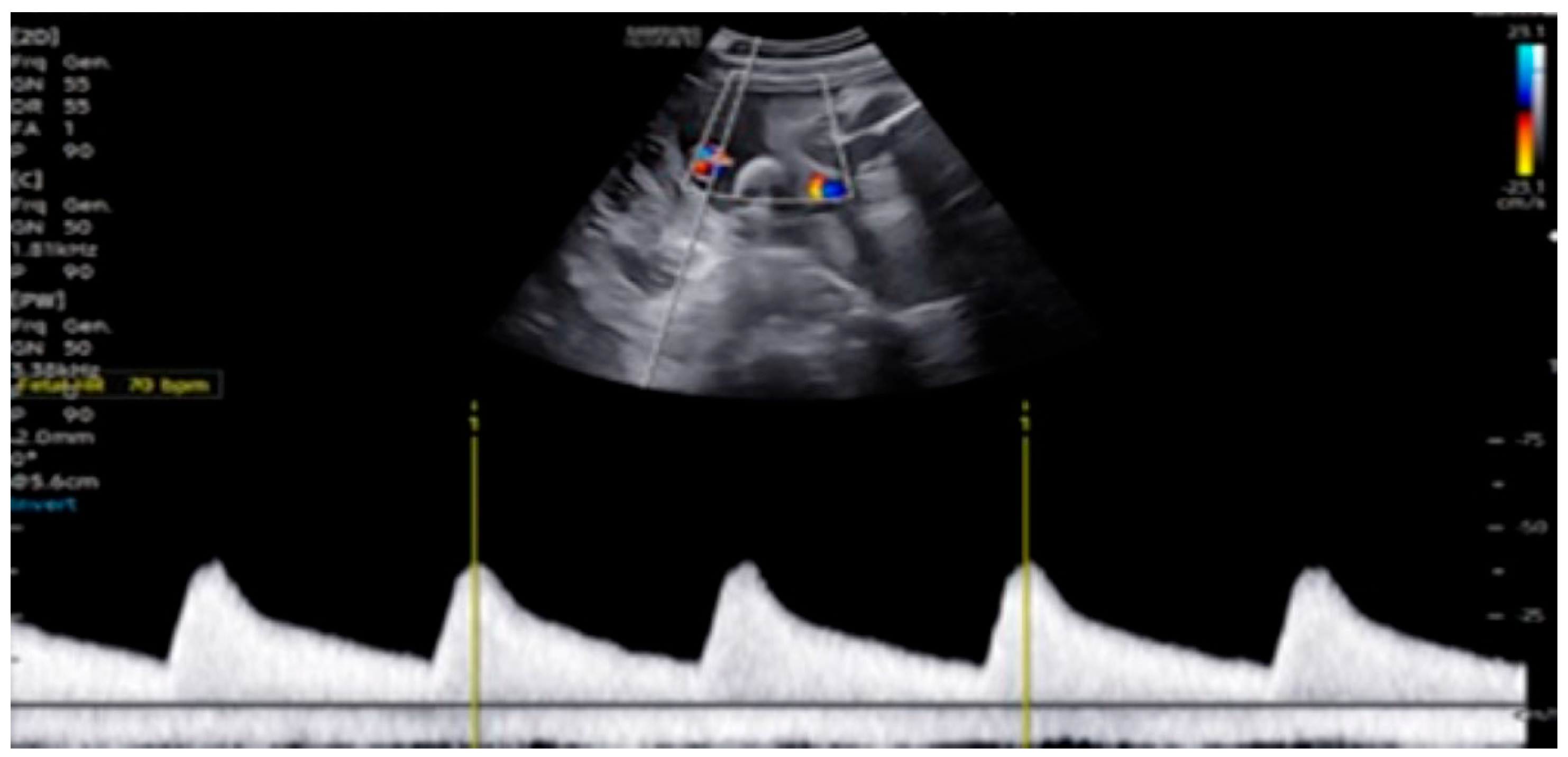
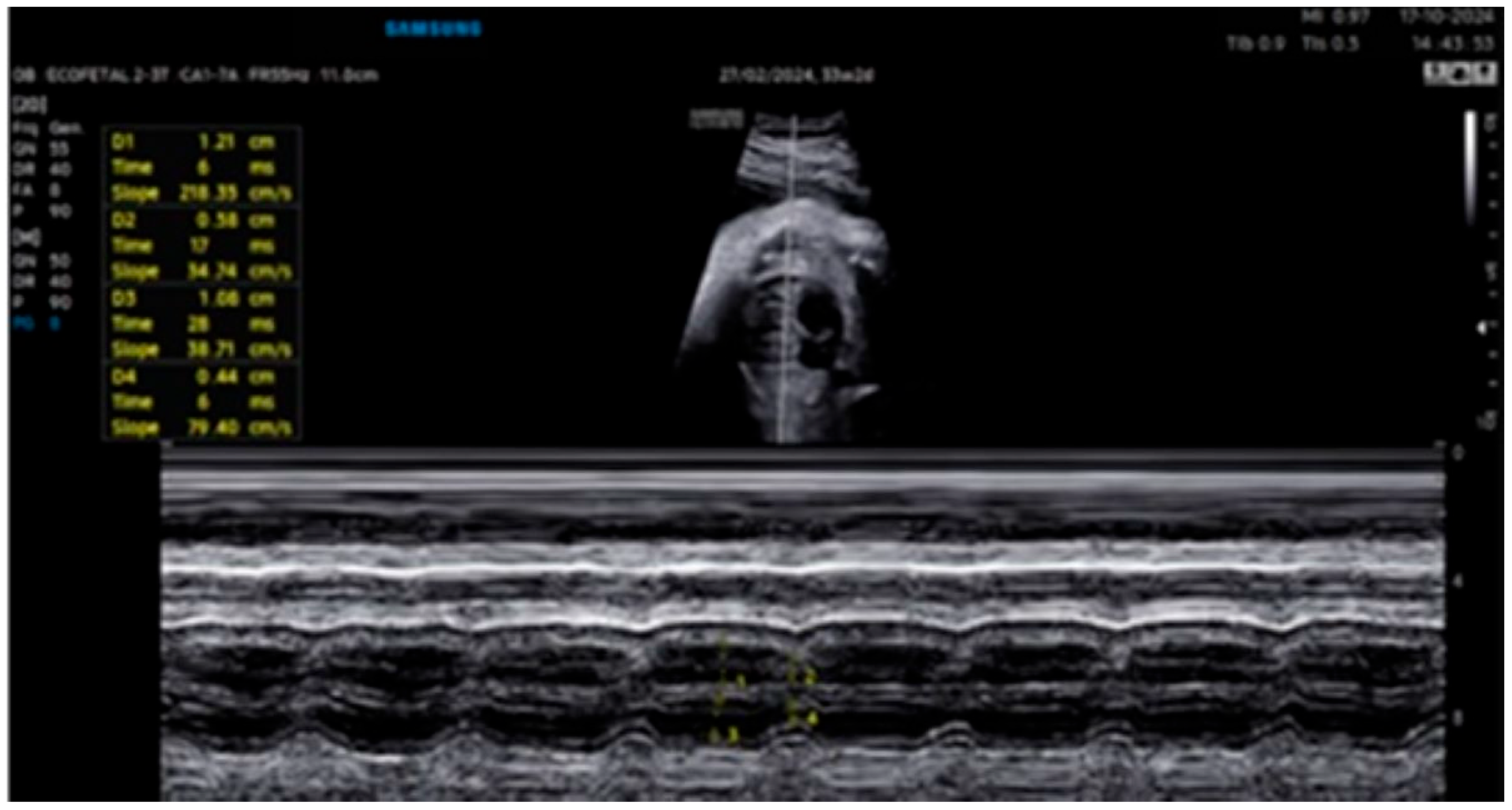
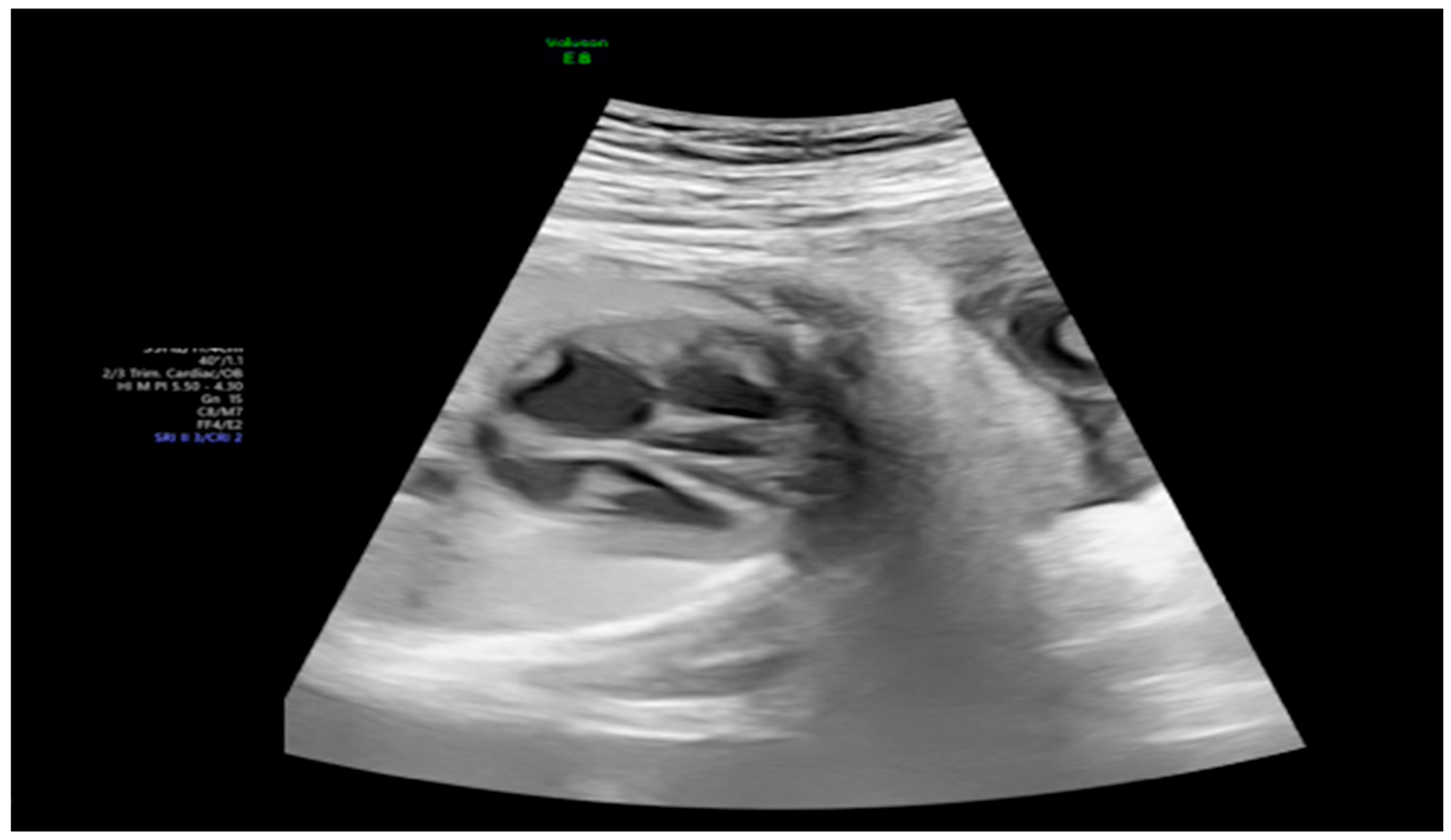
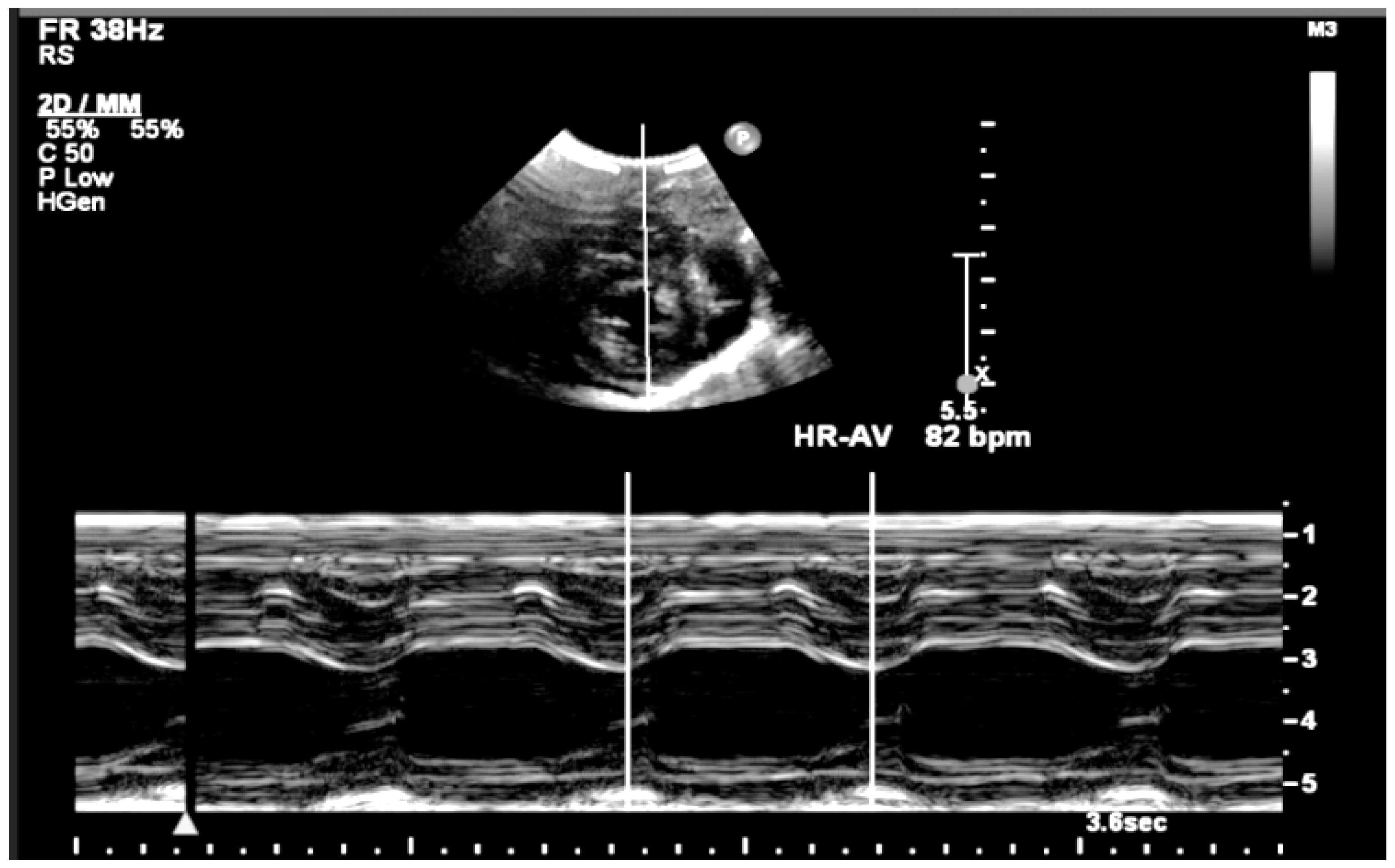
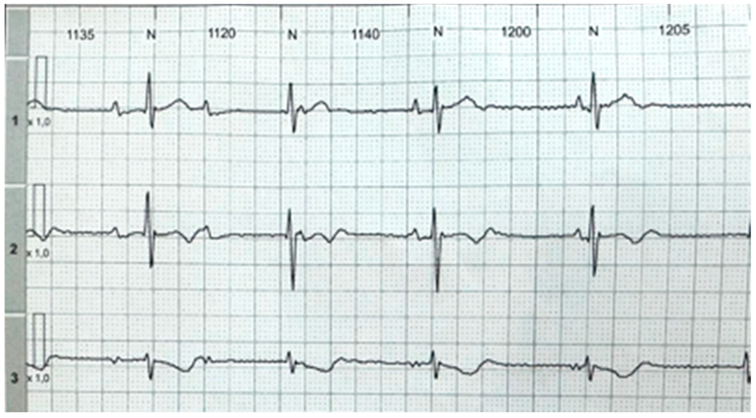
2. Detailed Case Description

A 20-year-old primigravida was diagnosed with rheumatoid arthritis at age 16, presenting with chronic polyarthralgia predominantly affecting the hands. She had been on hydroxychloroquine for one year. At 25 weeks of gestation, she was referred to the fetal cardiology clinic due to fetal bradycardia during a routine obstetrical ultrasound. Fetal echocardiography revealed a diagnosis of CAVB, with an atrial heart rate (AHR) of 139 bpm and a ventricular heart rate (VHR) of 62 bpm. The cardiovascular profile score was 8/10, with a shortening fraction (SF) of 34%. The fetal heart was anatomically normal except for a narrowed aortic isthmus measuring 2.2 mm (Z-score −2.6). Screening tests for systemic lupus erythematosus (SLE) were requested and came out positive. Figure 1 shows the fetal echocardiographic findings at the time of initial diagnosis.
At the time of pregnancy, the patient’s rheumatoid arthritis was clinically stable, with no active joint inflammation or systemic manifestations reported. Although hydroxychloroquine is not considered first-line for RA outside pregnancy, its continuation was maintained due to prior tolerance, disease control, and potential protective effects against antibody-mediated fetal cardiac injury.
The patient returned one week later with the test results: anti-SSB antibody >416 U/mL (positive >10 U/mL) and anti-SSA antibody >240 U/mL (positive >10 U/mL). Oral dexamethasone 4 mg/day was initiated to reduce inflammation associated with antibody-mediated myocardial injury, as suggested by existing evidence on autoimmune-associated congenital heart block [8]. She experienced mild lower limb edema on the first day, but the dose was maintained throughout the pregnancy. At follow-up, the AHR was 126 bpm, and the VHR was 60 bpm, with a cardiovascular profile score of 7/10. Qualitative assessment showed a significant reduction in myocardial contractility, despite an improved SF of 47%. Ambulatory follow-up was continued. One week later, the VHR increased to 75 bpm, with a stable SF, although signs of left ventricular endocardial fibroelastosis were noted. After multidisciplinary discussion and a review of the current literature [9], intravenous immunoglobulin (IVIG) therapy was initiated with the aim of halting the progression of myocardial injury, particularly endocardial fibroelastosis, and to improve myocardial function. Although the fetus remained hemodynamically stable, the presence of contractile dysfunction and evolving echocardiographic signs of fibroelastosis supported the decision to initiate immunomodulatory therapy (Figure 2, Figure 3, Figure 4 and Figure 5).
The patient was admitted to the intensive care unit for monitoring. Baseline renal function was normal. Premedication with 25 mg intravenous diphenhydramine was administered, and magnesium sulfate was replaced due to low serum levels. IVIG was administered at 1 g/kg (maximum 70 g), totaling 65 g via continuous infusion (13 vials of 5 g/100 mL) without dilution, at a rate of 24 mL/h, in two stages (600–700 mL). Daily fetal echocardiographic and obstetric evaluations were performed during hospitalization. No adverse effects occurred, and the patient was discharged after 48 h (Figure 6).
Three days after IVIG, outpatient follow-up showed improved left ventricular SF (52%), AHR of 138 bpm, VHR of 65 bpm, cardiovascular profile score of 8/10, and mild improvement in myocardial contractility and fibroelastosis. The aortic isthmus measured 2.6 mm (Z-score −2.66), with segmental hypoplasia of the aortic arch. After 10 days, AHR was 139 bpm, VHR was 63 bpm, SF was 56%, and further improvement in myocardial contractility and fibroelastosis were observed. Seventeen days after IVIG, AHR was 175 bpm, VHR was 70 bpm, cardiovascular score remained 8/10, with marked improvement in both right and left ventricular function, SF was 59% (left) and 52% (right), and the aortic isthmus was 3 mm (Z-score −1.65) (Figure 7 and Figure 8).
A second IVIG cycle was administered at 33 weeks, four weeks after the first, following the same protocol. The patient was discharged within 48 h without complications. Weekly outpatient follow-up was maintained. Similarly to the first cycle, peak improvement in myocardial function was observed between 14 and 21 days after infusion, with no observed impact on the fetal ventricular rate, indicating that IVIG did not exert a chronotropic effect. Visualization of the aortic isthmus remained limited due to suboptimal fetal acoustic windows at advanced gestational age (Figure 9, Figure 10 and Figure 11).
At 36 weeks and 3 days, the patient presented with preterm labor. Cesarean section was indicated due to fetal CAVB, which precluded reliable fetal well-being monitoring. A male neonate was delivered weighing 3025 g, with vigorous crying, good muscle tone, and Apgar scores of 8 and 9. Postnatal oxygen saturation was 87% with a heart rate of 84 bpm. Oxygen supplementation increased saturation to 94%. Electrocardiogram confirmed CAVB with a heart rate of 56 bpm. The infant required 24 h of intravenous dobutamine at 5 mcg/kg/h due to desaturation.
Postnatal echocardiography revealed mild localized narrowing of the transverse aortic arch, without evidence of aorta coarctation. Cardiac chambers were of normal size; atrioventricular valves and semilunar valves had normal morphology and function. The aorta and pulmonary artery had normal calibers. The pulmonary trunk was slightly enlarged (11.3 mm; Z-score +2.58), with a patent ductus arteriosus (3.1 mm). Measurements included the ascending aorta at 8.3 mm (Z-score +0.08), the transverse arch at 5.1 mm (Z-score −2.25), the distal aorta at 8.1 mm (Z-score +2.37), and the isthmus at 4.7 mm (Z-score −0.49) (Figure 12 and Figure 13).
A 24 h Holter monitor showed predominant CAVB with a mean heart rate of 71 bpm (range: 51–102 bpm), abnormal AV and intraventricular conduction, and no repolarization abnormalities or supraventricular arrhythmias. A total of 500 isolated ventricular ectopic beats were observed, with no pauses > 2 s or clinical symptoms (Figure 14).
Cardiothoracic surgery and pediatric cardiology teams evaluated the infant, and no indication for permanent pacemaker implantation was given due to hemodynamic stability. At six months of age, the child remains clinically stable and under routine outpatient follow-up, without requiring a pacemaker. Table 1 summarizes the therapeutic interventions, their timing, and subsequent echocardiographic responses, to aid reproducibility.

3. Discussion

We present a case of antibody-mediated fetal CAVB successfully managed with early dexamethasone and two cycles of IVIG, which resulted in significant myocardial recovery despite persistent conduction block. Despite advances in prenatal screening and fetal echocardiography, its management is still debated, particularly regarding immunomodulatory therapies. This case highlights a favorable clinical trajectory in a fetus with antibody-mediated CAVB treated with both dexamethasone and IVIG, suggesting a possible benefit in preventing progression of myocardial injury and improving cardiac function [9].
The presented case is notable for the rare combination of progressive myocardial dysfunction, endocardial fibroelastosis, and preserved biventricular function despite complete AV block. The use of two cycles of IVIG—initiated in response to contractile worsening—contributed to measurable improvements in myocardial performance [10]. To our knowledge, this sequence of interventions and favorable postnatal evolution remains uncommon and highlights the potential value of combining corticosteroids and IVIG in selected cases.
Current treatment options primarily include corticosteroids and beta-agonists. Dexamethasone is frequently used in cases of incomplete heart block or complete block with evidence of myocardial dysfunction or inflammation [11]. Although it may not reverse complete AV block once established, studies have reported significantly improved fetal and neonatal survival rates with corticosteroid use [11,12]. In this case, the combination of dexamethasone and IVIG was chosen following a multidisciplinary review of emerging evidence and similar case series [13]. Although no standard protocol was available, the observed progression of myocardial dysfunction despite steroid use justified additional immunomodulatory therapy. In our case, dexamethasone was initiated following confirmation of anti-SSA and anti-SSB positivity and was maintained throughout the pregnancy despite the presence of third-degree block. The initial response showed partial improvement in myocardial function but emerging signs of fibroelastosis prompted further therapeutic intervention.
Beta-agonists were not employed in this case, as the ventricular rate remained above the threshold typically used to indicate their use [14]. Hydroxychloroquine (HCQ), which has been suggested as a preventive agent in mothers with known anti-Ro/La antibodies, was already part of the maternal treatment regimen for rheumatoid arthritis but is not indicated for reversing established CAVB. Importantly, the evidence supporting HCQ for prevention is still limited and marked by high risk of bias [15].
Terbutaline, a beta-sympathomimetic, can be used in the management of autoimmune-mediated fetal CAVB, primarily to increase fetal heart rates, particularly when the heart rate is below 55 beats per minute or when there is associated cardiac dysfunction or hydrops [16]. The American Heart Association guidelines suggest that beta-sympathomimetics like terbutaline are reasonable to use in these scenarios, although they note that while terbutaline may increase fetal heart rates and prolong pregnancy, no studies have demonstrated a survival benefit [17].
IVIG, although more controversial and not routinely recommended, was employed in this case due to progressive myocardial dysfunction and echocardiographic signs of endocardial fibroelastosis [17]. The rationale was based on limited evidence suggesting that IVIG may neutralize circulating maternal antibodies and modulate fetal cardiac inflammation [15]. While randomized controlled data remain lacking, prior case series and observational studies have explored its potential in attenuating progression or improving biventricular function [8,15]. In this patient, two cycles of IVIG were associated with notable improvements in shortening fraction, contractility, and reversal of fibroelastosis, without adverse effects. Although the heart block persisted, the absence of hemodynamic deterioration and the stability of ventricular function through the neonatal period suggest a possible protective effect of IVIG against the development of cardiomyopathy. This is consistent with current literature on fetal arrhythmia treatment, which explores pharmacologic and immunologic approaches for autoimmune-mediated conduction abnormalities [18]. In comparison to reported cases managed solely with corticosteroids, this case exhibited greater recovery in myocardial function following the addition of IVIG, consistent with previous observational reports suggesting a potential synergistic effect, although complete reversal of heart block remains rare.

4. Conclusions

This case underscores several challenges in the management of autoimmune-mediated CAVB. First, the diagnosis often occurs after irreversible injury to the conduction system has already taken place, limiting the effectiveness of therapy. Second, treatment recommendations are largely based on retrospective studies, case series, and expert opinion. The use of corticosteroids and IVIG remains individualized, guided by fetal condition, presence of myocardial inflammation or dysfunction, and provider experience. The favorable evolution in this case—including preserved biventricular function, absence of hydrops, and no indication for pacemaker implantation at six months—raises important questions about the potential benefits of early combined immunomodulatory treatment, even in cases of established third-degree block. Despite notable improvement in myocardial contractility and the absence of clinical deterioration, the persistence of complete heart block underscores the limitations of current immunomodulatory strategies in reversing conduction system fibrosis.
Further studies are needed to establish standardized protocols and determine which fetuses may benefit most from interventions such as corticosteroids and IVIG. Until then, careful monitoring, early detection of myocardial changes, and individualized, multidisciplinary management remain essential for optimizing outcomes in pregnancies affected by autoimmune-associated CAVB.

Author Contributions

Conceptualization, E.A.J. and M.E.M.A.; methodology, N.J.B.-V. and M.G.B.; validation, G.Y.C.; formal M.E.M.A. and E.A.J.; investigation, N.J.B.-V. and M.G.B.; resources, E.A.J.; data curation, M.G.B.; writing—original draft preparation, G.Y.C.; writing—review and editing, E.A.J.; visualization, E.A.J., M.E.M.A., N.J.B.-V., M.G.B. and G.Y.C.; supervision, M.E.M.A.; project administration, E.A.J. All authors have read and agreed to the published version of the manuscript.

Funding

This article received no external funding.

Institutional Review Board Statement

Our investigations were carried out following the rules of the Declaration of Helsinki of 1975, revised in 2013. As this is a case report, it was not necessary to obtain ethics committee approval.

Informed Consent Statement

The patient gave consent for the case and images to be published.

Data Availability Statement

The data presented in this study are available on request from the corresponding author.

Conflicts of Interest

The authors report no conflict of interest.

References

  1. Bordachar, P.; Zachary, W.; Ploux, S.; Labrousse, L.; Haissaguerre, M.; Thambo, J.B. Pathophysiology, clinical course, and management of congenital complete atrioventricular block. Heart Rhythm. 2013, 10, 760–766. [Google Scholar] [CrossRef] [PubMed]
  2. Ambrosi, A.; Thorlacius, G.E.; Sonesson, S.E.; Wahren-Herlenius, M. Interferons and innate immune activation in autoimmune congenital heart block. Scand. J. Immunol. 2021, 93, e12995. [Google Scholar] [CrossRef] [PubMed]
  3. Wainwright, B.; Bhan, R.; Trad, C.; Cohen, R.; Saxena, A.; Buyon, J.; Izmirly, P. Autoimmune-mediated congenital heart block. Best Pract. Res. Clin. Obstet. Gynaecol. 2020, 64, 41–51. [Google Scholar] [CrossRef] [PubMed]
  4. Manolis, A.A.; Manolis, T.A.; Melita, H.; Manolis, A.S. Congenital heart block: Pace earlier (Childhood) than later (Adulthood). Trends Cardiovasc. Med. 2020, 30, 275–286. [Google Scholar] [CrossRef] [PubMed]
  5. Zhou, K.Y.; Hua, Y.M. Autoimmune-associated Congenital Heart Block: A New Insight in Fetal Life. Chin. Med. J. 2017, 130, 2863–2871. [Google Scholar] [CrossRef] [PubMed]
  6. Wahren-Herlenius, M.; Sonesson, S.E. Specificity and effector mechanisms of autoantibodies in congenital heart block. Curr. Opin. Immunol. 2006, 18, 690–696. [Google Scholar] [CrossRef] [PubMed]
  7. Eliasson, H.; Sonesson, S.E.; Sharland, G.; Granath, F.; Simpson, J.M.; Carvalho, J.S.; Jicinska, H.; Tomek, V.; Dangel, J.; Zielinsky, P.; et al. Isolated atrioventricular block in the fetus: A retrospective, multinational, multicenter study of 175 patients. Circulation 2011, 124, 1919–1926. [Google Scholar] [CrossRef] [PubMed]
  8. Kaplinski, M.; Cuneo, B.F. Novel approaches to the surveillance and management of fetuses at risk for anti-Ro/SSA mediated atrioventricular block. Semin. Perinatol. 2022, 46, 151585. [Google Scholar] [CrossRef] [PubMed]
  9. Brucato, A. Prevention of congenital heart block in children of SSA-positive mothers. Rheumatology 2008, 47 (Suppl. S3), iii35–iii37. [Google Scholar] [CrossRef] [PubMed]
  10. Zhao, L.; Zhou, Y.; Wang, C.; Li, Y.; Zhu, Q.; Hua, Y.; Qiao, L.; Wu, J.; Zhou, K. Successful Prevention of Fetal Autoimmune-Mediated Heart Block by Combined Therapies with Hydroxychloroquine and Intravenous Immunoglobulin: A Case Report. Front. Cardiovasc. Med. 2021, 8, 759260. [Google Scholar] [CrossRef] [PubMed]
  11. Batra, A.S.; Silka, M.J.; Borquez, A.; Cuneo, B.; Dechert, B.; Jaeggi, E.; Kannankeril, P.J.; Tabulov, C.; Tisdale, J.E.; Wolfe, D.; et al. Pharmacological Management of Cardiac Arrhythmias in the Fetal and Neonatal Periods: A Scientific Statement from the American Heart Association: Endorsed by the Pediatric & Congenital Electrophysiology Society (PACES). Circulation 2024, 149, e996. [Google Scholar]
  12. Ruffatti, A.; Marson, P.; Svaluto-Moreolo, G.; Marozio, L.; Tibaldi, M.; Favaro, M.; Calligaro, A.; Grava, C.; Hoxha, A.; Pengo, V.; et al. A combination therapy protocol of plasmapheresis, intravenous immunoglobulins and betamethasone to treat anti-Ro/La-related congenital atrioventricular block. A case series and review of the literature. Autoimmun. Rev. 2013, 12, 768–773. [Google Scholar] [CrossRef] [PubMed]
  13. Mawad, W.; Hornberger, L.; Cuneo, B.; Raboisson, M.; Moon-Grady, A.J.; Lougheed, J.; Diab, K.; Parkman, J.; Silverman, E.; Jaeggi, E. Outcome of Antibody-Mediated Fetal Heart Disease with Standardized Anti-Inflammatory Transplacental Treatment. J. Am. Heart Assoc. 2022, 11, e023000. [Google Scholar] [CrossRef] [PubMed]
  14. Jaeggi, E.T.; Fouron, J.C.; Silverman, E.D.; Ryan, G.; Smallhorn, J.; Hornberger, L.K. Transplacental fetal treatment improves the outcome of prenatally diagnosed complete atrioventricular block without structural heart disease. Circulation 2004, 110, 1542–1548. [Google Scholar] [CrossRef] [PubMed]
  15. Agarwal, A.; Skoutelis, N.; Zhou, Y.; Kawanami, R.; Charras, A.; Occhigrossi, F.; Agarwal, U.; Khan, S.; Donegan, S.; Hawcutt, D. Treatment options for preventing autoimmune-mediated congenital heart block: A systematic review. Arch Dis Child. 2025. [Google Scholar] [CrossRef] [PubMed]
  16. Albrecht, M.E.M.; de Miranda, N.R.D.C.R.; Giuberti, M.; Araujo Júnior, E. Fetal total atrioventricular block in transgender man with systemic lupus erythematosus—Literature review and establishment of a protocol with management and treatment with terbutaline. Ceska Gynekol. 2025, 90, 52–57. [Google Scholar] [CrossRef] [PubMed]
  17. Donofrio, M.T.; Moon-Grady, A.J.; Hornberger, L.K.; Copel, J.A.; Sklansky, M.S.; Abuhamad, A.; Cuneo, B.F.; Huhta, J.C.; Jonas, R.A.; Krishnan, A.; et al. Diagnosis and treatment of fetal cardiac disease: A scientific statement from the American Heart Association. Circulation 2014, 129, 2183–2242. [Google Scholar] [CrossRef] [PubMed]
  18. Veduta, A.; Panaitescu, A.M.; Ciobanu, A.M.; Neculcea, D.; Popescu, M.R.; Peltecu, G.; Cavoretto, P. Treatment of Fetal Arrhythmias. J. Clin. Med. 2021, 10, 2510. [Google Scholar] [CrossRef] [PubMed]
Figure 1. Fetal heart assessment using M-mode showing the movement during the systole and diastole of left ventricle and the measurement of heart rate (HR) = 62 bpm at 25 weeks and 2 days.
Women 05 00020 g001
Figure 2. M-mode of the left ventricle for the assessment of fetal cardiac function by shortening fraction of 47.04%.
Women 05 00020 g002
Figure 3. Fetal heart M-mode assessment showing the atrial rate of 146 bpm and left rate of 76 bpm at 29 weeks and 2 days.
Women 05 00020 g003
Figure 4. Fetal heart with hyperechogenic spots in the myocardial walls (yellow arrows).
Women 05 00020 g004
Figure 5. Fetal heart at four-chamber view showing the fibroelastosis in the left ventricle and dilatation and trabeculation in the right ventricle.
Women 05 00020 g005
Figure 6. Fetal artery and umbilical Doppler showing the ventricular rate (HR) = 70 bpm.
Women 05 00020 g006
Figure 7. Fetal heart (31 weeks and 2 days) at three-vessel and tracheal views showing a marked reduction in the diameter of the transverse aortic arch and an aortic isthmus of 2.6 mm (Z-score −2.66), suggesting aortic coarctation.
Women 05 00020 g007
Figure 8. Fetal heart M-mode at 31 weeks and 2 days showing the atrial rate of 65 bpm and ventricular rate of 138 bpm.
Women 05 00020 g008
Figure 9. Fetal heart M-mode at 33 weeks and 2 days the shortening fraction of right ventricle = 52% and left ventricle = 59%.
Women 05 00020 g009
Figure 10. Fetal heart at four-chamber view showing the improvement of fibroelastosis in the left ventricle and dilatation of the right ventricle.
Women 05 00020 g010
Figure 11. Fetal heart function using fetalQH (GE, Healthcare, Zipf, Austria), global ventricular contractility, showing the myocardial deformation of both right and left ventricle. Strain of left ventricle = −25.88% and strain of right ventricle = −39.53%.
Women 05 00020 g011
Figure 12. Transthoracic postnatal echocardiography showing the left ventricle and the ascending aorta without alterations.
Women 05 00020 g012
Figure 13. Postnatal heart M-mode at 20 days of life showing a ventricular rate = 82 bpm.
Women 05 00020 g013
Figure 14. 24 h Holter showing a predominant standard of congenital complete atrioventricular block with minimum heart rate = 51 bpm.
Women 05 00020 g014
Table 1. Timeline of maternal immunomodulatory therapy and echocardiographic follow-up.
GA (Weeks + Days)InterventionVHR (bpm)Cardiac Function ResponseNotes
26 + 0Start of dexamethasone 4 mg/day60SF increased from 34% to 47%, but
qualitative contractility decreased
Mild edema; no beta-
agonists used
28 + 0First IVIG cycle (65 g total)75SF increased to 52%; mild
improvement in contractility
Initiated due to signs
of fibroelastosis
31 + 2Post-IVIG peak response70SF: 59% (LV), 52% (RV); marked
biventricular improvement
Aortic isthmus 3 mm
(Z-score –1.65)
33 + 0Second IVIG cycle68Stable function maintainedRepeated full protocol; no complications
36 + 3Delivery84Stable neonate;
no pacemaker needed
VHR = ventricular heart rate; SF = shortening fraction. LV = left ventricle; RV = right ventricle. GA = gestational age. The table summarizes the timing and rationale of therapeutic interventions, as well as fetal cardiac responses observed throughout the pregnancy.
Disclaimer/Publisher’s Note: The statements, opinions and data contained in all publications are solely those of the individual author(s) and contributor(s) and not of MDPI and/or the editor(s). MDPI and/or the editor(s) disclaim responsibility for any injury to people or property resulting from any ideas, methods, instructions or products referred to in the content.

