Article

Development and Evaluation of Hyaluronic Acid-Chitosan Coated Liposomes for Enhanced Delivery of Resveratrol to Breast Cancer Cells

by
Yin Yin Myat
1,2,
Khin Khin Gyi
3,
Pornthida Riangjanapatee
4,
Chuda Chittasupho
2,5,
Songyot Anuchapreeda
2,3 and
Siriporn Okonogi
2,5,*
1
Office of Research Administration, Chiang Mai University, Chiang Mai 50200, Thailand
2
Center of Excellence in Pharmaceutical Nanotechnology, Faculty of Pharmacy, Chiang Mai University, Chiang Mai 50200, Thailand
3
Department of Medical Technology, Faculty of Associated Medical Sciences, Chiang Mai University, Chiang Mai 50200, Thailand
4
National Nanotechnology Center (NANOTEC), National Science and Technology Development Agency (NSTDA), Pathum Thani 12120, Thailand
5
Department of Pharmaceutical Sciences, Faculty of Pharmacy, Chiang Mai University, Chiang Mai 50200, Thailand
*
Author to whom correspondence should be addressed.
Polysaccharides 2025, 6(4), 93; https://doi.org/10.3390/polysaccharides6040093
Submission received: 29 July 2025 / Revised: 8 September 2025 / Accepted: 5 October 2025 / Published: 10 October 2025

Abstract

Resveratrol (RES), a naturally occurring polyphenolic compound with well-documented anticancer potential, is limited in clinical application due to its poor aqueous solubility and low bioavailability. This study aimed to develop RES-loaded liposomes coated sequentially with chitosan (CS) and hyaluronic acid-chitosan (HA) (RES-HA-CS-Lip) to enhance RES stability, delivery, and anticancer efficacy in breast cancer cells. HA-CS-coated liposomes were prepared using a thin-film hydration technique. Their physicochemical characteristics were thoroughly investigated through dynamic light scattering, transmission electron microscopy, Fourier transform infrared spectroscopy, and differential scanning calorimetry. The optimized RES-HA-CS-Lip exhibited spherical morphology with an average particle size of 212 nm, a narrow polydispersity index (<0.4), a zeta potential of +9.04 ± 1.0 mV, and high entrapment efficiency of 82.16%. Stability studies demonstrated superior retention of size, surface charge, and encapsulation efficiency over 28 days at both 4 °C and 25 °C. In vitro release profiles at physiological and acidic pH revealed sustained drug release, with enhanced release under acidic conditions mimicking the tumor microenvironment. Antioxidant activity, assessed via DPPH and ABTS radical-scavenging assays, indicated that RES retained its radical-scavenging potential upon encapsulation. Cytotoxicity assays demonstrated markedly improved anticancer activity against MCF-7 breast cancer cells, with an IC50 of 13.08 μg/mL at 48 h, while maintaining high biocompatibility toward normal HaCaT keratinocytes. RES-HA-CS-Lip demonstrated excellent stability against degradation and aggregation. Overall, these findings highlight HA-CS-coated liposomes as a promising polysaccharide-based nanocarrier that enhances stability, bioactivity, and therapeutic efficacy of RES, representing a potential strategy for targeted breast cancer therapy.

1. Introduction

Breast cancer remains the most frequently diagnosed malignancy and is recognized as the second leading cause of cancer-related mortality worldwide. Conventional therapeutic strategies generally involve a combination of surgery, radiation therapy, chemotherapy, and hormonal treatments. However, these approaches are often limited by suboptimal efficacy and significant adverse effects. Chemotherapy is particularly constrained by poor selectivity, high systemic toxicity, and the emergence of multidrug resistance, all of which reduce therapeutic effectiveness and increase damage to healthy tissues.
Resveratrol (RES) or trans-3,4′,5-trihydroxystilbene, is a naturally occurring phytoalexin found predominantly in grapes and has attracted considerable attention for its chemopreventive and chemotherapeutic potential across a range of cancer cell types and animal tumor models [1]. Notably, recent work by Sinha et al. emphasized its potential in both the prevention and treatment of breast cancer [2]. In breast cancer cells, RES has demonstrated the ability to inhibit cell migration and invasion [3], induce autophagy, and counteract chemotherapy resistance [4].
The clinical use of RES as an anticancer agent is constrained by its low aqueous solubility, leading to poor absorption and bioavailability (~1%). Moreover, it is unstable under UV light, heat, and pH variations, further reducing its stability and efficacy. Although its oral absorption rate is relatively high (~75%), rapid metabolism and excretion markedly diminish its systemic bioavailability. These challenges necessitate the development of advanced delivery systems to enhance their stability, bioavailability, and overall effectiveness [5]. To overcome these limitations, RES has been incorporated into liposomes. However, the practical application of liposomes is limited by their thermodynamic instability. One promising strategy to improve liposome delivery performance is to modify the liposomal membrane surface by forming bioadhesive and polymeric layers [6]. Further functionalization of liposomes with natural polysaccharides such as chitosan (CS) and hyaluronic acid (HA) offers additional benefits. CS is a biodegradable cationic polymer. It has been shown to enhance stability, prolong residence time, improve mucoadhesion, and facilitate cellular uptake across epithelial barriers [7]. Therefore, coating liposomes with CS is expected to improve delivery outcomes.
HA is a hydrophilic polysaccharide characterized by a high density of negatively charged carboxyl groups, which contribute to its ability to avoid phagocytosis by macrophages. Importantly, HA exhibits selective affinity for CD44 receptors, which are overexpressed on the surface of several cancer cell types. Consequently, HA has been widely incorporated into nanocarrier systems, such as liposomes, micelles, dendrimers, and nanoparticles, for active targeted drug delivery. Studies have demonstrated that HA-functionalized liposomal formulations exhibit enhanced cytotoxic abilities, prolonged systemic circulation, and facilitated cellular targeting compared to non-targeted liposomal drugs in CD44-overexpressing cancer cells [8,9].
A previous study reported that layer-by-layer modification of liposomes with HA and CS can combine the beneficial properties of both polymers, resulting in a delivery system with controlled release, improved targeting, low toxicity, and increased cellular uptake [10]. Therefore, in the present study, we prepared liposomes coated with CS by electrostatic interaction and further modified with HA using the positive charge of CS and the negative charge of HA. We aimed to encapsulate RES within the prepared liposomes. We hypothesized that the RES-loaded liposomes coated with CS and HA (RES-HA-CS-Lip) could efficiently encapsulate RES and enhance its antioxidant and anticancer activities. To test this hypothesis, RES-loaded liposomal formulations, including CS-coated and HA-CS layer-by-layer coated liposomes, were developed and thoroughly characterized. Their physicochemical characteristics, antioxidant potential, cellular biocompatibility, and cytotoxicity on HaCaT and MCF7 cancer cell lines were evaluated. In addition, the influence of CS and HA layer-by-layer coating on the performance of the encapsulated RES was systemically investigated to provide mechanistic insights and empirical evidence supporting the potential of HA-CS-based nutraceutical nanocarriers in enhancing the bioactivity of RES, particularly for cancer therapy. This study may contribute to the field of nano-drug delivery by presenting a HA-CS-modified liposome formulation that improves RES encapsulation efficiency. In addition, the system provides sustained drug release and enables active targeting through CD44 receptors, which are overexpressed in breast cancer. By addressing current challenges in liposome stability and targeted delivery, these findings may guide the future development of more effective and safer nanocarriers for hydrophobic anticancer agents.

2. Materials and Methods

2.1. Materials

CS (low molecular weight, 5–20 mPas) and RES were obtained from Tokyo Chemical Industry (TCI), Tokyo, Japan. Phosphatidylcholine (PC) was purchased from My Skin Recipes (Bangkok, Thailand). Linoleic acid (L), dimethyl sulfoxide (DMSO), 3-(4,5-dimethylthiazolyl-2)-2,5-diphenyl tetrazolium bromide (MTT), 2,2-diphenyl-1-picrylhydrazyl (DPPH), and 2,2′-azino-bis (3-ethylbenzothiazoline-6-sulfonic acid) (ABTS) were purchased from Sigma-Aldrich (St. Louis, MO, USA). Dulbecco’s modified Eagle’s medium (DMEM), penicillin, and streptomycin were obtained from Invitrogen™ Life (Carlsbad, CA, USA). Fetal bovine serum (FBS) was obtained from Capricorn Scientific, Ebsdorfergrund, Germany. Water was purified using a Millipore Milli-Q system (Billerica, MA, USA). All other chemicals and solvents were of the highest grade available.

2.2. Preparation of Liposome Formulations

2.2.1. Blank Liposomes

The blank uncoated liposomes (uncoated-Lip) were first prepared using the thin-film hydration method. Preliminary tests were conducted with PC and L at molar ratios of 15:1, 15:2, and 15:3. The 15:2 ratio was selected for further experiments according to size, size distribution, and zeta potential n. Briefly, a 15:2 molar ratio of PC and L was dissolved in chloroform/methanol (3:1) to yield a total lipid concentration of 12% w/w. Then, the solvent was evaporated using a rotary evaporator (Type N-1000, Tokyo Rikakikai Co., Ltd., Tokyo, Japan) until a thin film was formed. To ensure complete solvent evaporation, the thin film was placed in a desiccator for at least 6 h. The next day, obtained thin film (~60 mg) was hydrated with 5 mL of phosphate-buffered solution (PBS, pH 7.4). To obtain vesicles, the lipid suspension was sonicated for 10 min in a bath sonicator. Subsequently, the particle size of the liposomes was reduced using a probe sonicator (40% amplitude, 30 min) (VCX 600, Sonic & Materials Inc., Newtown, CT, USA) with a sequence of 2-s sonication and 2-s pause.
After that, CS was functionalized onto the uncoated-Lip by ionic interaction to form blank CS coated liposomes (CS-Lip) according to a previous method with some modifications [11]. Briefly, a freshly prepared CS aqueous solution, adjusted to a pH of approximately 5 using 0.1% (v/v) acetic acid, was slowly added dropwise into the blank uncoated liposomal suspension under magnetic stirring, and then incubated at 4 °C overnight.
For the preparation of blank HA-CS coated liposomes (HA-CS-Lip), a predetermined amount of HA solution (dissolved in DI water) was added dropwise at a rate of 0.5 mL/min to an equal volume of the selected CS-Lip, achieving a final HA concentration of 2 mg/mL under continuous magnetic stirring. The stirring was maintained for at least 2 h to allow complete ionic reaction between CS and HA. The coated suspension was then centrifuged at 4000 rpm for 15 min to remove the excess coated particles.

2.2.2. RES-Loaded Liposomes

Loading RES into uncoated-Lip, CS-Lip, and HA-CS-Lip to obtain RES-uncoated-Lip, RES-CS-Lip, and RES-HA-CS-Lip, respectively, was performed by dissolving various concentrations of RES in the lipid suspension prior to forming lipid thin films. The films were then hydrated with PBS, followed by centrifugation and sonication. Subsequent steps were carried out according to the procedure described in Section 2.2.1. Excess RES and unbound polymer were removed from the liposomes using ultra-centrifugation at 4000 rpm for 15 min. The resulting pellets were resuspended in PBS.

2.3. Characterization of Liposomes

2.3.1. Particle Size and Zeta Potential Analysis

The particle size, size distribution expressed as polydispersity index (PDI), and zeta potential of the liposomes were measured by dynamic light scattering (DLS) using a Zetasizer Nano ZS (Malvern Instruments, Malvern, UK). Before analysis, the liposome suspension was diluted with ultrapure water at a volume ratio of 1 to 100 and analyzed in triplicate at 25 °C.

2.3.2. Morphology Study

The morphology of the liposomes was observed under a transmission electron microscope (TEM) using Hitachi HT 7800 (Hitachi High-Technologies Corporation, Tokyo, Japan), operating at 80 kV accelerating voltage. The samples were diluted, stained with 1% (w/v) phosphotungstic acid, and embedded on a supporting copper grid before characterization.

2.3.3. Conformational Properties

The conformational characteristics of the samples were investigated using attenuated total reflectance Fourier-transformed infrared spectroscopy (ATR-FTIR). The FTIR spectra were recorded with a Bruker Invenio-R spectrometer (Bruker Optics GmbH, Ettlingen, Germany) equipped with a diamond ATR crystal. Each spectrum was obtained by averaging 32 scans at a spectral resolution of 4 cm−1 within the range of 4000–500 cm−1.

2.3.4. Thermal Analysis

Differential scanning calorimetry (DSC) is widely employed to investigate the phase transition behavior of phospholipid bilayers, providing key thermal parameters such as onset temperature, melting point, completion temperature, and associated enthalpy changes. In this study, DSC (DSC1, Mettler Toledo, Columbus, OH, USA) was used to characterize the thermal profiles of the various samples, including intact PC, L, RES, lyophilized formulations of CS-Lip, HA-CS-Lip, and RES- HA-CS-Lip. Approximately 10 ± 0.5 mg of each sample was sealed in an aluminum pan and heated from 50 °C to 300 °C at a rate of 10 °C/min, with thermal transitions recorded throughout the process.

2.4. Determination of the Entrapment Capacity of Liposomes

The entrapment capacity of the CS-Lip and HA-CS-Lip formulations was assessed after RES incorporation using an indirect method and expressed as the percentage of entrapment efficiency (EE). To qualify the total RES content, the liposomal dispersion was diluted 20-fold with methanol and mixed with 10% Triton X-100 (1:1, v/v) to disrupt the liposomal structure [12]. The mixture was centrifuged at 12,000 rpm for 10 min at 4 °C using a refrigerated centrifuge (MPW-352R, MPW Med. Instruments, Warsaw, Poland). The supernatant was then analyzed spectrophotometrically (UV-2600i, Shimadzu, Kyoto, Japan) at a wavelength of 305 nm, with methanol used as the blank. To determine the amount of unentrapped RES, a separate aliquot of the liposomal dispersion was centrifuged at 15,000 rpm for 20 min at 4 °C. The resulting supernatant was diluted 200-fold with methanol and analyzed at 305 nm. The RES content was quantified using a microplate fluorescence spectrometer. EE was calculated using Equation (1).
E E % = T o t a l   R E S F r e e   R E S T o t a l   R E S × 100

2.5. Release Behavior of RES in Different Release Media

The in vitro release behavior of RES from RES-loaded liposomal formulations was evaluated in comparison with free RES in solution. The RES solution was prepared by dissolving RES in DMSO and adjusting it to the same concentration as that of the drug-loaded liposomal formulations. The release study was conducted using a dialysis method. Briefly, 1 mL of each liposomal formulation was transferred into a Cellu Sep® dialysis bag (MWCO. = 3500 Da, Membrane Filtration Products, Inc., Seguin, TX, USA). The ends of the dialysis bags were securely tied with a dialysis tubing clamp to prevent leakage, and the bags were then immersed in 20 mL of PBS at pH 5.5 and pH 7.4, serving as the release media. The system was maintained at 37 °C with constant shaking at 100 rpm. At predetermined time intervals, 1 mL of the release medium was withdrawn and immediately replaced with an equal volume of fresh medium. The amount of RES released at each time point was subsequently quantified.

2.6. Antioxidant Activity

The antioxidant activities of the developed liposomes were investigated using two standard free radical-scavenging methods, DPPH and ABTS assays.

2.6.1. DPPH Assay

The DPPH radical-scavenging activity of RES-loaded liposomes was evaluated and compared with that of RES solution and blank liposomes. An aliquot of 1 mL of each liposomal formulation or RES solution was mixed thoroughly with 0.5 mL of DPPH solution (1 mM in ethanol), followed by incubation at 37 °C for 30 min in the dark. The control sample was prepared by replacing RES with distilled water, while the blank was prepared by replacing the DPPH solution with ethanol. After incubation, the absorbance of each sample was measured using a UV spectrophotometer at 525 nm. The DPPH scavenging activity was calculated using Equation (2). Where Asample = absorbance of the test sample, Ablanke = absorbance of the blank, and Acontrol = absorbance of the control.
D P P H   s c a v e n g i n g % = 1 A s a m p l e A b l a n k A c o n t r o l × 100

2.6.2. ABTS Assay

The ABTS radical-scavenging activity of the samples prepared at the same concentrations as those used in the DPPH assay was evaluated based on a previously reported method with slight modifications [13]. In brief, ABTS radicals were produced by oxidizing a 7 mM ABTS solution with 2.45 mM potassium persulfate. This reaction mixture was then incubated in the dark at room temperature for 12–16 h to allow complete radical formation. Subsequently, the ABTS radical solution was diluted with deionized water until an absorbance of approximately 0.9 ± 0.02 was achieved at 750 nm. To perform the assay, a 20 µL aliquot of each test sample, prepared at various concentrations, was mixed with 180 µL of the diluted ABTS radical solution in a 96-well microplate. A control sample was prepared by substituting the RES-containing sample with distilled water, while the blank was prepared by replacing the ABTS solution with ethanol. The resulting mixtures were incubated in the dark for 30 min at room temperature. After incubation, absorbance was measured at 750 nm using a microplate reader (Spectrostar Nano, BMG Labtech, Ortenberg, Germany). The ABTS radical-scavenging activity was then calculated using Equation (3), where Asample = absorbance of the test sample, Ablanke = absorbance of the blank, and Acontrol = absorbance of the control.
A B T S   s c a v e n g i n g % = 1 A s a m p l e   A b l a n k A c o n t r o l × 100

2.7. In Vitro Cell Study

In this experiment, HaCaT (human keratinocyte cell line) (American Type Culture Collection, Manassas, VA, USA) and MCF-7 cells (HTB-22, ATCC, Manassas, VA, USA) were used as models of normal cells and cancer cells, respectively. Both cell lines were cultured in DMEM supplemented with 10% FBS, 100 units/mL penicillin, and 100 µg/mL streptomycin, and maintained at 37 °C in a humidified incubator with 5% CO2. Cells from the 5th to 7th passage were used for all experiments, and the culture medium was replaced every 2–3 days. The cell study included biocompatibility and cytotoxicity.

2.7.1. Biocompatibility Study

In this study, the developed blank liposomes were assessed against normal HaCaT cells and MCF-7 cancer cells using the MTT assay. Cells were seeded at a density of 10,000 cells/well in 96-well plates and incubated under standard culture conditions. The liposomal formulations were prepared at concentrations ranging from 6.25 µg/mL to 100 µg/mL in DMEM and used to treat the cells for 48 h. After treatment, 100 µL of culture medium was removed from each well and replaced with 15 µL of MTT solution (5 mg/mL in PBS), followed by incubation for 4 h. The medium was then discarded, and the resulting formazan crystals were dissolved in 200 µL of DMSO. Absorbance was measured at 570 nm with a reference wavelength of 630 nm using a microplate reader. Cell viability was calculated as a percentage relative to untreated control cells.

2.7.2. Cytotoxicity

In this experiment, RES solution, RES-CS-Lip, and RES-HA-CS-Lip formulations were tested against MCF-7 cancer cells using the MTT assay. The procedure of this treatment was the same as that of the biocompatibility study, except that but the treatment time was 24 h and 48 h to investigate the effect of exposure time on cytotoxicity of RES. Relative cell viability was calculated, and IC50 values were determined from dose–response curves generated by plotting log(concentration) against relative cell viability.

2.8. Stability Study

The stability of the liposomal formulations was evaluated by storage at 4 °C and 25 °C for four weeks. Samples were collected at 0, 2, and 4 weeks, and the particle size and size distribution were analyzed using DLS. In addition, the ability of the coated liposomes to retain RES during the storage period was assessed by determining the EE of the samples.

2.9. Statistical Analysis

All experiments were carried out in triplicate. The data are presented as mean ± standard deviation (SD). Statistical analysis was calculated using Excel 2013. Differences between groups were evaluated using two-way ANOVA, with p-values < 0.05, <0.01, and <0.001 considered statistically significant.

3. Results and Discussion

3.1. Preparation and Characterization of Liposomes

Preliminary experiments using various molar ratios of PC to L indicated that liposomes could be successfully formulated at a PC/L ratio of 15:2. Coating the liposomes with different concentrations of CS, ranging from 0 to 3 mg/mL, resulted in measurable variations in particle characteristics, as summarized in Table 1. The positively charged CS molecules readily interact with the negatively charged phosphate groups of PC, thereby establishing stable electrostatic associations. This interaction is evidenced by the shift in zeta potential from negative to positive values (up to +24.8 mV), confirming the successful surface coating of the liposomes with the cationic polymer [14]. Among these formulations, the uncoated-Lip formulations coated with 1.5 mg/mL CS (CS-Lip) were selected as the optimal candidate due to their higher positive zeta potential, smaller particle size, and acceptable size distribution. This optimized formulation was subsequently conjugated with HA for further studies. To formulate HA-CS-Lip, different concentrations of HA were gradually added to the optimized CS-Lip formulation. It was observed that the addition of HA at 2 mg/mL yielded the most suitable formulation, which was therefore selected for subsequent RES-loading experiments.
Various concentrations of RES (0.5–1.5 mg/mL) were loaded into the selected blank liposomal formulations, and the effects of HA on the resulting liposomes were systematically investigated. Consequently, ten formulations were obtained: five formulations without HA (S1–S5) and five formulations with HA (F1–F5), as summarized in Table 2.

3.2. Encapsulation Capacity of Liposomes

After loading various concentrations of RES into the ten selected liposomal formulations, the resulting liposomes were characterized, and their EE was determined. The results are expressed in Table 3. The results demonstrated that RES-CS-Lip (without HA) exhibited an increase in particle size from 205 to 259 nm as the RES loading increased from 0.5 to 1.5 mg/mL, respectively. This trend is consistent with bilayer expansion upon drug incorporation [7]. These formulations also maintained a high positive zeta potential (+29 to +36 mV), indicating effective CS coating and stable electrostatic interactions [15]. Interestingly, the addition of HA led to a reduction in particle size, yielding slightly smaller vesicles (176–234 nm). This observation may be attributed to partial neutralization of the CS amine groups through electrostatic interaction with HA, resulting in surface compaction [10]. Furthermore, the zeta potential of RES-HA-CS-Lip was significantly reduced (+1.7 to +11.6 mV), supporting the successful formation of HA-coated surfaces via interaction between anionic HA and cationic CS. Such modification may confer stealth-like properties to the vesicles, potentially reducing nonspecific interactions in biological environment [14].
Regarding EE, it was found that the S1–S5 RES-CS-Lip formulations exhibited a sharp increase in EE from approximately 27% at low concentrations of RES (S1–S2) to a plateau of around 80% at RES concentrations above 1.25 mg/mL (S4–S5). This saturation trend reflects Michaelis–Menten-like loading kinetics, consistent with previous observations in lipid-based nanoparticle systems [16]. Interestingly, after HA coating (F1–F5), the EE of the resulting RES-HA-CS-Lip formulations was further enhanced. At lower RES concentrations, EE values increased from approximately 59–60% and reached up to 82% at the highest RES loading (F5). These findings suggest that HA not only modulates surface charge but also contributes to drug retention within the vesicles, likely by minimizing drug leakage [17]. All formulations exhibited low PDI (≤0.42), indicating uniform size distributions suitable for reproducible drug delivery.

3.3. Morphology Study

The morphology of the developed liposomes, examined using TEM, is shown in Figure 1. Blank liposomes revealed a population of spherical vesicles with diverse sizes. The vesicles are well-dispersed throughout the field, highlighting uniformity of shape despite size variation, as shown in Figure 1A. Interestingly, CS coating produced a faint, uneven peripheral layer and a slight increase in particle size compared to uncoated liposomes, as observed in Figure 1B, suggesting successful polymer adsorption [18]. Upon subsequent deposition of HA over CS-Lip, the vesicles displayed a faint ring-shaped outer layer along with a slight decrease in particle size, as shown in Figure 1C. This reduction in size has been reported previously for HA-CS-modified remdesivir-loaded liposomes, where electrostatic attraction between the positively charged CS and negatively charged HA produced a more compact and reorganized outer layer, rather than a loose shell [19]. Such compaction supports the successful sequential deposition of HA over CS, while maintaining vesicular integrity. This confirms that polyelectrolyte layering not only stabilizes the liposomes but also refines their structure.

3.4. Conformational Properties

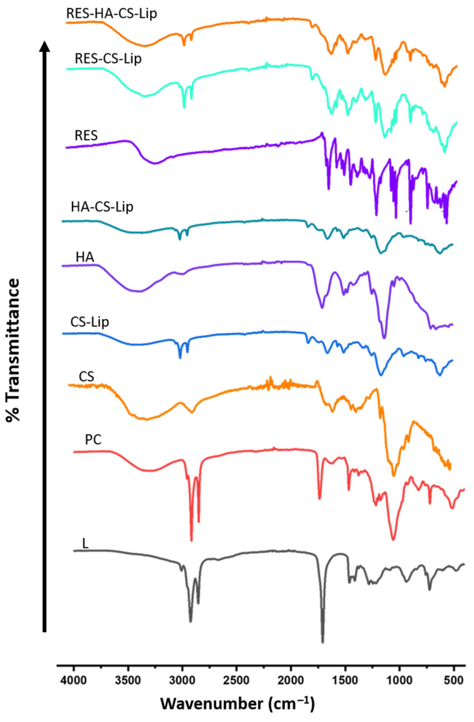
FTIR spectroscopy was employed to investigate molecular interaction and to confirm the successful encapsulation of RES within CS- and HA-coated liposomes. The results are shown in Figure 2. The FTIR spectrum of intact RES exhibited characteristic absorption bands at 3200–3500 cm−1, corresponding to phenolic O–H stretching vibrations. Additional peaks were observed at 1600–1580 cm−1, attributed to C=C stretching of the aromatic systems, and at 1300–1100 cm−1, corresponding to ring skeletal vibrations. [20].
The FTIR spectrum of CS revealed a broad absorption band between 3000 and 3500 cm−1, corresponding to O–H and N–H stretching vibrations. The peak appearing at approximately 1588 cm−1 was assigned to N–H bending vibration of the primary amine groups of the glucosamine units. Additionally, N–H stretching vibrations were also observed at 3286–3361 cm−1. A weak absorption band at 1646 cm−1 indicated the C=O stretching from secondary amide groups, while additional peaks detected at 2874, 1417, 1323, and 1248 cm−1 corresponded to CH2 bending and stretching vibrations, consistent with the polysaccharide backbone structure of CS [21].
PC and L exhibited different characteristic peaks. The FTIR spectrum of PC showed CH2/CH3 stretching at 2922 and 2822 cm−1, ester C=O peak at 1733 cm−1. Peaks around 1257–1055 cm−1 were due to phosphate group vibrations [22]. The spectra of L exhibited prominent FTIR absorption bands near 2925 and 2854 cm−1 were corresponding to aliphatic CH2/CH3 stretching, a strong C=O stretch at 1745 cm−1. A peak at around 3011 cm−1 was according to an unsaturated =C–H band [23].
In the spectra of RES-CS-Lip and RES-HA-CS-Lip, characteristic peaks of intact RES were markedly reduced or completely disappeared, indicating successful encapsulation of RES within the lipid bilayer and polymer coating. Meanwhile, the absorption bands corresponding to CS and HA were retained but exhibited slight shifts and peak broadening. These changes suggest the formation of hydrogen bonding and electrostatic interactions between polymers and the liposomal lipids, thereby confirming the successful formation of a stable polymer–lipid complex in RES-HA-CS-Lip [10,24].

3.5. Thermal Analysis

DSC was conducted to investigate thermal transitions, potential drug–excipient interactions, and the physical state of RES within the formulated liposomal system. This analysis also provides insight into the thermal stability and structural integrity of the liposomal bilayer under physiological and storage conditions. As shown in Figure S1, intact RES exhibited a sharp endothermic peak at approximately 266 °C, corresponding to its melting point and confirming its crystalline characteristics [25]. In contrast, this distinct melting peak was either absent or significantly broadened in the thermograms of both RES-CS-Lip and RES-HA-CS-Lip, suggesting that RES was molecularly dispersed or amorphously encapsulated within the lipid–polymer matrix [26]. This is an indication of successful liposomal encapsulation. The thermogram of CS showed a broad endothermic peak around 80–100 °C, attributed to moisture loss. Additionally, a minor transition above 280 °C was likely associated with polymer degradation [27]. HA, consistent with its amorphous polysaccharide nature, showed only moisture loss below 150 °C [28]. In the composite liposomal formulations, only broad thermal transitions below 150 °C were observed, attributed to bound water evaporation and polymer–lipid rearrangements. These findings confirm the formation of a stable HA-CS coating layer, which efficiently masks individual thermal transitions of the components and supports improved solubility and potential bioavailability of RES.
In addition, the distinct thermal behavior observed between RES-HA-CS-Lip and RES-CS-Lip indicates enhanced polymer–polymer and polymer–lipid interactions in the layer-coated system. These observations support the concept of sequential layer assembly, reinforcing both electrostatic anionic–cationic crosslinking and bilayer cohesion, consistent with previous reports on multilayer liposomal formulations [29,30].

3.6. Release Behavior of RES in Different Release Media

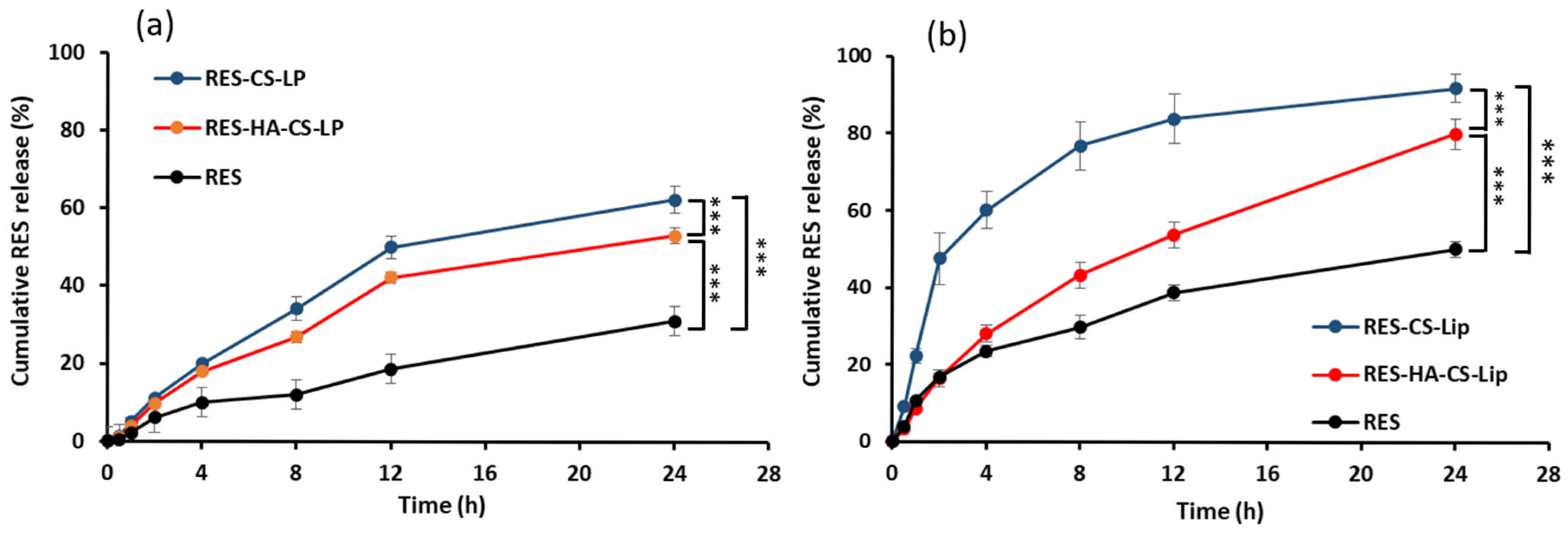
The in vitro release of RES from the developed liposomal formulations was investigated using the dialysis method, and the results are presented in Figure 3. It was found that among the three formulations, the RES solution exhibited the slowest release profile during 24 h under both pH conditions. It is considered that this behavior is likely attributed to the poor aqueous solubility of RES and the absence of a delivery vehicle, which limits sustained diffusion [31].
At physiological condition (pH 7.4), the release profiles of RES from both RES-CS-Lip and RES-HA-CS-Lip demonstrated sustained and controlled release profiles, with cumulative release at 24 h of approximately 62% and 53%, respectively, obviously higher than that of RES solution, which exhibited only 31%. These findings are in accordance with the typical non-Fickian, pH-dependent release behavior previously reported [32], in which polymeric shells could modulate drug diffusion and matrix relaxation, thereby prolonging drug release.
Under acidic conditions (pH 5.5), which mimic the tumor microenvironment or endosomal compartments, the release rate of RES increased markedly shown in Figure 3b. Within 24 h, the accumulative release of RES reached approximately 90% from RES-CS-Lip and around 80% from RES-HA-CS-Lip, both of which were obviously higher than the RES release from the RES solution, which was only 50%.
Considering the difference between RES-CS-Lip and RES-HA-CS-Lip, RES-CS-Lip showed a more rapid and sustained drug release than RES-HA-CS-Lip. The CS coating stabilizes liposomal structure while imparting mucoadhesive properties and pH-responsive swelling behavior in acidic environments [33]. Comparing the two pH conditions of the release media, it was found that the release of RES from both liposomal formulations in the media at pH 5.5 is significantly faster and more pronounced than that at pH 7.4. This enhanced release under acidic conditions can be attributed to the protonation of amine groups in CS, which leads to destabilization of the coating and increased liposomal permeability, a mechanism frequently exploited for pH-triggered drug delivery [34].
RES-HA-CS-lip demonstrated a moderate yet controlled release profile under an acidic cancer environment, reaching approximately 80% at 24 h. Although the RES release from RES-HA-CS-Lip was slower than that from RES-CS-Lip, it was still faster than that from the RES solution. HA, an anionic polymer, may interact electrostatically with cationic CS to form a denser outer layer, thus delaying the initial burst and enabling a more gradual release. This HA coating over CS-Lip may modulate drug release by balancing permeability and retention, which is crucial for the delivery of poorly bioavailability drugs [33,35].

3.7. Antioxidant Activity of RES

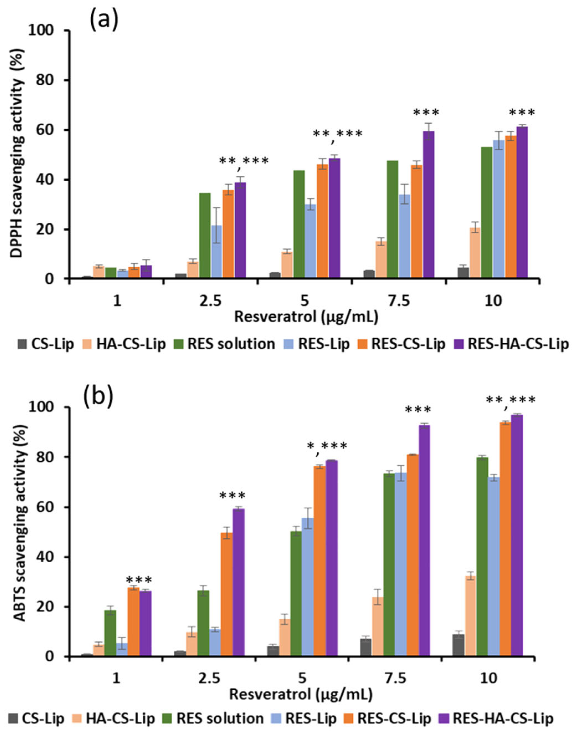
The antioxidant activity of RES loaded in the developed liposomes is presented in Figure 4. Two standard free radical-scavenging antioxidant assays, DPPH and ABTS, were employed to evaluate the antioxidant potential, with the corresponding results shown in Figure 4a,b, respectively. In both methods, RES was tested at concentrations ranging from 1 to 10 µg/mL, and comparisons were made with blank liposomes. Although the blank liposomal formulations contributed negligible scavenging activity, the slightly elevated ABTS activity observed in RES-HA-CS-Lip may partially arise from the antioxidant behavior of the HA coating itself. In both assays, the antioxidant activity of RES exhibited a clear dose-dependent trend across all formulations. Importantly, RES-CS-Lip and RES-HA-CS-Lip demonstrated significantly higher free radical-scavenging activity compared to both the RES solution and the RES-loaded uncoated liposomes. This higher activity happened even at low concentrations of RES. The enhancement is attributed to the presence of CS and HA coating, which not only improves the physicochemical stability of the liposomes but also enhances the radical-scavenging potential of RES. These findings align with the previous reports indicating that encapsulated RES in niosomes retains or improves its antioxidant activity, with the DPPH and ABTS scavenging results exceeding those of the intact drug [36,37]. Moreover, RES-HA-CS-Lip exhibits statistically significant improved antioxidant activity in both assays compared to RES solution, RES-Lip, and RES-CS-Lip (p < 0.05, 0.01, and 0.001). Comparison between RES-CS-Lip and RES-HA-CS-Lip, both assays consistently showed that the antioxidant activity of RES-HA-CS-Lip was clearly higher than that of RES-CS-Lip. At the highest tested concentration (10 µg/mL), RES-HA-CS-Lip showed approximately 60% DPPH inhibition and 95% ABTS inhibition. This is consistent with the previous study in which RES encapsulated in CS–pectin core–shell nanoparticles exhibited enhanced antioxidant activity compared to the unencapsulated RES [38]. The improved antioxidant activity observed with CS and HA coatings can be attributed to their water-compatible nature, especially given that both polymers used in this study were of low molecular weight. The progressive enhancement in antioxidant performance from uncoated liposomes to CS-coated and finally to HA-CS-coated liposomes highlights the synergistic benefit of polysaccharide layering. Specifically, CS contributes to vesicle stability and imparts a positive surface charge, while the HA layer further reinforces the structure and may facilitate more effective interaction with free radicals. Although DPPH and ABTS assays are extracellular in nature, structural preservation of RES through encapsulation likely enhances its measured antioxidant potential [39]. In contrast, the uncoated liposomes, composed mainly of lipid compartments with limited water miscibility, exhibited the lowest antioxidant activity. This may be due to the poor solubility of the carrier and RES as well as partial degradation of RES. Overall, these findings suggest that coating liposomes with CS and HA not only protects RES from degradation but also improves its antioxidant efficacy.

3.8. Biocompatibility Study

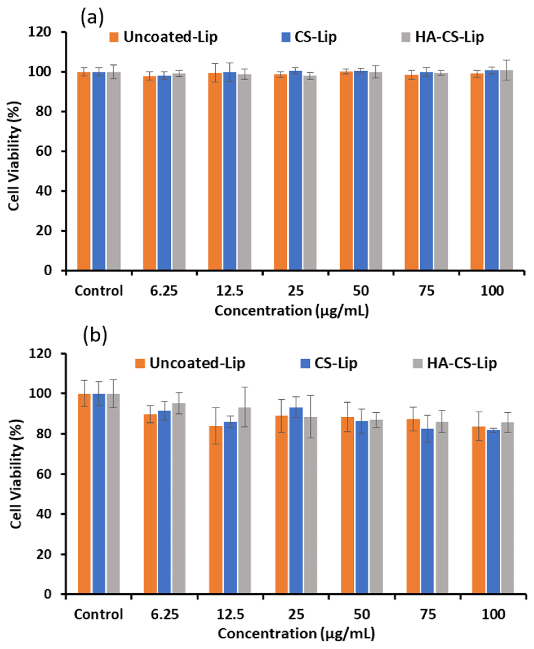
To assess the biocompatibility of the blank liposomal formulations, cellular mitochondrial metabolic activity was evaluated after 48 h of exposure using the MTT assay in two cell lines: human normal keratinocytes (HaCaT) and human breast cancer cells (MCF-7). As shown in Figure 5, all three types of blank liposomes, including uncoated liposomes, CS-Lip, and HA-CS-Lip, demonstrate excellent compatibility, maintaining cell viability above 80% across all tested concentrations, even at 100 µg/mL. These results suggest that both the lipid-based vesicles and the polymeric coatings exert minimal cytotoxic effects in the absence of RES loading.
According to Figure 5a, HaCaT cell viability remained consistently high (>95%) at all tested concentrations, including the highest concentration of 100 µg/mL. This prominent biocompatibility lines up with previous studies reporting that CS-coated liposomes are non-toxic to keratinocytes and may even promote cell proliferation. Such effects are likely contributed to the bioadhesive properties and well-documented biocompatibility of CS, which facilitate favorable interactions with epithelial cells [40].
In contrast, as shown in Figure 5b, MCF-7 cells exhibited a slight, concentration-dependent decrease in viability, with cell survival rate remaining around 80% at the highest concentration. This modest reduction may reflect minor cellular stress responses rather than overt cytotoxicity. Our findings are consistent with earlier reports demonstrating that CS coatings enhance nanoparticle stability while preserving compatibility with epithelial and tumor cell lines. Furthermore, the additional HA layer over the CS layer appears to further support favorable cellular responses, in line with the widely recognized biocompatibility of HA and its role in modulating cell–surface interactions [41].
The enhanced biocompatibility of HA-CS-Lip observed in both cell lines can be further attributed to the presence of HA, which is known to mitigate the potential cytotoxic effects associated with CS. In addition, HA contributes to improving the overall cellular compatibility of the liposomal formulation by providing a hydrophilic and biocompatible interface that supports favorable cell interactions [42].

3.9. Cytotoxicity Study

The cytotoxic effects of RES solution, RES-CS-Lip, and RES-HA-CS-Lip were assessed against MCF-7 breast cancer cells using the MTT assay over 24 h and 48 h treatments. The results reveal both dose-dependent and time-dependent reduction in cell viability across all tested formulations, as demonstrated in Figure 6.
After 24 h of treatment, the results, as shown in Figure 6a, demonstrated that both RES-loaded liposomal formulations exhibited markedly greater cytotoxicity against MCF-7 cells compared to the free RES solution. At a concentration of 50 µg/mL, cell viability decreased to approximately 45% with RES-CS-Lip and 25% with RES-HA-CS-Lip, while RES solution reduced viability to only 80%. This pronounced early effect aligns with previous reports in which liposomal RES formulations enhanced cytotoxicity to cancer cells even after short incubation periods [43]. It is clearly indicated that among them, RES-HA-CS-Lip possessed the highest cytotoxic efficacy. These findings suggest that the sequential HA and CS coating facilitates more efficient intracellular delivery of RES, possibly due to synergistic polymer interactions and HA-mediated active targeting via CD44 receptor-mediated endocytosis, thereby accelerating apoptotic responses. In contrast, the comparatively lower cytotoxicity of the free RES solution is likely due to its limited aqueous solubility and poor cellular internalization, which is consistent with previous reports describing the low bioavailability and weak membrane permeability of the active compounds [31].
After 48 h of treatment, the results presented in Figure 6b revealed that the cytotoxic effects were stronger compared to the 24 h exposure. At 50 μg/mL, the RES solution slightly reduced cell viability to approximately 55%, whereas RES-CS-Lip reduced viability to about 25%. Importantly, RES-HA-CS-Lip displayed an even higher cytotoxic potential than RES-CS-Lip, with a viability reduction to roughly 15%. This time-dependent enhancement of cytotoxicity further supports the beneficial role of liposomal encapsulation in promoting sustained drug retention and improved cellular uptake. Similar outcomes have been reported with other RES-loaded nanocarriers, where encapsulation leads to superior anticancer activity compared to free RES [44].
The markedly lower IC50 value of 13.08 µg/mL observed for the HA-CS layer-by-layer liposomes at 48 h supports the hypothesis that dual functionalization enhances cellular uptake via CD44-mediated endocytosis and sustains intracellular release, corroborating earlier findings in HA-CS nanocarriers loaded with hydrophobic anticancer agents such as α-mangostin (IC50 4.37 µg/mL) [45]. Additionally, the improved cytotoxicity relative to chitosan-only coated formulations (IC50 34.12 µg/mL) is in good agreement with previous studies demonstrating the role of CS in enhancing liposomal delivery efficiency. Lastly, the efficacy of conventional RES in our MCF-7 model (IC50 ~28.6 µg/mL) mirrors documented values for free RES nanoformulations in cancer cell lines [46].

3.10. Stability Study

The stability of the liposome formulations was evaluated by storing the samples at 4 °C and 25 °C for a period of four weeks. The particle size, size distribution expressed as PDI, and zeta potential values are summarized in Table S1. At 4 °C, all formulations exhibited relatively stable particle sizes and surface charges throughout the 28 day storage period. Slight increases in size were observed in blank and RES-loaded uncoated liposomes. Specifically, the particle size of RES-Lip increased from 132.7 nm to 156.5 nm, and that of blank liposomes from 90.6 nm to 125.9 nm. Despite this size increase, the vesicles remained negatively charged, which may be attributed to minor lipid rearrangements or increased hydration over time.
In contrast, RES-CS-Lip and RES-HA-CS-Lip maintained consistent particle sizes, with final sizes of 210.1 nm and 206.8 nm, respectively. Their PDI values remained below 0.5 for most systems, indicating a uniform and monodisperse distribution without evidence of severe aggregation. RES-CS-Lip maintained a positive surface charge, declining slightly from 14.8 mV to 7.9 mV, whereas RES-HA–CS-Lip showed a stable, moderately zeta potential ranging from +11.9 mV to +10.3 mV. These zeta potential values suggest good colloidal stability, as values beyond ±10 mV are generally sufficient to provide electrostatic repulsion and prevent vesicle aggregation. These findings are aligned with previous reports demonstrating the ability of CS and HA coatings to preserve nanocarrier size and suppress vesicle fusion under refrigerated conditions [10].
At ambient temperature (25 °C), as shown in Table S2, the liposomal formulations demonstrated more noticeable changes in structural stability. The reductions in surface charge were more pronounced, likely due to thermal effects on surface-exposed functional groups. It is noted that uncoated RES-Liposomes exhibited significant instability, with particle size decreasing from approximately 133 nm to 90 nm, along with a considerable decline in zeta potential over a period of 28 days. This is likely due to vesicle collapse or drug leakage during storage. RES-CS-Lip showed a biphasic size behavior, initially swelling from about 234 nm to 279 nm by week 2, then shrinking to around 192 nm by week 4. This pattern suggests transient swelling followed by partial polymer degradation or erosion, supported by a concurrent reduction in zeta potential. Conversely, RES-HA-CS-Lip showed remarkable structural stability at 25 °C, with particle size only slightly decreasing from approximately 212 nm to 198 nm and a consistent zeta potential of about +9 to +11 mV. The PDI remained low at <0.4, indicating minimal aggregation or vesicle fusion. These results confirm the effectiveness of the HA-CS polyelectrolyte layer in maintaining liposomal integrity and preventing thermally induced membrane disruption at room temperature [24].
In addition to structural stability, the ability of the coated liposomal formulations to retain RES during the storage period was evaluated by measuring the EE, with the results shown in Figure 7. It was found that over 28 days at 4 °C, both RES-CS-Lip and RES-HA-CS-Lip retained high EE values (>80–85%), whereas the EE of uncoated RES-Lip declined significantly to approximately 55% as shown in Figure 7a. The difference was more pronounced at 25 °C, where RES-Lip exhibited the greatest instability, with EE% dropping to approximately 35% by day 28, as shown in Figure 7b. Meanwhile, RES-CS-Lip maintained moderate retention of EE to approximately 65%. Importantly, RES-HA-CS-Lip exhibited the highest stability, maintaining EE above 80%. The superior performance of polymer-coated liposomal formulations is attributed to the stabilizing role of the surface polymeric coatings. CS improves liposomal membrane integrity by forming electrostatic interactions with phospholipid headgroups, thereby reducing membrane permeability and minimizing drug leakage [7]. Furthermore, when HA is applied as an outer layer in a layer-by-layer assembly over CS, it forms a polyelectrolyte complex with CS, creating an additional steric barrier and electrostatic crosslinking. Consequently, this not only enhances bilayer rigidity but also protects the vesicles against thermal degradation, leading to sustained drug retention during storage [30].

4. Conclusions

This study demonstrates that sequential surface modification of liposomes with CS and HA significantly enhances the delivery efficiency, stability, and bioactivity of RES for potential application in breast cancer therapy. The use of these biocompatible polysaccharides not only improved encapsulation efficiency and colloidal stability of the liposomes but also provided sustained release, particularly under acidic conditions resembling the tumor microenvironment. Among the tested formulations, HA-CS-coated liposomes exhibited superior antioxidant activity, enhanced cytotoxicity against MCF-7 breast cancer cells, and excellent biocompatibility toward normal human keratinocytes, HaCaT cells. The formation of a stable polyelectrolyte complex between HA and CS created a robust barrier that minimized RES leakage, improved intracellular uptake, and potentially facilitated CD44 receptor-mediated endocytosis. These findings highlight the promise of HA-CS-coated liposomes as a stable, biocompatible, and effective nanocarrier for delivering RES and other poorly soluble anticancer agents. However, the present study is limited to in vitro experiments, and further in vivo evaluation is required to validate therapeutic potential and pharmacokinetic behavior.

Supplementary Materials

The following supporting information can be downloaded at: https://www.mdpi.com/article/10.3390/polysaccharides6040093/s1, Figure S1: DSC thermograms of CS, CS-Lip, HA, HA-CS-Lip, RES, RES-CS-Lip, RES-HA-CS-Lip; Table S1: Stability study at 4 °C; Table S2: Stability study at 25 °C.

Author Contributions

Conceptualization, Y.Y.M. and S.O.; Data curation, Y.Y.M., S.A. and S.O.; Formal analysis, Y.Y.M. and P.R.; Investigation, Y.Y.M., P.R. and C.C.; Methodology, Y.Y.M., K.K.G. and S.A.; Project administration, S.O.; Resources, Y.Y.M. and S.O.; Supervision, S.O.; Writing—original draft, Y.Y.M. and P.R.; Writing—review & editing, S.A. and S.O. All authors have read and agreed to the published version of the manuscript.

Funding

This research was funded by Chiang Mai University under the CMU Proactive Researcher Program (2024) for Yin Yin Myat, Grant No. 628/2567.

Institutional Review Board Statement

Not applicable as no patients or animals were involved in this study.

Informed Consent Statement

Not applicable as no patients were involved in this study.

Data Availability Statement

The original contributions presented in this study are included in the article and Supplementary Materials. Further inquiries can be directed to the corresponding author.

Acknowledgments

Financial support from the CMU Proactive Researcher Program (2024), Chiang Mai University is gratefully acknowledged. The authors are grateful to the Center of Excellence in Pharmaceutical Nanotechnology, Faculty of Pharmacy, Chiang Mai University, Thailand, for their equipment, facility, and partial financial support.

Conflicts of Interest

The authors declare no conflicts of interest.

Abbreviations

The following abbreviations are used in this manuscript:
ABTS2,2′-azino-bis (3-ethylbenzothiazoline-6-sulfonic acid
ATR-FTIRAttenuated total reflectance Fourier-transformed infrared spectroscopy
CSChitosan
CS-LipBlank liposomes coated with chitosan
DLSDynamic light scattering
DPPH2,2-diphenyl-1-picrylhydrazyl
FTIRFourier-transformed infrared spectroscopy
HAHyaluronic acid
HA-CS-LipBlank CS-Lip coated with hyaluronic acid
LLinoleic acid
MTT3-(4,5-dimethylthiazol-2-yl)-2,5-diphenyltetrazolium bromide
PCPhosphatidylcholine
RESResveratrol
RES-CS-LipCS-Lip loaded with resveratrol
RES-HA-CS-LipHA-CS-Lip loaded with resveratrol
TEMTransmission electron microscope

References

  1. Kundu, J.K.; Surh, Y.-J. Cancer chemopreventive and therapeutic potential of resveratrol: Mechanistic perspectives. Cancer Lett. 2008, 269, 243–261. [Google Scholar] [CrossRef]
  2. Sinha, D.; Sarkar, N.; Biswas, J.; Bishayee, A. Resveratrol for breast cancer prevention and therapy: Preclinical evidence and molecular mechanisms. Semin. Cancer Biol. 2016, 40–41, 209–232. [Google Scholar] [CrossRef]
  3. Azios, N.G.; Krishnamoorthy, L.; Harris, M.; Cubano, L.A.; Cammer, M.; Dharmawardhane, S.F. Estrogen and Resveratrol Regulate Rac and Cdc42 Signaling to the Actin Cytoskeleton of Metastatic Breast Cancer Cells. Neoplasia 2007, 9, 147–158. [Google Scholar] [CrossRef]
  4. Ito, Y.; Mitani, T.; Harada, N.; Isayama, A.; Tanimori, S.; Takenaka, S.; Nakano, Y.; Inui, H.; Yamaji, R. Identification of Carbonyl Reductase 1 as a Resveratrol-Binding Protein by Affinity Chromatography Using 4’-Amino-3,5-dihydroxy-trans-stilbene. J. Nutr. Sci. Vitaminol. 2013, 59, 358–364. [Google Scholar] [CrossRef]
  5. Szulc-Musioł, B.; Sarecka-Hujar, B. The Use of Micro- and Nanocarriers for Resveratrol Delivery into and across the Skin in Different Skin Diseases-A Literature Review. Pharmaceutics 2021, 13, 451. [Google Scholar] [CrossRef] [PubMed]
  6. Bohara, R.A.; Tabassum, N.; Singh, M.P.; Gigli, G.; Ragusa, A.; Leporatti, S. Recent Overview of Resveratrol’s Beneficial Effects and Its Nano-Delivery Systems. Molecules 2022, 27, 5154. [Google Scholar] [CrossRef] [PubMed]
  7. Sebaaly, C.; Trifan, A.; Sieniawska, E.; Greige-Gerges, H. Chitosan-Coating Effect on the Characteristics of Liposomes: A Focus on Bioactive Compounds and Essential Oils: A Review. Processes 2021, 9, 445. [Google Scholar] [CrossRef]
  8. Sun, X.; He, Z.; Lu, R.; Liu, Z.; Chiampanichayakul, S.; Anuchapreeda, S.; Jiang, J.; Tima, S.; Zhong, Z. Hyaluronic acid-modified liposomes Potentiated in-vivo anti-hepatocellular carcinoma of icaritin. Front. Pharmacol. 2024, 15, 1437515. [Google Scholar] [CrossRef]
  9. Louderbough, J.M.; Schroeder, J.A. Understanding the dual nature of CD44 in breast cancer progression. Mol. Cancer Res. 2011, 9, 1573–1586. [Google Scholar] [CrossRef]
  10. Bonechi, C.; Tamasi, G.; Donati, A.; Leone, G.; Consumi, M.; Cangeloni, L.; Volpi, V.; Magnani, A.; Cappelli, A.; Rossi, C. Physicochemical Characterization of Hyaluronic Acid and Chitosan Liposome Coatings. Appl. Sci. 2021, 11, 12071. [Google Scholar] [CrossRef]
  11. Yang, K.K.; Kong, M.; Wei, Y.N.; Liu, Y.; Cheng, X.J.; Li, J.; Park, H.J.; Chen, X.G. Folate-modified–chitosan-coated liposomes for tumor-targeted drug delivery. J. Mater. Sci. 2013, 48, 1717–1728. [Google Scholar] [CrossRef]
  12. Rodwattanagul, S.; Sasarom, M.; Riangjanapatee, P.; Anuchapreeda, S.; Okonogi, S. Antioxidant activity of Sophora exigua and liposome development of its powerful extract. Drug Discov. Ther. 2024, 18, 150–159. [Google Scholar] [CrossRef]
  13. Sasarom, M.; Wanachantararak, P.; Chaijareenont, P.; Okonogi, S. Antioxidant, antiglycation, and antibacterial of copper oxide nanoparticles synthesized using Caesalpinia Sappan extract. Drug Discov. Ther. 2024, 18, 167–177. [Google Scholar] [CrossRef] [PubMed]
  14. Manconi, M.; Manca, M.L.; Valenti, D.; Escribano, E.; Hillaireau, H.; Fadda, A.M.; Fattal, E. Chitosan and hyaluronan coated liposomes for pulmonary administration of curcumin. Int. J. Pharm. 2017, 525, 203–210. [Google Scholar] [CrossRef] [PubMed]
  15. Imam, S.S.; Alshehri, S.; Altamimi, M.A.; Hussain, A.; Qamar, W.; Gilani, S.J.; Zafar, A.; Alruwaili, N.K.; Alanazi, S.; Almutairy, B.K. Formulation of Piperine–Chitosan-Coated Liposomes: Characterization and In Vitro Cytotoxic Evaluation. Molecules 2021, 26, 3281. [Google Scholar] [CrossRef] [PubMed]
  16. Hayward, S.L.; Francis, D.M.; Sis, M.J.; Kidambi, S. Ionic Driven Embedment of Hyaluronic Acid Coated Liposomes in Polyelectrolyte Multilayer Films for Local Therapeutic Delivery. Sci. Rep. 2015, 5, 14683. [Google Scholar] [CrossRef]
  17. Park, S.N.; Jo, N.R.; Jeon, S.H. Chitosan-coated liposomes for enhanced skin permeation of resveratrol. J. Ind. Eng. Chem. 2014, 20, 1481–1485. [Google Scholar] [CrossRef]
  18. Channarong, S.; Chaicumpa, W.; Sinchaipanid, N.; Mitrevej, A. Development and evaluation of chitosan-coated liposomes for oral DNA vaccine: The improvement of Peyer’s patch targeting using a polyplex-loaded liposomes. AAPS PharmSciTech 2011, 12, 192–200. [Google Scholar] [CrossRef]
  19. Milkova, V.; Vilhelmova-Ilieva, N.; Gyurova, A.; Kamburova, K.; Dimitrov, I.; Tsvetanova, E.; Georgieva, A.; Mileva, M. Remdesivir-Loaded Nanoliposomes Stabilized by Chitosan/Hyaluronic Acid Film with a Potential Application in the Treatment of Coronavirus Infection. Neurol. Int. 2023, 15, 1320–1338. [Google Scholar] [CrossRef]
  20. Gu, H.; Chen, P.; Liu, X.; Lian, Y.; Xi, J.; Li, J.; Song, J.; Li, X. Trimethylated chitosan-coated flexible liposomes with resveratrol for topical drug delivery to reduce blue-light-induced retinal damage. Int. J. Biol. Macromol. 2023, 252, 126480. [Google Scholar] [CrossRef]
  21. Myat, Y.Y.; Vollrath, M.K.; Sahatspan, N.; Ngawhirunpat, T.; Pornpitchanarong, C.; Patrojanasophon, P. Development of mucoadhesive chitosan-iodoacetamide buccal tablets for local delivery of dextromethorphan. Nat. Life Sci. Commun. 2023, 22, e2023041. [Google Scholar] [CrossRef]
  22. Yeo, S.; An, J.; Park, C.; Kim, D.; Lee, J. Design and Characterization of Phosphatidylcholine-Based Solid Dispersions of Aprepitant for Enhanced Solubility and Dissolution. Pharmaceutics 2020, 12, 407. [Google Scholar] [CrossRef] [PubMed]
  23. Vongsvivut, J.; Heraud, P.; Zhang, W.; Kralovec, J.A.; McNaughton, D.; Barrow, C.J. Quantitative determination of fatty acid compositions in micro-encapsulated fish-oil supplements using Fourier transform infrared (FTIR) spectroscopy. Food Chem. 2012, 135, 603–609. [Google Scholar] [CrossRef] [PubMed]
  24. Sun, Y.; Shen, X.; Yang, J.; Tan, C. Hyaluronic Acid-Coated Nanoliposomes as Delivery Systems for Fisetin: Stability, Membrane Fluidity, and Bioavailability. Foods 2024, 13, 2406. [Google Scholar] [CrossRef]
  25. Balanč, B.; Trifković, K.; Đorđević, V.; Marković, S.; Pjanović, R.; Nedović, V.; Bugarski, B. Novel resveratrol delivery systems based on alginate-sucrose and alginate-chitosan microbeads containing liposomes. Food Hydrocoll. 2016, 61, 832–842. [Google Scholar] [CrossRef]
  26. Sharma, S.; Gupta, P.; Kawish, S.M.; Ahmad, S.; Iqbal, Z.; Vohora, D.; Kohli, K. Novel Chitosan-Coated Liposomes Coloaded with Exemestane and Genistein for an Effective Breast Cancer Therapy. ACS Omega 2024, 9, 9735–9752. [Google Scholar] [CrossRef]
  27. Ramasamy, T.; Haidar, Z.S.; Tran, T.H.; Choi, J.Y.; Jeong, J.-H.; Shin, B.S.; Choi, H.-G.; Yong, C.S.; Kim, J.O. Layer-by-layer assembly of liposomal nanoparticles with PEGylated polyelectrolytes enhances systemic delivery of multiple anticancer drugs. Acta Biomater. 2014, 10, 5116–5127. [Google Scholar] [CrossRef]
  28. Collins, M.N.; Birkinshaw, C. Physical properties of crosslinked hyaluronic acid hydrogels. J. Mater. Sci. Mater. Med. 2008, 19, 3335–3343. [Google Scholar] [CrossRef]
  29. de Souza, J.B.; de Lacerda Coriolano, D.; Dos Santos Silva, R.C.; da Costa Júnior, S.D.; de Almeida Campos, L.A.; Cavalcanti, I.D.L.; Lira Nogueira, M.C.B.; Pereira, V.R.A.; Brelaz-de-Castro, M.C.A.; Cavalcanti, I.M.F. Ceftazidime and Usnic Acid Encapsulated in Chitosan-Coated Liposomes for Oral Administration against Colorectal Cancer-Inducing Escherichia coli. Pharmaceuticals 2024, 17, 802. [Google Scholar] [CrossRef]
  30. Jeon, S.; Yoo, C.Y.; Park, S.N. Improved stability and skin permeability of sodium hyaluronate-chitosan multilayered liposomes by Layer-by-Layer electrostatic deposition for quercetin delivery. Colloids Surf. B Biointerfaces 2015, 129, 7–14. [Google Scholar] [CrossRef]
  31. Walle, T. Bioavailability of resveratrol. Ann. N. Y. Acad. Sci. 2011, 1215, 9–15. [Google Scholar] [CrossRef]
  32. Li, Y.; Sun, K.; Chen, S.; Zhao, J.; Lei, Y.; Geng, L. Nano-Resveratrol Liposome: Physicochemical Stability, In Vitro Release, and Cytotoxicity. Appl. Biochem. Biotechnol. 2023, 195, 5950–5965. [Google Scholar] [CrossRef] [PubMed]
  33. Yan, L.; Crayton, S.H.; Thawani, J.P.; Amirshaghaghi, A.; Tsourkas, A.; Cheng, Z. A pH-Responsive Drug-Delivery Platform Based on Glycol Chitosan-Coated Liposomes. Small 2015, 11, 4870–4874. [Google Scholar] [CrossRef] [PubMed]
  34. Beheshtkhoo, N.; Jadidi Kouhbanani, M.A.; Daghighi, S.M.; Shakouri Nikjeh, M.; Esmaeili, Z.; Khosravani, M.; Adabi, M. Effect of oral resveratrol-loaded nanoliposomes on hyperlipidemia via toll-like receptor 3 and TIR domain-containing adaptor inducing interferon-β protein expression in an animal model. J. Liposome Res. 2025, 35, 225–251. [Google Scholar] [CrossRef] [PubMed]
  35. Ariga, K.; McShane, M.; Lvov, Y.M.; Ji, Q.; Hill, J.P. Layer-by-layer assembly for drug delivery and related applications. Expert. Opin. Drug Deliv. 2011, 8, 633–644. [Google Scholar] [CrossRef] [PubMed]
  36. Zhang, X.; Wu, Z.; Zhang, W.; Wang, L.; Zhao, P.; Lv, X.; Guo, P.; Chen, J. Surface modification by chitosan for improving stability and antioxidative activity of astaxanthin-loaded liposomes. LWT 2024, 198, 116033. [Google Scholar] [CrossRef]
  37. Machado, N.D.; Gutiérrez, G.; Matos, M.; Fernández, M.A. Preservation of the Antioxidant Capacity of Resveratrol via Encapsulation in Niosomes. Foods 2021, 10, 988. [Google Scholar] [CrossRef]
  38. Sarma, S.; Agarwal, S.; Bhuyan, P.; Hazarika, J.; Ganguly, M. Resveratrol-loaded chitosan-pectin core-shell nanoparticles as novel drug delivery vehicle for sustained release and improved antioxidant activities. R. Soc. Open Sci. 2022, 9, 210784. [Google Scholar] [CrossRef]
  39. Jøraholmen, M.W.; Škalko-Basnet, N.; Acharya, G.; Basnet, P. Resveratrol-loaded liposomes for topical treatment of the vaginal inflammation and infections. Eur. J. Pharm. Sci. 2015, 79, 112–121. [Google Scholar] [CrossRef]
  40. Alomrani, A.; Badran, M.; Harisa, G.I.; Lshehry, A.M.; Alhariri, M.; Alshamsan, A.; Alkholief, M. The use of chitosan-coated flexible liposomes as a remarkable carrier to enhance the antitumor efficacy of 5-fluorouracil against colorectal cancer. Saudi Pharm. J. 2019, 27, 603–611. [Google Scholar] [CrossRef]
  41. Hasan, M.; Elkhoury, K.; Belhaj, N.; Kahn, C.; Tamayol, A.; Barberi-Heyob, M.; Arab-Tehrany, E.; Linder, M. Growth-Inhibitory Effect of Chitosan-Coated Liposomes Encapsulating Curcumin on MCF-7 Breast Cancer Cells. Mar. Drugs 2020, 18, 217. [Google Scholar] [CrossRef] [PubMed]
  42. Yao, Z.-A.; Wu, H.-G. Characterization of chitosan-hyaluronic acid blended membranes and their biocompatibility with keratocytes. In Proceedings of the 2010 3rd International Conference on Biomedical Engineering and Informatics, Yantai, China, 16–18 October 2010; Volume 4, pp. 1650–1654. [Google Scholar]
  43. Dana, P.; Thumrongsiri, N.; Tanyapanyachon, P.; Chonniyom, W.; Punnakitikashem, P.; Saengkrit, N. Resveratrol Loaded Liposomes Disrupt Cancer Associated Fibroblast Communications within the Tumor Microenvironment to Inhibit Colorectal Cancer Aggressiveness. Nanomaterials 2022, 13, 107. [Google Scholar] [CrossRef] [PubMed]
  44. Firouzi Amandi, A.; Bahmanyar, Z.; Dadashpour, M.; Lak, M.; Natami, M.; Döğüş, Y.; Alem, M.; Adeli, O.A. Fabrication of magnetic niosomal platform for delivery of resveratrol: Potential anticancer activity against human pancreatic cancer Capan-1 cell. Cancer Cell Int. 2024, 24, 46. [Google Scholar] [CrossRef]
  45. Meylina, L.; Muchtaridi, M.; Joni, I.M.; Elamin, K.M.; Wathoni, N. Hyaluronic Acid-Coated Chitosan Nanoparticles as an Active Targeted Carrier of Alpha Mangostin for Breast Cancer Cells. Polymers 2023, 15, 1025. [Google Scholar] [CrossRef]
  46. Sharifi-Rad, J.; Quispe, C.; Mukazhanova, Z.; Knut, E.; Turgumbayeva, A.; Kipchakbayeva, A.; Seitimova, G.; Mahomoodally, M.F.; Lobine, D.; Koay, A.; et al. Resveratrol-Based Nanoformulations as an Emerging Therapeutic Strategy for Cancer. Front. Mol. Biosci. 2021, 8, 649395. [Google Scholar] [CrossRef]
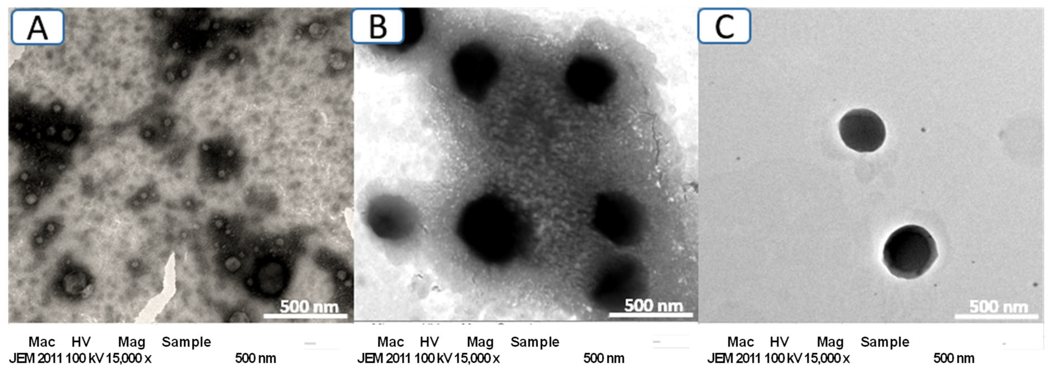
Figure 1. TEM images of blank uncoated-Lip (A), CS-Lip (B), and HA-CS-Lip (C).
Polysaccharides 06 00093 g001
Figure 2. FTIR spectra of intact substances and liposomes.
Polysaccharides 06 00093 g002
Figure 3. Release profiles of RES from RES-CS-Lip and RES-HA-CS-Lip in comparison with that from RES solution in PBS pH 7.4 (a) and pH 5.5 (b). (*** Significant difference from RES solution, and RES-CS-Lip, p < 0.001).
Polysaccharides 06 00093 g003
Figure 4. DPPH (a) and ABTS (b) radical-scavenging activity of RES-loaded liposomes. (Significant difference from RES solution, RES-Lip and RES-CS-Lip, * signifies p < 0.05, ** signifies p < 0.01, *** signifies p < 0.001).
Polysaccharides 06 00093 g004
Figure 5. Biocompatibility study of blank liposomes by MTT assay on HaCaT cells (a) and MCF7 cells (b).
Polysaccharides 06 00093 g005
Figure 6. Cytotoxic activity against MCF7 breast cancer cells of RES-CS Lip and RES-HA-CS-Lip compared with RES solution at 24 h (a) and 48 h (b) treatment period.
Polysaccharides 06 00093 g006
Figure 7. RES retention in different types of RES-loaded liposomes after storage at 4 °C (a) and 25 °C (b) for 28 days. (* Significant difference from RES-uncoated-Lip and RES-CS-Lip, p < 0.05).
Polysaccharides 06 00093 g007
Table 1. Size, PDI and Zeta potential of non-coated and CS-Lip.
LipidsMolar RatioCS (mg/mL)Size
(nm)
PDIZeta Potential (mV)
PC:L15:20.0111.7 ± 4.10.31 ± 0.14−41.13 ± 2.4
PC:L15:21.0189.03 ± 13.30.3 ± 0.06−13.4 ± 1.4
PC:L15:21.5206.5 ± 0.6 *0.23 ± 0.17+24.8 ± 2.5 *
PC:L15:22.0306.6 ± 5.10.27 ± 0.24+17.63 ± 0.2
PC:L15:22.5368.9 ± 1.70.25 ± 0.01+13.77 ± 0.5
PC:L15:23.0501.1 ± 5.90.35 ± 0.05+5.98 ± 1.1
* Significant difference from others (p < 0.05).
Table 2. Composition of ten selected liposomal formulations.
LiposomesHA (mg/mL)CS (mg/mL)RES (mg/mL)
S101.50.50
S201.50.75
S301.51.00
S401.51.25
S501.51.50
F121.50.50
F221.50.75
F321.51.00
F421.51.25
F521.51.50
Table 3. Size, PdI, zeta potential, and EE of RES-CS-Lip and RES-HA-CS-Lip.
LiposomesSize (nm)PDIZeta Potential (mV)EE (%)
S1205.4 ± 14.70.28 ± 0.01+32.3 ± 0.227.01 ± 0.05
S2213.3 ± 4.10.19 ± 0.01+29.4 ± 0.628.23 ± 0.03
S3217.5 ± 5.40.21 ± 0.02+35.6 ± 0.553.41 ± 0.02
S4258.9 ± 11.70.42 ± 0.04+34.8 ± 1.380.92 ± 0.02
S5246.6 ± 7.30.22 ± 0.01+32.1 ± 1.680.73 ± 0.31 *
F1215.4 ± 1.50.15 ± 0.00+1.7 ± 0.260.53 ± 0.01
F2228.4 ± 1.40.17 ± 0.01+8.0 ± 0.659.28 ± 0.15
F3234.2 ± 1.20.21 ± 0.00+9.6 ± 0.260.52 ± 2.75
F4176.0 ± 0.50.22 ± 0.00+11.6 ± 1.364.05 ± 4.64
F5212.5 ± 7.30.16 ± 0.01+9.0 ± 1.082.16 ± 0.03 **
* Significant difference from S1–S3, ** Significant difference from F1–F4), n = 3.
Disclaimer/Publisher’s Note: The statements, opinions and data contained in all publications are solely those of the individual author(s) and contributor(s) and not of MDPI and/or the editor(s). MDPI and/or the editor(s) disclaim responsibility for any injury to people or property resulting from any ideas, methods, instructions or products referred to in the content.