Article

Role of Chitosan Characteristics on the Properties of Curcumin-Loaded Carriers and Their Potential Application in Ophthalmologic Infection Therapy

1
Rostislaw Kaischew Institute of Physical Chemistry, Bulgarian Academy of Sciences, 1113 Sofia, Bulgaria
2
Stephan Angeloff Institute of Microbiology, Bulgarian Academy of Sciences, 1113 Sofia, Bulgaria
3
Institute of Experimental Morphology, Pathology and Anthropology with Museum, Bulgarian Academy of Sciences, 1113 Sofia, Bulgaria
*
Author to whom correspondence should be addressed.
Polysaccharides 2025, 6(1), 22; https://doi.org/10.3390/polysaccharides6010022
Submission received: 4 December 2024 / Revised: 21 February 2025 / Accepted: 11 March 2025 / Published: 17 March 2025

Abstract

:
This study is a comparative investigation of the activity of unloaded and curcumin-loaded oil-in-water emulsion or chitosan-based capsules on rabbit retinal cells (RRC), coronavirus HCoV-OC43, and virus HSV-1 virus in relation to their potential ophthalmologic applications. The carriers are developed by using well-established experimental procedures. The characterization of their surface properties and stability in simulated ocular fluids (tear fluid, aqueous humor, and vitreous humor) is performed using the dynamic light scattering method and UV–vis spectrophotometry. In vitro tests are performed to determine the cytotoxicity and phototoxicity of pure curcumin (CR) and selected CR-containing carriers on RRC. The effect of the unloaded and CR-loaded carriers on the antiviral activity, the behavior of the extracellular virions, and the influence on viral adsorption is evaluated against coronavirus HCoV-OC43 and HSV-1 virus by using suitable microbiological assays. In accordance with the obtained experimental results, the toxicity of carriers containing CR is significantly reduced compared to pure compound and unloaded carriers. Moreover, the activity of the unloaded carriers can be increased several times by incorporating CR. The experimental results demonstrate that the variation in the properties of even one component of the structural composition can provoke the different activity of the carriers.

1. Introduction

Viruses are among the most prevalent ocular pathogens. Acute or chronic ocular damage can occur following viral infection [1,2,3]. Viral RNA is detected in the conjunctival secretions of patients with confirmed coronavirus 2 (laboratory-confirmed COVID-19) [4]. The most common manifestations of ocular viral infection are conjunctivitis and keratitis [5,6,7]. In many cases, certain viral infections (e.g., herpes viruses such as herpes simplex virus, varicella-zoster virus, cytomegalovirus, as well as other viruses such as Ebola virus, Zika virus, Chikungunya virus, dengue virus, and coronavirus (SARS-CoV-2)) have also been associated with sequelae of primary or secondary glaucoma [2,3,4,5,6,7,8,9,10,11].
Particular delivery systems (emulsions, liposomes, nanocapsules, etc.) can improve the solubility and stability of the drugs in a physiological medium. The type of carrier can be selected based on the characteristics of the active component.
In addition to the many chemical-produced compounds (timolol, travoprost, latanoprost, levobunolol, and pilocarpine), different natural compounds can be used as an alternative approach in the medical treatment of ophthalmologic diseases because of their remarkable antioxidant and anti-inflammatory properties—curcumin, caffeine, saffron anthocyanins, astaxanthin, vitamins, and extracts from medical plants (Ginkgo biloba, Lyceum barbarum, etc.) [2,12].
The present study uses curcumin (CR) for the formation of drug-loaded oil-in-water emulsion and polysaccharide CR-loaded capsules. CR is chosen because of its remarkable antioxidant, anti-inflammatory, anti-cancer, anti-asthmatic, antimicrobial, antiviral, and antifungal properties [13,14,15,16,17,18]. Therefore, CR can be a potential candidate in the medical treatment of glaucoma by reducing oxidative stress. Despite the powerful biological activity of CR, its very low solubility in water (11 ng/mL) is a disadvantage in the possible application of the substance in the pharmacy [https://pubchem.ncbi.nlm.nih.gov/compound/curcumin (accessed on 1 December 2024)]. This is why developing oil-core composite carriers is an appropriate and well-established approach for encapsulating poorly soluble substances. The additional adsorption of biopolymers on the surface of produced structures will improve the stability of the carriers.
In the present study, the produced structures were stabilized with chitosan. Chitosans are a family of poly- or oligosaccharides obtained by deacetylation of their parent polymer chitin, a compound widely distributed in nature and found in the exoskeletons of insects and crustaceans, and in certain fungi. Chitosan is a weak polycation that enables the formation of polyelectrolyte complexes with negatively charged biomolecules, allowing them to interact with cell membranes and other oppositely charged species. The high charge density of chitosan (at pH < 6.5) favors polyelectrolyte behavior, whereas a low charge density (at neutral pH) contributes to its low cytotoxicity and facilitates the intracellular release of biomolecules. Chitosan is biocompatible, non-toxic to living tissues, and has antibacterial, antifungal, and antitumor activity. The polysaccharide is a very promising compound for ophthalmological applications because of its favorable biological properties (low toxicity, bio adhesion, biocompatibility biodegradability, and ocular tolerance), as well as its inherent biological activity, which can have an impact on the development of ocular drug delivery platforms [13,14,15,16,17,18,19]. The unique properties of CS make it a good candidate for the ocular delivery of many ingredients, including immune-modulating agents, antibiotics, ocular hypertension medications, etc. CS-based nano-systems have been reported to successfully modulate ocular diseases by penetrating biological ocular barriers and targeting and controlling drug release [20,21].
The present research aims to compare the activity of unloaded and CR-loaded carriers. The influence of the composition of the carriers on their properties and stability was evaluated in correspondence to the potential ophthalmological application. In vitro ocular infection of rabbit retinal cells (RRC) was induced by herpes simplex virus type 1 (HSV-1) or coronavirus strain OC-43 (HCoV-OC43). The respective viral strains are treated at different stages of viral reproduction with the developed unloaded or CR-loaded carriers and pure curcumin solution.

2. Materials and Methods

2.1. Materials

2.1.1. Materials for the Formation of the Capsules

Chitosan with low (50–190 kDa), CS-L, and medium (190–310 kDa), CS-M, molecular weight and degree of deacetylation (DDA) of 75–85%, and chitosan oligosaccharide, COS (Mw 5 kDa, DDA > 90%), were purchased from Sigma Aldrich. The stock polymer solutions were prepared with a concentration of 1 mg/mL in hydrochloric acid (10−4 M HCl) and were filtered through a 5 µm filter (Minisart®, Sartorius, Göttingen, Germany) to remove possible aggregates.
Curcumin was purchased also from Sigma Aldrich, Steinheim, Germany (product number C-1386-10G).
For the formation of the core of the capsules, soybean lecithin, PC 40% (Avanti Polar Lipids Inc., Birmingham, UK, 341602G). Miglyol 812 N® was kindly provided by Sasol Germany GmbH, Witten, Germany.

2.1.2. Materials for the Implementation of Microbiological Studies

  • Cell culture
The adherent cell line of rabbit retinal cells (RRC) (from the collection of the Institute of Experimental Morphology, Pathology and Anthropology with Museum, Bulgarian Academy of Sciences, Sofia, Bulgaria) was cultured in DMEM/Nutrient Mixture F12 Ham medium (4.5 mg/mL glucose), 10% fetal calf serum, 100 U/mL penicillin, and 0.1 mg/mL streptomycin in 25 cm2 and 75 cm2 plastic cell culture dishes. Cells were cultured at 37 °C and a 5% CO2 atmosphere.
  • Viruses
Human herpes simplex virus type 1, strain Victoria (HSV-1), was obtained from the National Center for Infectious and Parasitic Diseases (Sofia, Bulgaria). For virus replication, RRC cells are used in maintenance solution DMEM/Nutrient Mixture F12 Ham medium (4.5 mg/mL glucose), containing 100 IU/mL penicillin, 100 μg/mL streptomycin, and 0.5% fetal bovine serum (Gibco BRL, Scotland, UK). The virus incubation was performed at 37 °C with a 5% CO2 atmosphere. The resulting viral yield was aliquoted and stored at −80 °C. The infectious viral titer was determined to be 106.5 CCID50/mL. Human Coronavirus OC-43 (HCoV-OC43, ATCC: VR-1558) strain was propagated in the RRC cell line in DMEM/Nutrient Mixture F12 Ham medium (4.5 mg/mL glucose) supplemented with 0.5% fetal bovine serum (Gibco BRL, Scotland, UK), 100 U/mL penicillin, and 100 μg/mL streptomycin. The infectious virus titer was determined to be 105.5 CCID50/mL according to the formula of Reed and Muench [22]. The resulting virus stock was stored at −80 °C.
  • Reference substances
Acyclovir {ACV, [9-(2-hydroxyethoxymethyl)-guanine]} was kindly provided by the Deutsches Kresforschung Zentrum, Heidelberg, with a stock concentration of 3 mM solution in DMSO. Then, falling dilutions were made in DMEM medium to the required concentration. The stock solution of Veklury® (Gilead Science Inc. Ireland UC, IDA Business & Technology Park, County Cork, Ireland) with a concentration of 150 mg/mL was prepared in double-distilled water and the concentration of remdesivir (REM) in the stock solution was estimated at 8.3 × 10⁻³ M.

2.2. Methods and Procedures

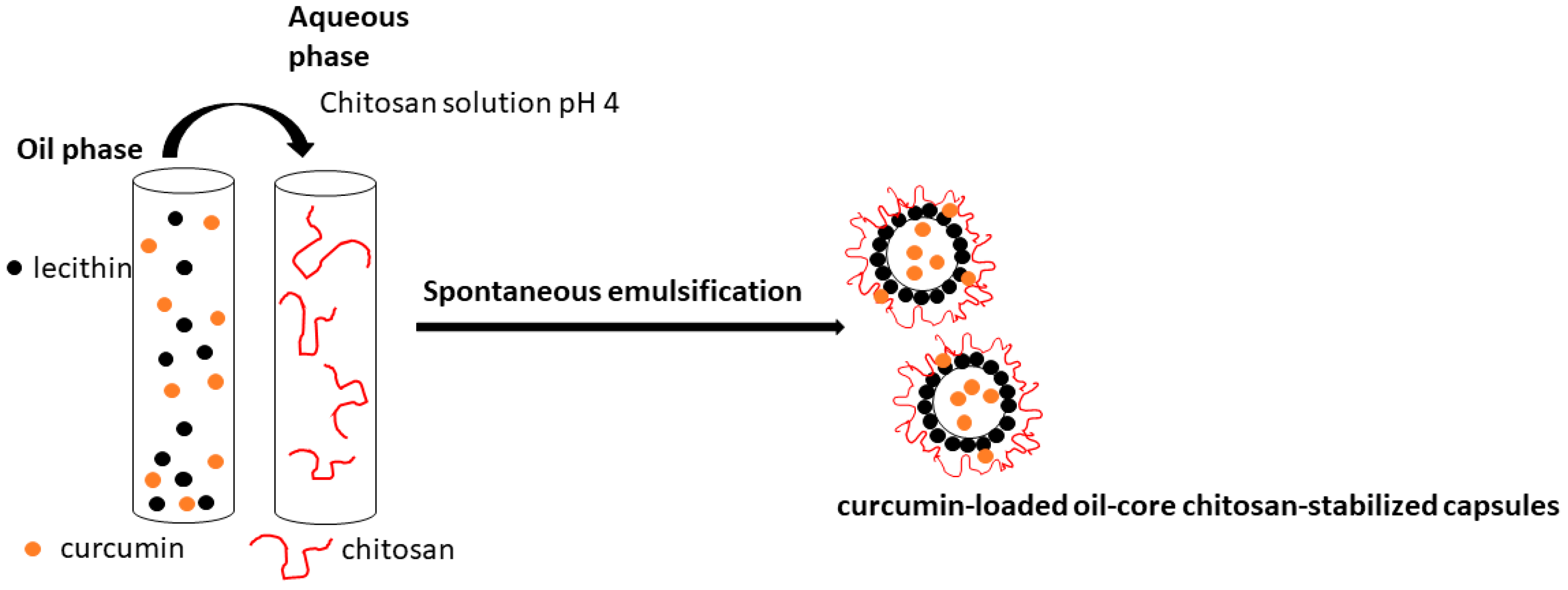
2.2.1. Preparation of the Curcumin-Loaded Capsules

The procedure for encapsulation of hydrophobic bioactive substances in the composite oil-in-water emulsion was originally proposed by Calvo et al. [23]. Briefly, the oil phase was prepared from 0.5 mL of ethanolic lecithin solution (80 mg/mL) and subsequently mixed with 0.512 mL ethanolic curcumin solution (12 mg/mL), 0.125 mL Miglyol 812 N®, and 9.5 mL ethanol. The aqueous phase was a chitosan solution (20 mL, 0.5 mg/mL, and pH 4). After mixing, the organic solvent was evaporated at 40 °C for ca. 30 min. The volume of the final dispersion is ca. 20 mL. For comparison, following the same procedure, an oil-in-water emulsion was prepared by replacing the chitosan solution with a solution of hydrochloric acid (pH 4). In order to prevent a possible aggregation in the subsequent steps, the stock emulsion was diluted 1:19 with a solution of HCl (pH 4) or simulated ocular fluids. The experimental procedure for encapsulation of curcumin is presented in Figure 1.

2.2.2. Preparation of Simulated Ocular Fluids

  • Simulated tear fluid (STF)
Freshly prepared STF was made according to the composition proposed by Kurniawansyah et al. [24]. To prepare 100 mL of solution, we mixed NaCl (670 mg), NaHCO3 (200 mg), CaCl2·2H2O (8 mg), and 100 mL of double-distilled water. The final concentration of the components was, respectively, NaCl (6.7 mg/mL), NaHCO3 (2 mg/mL), and CaCl2·2H2O (0.08 mg/mL).
  • Simulated aqueous humor (SAH)
Aqueous humor is an extremely multicomponent composition of inorganic salt, proteins, and amino acids [25]. The preparation of SAH with completely the same composition for laboratory purposes is almost impossible. The present study used the sterile ocular solution containing sodium hyaluronate, sodium chloride, boric acid, sodium tetraborate, sodium edetate, polyhexanide (PHMB), and highly purified water (Polfa Warszawa S.A., Warszawa, Poland).
  • Simulated vitreous humor (SVH)
The simulated fluid was prepared according to the procedure proposed by Kummer et al. [26]. To prepare the solution (5 mL), the desired amount of Agar (15 mg) and hyaluronic acid (15 mg) were weighed and mixed in a vial. For the formation of SVH, hyaluronic acid sodium salt (1000 kDa) products of Fluka were used. Double-distilled water (ca. 50 mL) was brought to a boil and then rested until no bubbles were forming anymore. Then, we added 5 mL of water in a vial to achieve a 3 mg/mL concentration of Agar and hyaluronic acid. The solution was mixed well by using Vortex for a minute. To ensure full dissolution of Agar, the vial was placed in a water bath at 100 °C for 10 min. The solution was periodically stirred (Vortex). Upon the removal of the produced solution from the water bath, it was stirred for the last time and placed to rest without stirring for 24 h at room temperature (24 °C) before use in the next experiments.

2.2.3. Evaluation of the Loaded Amount of Curcumin

The concentration of CR loaded into the different types of capsules was determined by the difference between the initial concentration of the compound added to the dispersion (0.31 mg/mL) and the concentration in the supernatant after centrifugation of the dispersions (15,000 rpm, 21,382× g, at 15 °C for 90 min). After centrifugation of the sample, 1 mL from the supernatant was mixed with 1 mL ethanol, and CR concentration was estimated by monitoring with a T60 UV-visible spectrophotometer (PG Instruments Limited, UK, Wood wayLane, Alma park, Leicestershire, UK).
The compound was detected at a wavelength of 425 nm corresponding to the maximum absorbance peak of CR and the concentration of free CR in solution was calculated by using an appropriate calibration curve of a compound in ethanol [27]. The encapsulation efficiency (EE%) was estimated by using the following equation:
EE% = (Ctotal − Cfree)·100/Ctotal
where the Ctotal is the initial concentration of CR added to the dispersion and Cfree is the determined concentration of the compound in the supernatant.
The drug loading capacity (in %) was calculated with the following equation:
LC% = mencapsulated·100/mcapsules
where mencapsulated is the concentration of drug successfully loaded into the capsules (in mg/mL) and mcapsules is the concentration of capsules (0.24 mg/mL).

2.2.4. Stability of the Capsules in Simulated Ocular Fluids

To investigate the stability of the produced capsules loaded with CR, we prepared samples of stock dispersion and simulated ocular fluids: 52 µL from each dispersion was mixed with 1000 µL of STF, SAH, or SVH. The volume ratio between the dispersion and simulated ocular fluids was 1:19. The produced dispersions were sonicated in an ultrasonic bath for 15 min. The vials were incubated at 37 °C for 20 h at 100 rpm in an incubator (ES-20, Biosan SIA, Riga, Latvia). Then, the samples for measuring the charge, size, and determining the released amount of CR were taken. For evaluation of the concentration of free CR in the dispersion, the samples were centrifuged (15,000 rpm, 21,382× g, at 15 °C for 90 min) and the concentration of CR in the supernatant was determined by monitoring with a spectrophotometer (see Section 2.2.3).

2.2.5. Characterization of the Charge and Size of the Capsules

The electrokinetic charge and hydrodynamic diameter of the produced structures were evaluated by using dynamic light scattering with non-invasive backscattering (DLS-NIBS, measuring angle 173°). The measurements were carried out using Zatasizer Pro (Malvern Panalytical Ltd., Malvern, UK) equipped with a He-Ne laser with a maximum power of 10 mW operating at a wavelength of 633 nm with a fixed scattering light angle of 173°. The measurements were performed at 24.0 ± 0.1 °C and 37 ± 0.1 °C. After five measurements, the average value was taken as the surface charge and size of the capsules.

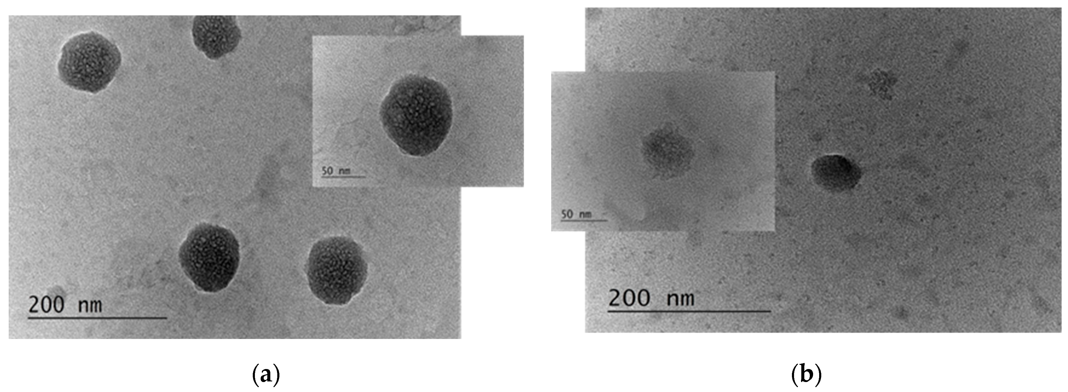
2.2.6. Visualization of the Produced Capsules

The produced capsules were visualized by TEM. The samples for the TEM studies were prepared by dropping and drying a drop of suspension of capsules on formvar-covered TEM grids. The images were captured using a High-Resolution Scanning Transmission electron microscope HR STEM JEOL JEM 2100 for investigations of surface morphology.

2.3. Microbiology Studies

2.3.1. Determination of Cytotoxicity and Phototoxicity

In vitro tests were performed to determine the cytotoxicity and phototoxicity of CR and selected types of CR-containing capsules on rabbit retinal cells (RRC). The obtained results are processed mathematically, statistically, and graphically. Cell viability was determined by the neutral red uptake test (NRU assay) [28]. The test is used to determine the cytotoxic concentrations of substances that reduce cell viability by 50% (CC50). Cells are plated in 96-well cell culture plates at a density of 104 cells/well and incubated for 24 h at 37 °C and 5% CO2. The cells were then treated with a solution of the test products in decreasing concentrations. The phototoxicity test was performed in parallel on a second plate with 96 wells, which was irradiated with a solar simulator (LED lamp—Helios-iO (SERIC Ltd., Tokyo, Japan)) for 10 min (irradiation dose 0.86 J). After 24 h, the effect was determined by NRU assay. After 3 h of incubation, the wells were washed with PBS, pH 7.4, and a solution of ethanol/acetic acid/dH2O = 49/1/50 was added. The optical density was measured with a TECAN microplate reader at λ = 540 nm and the percentage of Cytotoxicity was calculated according to the following equation:
Cytotoxicity (%) = (1 − (OD570 (sample)/OD570 (negative control)) × 100
The photoirradiation factor (PIF) was calculated according to the following equation:
PIF = (CC50 − Irr)/(CC50 + Irr*)

2.3.2. Determination of Infectious Viral Titers

In 96-well plates, a monolayer of RRC cells was infected with 0.1 mL of virus suspension at tenfold decreasing dilutions [29]. After the virus adsorption period, the unabsorbed virus was removed and the DMEM medium was added. This was followed by incubation at 37 °C and 5% CO2 in a HERA cell 150 CO2 incubator (Radobio Scientific Co., Ltd., Shanghai, China) for 48 h (for HSV-1) and 120 h (for HCoV-OC43). Cells infected with the maximum concentration of the virus and demonstrating the maximum cytopathic effect were used as controls. The resulting cytopathic effect (CPE) was monitored by microscopic observation of the cell monolayer and confirmed by neutral red uptake assay. The development of CPE as a percentage of controls was calculated for each virus dilution according to the following equation:
% CPE = 100 − {[(a − b)/c] × 100}
where a—mean optical density of a given virus dilution, b—mean optical density of the maximum CPE control, and c—mean optical density of a cell control. Viral titers are expressed as lg IU (infectious units) (CCID50)/0.1 mL.

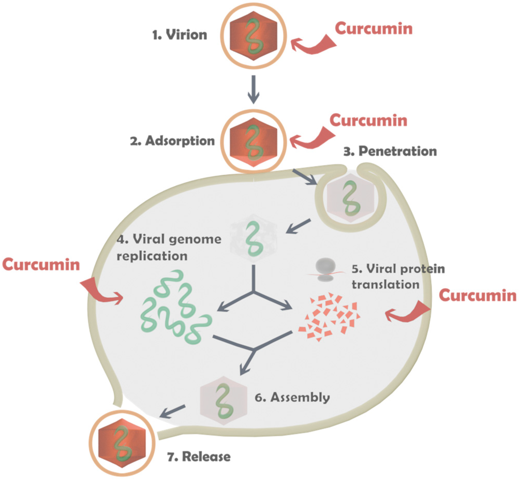
2.3.3. Antiviral Activity Assay

The cytopathic effect inhibition (CPE) test was used to evaluate the antiviral activity of the study products [30]. A monolayer of RRC cells in 96-well plates was infected with 100 cell culture infectious dose of 50% (CCID50) in 0.1 mL. After the viral adsorption time, the unadsorbed virus was removed and the test sample was added at different concentrations. The cells were incubated for 48 h (for HSV-1) and 120 h (for HCoV-OC43) at 37 °C and 5% CO2. Cytopathic effect inhibition was determined by NRU assay for each concentration of the test sample and was calculated according to the following equation:
% CPE = [ODtest sample − ODvirus control]/[ODtoxicity control − ODvirus control] × 100
where ODs (optical density) test sample—containing virus treated with the test sample at the appropriate concentration, ODs virus control—containing virus but no test product in the medium, and ODs toxicity control—not inoculated with a virus but treated with the corresponding concentration of the tested sample.
The concentration of the tested sample at which 50% of the cytopathic effect is inhibited compared to the viral control is determined and defined as the inhibitory concentration of 50% (IC50). From the cytotoxic concentration 50% and inhibitory concentration 50% values determined so far, the selectivity index (SI) of each test sample was determined using the CC50/IC50 ratio.

2.3.4. Determination of the Effect on Extracellular Virions

Samples containing the virus (105 CCID50) and test product at its MTC were prepared, mixed in a 1:1 ratio, and then stored at room temperature for various time intervals (15, 30, 60, 90, and 120 min). The content of residual infectious virus in each sample was then determined by the endpoint dilution method, and the resulting cytopathic effect was reported [30]. The decrease in viral titer in the samples treated with the test products and the untreated viral control was evaluated and ∆lgs were determined.

2.3.5. Effect on Viral Adsorption

A monolayer of RRC cells in 24-well plates was pre-chilled to 4 °C. After cooling, it was inoculated simultaneously with 104 CCID50 of HSV-1 or HCoV-OC43 and the test samples at their maximum non-toxic concentration (MTC) at which they did not affect the cell monolayer. Adsorption of the virus is carried out at 4 °C [31]. After the end of the viral adsorption, the tested samples together with the unadsorbed virus are removed and the cells are washed with PBS at different time intervals (15, 30, 45, and 60 min). Add maintenance medium and followed by incubation at 37 °C for 24 h. The resulting samples were frozen and thawed three times and the viral titer of each sample was determined. The differences in viral titers compared to the viral control (untreated with the products) were used to calculate ∆ lg. Each sample was prepared in quadruplicate.

2.3.6. Statistical Analysis

The values of CC50 were calculated using non-linear regression analysis (GraphPad Software, San Diego, CA, USA). The values were presented as means ± SD from three independent experiments. The differences’ significance between the cytotoxicity values of the samples and the reference substance ACV, as well as between the effects of the test products on the viral replication, was determined by Student’s t-test. p-values of <0.05 were considered significant.

3. Results

3.1. Characterization of the Properties and Stability of the Produced Capsules

The measured size and electrokinetic potential of the produced carriers are summarized in Table 1. According to the presented data, the size of the unloaded capsules depends on the molecular weight of the chitosan used for the stabilization of the structures.
The thickness of the polysaccharide layer can be estimated from the differences in the size values of emulsion and capsules. The thicknesses of the layer of unloaded carriers are 29 nm (CS-L), 83 nm (CS-M), and 62 nm (COS), whereas for the CR-loaded 7 nm (CS-L), 44 nm (CS-M) and 20 nm (COS), respectively. The estimation indicates that the chitosan layer on the unloaded carriers is significantly higher compared to the film formed on the CR-loaded ones.
The dependence of the hydrodynamic size of the capsules on the molecular weight of chitosan is also observed for the CR-loaded capsules. However, a correlation between the size of unloaded and CR-loaded capsules did not register and it was supposed that the size of the capsules almost does not depend on the presence of CR into the core. The evaluated hydrodynamic size (which is almost two times higher compared to the real one) is in good correspondence with the results from the electron microscopy (Figure 2).
The electrokinetic charge of the unloaded emulsion is highly negative and governed by the lecithin membrane on the surface [23,32]. The incorporation of the CR results in a slight decrease in the charge and it was supposed that the reason is the deposition of the CR molecules on the droplet surface, not only in the core of the produced structures. It was supposed that this is also the reason for the registered difference in the thickness of a polymer layer adsorbed on unloaded or CR-loaded carriers. According to the data from the literature, the curcumin molecule is neutral. However, in the presence of acids (low pH), the molecules can accept protons from the solution and the charge of the molecules will be positive. At high pH, the molecule can lose protons, leading to deprotonation and the molecule becoming more negatively charged or neutral [33,34,35].
The experimental values for ζ-potential from the dispersion of capsules stabilized with chitosan indicate the achievement of positive charge of the structures after the polymer adsorption but the ζ-potential of the capsules almost does not depend on the physicochemical characteristics of chitosan. The increase in the positive charge is registered for the CR-loaded capsules.
The amount of CR loaded into the carriers is evaluated by using the experimental procedure as described in Section 2.2.3. The results indicate a very high encapsulation efficiency of the compound, above 94% (ca. 0.3 mg/mL), which almost does not depend on the chitosan characteristics (Table 1). An approximate estimation shows that the CAR concentration per capsule is ca. 4.29 fg and the loading capacity is ca. 125% (Equation (2)).
The stability of the produced carriers is investigated in three artificial ocular fluids. An aggregation is observed in all dispersions of unloaded and CR-loaded capsules. However, despite the very high ionic strength of the fluids, the unloaded and CR-loaded emulsions show remarkable stability. The centrifugation of the dispersion after the stability experiments allows us to evaluate the release amount of the compound in each fluid. According to the presented in Table 2 results, the released amount of CR is in the range of 0.5 to 1% or ca. 1 µg/mL.

3.2. Analysis of the Microbiology Studies

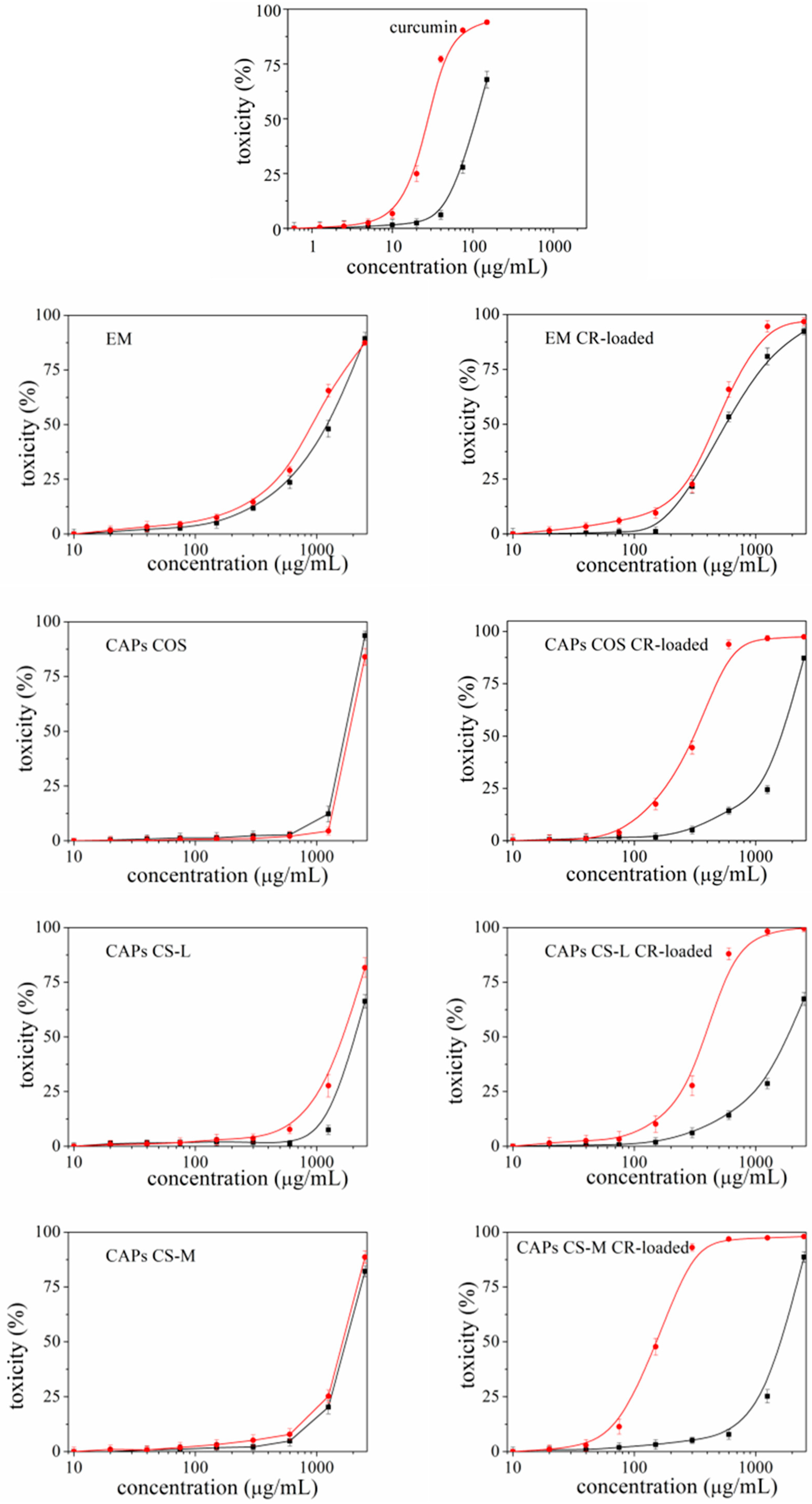
3.2.1. Cytotoxicity and Phototoxicity of the Carriers

The observed effect (cytotoxicity/phototoxicity) was of a dose-dependent type (Figure 3).
Based on the obtained sigmoidal curves, the average C50 values for each tested sample, determined for 24 h, were calculated (Table 3). The calculated PIF factor for CR is 3.96, which corresponds to substances with probable phototoxicity. These substances can be used in pharmacy, through systemic application. For local surface application, exposure to direct sunlight should be avoided. According to the experimental results, the unloaded capsules containing COS, CS-L, and emulsion are non-toxic, and the determined CC50 values are above 1000 µg/mL. Furthermore, the calculated PIF factor is <2, which corresponds to the carriers as non-phototoxic. The introduction of CR into the carriers leads to a slight increase in their toxicity, but the phototoxicity is significantly enhanced (Table 3).
To perform the antiviral experiments at cell-safe, non-toxic concentrations, the cytotoxic effect of the tested samples on RRC cells is investigated. The effect is reported at two time intervals—48 and 120 h from the first registration of the effect. These are the same time intervals as in the antiviral experiments reported (because the cytopathic effect is most pronounced, see Section 3.2.2). For each sample, two values of CC50 are determined—the first CC50 is the concentration of the substance where the cell monolayer is affected by 50% and participates in the determination of the SI of the investigated samples (if it can be determined). The second value CC50 corresponds to the MTC (the highest concentration at which the cell monolayer does not appear to be visually affected by the test component) (used as the dose of product administered in most antiviral experiments) (Table 4).
According to the results presented in Table 4 for the toxicity 48 h after the beginning of the experiment, the curcumin shows the highest toxicity, as expected, with CC50 = 105.2 µg/mL. Among the produced structures, the lowest toxicity is registered for the capsules stabilized with CS-L (for unloaded and CR-loaded capsules ca. 2500 µg/mL and ca. 2403.0 µg/mL, respectively). The highest toxicity is registered for unloaded and loaded emulsions (CC50 is ca 1350.0 µg/mL and ca. 600.0 µg/mL, respectively).
Moreover, the results indicate that 120 h after exposure, the same toxicology behavior of the samples can be observed. Pure curcumin solution showed the strongest toxicity (CC50 is ca. 94.4 µg/mL). The lowest toxicity from unloaded and CR-loaded capsules is shown for CS-L, whereas the high toxicity is registered for emulsion. According to the presented data, the encapsulation of CR in capsules results in a decrease in their toxicity.

3.2.2. Antiviral Activity of the Capsules

Concerning the antiviral activity of the capsules, their effect on HSV-1 and HCoV-OC43 was determined at 48 h and 120 h of exposure, respectively. A stronger inhibitory effect is registered against the replication of HCoV-OC43 (SI = 11.5), while for HSV-1 the effect is significantly weaker (SI = 5.1) (Table 5).
The presence of unloaded capsules did not show any effect on the replication of HSV-1 or HCoV-OC43 strains, but the addition of CR-loaded capsules was found a stronger effect against HCoV-OC43. The most distinct effect is noted for capsules stabilized with CS-L (SI = 21.4) and for EM (SI = 20.8). Moreover, the effectiveness increases nearly two times compared to the effect registered for pure curcumin. The influence of capsules containing CS-M (SI = 6.9) and COS (SI = 5.4), where the effect is weaker than that of pure curcumin, was significantly more moderate. In general, the inhibition of HSV-1 replication is weaker, with the most pronounced effect at CS-L (SI = 9.8), nearly two times more pronounced than pure curcumin. The addition of EM and capsules stabilized with COS showed activity close to that of curcumin.
Moreover, by directly acting on virions, curcumin significantly inhibits viral particles and reduces their ability to enter susceptible cells (Table 6). For HSV-1 virions, a weak effect (Δlg = 1.5) is observed as early as 15 min of exposure, but at the end of the experiment, a decrease in the viral titer is achieved (Δlg = 3.25).
According to the results presented in Table 7, the influence of the capsules on HCoV-OC43 virions is similar. At 15 min of exposure, the decrease in viral titer is slightly weaker than for HSV-1 (Δlg = 1.25). With increasing exposure time it also intensifies, and at 120 min it is slightly stronger than for HSV-1 (Δlg = 3.5).
Neither the unloaded nor the curcumin-loaded capsules showed a statistically significant effect on viral titer reduction upon direct interaction with virions (Table 6 and Table 7).
It was supposed that the individual stages of viral replication to which curcumin was applied in its pure state or included in the composition of carriers are presented in Figure 4.
Having determined that curcumin affects both the viral replication step and extracellular virions, that unloaded capsules have no effect on viral replication or virions, and that CR-loaded capsules only differentially affect viral replication but not the virions, we decided to check whether any of the investigated samples affected the step of virus adsorption to susceptible cells.
Curcumin exerts a weak effect on both tested virus strains (Δlg = 1.5) as early as 15 min after exposure, and this effect remains for up to 30 min. This is followed by a strengthening of the effect to Δlg = 2.0 from 45 to 60 min. In HCoV-OC43, the process of adsorption is followed up to 120 min and the influence increases up to Δlg = 2.25. The unloaded capsules did not have a significant effect on the virus adsorption stage. The curcumin-loaded capsules at 15 and 30 min had no effect, and at the later investigated time steps, the effect was weak (Table 8 and Table 9).

4. Discussion

Curcumin is a natural product known for its many biological activities. However, its low solubility and low bioavailability make it difficult to manifest these activities [28,29,30]. The possibilities for loading into functional carriers can significantly overcome these obstacles in applying CR [36,37,38,39,40,41]. In the present study, three main advantages of the incorporation of the compound are identified:
(1)
The toxicity of carriers containing CR is significantly reduced compared to pure compound (previous studies have reported similar findings for other CR-containing delivery systems [42,43]);
(2)
The activity of the carriers can be increased several times by incorporating CR [43,44,45]. For example, in the present study, the microbiological experiments showed that the CR-loaded emulsion and capsules produced from CS-L have a few times higher activity compared to the unloaded ones. The experiments demonstrate that changing even one form of the components of the structural composition may not lead to the desired effect and the system may not be fully productive. In the present study, the variation in the molecular weight of the chitosan results in different activity of the structures. The capsules containing CS-L showed the highest activity of all the tested samples, while the registered activity for the capsules with CS-H was almost three times lower.
(3)
CR is known to possess some phototoxicity. An ability for which it is being studied (but not yet recognized as an official therapeutic) as a photosensitizer most often in the treatment of cancers [46,47,48]. However, when it is not a matter of antitumor activity, the manifestation of this phototoxicity can often turn out to be negative, especially if the aim is to apply CR externally on the skin or eyes—areas that have direct contact with sunlight. The present study proved that, depending on the type of carriers used, it is possible to reduce the unwanted phototoxicity of the compound. The most effective in this regard was EM-CR-loaded, which reduced the manifestation of CR phototoxicity more than three times.
For decades, numerous studies have been conducted to develop therapeutics affecting viral ocular infections. The first antiviral agent introduced for the successful treatment of ocular viral infection was idoxuridine in the healing of herpes simplex keratitis. Its use was quite often accompanied by pronounced toxicity and serious hypersensitivity reactions. Significant progress in the treatment of herpetic ophthalmic infections was achieved after the discovery of acyclovir. This was followed by the development of other therapeutics such as valacyclovir, famciclovir, ganciclovir, valganciclovir, cidofovir, and foscarnet, which made it possible to treat a larger number of viral ophthalmic diseases [49,50,51]. However, the formation of resistance of viruses to the used therapy creates the need for the development of new therapeutics.
The presented experimental results demonstrated that CR inhibited the replication of HCoV-OC43 (SI = 11.5) and HSV-1 (SI = 5.1) to varying degrees. At the same time, it also has a remarkable ability to inhibit the extracellular virions of HCoV-OC43 and HSV-1 by lowering viral titers by Δlg < 3. The effect of inhibiting the step of virus adsorption to RRC cells is also significant. Similar results are reported by other authors [51,52]. The antiviral and virucidal activity of CR against transmissible gastroenteritis virus (TGEV), α-COV, which causes infection in pigs, was analyzed. The study showed that CR has direct inactivating effects on TGEV, as well as antiviral activity, represented mainly by inhibition of the viral adsorption step [53]. Another study conducted with the Zika and Chikungunya viruses (CHIKV) also demonstrated a direct reduction in virus infectivity in a dose-dependent manner, suggesting that CR’s antiviral activity is most likely due to inhibition of virus binding to cells [54].
By following the direct influence of unloaded and CR-loaded carriers, we found that they did not affect extracellular virions. This is another proof of the stability of loaded formulations. This proves that the encapsulated CR was not released from the carriers before reaching the target cell. Otherwise, the virions would be affected. The fact that unloaded and CR-loaded carriers did not possess virucidal activity indicates that they are inactive carriers outside the cell with low cytotoxicity, whose one function is to deliver the active substance to its site of action—inside the cell.
The weak effect shown by CAPs-COS-CR-loaded and EM-CR-loaded on the virus adsorption stage is probably due to a weak non-specific influence (most likely by steric interference) on the recognition of the cell receptors by the virus-specific proteins responsible for attachment and penetration into the cell. The observed effect excludes a virucidal effect because, in the previous study, its absence was proved.
In our previous studies [55,56], we investigated the antiviral activity (against HCoV-OC43) of the different types of chitosan used to construct the capsules in the present study. We observed a dependence of the activity of chitosan depending on the molecular weight. The one with the lowest molecular weight (COS) showed remarkable inhibitory activity against virus replication (SI = 52.9). The other two types of chitosan, CS-L and CS-M, did not show any effect on virus replication.

5. Conclusions

The study presents a comparative investigation of the activity of unloaded and CR-loaded oil-in-water emulsion or chitosan-based capsules loaded with curcumin on rabbit retinal cells (RRC), coronavirus HCoV-OC43, and virus HSV-1 virus concerning their potential ophthalmological applications.
The main conclusions can be summarized as follows:
(1)
The size of the unloaded capsules depends on the characteristics of the chitosan used for the stabilization of the structures. The size of the capsules formed with chitosan with a higher molecular weight is larger compared to capsules produced with the low-molecular polymer samples (at a similar degree of acetylation of the polymer). The thickness of the chitosan layer on the unloaded carriers is significantly greater compared to the film formed on the CR-loaded ones. However, a correlation between the size of unloaded and CR-loaded capsules did not register, and it was supposed that the size of the capsules almost does not depend on the presence of CR in the core.
(2)
The registered ζ-potential from the dispersion of capsules stabilized with chitosan indicates the achievement of a positive charge of the structures after the chitosan adsorption but the ζ-potential of the capsules almost does not depend on the physicochemical characteristics of the polymer. The increase in the positive charge is registered for the CR-loaded capsules.
(3)
The encapsulation efficiency of CR into the carriers is evaluated at above 94% and almost does not depend on the characteristics of chitosan.
(4)
The aggregation is registered in all samples of chitosan-stabilized capsules (unloaded and CR-loaded) when the carriers are redispersed in artificial ocular fluids (STF, SAH, and SVH) and the released amount of CR is in the range of 0.8 to 1% (or ca. 1.5 µg/mL). It is assumed that there is a correlation between the stability of the capsules and the released amount of CR. The capsules stabilized by CAPs COS CR-loaded are more stable because they released the lowest amount of CR.
(5)
The characterization of the emulsions indicates that the size of the CR-loaded droplets is larger compared to the unloaded ones. Moreover, the electrokinetic charge of the unloaded droplets is highly negative but the incorporation of the CR results in a slight decrease in the charge because of the deposition of the positively charged CR molecules on the droplet surface, not only in the core of the structures. The encapsulation efficiency of CR in emulsion droplets is slightly higher compared to the chitosan-stabilized capsules. The emulsions are shown significant stability when the carriers are redispersed in artificial ocular fluids and the released amount of CR is ca. 0.61% (or ca. 0.93 µg/mL).
(6)
The registered cyto- and phototoxicity of carriers containing CR is significantly reduced compared to the solution of pure CR. Moreover, the activity of the unloaded carriers can be increased several times by incorporating the compound. The experimental results demonstrate that the variation in the properties of even one component of the structural composition can provoke different activities in the carriers.

Supplementary Materials

The following supporting information can be downloaded at: https://www.mdpi.com/article/10.3390/polysaccharides6010022/s1, Figure S1: Visualisation of the produced capsules: (a) unloaded CAPs CS-L and CR-loaded capsules (smaller figure), (b) unloaded CAPs-COS and CR-loaded capsules (smaller figure).

Author Contributions

Conceptualization, V.M. and N.V.-I.; methodology, V.M., N.V.-I. and I.I.; software, N.V.-I. and I.I.; validation, V.M.; formal analysis, V.M., N.V.-I. and I.I.; investigation, V.M., P.M., N.V.-I. and I.I.; resources, V.M., N.V.-I. and I.I.; data curation, V.M. and N.V.-I.; writing—original draft preparation, V.M. and N.V.-I.; writing—review and editing, N.V.-I.; visualization, N.V.-I. and I.I.; supervision, N.V.-I. and V.M.; project administration, V.M. and N.V.-I.; funding acquisition, V.M. and N.V.-I. All authors have read and agreed to the published version of the manuscript.

Funding

This research received no external funding.

Institutional Review Board Statement

The study was conducted according to the guidelines of the Declaration of Helsinki, and approved by the Head of the Stephan Angeloff Institute of Microbiology, Bulgarian Academy of Sciences and the Head of the Department of Microbiology and Virology at the Institute (protocol 76/20.01.2025).

Data Availability Statement

The original contributions presented in this study are included in the article and Supplementary Materials. Further inquiries can be directed to the corresponding author.

Acknowledgments

The authors thank D. Karashanova for the visualization and analysis of the TEM images of the produced samples. Research equipment of the Distributed Research Infrastructure INFRAMAT, part of the Bulgarian National Roadmap for Research Infrastructures, supported by the Bulgarian Ministry of Education and Science, was used for some investigations in the present study. The authors acknowledge the support of European Regional Development Fund within the OP Science and Education for Smart Growth 2014–2020, Project CoE National centre for mechatronics and clean technologies, No. BG05M2OP001-1.001-0008.

Conflicts of Interest

The authors declare no conflicts of interest.

References

  1. Petrillo, F.; Petrillo, A.; Sasso, F.P.; Schettino, A.; Maione, A.; Galdiero, M. Viral Infection and Antiviral Treatments in Ocular Pathologies. Microorganisms 2022, 10, 2224. [Google Scholar] [CrossRef] [PubMed]
  2. Clare, G.; Kempen, J.H.; Pavésio, C. Infectious eye disease in the 21st century—An overview. Eye 2024, 38, 2014–2027. [Google Scholar] [CrossRef] [PubMed]
  3. Vinod, K.; Sidoti, P.A. Glaucoma care during the coronavirus disease 2019 pandemic. Curr. Opin. Ophthalmol. 2021, 32, 75–82. [Google Scholar] [CrossRef] [PubMed]
  4. Van Der Hoek, L.; Pyrc, K.; Jebbink, M.F.; Vermeulen-Oost, W.; Berkhout, R.J.M.; Wolthers, K.C. Identification of a New Human Coronavirus. Nat. Med. 2004, 10, 368–373. [Google Scholar] [CrossRef]
  5. Sen, M.; Honavar, S.G.; Sharma, N.; Sachdev, M.S. COVID-19 and Eye: A Review of Ophthalmic Manifestations of COVID-19. Indian J. Ophthalmol. 2021, 69, 488–509. [Google Scholar] [CrossRef]
  6. Bagga, B.; Kate, A.; Joseph, J.; Dave, V.P. Herpes simplex infection of the eye: An introduction. Community Eye Health 2020, 33, 68–70. [Google Scholar]
  7. Aldaas, K.; Challa, P.; Weber, D.J.; Fleischman, D. Infections and glaucoma. Surv. Ophthalmol. 2022, 67, 637–658. [Google Scholar] [CrossRef]
  8. Singh, R.B.; Ichhpujani, P.; Thakur, S.; Jindal, S. Promising therapeutic drug delivery systems for glaucoma: A comprehensive review. Ther. Adv. Ophthalmol. 2020, 12, 2515841420905740. [Google Scholar] [CrossRef]
  9. Pater, K.D.; Silva, L.B.; Park, Y.; Shakouri, T.; Keskin-Erdogan, Z.; Sawadkar, P.; Cho, K.J.; Knowles, J.C.; Chau, D.Y.S.; Kim, H.-W. Recent advances in drug delivery systems for glaucoma treatment. Mater. Today Nano 2022, 18, 100178. [Google Scholar]
  10. Cvenkel, B.; Kolko, M. Devices and treatment address low adherence in glaucoma patients: A narrative review. J. Clin. Med. 2023, 12, 151. [Google Scholar] [CrossRef]
  11. Giannaccare, G.; Pellegrini, M.; Senni, C.; Bernabei, F.; Scorca, V.; Cicero, A.F.G. Clinical applications of astaxantin in the treatment of ocular diseases: Emerging insights. Mar. Drugs 2020, 18, 239. [Google Scholar] [CrossRef] [PubMed]
  12. Wang, L.; Li, C.; Guo, H.; Kern, T.S.; Huang, K.; Zheng, L. Curcumin inhibits neuronal and vascular degeneration in retina after ischemia and reperfusion injury. PLoS ONE 2011, 6, e23194. [Google Scholar] [CrossRef] [PubMed]
  13. Esfandiari, A.; Hashemi, F. Protective effects of curcumin on ischemic reperfusion of rat retina. Comp. Clin. Pathol. 2019, 28, 89–95. [Google Scholar] [CrossRef]
  14. Kevin, T.T.M.; Nur Idanis, A.S.; Anastasha, B.; Mohd Faris, M.R.; Faizah, O.; Taty Anna, K. Curcumin minimises histopathological and immunological progression in the ankle joints of collagen-induced arthritis rats. Med. Health 2020, 15, 26–36. [Google Scholar]
  15. Kamal, D.A.M.; Salamt, N.; Yusuf, A.N.M.; Kashim, M.; Mokhtar, M.H. Potential health benefits of curcumin on female reproductive disorders: A review. Nutrients 2021, 13, 3126. [Google Scholar] [CrossRef]
  16. Buccarello, L.; Dragotto, J.; Hassanzadeh, K.; Maccarone, R.; Corbo, M.; Feligioni, M. Retinal ganglion cell loss in an ex vivo mouse model of optic nerve cut is prevented by curcumin treatment. Cell Death Discov. 2021, 7, 394. [Google Scholar] [CrossRef]
  17. Stohs, S.J.; Chen, O.; Ray, S.D.; Ji, J.; Bucci, L.R.; Preuss, H.G. Highly Bioavailable Forms of Curcumin and Promising Avenues for Curcumin-Based Research and Application: A Review. Molecules 2020, 25, 1397. [Google Scholar] [CrossRef]
  18. Alonso, M.J.; Sanchez, A. The potential of chitosan in ocular drug delivery. JPP 2003, 55, 1451–1463. [Google Scholar] [CrossRef]
  19. Sable, A.A.; Kunwar, A.; Barik, A. Alginate and chitosan-based delivery systems for improving the bioavailability and therapeutic efficacy of curcumin. Pharmaceutics 2024, 16, 423. [Google Scholar] [CrossRef]
  20. Dmour, I. Absorption enhancement strategies in chitosan-based nanosystems and hydrogels intended for ocular delivery: Latest advances for optimization of drug permeation. Carbohydr. Polym. 2024, 343, 122486. [Google Scholar] [CrossRef]
  21. Haider, A.; Khan, S.; Iqbal, D.N.; Shrahili, M.; Haider, S.; Mohammad, K.; Mohammad, A.; Rizwan, M.; Kanwal, Q.; Mustafa, G. Advances in chitosan-based drug delivery systems: A comprehensive review for therapeutic applications. Eur. Polym. J. 2024, 210, 112983. [Google Scholar] [CrossRef]
  22. Reed, L.J.; Muench, H. A simple method of estimating fifty percent endpoints. Am. J. Hyg. 1938, 27, 493–497. [Google Scholar]
  23. Calvo, P.; Remufifin-Lopez, C.; Vila-Jato, J.L.; Alonso, M.J. Development of positively charged colloidal drug carriers: Chitosan-coated polyester nanocapsules and submicron-emulsions. Colloid Polym. Sci. 1997, 275, 46–53. [Google Scholar] [CrossRef]
  24. Kurniawansyah, I.S.; Rusdiana, T.; Sopyan, I.; Ramoka, H.; Wahab, H.A.; Subarnas, A. In situ ophthalmic gel forming of poloxamer 407 and hydroxypropyl methylcellulose mixtures for sustained ocular delivery of chloramphenicol: Optimization study by factorial design. Heliyon 2020, 11, e05365. [Google Scholar]
  25. Hayashi, R.; Hayashi, S.; Arai, K.; Yoshida, S.; Chikuda, M.; Machida, S. Evaluating the Biostability of Yellow and Clear Intraocular Lenses with a System Simulating Natural Intraocular Environment. Transl. Vis. Sci. Technol. 2016, 5, 11. [Google Scholar] [CrossRef]
  26. Kummer, M.P.; Abbott, J.J.; Dinser, S.; Nelson, B.J. Artificial vitreous humor for in vitro experiments. In Proceedings of the Annual International Conference of the IEEE Engineering in Medicine and Biology Society, Lyon, France, 22–26 August 2007; pp. 6407–6410. [Google Scholar]
  27. Nong, H.V.; Hung, L.X.; Thang, P.N.; Chinh, V.D.; Vu, L.V.; Dung, P.T.; Trung, T.V.; Nga, P.T. Fabrication and vibration characterization of curcumin extracted from turmeric (Curcuma longa) rhizomes of northern Vietnam. SpringerPlus 2016, 5, 1147. [Google Scholar] [CrossRef]
  28. Borenfreund, E.; Puerner, J.A. Toxicity determination in vitro by morphological alterations and neutral red absorption. Toxicol. Lett. 1985, 24, 119–124. [Google Scholar] [CrossRef]
  29. Cytopathic Effect Inhibition Assay. Creative Diagnostics. 2025. Available online: https://antiviral.creative-diagnostics.com/cc50-ic50-assay.html (accessed on 15 January 2025).
  30. Rabenau, H.F.; Schwebke, I.; Blümel, J. Guideline for testing chemical disinfectants regarding their virucidal activity within the field of human medicine. Bundesgesundheitsbl 2020, 63, 645–655. [Google Scholar] [CrossRef]
  31. Lopes, N.; Ray, S.; Espada, S.F.; Bomfim, W.A.; Ray, B.; Faccin-Galhardi, L.C.; Linhares, R.E.C.; Nozawa, C. Green seaweed Enteromorpha compressa (Chlorophyta, Ulvaceae) derived sulphated polysaccharides inhibit herpes simplex virus. Int. J. Biol. Macromol. 2017, 102, 605–612. [Google Scholar] [CrossRef]
  32. Milkova, V. Electrosteric stabilization of oil/water emulsions by adsorption of chitosan oligosaccharides—An electrokinetic study. Carbohydr. Polym. 2021, 265, 118072. [Google Scholar] [CrossRef]
  33. Bernabé-Pineda, M.; Ramírez-Silva, M.; Romero-Romo, M.; Gonzalez Vergara, E.; Rojas-Hernandez, A. Determination of acidity constants of curcumin in aqueous solution and apparent rate constant its decomposition. Spectrochim. Acta. Part A Mol. Biomol. Spectrosc. 2024, 60, 1091–1097. [Google Scholar] [CrossRef] [PubMed]
  34. Zsila, F.; Bikádi, Z.; Simonyi, M. Circular dichroism spectroscopic studies reveal pH dependent binding of curcumin in the minor groove of natural and synthetic nucleic acids. Org. Biomol. Chem. 2004, 2, 2902–2910. [Google Scholar] [CrossRef] [PubMed]
  35. Jen, M.; Lee, S.; Lee, G.; Lee, D.; Pang, Y. Intramolecular Charge Transfer of Curcumin and Solvation Dynamics of DMSO Probed by Time-Resolved Raman Spectroscopy. Int. J. Mol. Sci. 2022, 23, 1727. [Google Scholar] [CrossRef]
  36. Md Saari, N.H.; Chua, L.S.; Hasham, R.; Yuliati, L. Curcumin-Loaded Nanoemulsion for Better Cellular Permeation. Sci. Pharm. 2020, 88, 44. [Google Scholar] [CrossRef]
  37. Derman, S.; Uzunoglu, D.; Acar, T.; Arasoglu, T.O.; Ucak, S.; Ozalp, V.; Mansuroglu, B. Antioxidant Activity and Hemocompatibility Study of Quercetin Loaded Plga Nanoparticles. Iran. J. Pharm. Res. 2020, 19, 424–435. [Google Scholar]
  38. Vaiss, D.P.; Rodrigues, J.L.; Yurgel, V.C.; Guedes, F.C.; Mendonça da Matta, L.L.; Barros, P.A.B.; Vaz, G.R.; dos Santos, R.N.; Matte, B.F.; Kupski, L.; et al. Curcumin and quercetin co-encapsulated in nanoemulsions for nasal administration: A promising therapeutic and prophylactic treatment for viral respiratory infections. Eur. J. Pharm. Sci. 2024, 197, 106766. [Google Scholar] [CrossRef]
  39. Ma, Z.; Haddadi, A.; Molavi, O.; Lavasanifar, A. Micelles of poly (ethylene oxide) b-poly(epsilon-caprolactone) as vehicles for the solubilization, stabilization, and controlled delivery of curcumin. J. Biomed. Mater. Res. 2008, 86, 300–310. [Google Scholar] [CrossRef]
  40. Shaikh, J.; Ankola, D.D.; Beniwal, V.; Singh, D.; Ravi Kumar, M.N.V. Nanoparticle encapsulation improves oral bioavailability of curcumin by at least 9-fold when compared to curcumin administered with piperine as absorption enhancer. Eur. J. Pharm. Sci. 2009, 37, 223–230. [Google Scholar] [CrossRef]
  41. do Bonfim, C.M.; Monteleoni, L.F.; Calmon, M.d.F.; Cândido, N.M.; Provazzi, P.J.S.; Lino, V.d.S.; Rabachini, T.; Sichero, L.; Villa, L.L.; Quintana, S.M.; et al. Antiviral activity of curcumin nanoemulsion associated with photodynamic therapy in vulvar cell lines transducing different variants of HPV-16, Artificial Cells. Nanomed. Biotechnol. 2020, 48, 515–524. [Google Scholar]
  42. Yang, K.C.; Lin, J.C.; Tsai, H.H.; Hsu, C.Y.; Shih, V.; Hu, C.M.J. Nanotechnology advances in pathogen- and host-targeted antiviral delivery: Multipronged therapeutic intervention for pandemic control. Drug Deliv. Transl. Res. 2021, 11, 1420–1437. [Google Scholar] [CrossRef]
  43. Khizar, S.; Alrushaid, N.; Khan, F.A.; Zine, N.; Jaffrezic-Renault, N.; Errachid, A.; Elaissari, A. Nanocarriers based novel and effective drug delivery system. Int. J. Pharm. 2023, 632, 122570. [Google Scholar] [CrossRef] [PubMed]
  44. Allard, E.; Passirani, C.; Benoit, J.P. Convection-enhanced delivery of nanocarriers for the treatment of brain tumors. Biomaterials 2009, 30, 2302–2318. [Google Scholar] [CrossRef] [PubMed]
  45. Trigo Gutierrez, J.K.; Zanatta, G.C.; Ortega, A.L.M.; Balastegui, M.I.C.; Sanita, P.V.; Pavarina, A.C.; Barbugli, P.A.; de Oliveira Mima, E.G. Encapsulation of curcumin in polymeric nanoparticles for antimicrobial Photodynamic Therapy. PLoS ONE 2017, 12, e0187418. [Google Scholar] [CrossRef]
  46. Krausz, A.E.; Adler, B.L.; Cabral, V.; Navati, M.; Doerner, J.; Charafeddine, R.A.; Chandra, D.; Liang, H.; Gunther, L.; Clendaniel, A.; et al. Curcumin-encapsulated nanoparticles as innovative antimicrobial and wound healing agent. Nanomedicine 2015, 11, 195–206. [Google Scholar] [CrossRef]
  47. Ailioaie, L.M.; Ailioaie, C.; Litscher, G. Latest Innovations and Nanotechnologies with Curcumin as a Nature-Inspired Photosensitizer Applied in the Photodynamic Therapy of Cancer. Pharmaceutics 2021, 13, 1562. [Google Scholar] [CrossRef]
  48. Buggiani, G.; Troiano, M.; Rossi, R.; Lotti, T. Photodynamic therapy: Off-label and alternative use in dermatological practice. Photodiagnosis Photodyn Ther. 2008, 5, 134–138. [Google Scholar] [CrossRef]
  49. Skevaki, C.L.; Galani, I.E.; Pararas, M.V.; Giannopoulou, K.P.; Tsakris, A. Treatment of Viral Conjunctivitis with Antiviral Drugs. Drugs 2011, 71, 331–347. [Google Scholar] [CrossRef]
  50. Hoffman, J. Overview of antiviral medications used in ophthalmology. Community Eye Health 2020, 33, 85–88. [Google Scholar]
  51. MacCormack, M.A. Photodynamic therapy in dermatology: An update on applications and outcomes. Semin. Cutan. Med. Surg. 2008, 27, 52–62. [Google Scholar] [CrossRef]
  52. Agostinis, P.; Berg, K.; Cengel, K.A.; Foster, T.H.; Girotti, A.W.; Gollnick, S.O.; Hahn, S.M.; Hamblin, M.R.; Juzeniene, A.; Kessel, D.; et al. Photodynamic therapy of cancer: An update. CA Cancer J. Clin. 2011, 61, 250–281. [Google Scholar] [CrossRef]
  53. Li, Y.; Wang, J.; Liu, Y.; Luo, X.; Lei, W.; Xie, L. Antiviral and virucidal effects of curcumin on transmissible gastroenteritis virus in vitro. J. Gen. Virol. 2020, 101, 1079–1084. [Google Scholar] [CrossRef] [PubMed]
  54. Mounce, B.C.; Cesaro, T.; Carrau, L.; Vallet, T.; Vignuzzi, M. Curcumin inhibits Zika and chikungunya virus infection by inhibiting cell binding. Antivir. Res. 2017, 142, 148–157. [Google Scholar] [CrossRef] [PubMed]
  55. Milkova, V.; Vilhelmova-Ilieva, N.; Gyurova, A.; Kamburova, K.; Dimitrov, I.; Tsvetanova, E.; Georgieva, A.; Mileva, M. Remdesivir-loaded Nanoliposomes Stabilized by Chitosan/Hyaluronic Acid Film with a Potential Application in the Treatment of Coronavirus Infection. Neurol. Int. 2023, 15, 1320–1338. [Google Scholar] [CrossRef] [PubMed]
  56. Milkova, V.; Kamburova, K.; Martinov, P.; Vilhelmova-Ilieva, N.; Rashev, V. Chitosan-Based Nanocarriers for Delivery of Remdesivir. Sci. Pharm. 2023, 91, 37. [Google Scholar] [CrossRef]
Figure 1. Procedure for formation of the capsules.
Polysaccharides 06 00022 g001
Figure 2. Visualization of the produced capsules: (a) unloaded (CAPs CS-M) and (b) CR-loaded capsules (CAPs CS-M CR-loaded) stabilized with chitosan with medium molecular weight (CS-M).
Polysaccharides 06 00022 g002
Figure 3. Cytotoxicity (■) and phototoxicity () of curcumin and different types of carriers determined in the RRC cell line, n = 6.
Polysaccharides 06 00022 g003
Figure 4. Stages of viral replication to which curcumin is applied.
Polysaccharides 06 00022 g004
Table 1. Experimental data for hydrodynamic size (diameter) and electrokinetic ζ-potential of emulsions (EM), unloaded or CR-loaded capsules stabilized with chitosan CS-L, CS-M, or COS. An encapsulation efficiency EE% of CR in emulsion and capsules. The measurements are performed at 24 °C.
SamplesSize *
[nm]
Polydispersity Indexζ-Potential
[mV]
EE%
PDI
EM145.5 ± 5.50.12−55.4 ± 1.3-
EM CR-loaded192.7 ± 4.80.17−47.7 ± 3.098.6
CAPs CS-L204.4 ± 3.90.2268.6 ± 0.9-
CAPs CS-L CR-loaded207 ± 3.60.1889.8 ± 7.098.0
CAPs CS-M312.5 ± 0.20.3470.7 ± 1.2-
CAPs CS-M CR-loaded280.3 ± 8.90.40106.7 ± 5.994.6
CAPs COS269.4 ± 3.50.1158.7 ± 0.8-
CAPs COS CR-loaded233.4 ± 8.20.2586.3 ± 2.196.4
* mean by intensity.
Table 2. Experimental data for the stability of the produced CR-loaded carriers in different simulated ocular fluids. The measurements are performed at 37 °C.
SampleStability in STFStability in SAHStability in SVH
Release Amount CR
in [%] and [μg/mL]
Release Amount CR
in [%] and [μg/mL]
Release Amount CR
in [%] and [μg/mL]
EM CR-loaded0.56
(0.86)
0.68
(1.04)
0.58
(0.88)
CAPs CS-L CR-loaded0.80
(1.52)
0.96
(1.46)
0.70
(1.06)
CAPs CS-M CR-loaded0.93
(1.37)
0.78
(1.16)
0.63
(0.93)
CAPs COS CR-loaded0.73
(1.09)
0.66
(0.99)
0.53
(0.8)
Table 3. Cytotoxicity and phototoxicity in a cell line of rabbit retinal cells (RRC): CC50 and photoirritation factor (PIF) values.
SampleMean CC50 ± SD (µg/mL)PIF *
−Irr ***+Irr ****
CR110.49 ± 5.99927.88 ± 0.5813.96
CAPs COS1725.17 ± 22.4861862.36 ± 45.2710.93
CAPs COS CR-loaded1657.98 ± 25.087323.11 ± 12.3935.13
CAPs CS-L2496.61 ± 53.4612012.59 ± 58.4261.24
CAPs CS-L CR-loaded2366.59 ± 84.875506.93 ± 14.1704.67
EM1280.38 ± 88.110916.04 ± 41.451.4
EM CR-loaded557.62 ± 28.65466.10 ± 15.2721.2
CAPs CS-M1745.38 ± 58.631638.61 ± 53.271.07
CAPs CS-M CR-loaded1638.59 ± 47.577154.97 ± 8.55810.57
Chlorpromazine **16.98 ± 0.3162.59 ± 0.1666.56
* Photoirritation factor: PIF < 2 not phototoxic, 2 < PIF < 5 probable phototoxicity, PIF > 5 phototoxic; ** Positive control, *** Non-irradiated plates; **** Irradiated plates.
Table 4. Cytotoxicity of the tested samples.
Sample48 h120 h
CC50 Mean ± SD
(μg/mL)
MTC
(μg/mL)
CC50 Mean ± SD
(μg/mL)
MTC
(μg/mL)
CR105.2 ± 6.3 ***10.094.4 ± 6.2 ***10.0
CAPs COS1780.5 ± 12.3 ***1000.01548.2 ± 12.81000.0
CAPs COS CR-loaded1721.7 ± 13.2 ***1000.01597.2 ± 14.9 **1000.0
EM1350.3 ± 10.6 ***1000.01123.8 ± 12.1 ***1000.0
EM CR-loaded611.4 ± 12.7 ***320.0520.8 ± 9.2 ***320.0
CAPs CS-L˃2500.0 ***1000.02320.6 ± 16.7 ***1000.0
CAPs CS-L CR-loaded2403.8 ± 15.6 ***1000.01800.0 ± 14.3 ***1000.0
CAPs CS-M1708.6 ± 13.7 ***1000.01640.9 ± 13.3 ***1000.0
CAPs CS-M CR-loaded1658 ± 11.4 ***1000.01448 ± 10.8 ***1000.0
ACV182.4 ± 6.8100.0ndnd
REMndnd1543.6 ± 5.4640.0
CC50—cytotoxic concentrations 50%; SD—standard deviation; MTC—maximum tolerable concentration; nd—no data; ** p < 0.001—when comparing the value of each compound with the corresponding reference substance; *** p < 0.0001—when comparing the value of each compound with the corresponding reference substance.
Table 5. Antiviral activity of the tested samples.
SampleHSV-1 (Victoria Strain)HCoV-OC43
IC50 Mean ± SD (μg/mL)SIIC50 Mean ± SD (μg/mL)SI
CR20.6 ± 2.3 ***5.18.2 ± 2.2 ***11.5
CAPs COS----
CAPs COS CR-loaded389.7 ± 4.8 ***4.4295.4 ± 2.9 ***5.4
EM----
EM CR-loaded124.7 ± 4.2 ***4.925.8 ± 3.220.8
CAPs CS-L----
CAPs CS-L CR-loaded245.4 ± 5.7 ***9.884.2 ± 5.1 ***21.4
CAPs CS-M----
CAPs CS-M CR-loaded364.7 ± 6.1 ***4.7238.1 ± 4.8 ***6.9
ACV1.6 ± 0.3114.0ndnd
REMndnd28.753.8
SD—standard deviation; IC50—inhibitory concentration 50%; SI—selectivity index is calculated from the CC50/IC50 ratio; nd—no data; *** p < 0.0001—when comparing the value of each compound with the corresponding reference substance.
Table 6. Virucidal activity of the tested samples against HSV-1 virions (Victoria strain).
SampleΔlg
15 min30 min45 min60 min120 min
CR1.52.03.253.253.25
CAPs COS0.250.250.50.50.5
CAPs COS CR-loaded0.250.50.750.750.75
EM0.250.250.250.250.25
EM CR-loaded0.250.50.750.750.75
CAPs CS-L0.250.250.250.50.5
CAPs CS-L CR-loaded0.50.50.50.750.75
CAPs CS-M0.250.250.250.250.25
CAPs CS-M CR-loaded0.50.50.750.750.75
70% etanol6.256.256.256.256.0
Table 7. Virucidal activity of the tested samples against HCoV-OC43.
SampleΔlg
15 min30 min45 min60 min120 min
CR1.251.752.53.03.5
CAPs COS0.250.50.50.50.5
CAPs COS CR-loaded0.50.50.50.750.75
EM0.250.250.50.50.5
EM CR-loaded0.50.50.50.50.75
CAPs CS-L0.250.50.50.50.5
CAPs CS-L CR-loaded0.50.750.750.750.75
CAPs CS-M0.250.250.50.50.5
CAPs CS-M CR-loaded0.50.750.750.750.75
70% etanol5.55.255.255.255.0
Table 8. Influence of the tested samples on the stage of adsorption of HSV-1 (Victoria strain) to sensitive RRC cells.
SampleΔlg
15 min30 min45 min60 min
CR1.51.52.02.0
CAPs COS0.50.50.50.5
CAPs COS CR-loaded0.50.51.51.5
EM0.50.50.50.5
EM CR-loaded0.50.51.01.5
CAPs CS-L0.50.50.50.5
CAPs CS-L CR-loaded0.50.50.751.0
CAPs CS-M0.50.50.50.5
CAPs CS-M CR-loaded0.250.250.750.75
Table 9. Influence of the tested samples on the stage of adsorption of HCoV-OC43 to sensitive RRC cells.
SampleΔlg
15 min30 min60 min90 min120 min
CR1.51.52.02.252.25
CAPs COS0.50.50.50.50.5
CAPs COS CR-loaded0.50.51.251.51.5
EM0.50.50.50.50.5
EM CR-loaded0.50.51.01.51.5
CAPs CS-L0.50.50.50.50.5
CAPs CS-L CR-loaded0.50.51.01.251.25
CAPs CS-M0.50.50.50.50.5
CAPs CS-M CR-loaded0.50.50.751.01.0
Disclaimer/Publisher’s Note: The statements, opinions and data contained in all publications are solely those of the individual author(s) and contributor(s) and not of MDPI and/or the editor(s). MDPI and/or the editor(s) disclaim responsibility for any injury to people or property resulting from any ideas, methods, instructions or products referred to in the content.

