Article

Auricularia Auricula Polysaccharide-Mediated Green Synthesis of Highly Stable Au NPs

Xinjiang Key Laboratory of Biological Resources and Genetic Engineering, College of Life Science & Technology, Xinjiang University, Urumqi 830046, China
*
Author to whom correspondence should be addressed.
These authors contributed equally to this work.
Polysaccharides 2024, 5(4), 643-655; https://doi.org/10.3390/polysaccharides5040041
Submission received: 29 July 2024 / Revised: 21 October 2024 / Accepted: 31 October 2024 / Published: 2 November 2024
(This article belongs to the Special Issue Latest Research on Polysaccharides: Structure and Applications)

Abstract

:
Polysaccharide-functionalized gold nanoparticles (Au NPs) exhibit a promising application in biomedical fields due to their excellent stability and functional properties. The Au NPs from Auricularia auricula polysaccharide (AAP) were successfully synthesized using a straightforward method. By controlling the mass fraction of AAP, pH, reaction temperature, reaction time, and concentration of gold precursor, the highly dispersed spherical AAP-functionalized Au NPs (AAP-Au NPs) were prepared. The Fourier transform infrared spectrometer (FT-IR) and X-ray photoelectron spectroscopy (XPS) indicated that the synthesis mechanism of AAP-Au NPs was as follows: the molecular chain of AAP undergoes a glycosidic bond breakage to expose the reduction terminus in the presence of gold precursor, which reduced Au(III) to Au(0), and itself was oxidized to carboxylate compounds for maintaining the stability of AAP-Au NPs. Additionally, based on the electrostatic interactions and steric forces, as-prepared AAP-Au NPs exhibit excellent stability at various pH (5–11), temperature (25–60 °C), 5 mmol/L glutathione, and 0.1 mol/L Na+ and K+ solutions. Furthermore, AAP-Au NPs retained the ability to scavenge DDPH and ABTS radicals, which is expected to expand the application of Au NPs in biomedical fields.

Graphical Abstract

1. Introduction

Gold nanoparticles (Au NPs) have great potential for biomedical applications due to their excellent optical properties, unique physicochemical properties, and good biocompatibility [1,2]. For instance, Au NPs could be employed as vehicles for drugs to transport pharmaceuticals accurately to specific locations, thus enhancing the effectiveness of the drugs and minimizing any adverse effects [3,4]. Additionally, the rod and star-shaped Au NPs have been used as potential photosensitizers to enhance antitumor therapy under laser irradiation due to their ability to efficiently convert absorbed light into localized heat [5,6]. Another notable use of Au NPs was as contrast agents in cellular and biological imaging. The sensitivity and diagnostic capabilities of imaging modalities are enhanced by site-specific labeling of tissues or cells of interest [7,8,9]. Nevertheless, the synthesis of Au NPs mostly involves the use of toxic or biologically inactive chemicals (e.g., dodecane thiol and trisodium citrate). Additionally, the instability of citrate-terminated Au NPs in physiological environments further hinders their biomedical applications [10,11]. Therefore, it is imperative to create eco-friendly and secure approaches for synthesizing Au NPs.
Green synthesis methods have become a popular research topic due to the increasing demand for sustainable materials in recent years. Polysaccharides, as natural biomolecules, have become ideal materials for preparing Au NPs in an environmentally friendly manner due to their abundant sources, biodegradability, and biocompatibility [12,13,14]. At present, natural polysaccharides (including heparin, chitosan, dextran, cellulose, guar gum, and xanthan gum) have been utilized to prepare Au NPs with various shapes and sizes for cancer therapy [15,16,17]. However, these natural polysaccharides suffer from fundamental problems such as low solubility and high viscosity and require harsh reaction conditions (e.g., alkaline) [18]. Furthermore, the shape and size of Au NPs prepared from polysaccharides often exhibit variability, and the synthesis mechanism remains undetermined and needs further investigation [19,20,21]. Currently, natural active polysaccharides have been considered promising options for preparing Au NPs due to their favorable functional activities. Hence, it is imperative to devise additional methods for producing exceptionally stable and biologically active Au NPs utilizing naturally occurring polysaccharides in gentle environments.
Auricularia auricula polysaccharide (AAP) is one of the key bioactive components in Auricularia auricula with a wide range of biological activities, including antioxidant, immunomodulatory, and antitumor [22]. The preparation strategy, structural features, and chemical modification of AAP have been extensively studied, and its chemical composition usually consists of glucose, mannose, and galactose [23,24,25]. More importantly, the molecular chain of AAP is rich in abundant hydroxyl groups, which can be used as a template for the growth of Au NPs. AAP-Au NPs were successfully prepared using AAP as a reducing and stabilizing agent. Then it was characterized using UV–vis, FT-IR, XPS, TEM, and zeta potential. The possible synthesis mechanism was proposed, and the stability of AAP-Au NPs was investigated over a storage period at various pH values, temperatures, and in 5 mmol/L glutathione and different concentrations of ions. The AAP-Au NPs showed excellent stability and antioxidant activity, which is expected to expand the application of Au NPs in the biomedical field.

2. Materials and Methods

2.1. Materials

Auricularia auricula polysaccharides (AAP, 90%) were purchased from Shaanxi Ciyuan Biotechnology Co. (Yangling, China) and used without further purification. Chloroauric acid (HAuCl4) was acquired from Tianjin Beilian Fine Chemicals Development Co., Ltd. (Tianjin, China). All other reagents purchased were of analytical grade. Water (conductivity: 19.84 μS/cm) used was from the Milli-Q Plus system (Sichuan Youpu Ultra-Pure Technology Co., Ltd. Chengdu, China).

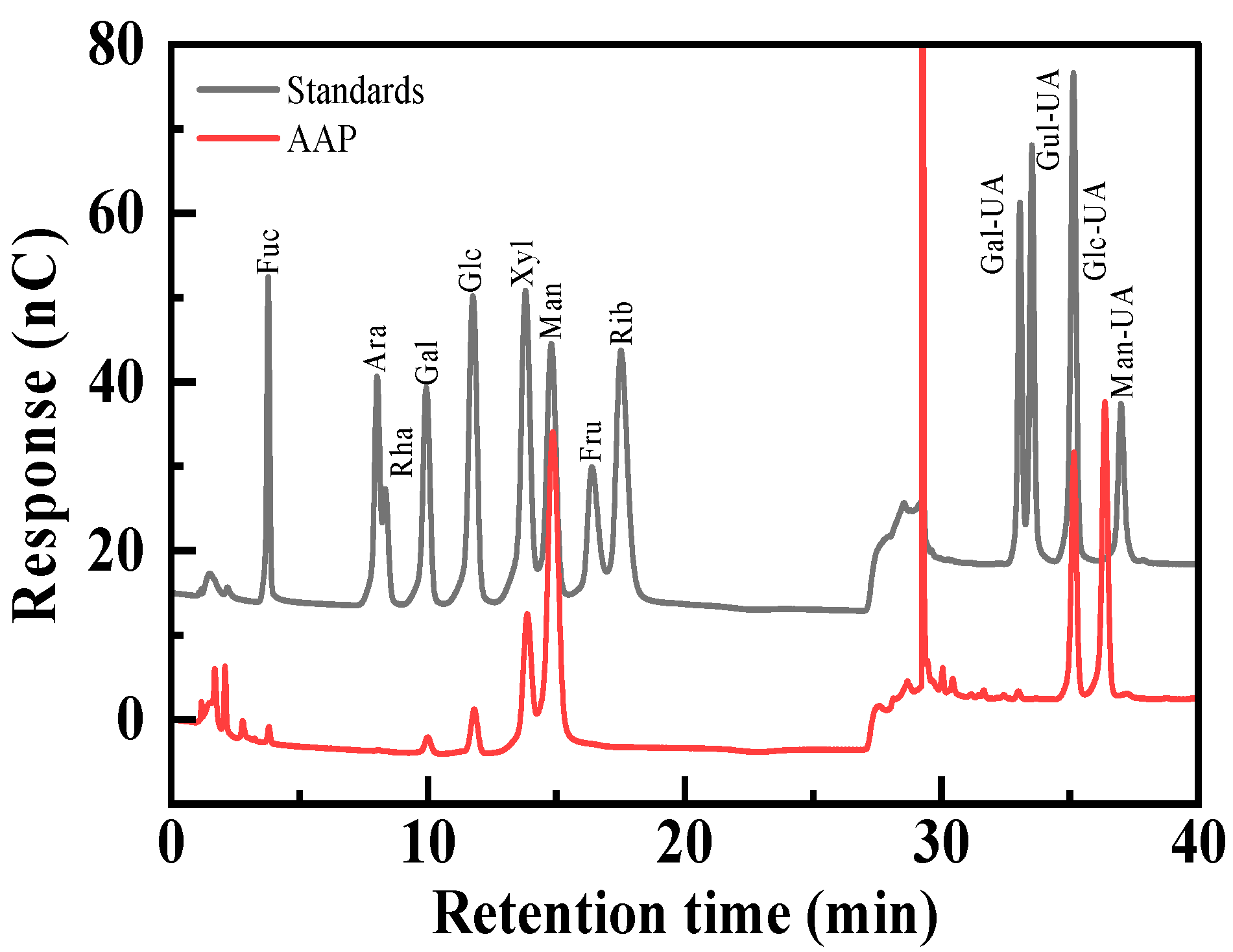
2.2. Preliminary Characterization of AAP

The molecular weight of AAP was determined by high performance gel permeation chromatography (HPGPC) using a Waters HPLC system (Waters Corporation, MA, USA) with tandem columns of UlyrahydrogelTM 500 (7.8 × 300 nm) (Waters Corporation, MA, USA), UlyrahydrogelTM 1000 (7.8 × 300 nm) (Waters Corporation, MA, USA), and UlyrahydrogelTM 2000 (7.8 × 300 nm) (Waters Corporation, MA, USA) matched on a series of columns and detected by differential refraction index detector (RID-2414) (Waters Corporation, MA, USA) at 40 °C. The mobile phase was 3 mM sodium acetate solution at a flow rate of 0.5 mL/min with an injection volume of 50 μL. Calibration curves were plotted using different standard dextran with known molecular weights (5.2, 23.8, 48.6, 148, and 668 kDa).
The monosaccharide composition of AAP was determined by ion chromatography. Five milligrams of AAP was weighed precisely in an ampoule, 2 mL of 2 mol/L trifluoroacetic acid was added, hydrolyzed at 121 °C for 3 h, blown dry under nitrogen, and washed with methanol for 2–3 times. Next, the sterile water was dissolved and transferred to a chromatographic vial for measurement. The monosaccharide composition was analyzed by a Thermo ICS 5000+ ion chromatography system (ICS 5000+, Thermo Fisher Scientific, MA, USA) and detected using an electrochemical detector. Chromatographic conditions: column: Dionex™ CarboPac™ PA20 (Thermo Fisher Scientific, MA, USA) (150 × 3.0 mm, 10 μm); mobile phases: A: H2O; B: 0.1 M NaOH; C: 0.1 M NaOH: 0.2 M sodium acetate (NaAC); flow rate: 0.5 mL/min; sample volume: 5 µL.

2.3. Synthesis of AAP-Au NPs

The stock solutions of 1% (w/v) of AAP, 10 mM of HAuCl4, 0.1 M of hydrochloric acid (HCl), and 0.1 M of sodium hydroxide (NaOH) were prepared in deionized water before the experiment for the synthesis of AAP-Au NPs. For optimal conditions, 3 mL of various concentrations of AAP (0.3, 0.5, 0.7, and 1% w/v) were incubated with HAuCl4 solution (3 mL, 1 mM), stirred, and heated at 85 °C for 1 h to study the effect of mass fraction of AAP on the synthesis of Au NPs. Next, the effect of pH on the formation of Au NPs was investigated by adjusting the reaction mixture to pH 3, 5, 7, 9, 11 using 0.1 M HCl and NaOH. Subsequently, the effect of the initial HAuCl4 concentration was determined by changing the concentration of HAuCl4 to 0.25, 0.5, 1, 2, and 3 mM, and the effect of the reaction temperature (55, 85, and 95 °C) and the reaction time (0.5, 1, 1.5, and 2 h) was further assessed. Only one condition was changed in each experiment.

2.4. Characterization of AAP-Au NPs

The absorption peaks of AAP-Au NPs were detected using an ultraviolet spectrophotometer (UV–vis) (Mapped, Shanghai, China), TEM images of AAP-Au NPs were obtained using a field emission transmission electron microscopy (Thermo, MA, USA), zeta potential maps of AAP-Au NPs were obtained using a zeta potentiometer (Malvern, UK), the reaction process of AAP-Au NPs was explored using a Fourier transform infrared spectrometer (FT-IR) (Bruker, Germany), and XPS maps of AAP and AAP-Au NPs were recorded using an X-ray photoelectron spectrometer (XPS) (Thermo, MA, USA).

2.5. The Stability Analysis of AAP-Au NPs

The storage stability of AAP-Au NPs was explored by storing the solutions at 4 °C. To clarify the effect of acidic or alkaline conditions, the pH of AAP-AuNPs solutions was adjusted from 3 to 11 by HCl and NaOH. The effect of temperature and salt ion solutions on the stability of AAP-Au NPs was investigated by exposing them to temperatures ranging from 25 to 60 °C and 5 mmol/L glutathione or various ionic (Na+, K+) environments.

2.6. The Antioxidant Properties of AAP-Au NPs

The 2,2′-azino-bis-(3-ethylbenzthiazoline-6-sulphonic acid) (ABTS) and 2,2-diphenyl-1-picrylhydrazyl (DPPH) radicals scavenging activities of AAP-Au NPs were evaluated using the method described by Pu et al. [26] with slight modification. Vitamin C (Vc) (Shanghai Yuanye Biotechnology Co., Ltd. Shanghai, China) was used as a positive control.

3. Results and Discussion

3.1. Optimization of Conditions for the Green Synthesis of AAP-Au NPs

The results of ion chromatography showed that AAP is a heteropolysaccharide consisting mainly of mannose (56.96%), xylose (13.43%), and glucuronic acid (20.66%) (Figure 1) with a molecular weight of 1815 kDa (Figure S1 in Supplementary Materials). This indicates that AAP consists of reducing monosaccharides and can be used as a template for the synthesis of Au NPs [27,28]. Notably, uronic acid can be used to maintain the stability of Au NPs [29,30]. Therefore, to obtain highly stable, high-yield, monodisperse Au NPs, the effects of mass fraction of AAP, pH, reaction temperature and time, and concentration of gold precursor on the synthesis of AAP-Au NPs were systematically investigated.

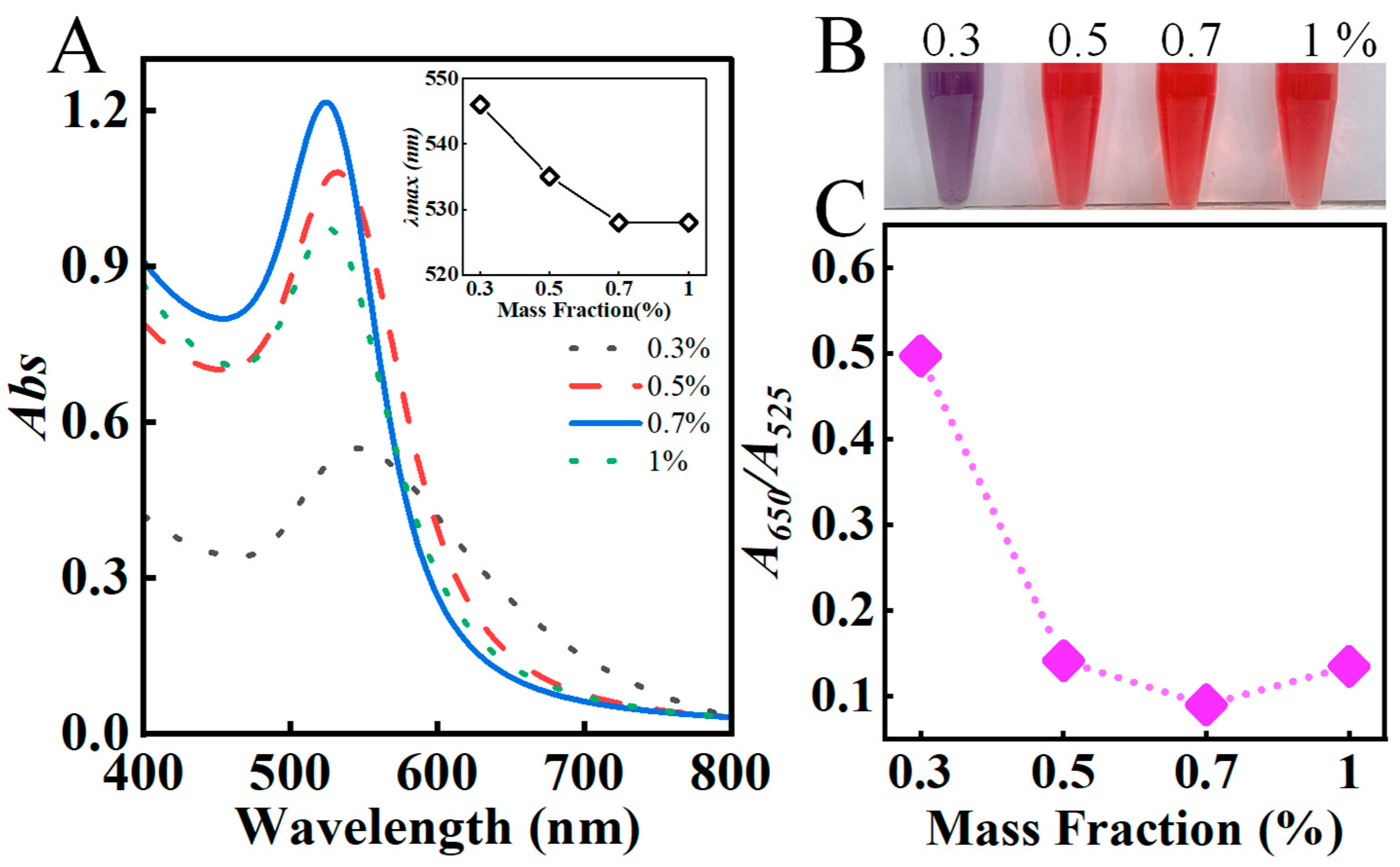
3.1.1. Effect of Various Mass Fractions of AAP

UV–vis spectroscopy is commonly employed to characterize Au NPs [31]. The surface plasmon resonance (SPR) of Au NPs arises from the collective oscillation of the surface electron cloud, which is responsible for the coloration of the solution [32]. Additionally, the peak position, spectral bandwidth, and intensity of the SPR provide insights into the size, shape, and yield of Au NPs [33]. Therefore, the synthesis of Au NPs can be assessed through the UV–vis spectrum and the color of the colloidal solution.
The mass fraction of polysaccharides is a key parameter affecting the morphology and size of Au NPs [34]. As the AAP mass fraction increased from 0.3% to 0.7% (Figure 2A), the SPR intensity of Au NPs increased, with a blue shift of the peak position, the half-peak width gradually narrowed, and the solution color changed from purple to burgundy (Figure 2B), indicating the size of the generated Au NPs gradually decreased. However, when the mass fraction of AAP was higher than 0.7%, the SPR intensity of Au NPs decreased due to the steric effect of AAP [35]. Generally, the SPR at 525 and 650 nm was related to the number of dispersed Au NPs (red) and aggregated Au NPs (purple), respectively. Therefore, the dispersion of as-prepared Au NPs was expressed by the ratio of extinction coefficients A650/A525 [36]. When the mass fraction was 0.7%, the A650/A525 value of Au NPs was the lowest (Figure 2C), indicating the synthesized AAP-Au NPs possess excellent stability and dispersion. Consequently, 0.7% of AAP was selected for the subsequent preparation of AAP-Au NPs.

3.1.2. Effect of pH

The pH value significantly influences the morphology, size, surface charge, and other properties of Au NPs [37]. When the pH was 3, the solution was purple-red (Figure 3B), indicating that larger particle-size Au NPs have been synthesized. At pH 5, the solution turned wine red, with the SPR peak of Au NPs observed at 525 nm and a narrow half-peak width, suggesting a uniform particle size distribution. As the pH continued to increase, the solution color darkened, and the maximum absorption peak of Au NPs remained around 525 nm (Figure 3). However, the absorbance gradually decreased, indicating a lower yield of Au NPs. This may be because as the pH increases, the excess O H in the solution is adsorbed on the surface of the Au clusters, hindering the formation of Au NPs from the clusters [38]. On the other hand, the excess of NaOH leads to the substitution of C l by O H in A u C l 4 to form Au(OH)3, which is ultimately converted to sodium aureate (NaAuO2) decreasing the formation of Au NPs [39,40]. Since the absorbance value of Au NPs was the highest and A650/A525 was the smallest at pH 5 (Figure 3C), it was used as the optimal pH value.

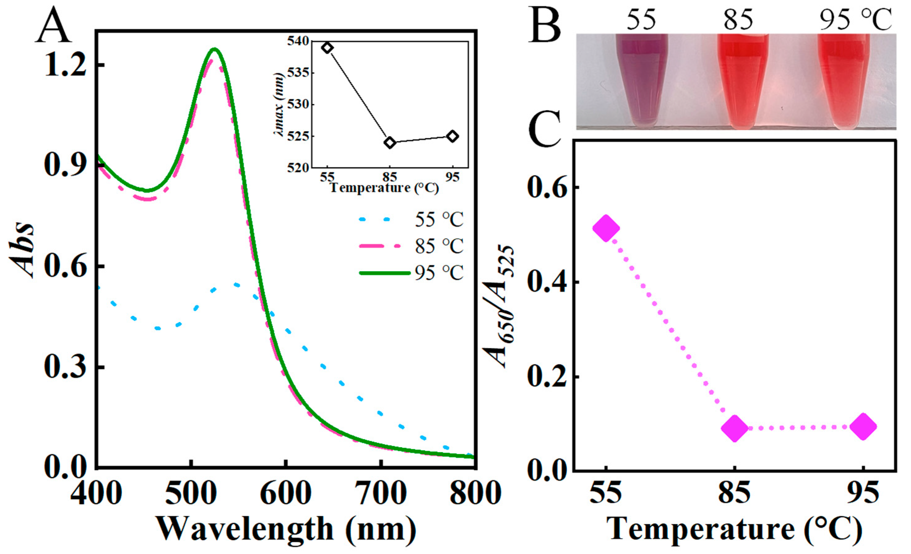
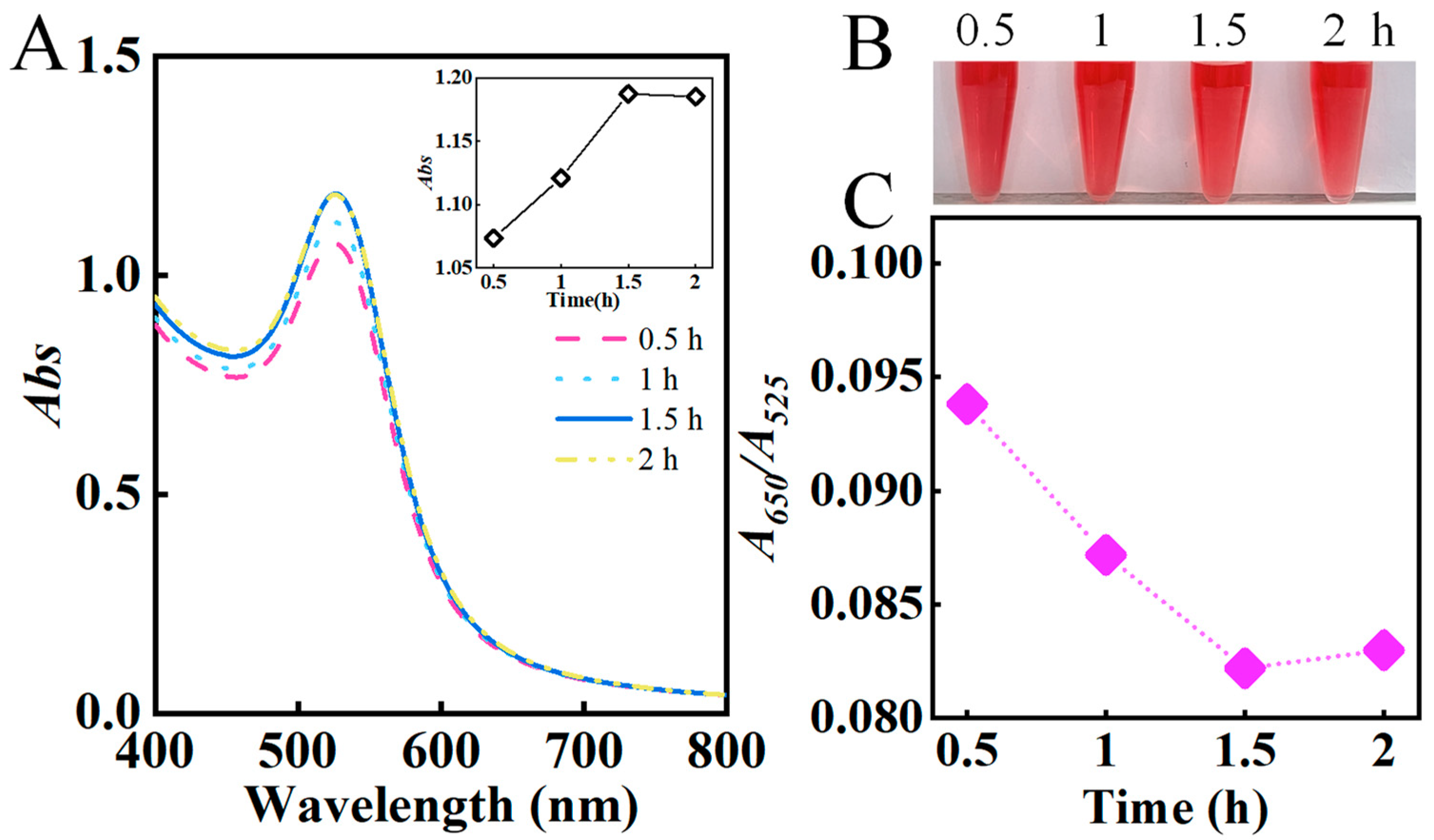
3.1.3. Effect of Reaction Temperature and Time

Temperature is a critical parameter affecting the particle size of Au NPs. When the temperature increased from 55 °C to 95 °C, the solution color changed from purple to wine red (Figure 4B), and the SPR showed a blue shift and intensity increased (Figure 4A), indicating that the particle size of Au NPs became smaller and the yield increased. The A650/A525 was minimized at 85 °C, indicating better dispersion of the synthesized AAP-Au NPs (Figure 4C). Time is also an important factor in synthesizing Au NPs [41]. When the reaction time increased from 0.5 to 2 h, the position of the Au NPs absorption peak always remained at 525 nm, while the intensity of the absorption peak first increased and then remained unchanged (Figure 5A). This was because the continued reduction of Au(III) in the early stage of the reaction increased the number of Au NPs, indicating that the reaction had not yet been completed. When the reaction time reached 1.5 h, the absorption peak’s intensity remained unchanged (Figure 5B), indicating the complete reduction reaction. In addition, the A650/A525 was the smallest when the reaction was carried out for 1.5 h (Figure 5C) and was selected as the optimal reaction time for subsequent experiments.

3.1.4. Effect of Concentrations of Gold Precursor

The reduction kinetics of A u 3 + , nucleation, and growth rate of Au NPs highly depend on the concentration of gold precursors [42]. When the concentrations of gold precursors were 0.25 and 0.5 mmol/L, respectively, the solution was almost colorless and the SPR intensity was low, indicating the generation of small amounts of Au NPs (Figure 6A). When the concentration increased to 1 mmol/L, the SPR was the highest and the A650/A525 value was the smallest (Figure 6C), indicating that Au NPs with uniform particle size distribution were generated. However, the solution turned purple at a concentration of 2 mmol/L (Figure 6B), indicating that Au NPs with larger particle sizes were formed. Further increasing the concentration to 3 mmol/L resulted in a significant red shift in the SPR and a marked decrease in intensity. This was because a large amount of Au(III) in the solution was reduced to Au(0), resulting in the inability of AAP to prevent the aggregation and precipitation of Au NPs. These results demonstrated that the concentration of gold precursor affects the size of Au NPs and determines their yield and stability in solution. Therefore, the optimal concentration of gold precursor was established to be 1 mmol/L.

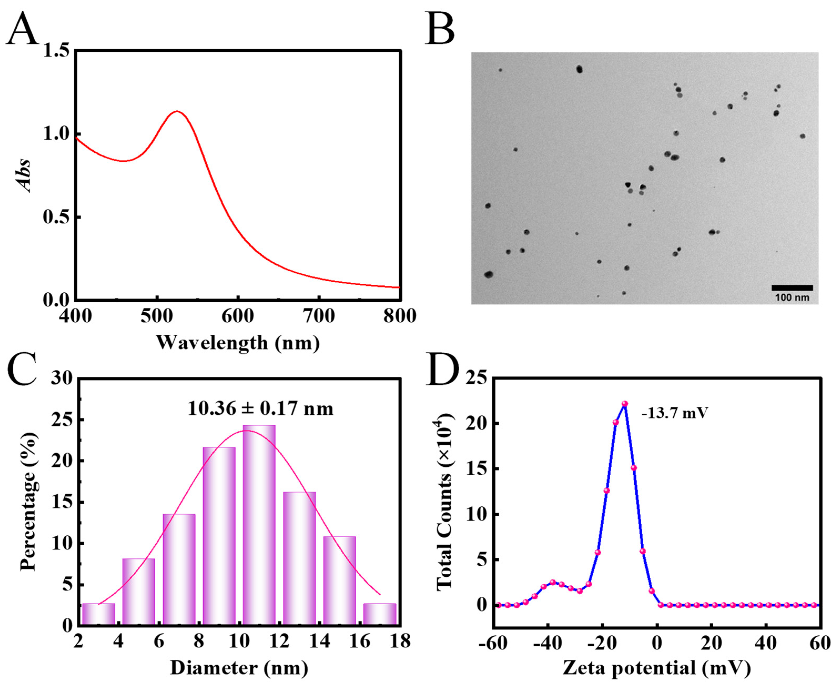
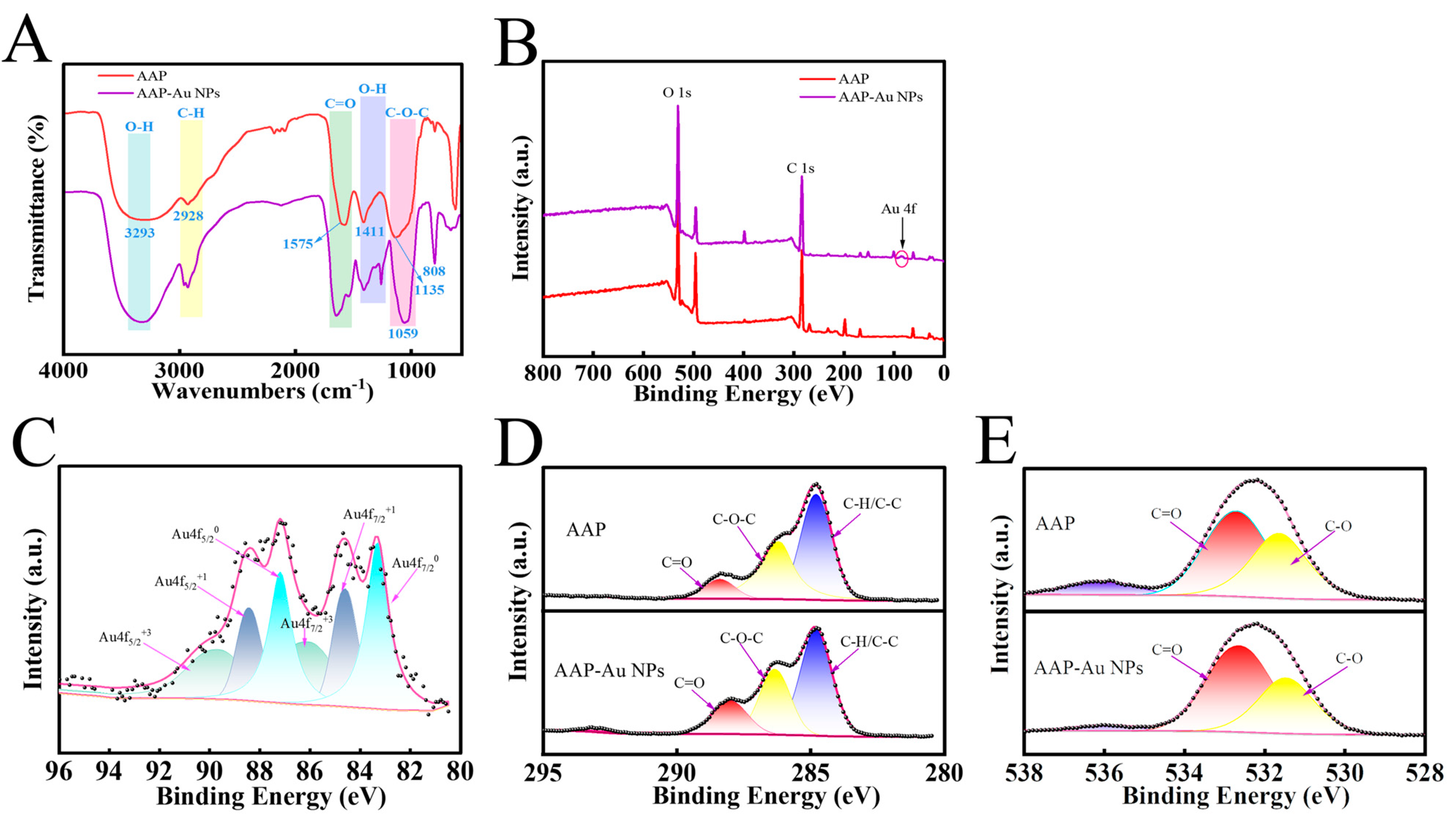
3.2. The Structural and Morphological Characterisation of AAP-Au NPs

AAP-Au NPs were prepared under the optimal reaction conditions (0.7% of AAP, 1.0 mmol/L of HAuCl4, pH 5, 85 °C, 1.5 h). As shown in Figure 7A, the absorption peak of the AAP-Au NPs was at 525 nm with a narrow half-peak width, which indicated that monodisperse spherical AAP-Au NPs were successfully synthesized. The TEM images further confirmed that AAP-Au NPs were spherical (Figure 7B), with a particle size distribution in the range of 2–18 nm (the average size was 10.36 ± 0.17 nm) (Figure 7C), and the polydispersity index (PDI) was 0.249 ± 0.015 (Figure S2 in Supplementary Materials), which indicated the excellent dispersion of AAP-Au NPs. Furthermore, the zeta potential of AAP-Au NPs was −13.7 mV (Figure 7D). Ultimately, the AAP-Au NPs were kept stable via electrostatic interactions and steric forces [43,44,45].

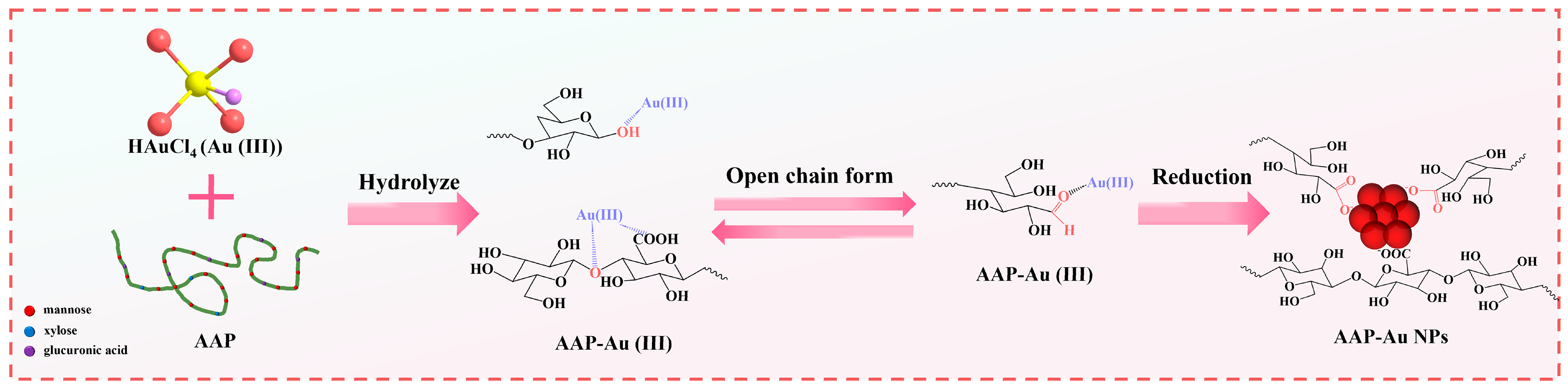
3.3. Synthesis Mechanism of AAP-Au NPs

To gain a deeper understanding of the synthesis mechanism of AAP-Au NPs, the generation of interaction sites and bonding environments between AAP and Au NPs was investigated. The FT-IR spectroscopy was used to preliminarily explore the reaction process of AAP-Au NPs (Figure 8A). AAP-Au NPs retained the key FT-IR peaks of polysaccharides (e.g., -OH, -CH, -C=O, and C-O-C). Notably, the relatively strong absorption bands were observed at 1647 and 1410 cm−1 (Figure 8A, purple line), consistent with the carboxylate (Au- C O O ) groups. Moreover, XPS was used to further characterize the changes in the chemosynthetic state during the prepared AAP-Au NPs. XPS spectra of AAP-Au NPs clearly showed the occurrence of relevant peaks for C, O, and Au (Figure 8B,C). The binding energy positions of the relevant peaks in the C1s and O1s spectra of the AAP-Au NPs were nearly the same compared with AAP (Figure 8D,E, Table 1 and Table 2). Nevertheless, the contents of C1s and O1s-related functional groups changed before and after the synthesis of AAP-Au NPs (Table 1 and Table 2). The results showed that after the synthesis of AAP-Au NPs, the C=O content in the C-related peak increased from 9.44% to 18.67%, and the C=O content in the O-related peak increased from 57.71% to 63.18%. In addition, the contents of C-O-C/C-OH in the C-related peak and C-O in the O-related peak decreased from 38.64% and 42.29% to 30.8% and 36.82%, respectively, indicating that there was a charge transfer between Au and O.
Based on these results, the synthesis mechanism of AAP-Au NPs was proposed (Figure 9): the Au(III) ions act as a strong Lewis acid, actively promoting intra- or intermolecular attacks of a nucleophile and helping break the glycosidic bond to expose the reduction terminus, which reduced Au(III) to Au(0), and itself was oxidized to carboxylate compounds. In addition, the carboxylate anions ( C O O ) self-assemble on the Au NPs to form a negatively charged surface, acting as a stabilizer to passivate the Au NPs to prevent particle aggregation, which was confirmed via zeta potential of the AAP-Au NPs.

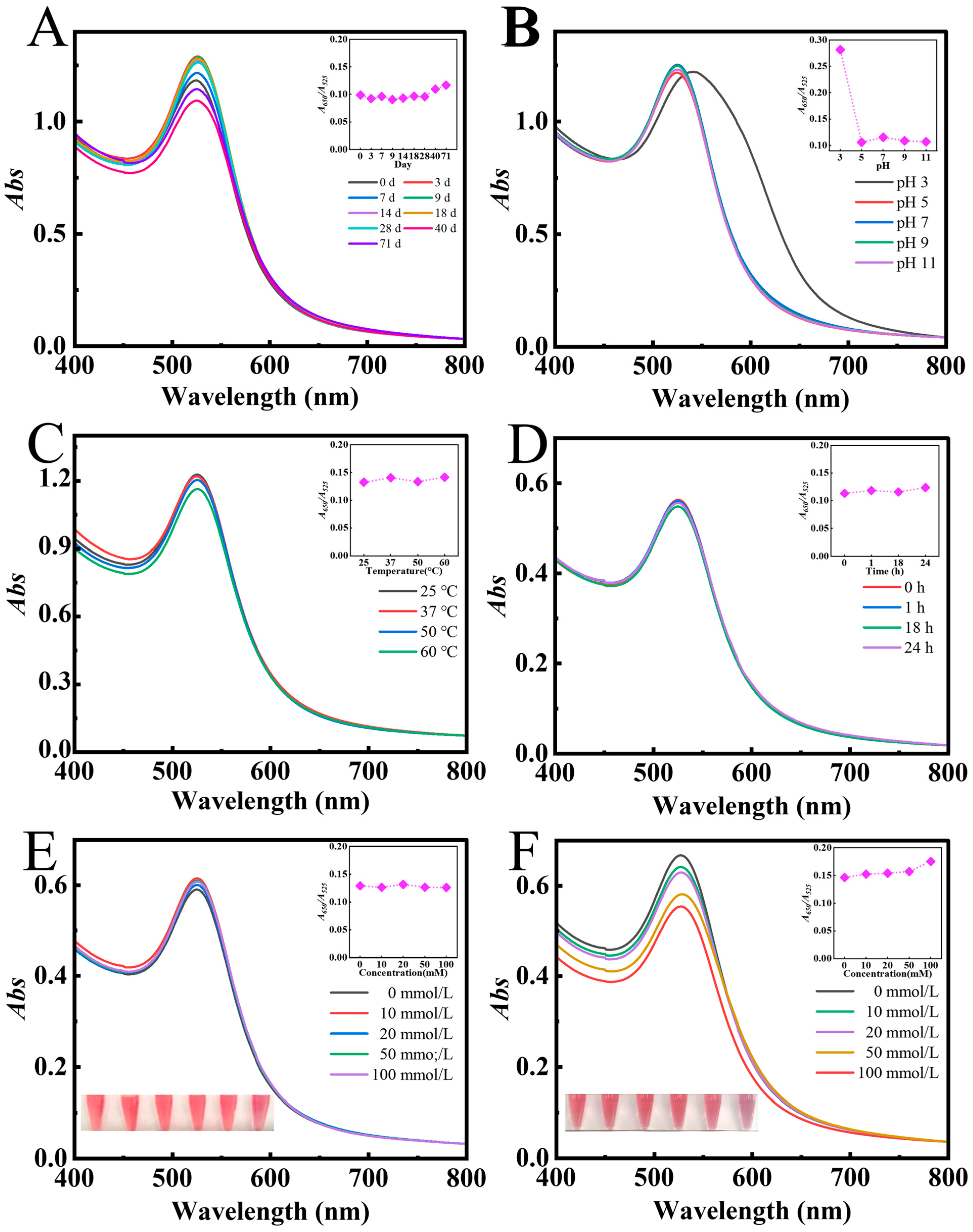
3.4. The Stability of AAP-Au NPs

Au NPs are commonly used for disease treatment via oral or intravenous administration. Thus, exposure to acidic media or complex physiological media consisting of high concentrations of salts and various proteins is inevitable. These conditions usually cause Au NPs to aggregate and deteriorate their biological activity [46]. Therefore, the stability of AAP-Au NPs in various external complex conditions was investigated.
As shown in Figure 10A, when AAP-Au NPs were placed at 4 °C for 71 d, the UV–vis spectroscopy and the ratio of A650/A525 remained unchanged, indicating that AAP-Au NPs had good storage stability. pH is one of the critical factors affecting the stability of Au NPs. At pH 3, the SPR of AAP-Au NPs was from 525 nm red-shifted to 542 nm, and the width at half maximum increased slightly, indicating that AAP-Au NPs aggregated under acidic conditions (Figure 10B). As the pH increased from 5 to 11, the intensity and peak position of the absorption peaks remained unchanged, and the absolute value of the zeta potential of AAP-Au NPs became gradually higher with the increase in pH (Figure S3 in Supplementary Materials), indicating that AAP-Au NPs had good stability in slightly acidic, neutral, and alkaline environments. Additionally, whether temperature affects the stability of Au NPs was investigated. As shown in Figure 10C, the absorption peaks of AAP-Au NPs remained consistent under 25–60 °C conditions, indicating that AAP-Au NPs had good temperature tolerance. On the other hand, glutathione, which is widely present in various cells, can bind to Au NPs through the sulfhydryl group, causing the aggregation of Au NPs [47]. When AAP-Au NPs were mixed with 5 mmol/L glutathione solution, the absorption peak remained unchanged (Figure 10D), indicating that AAP-Au NPs had good stability in glutathione solution. Furthermore, AAP-Au NPs could still maintain good stability in high concentrations of metal ions (Na+, K+) (Figure 10E,F), which was beneficial for expanding Au NPs applications in biomedicine.

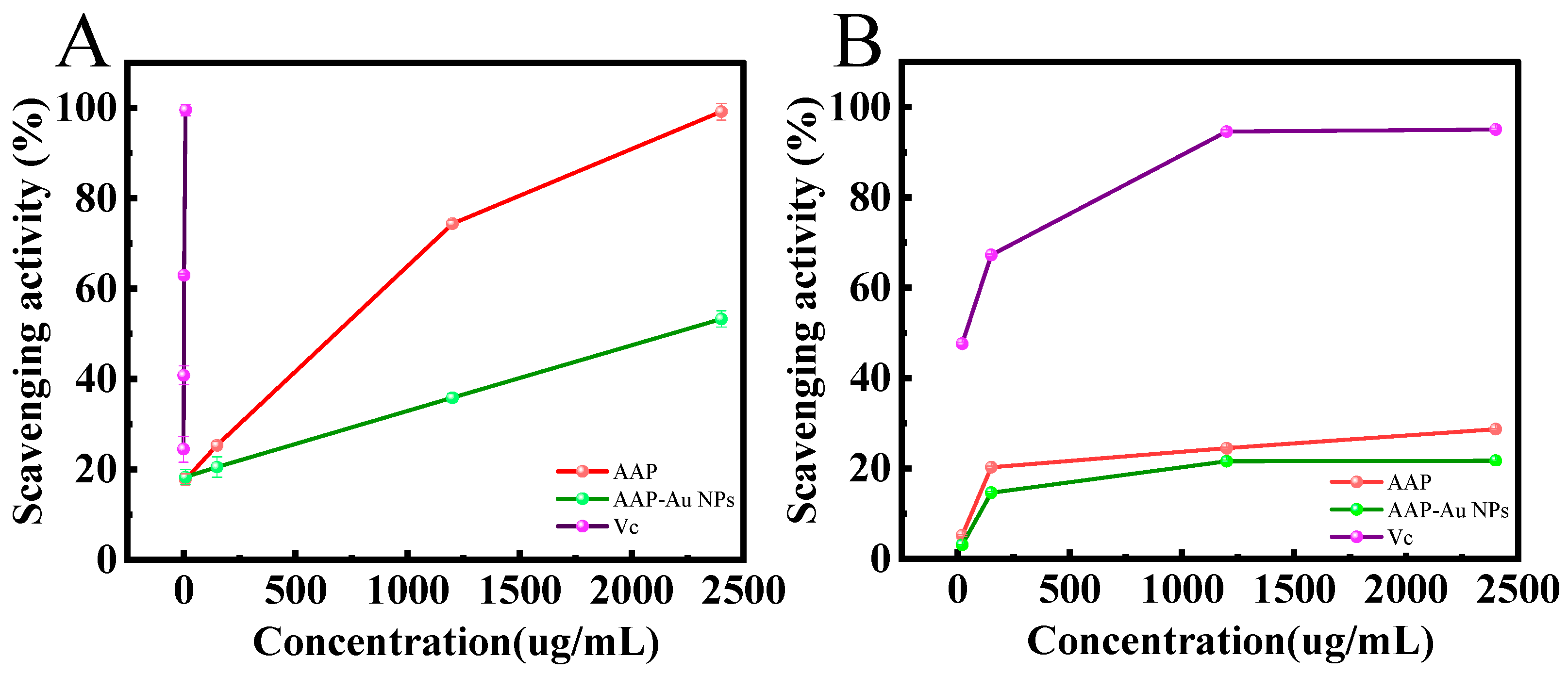
3.5. Scavenging Activity of AAP-Au NPs on ABTS and DPPH Free Radicals

It is well known that removing excess free radicals is essential for maintaining biological homeostasis. Therefore, the scavenging activity of AAP-Au NPs on ABTS and DDPH free radicals was investigated. The ABTS radical scavenging activity of AAP-Au NPs increased from 18.29% to 53.29% with increasing concentration (Figure 11A), indicating that AAP-Au NPs retained excellent antioxidant properties, but were weaker than AAP (99.18%). In addition, the AAP-Au NPs also exhibited a certain scavenging activity against DPPH radicals (Figure 11B). These results indicated that as-prepared AAP-Au NPs have good antioxidant activity.

4. Conclusions

In summary, monodisperse spherical Au NPs were successfully prepared by a straightforward method using AAP as both a reducing and stabilizing agent. By optimizing the conditions (mass fraction of AAP, pH, reaction temperature, reaction time, and concentration of gold precursor), AAP-Au NPs with an average particle size of 10.36 ± 0.17 nm were prepared, with a localized surface plasmon resonance absorption peak position of 525 nm and a zeta potential value of −13.7 mV, and the mechanism of action of preparing AAP-Au NPs was proposed. Furthermore, as-prepared AAP-Au NPs exhibited good storage stability and excellent tolerance to acid–base solutions and salt-ion solutions. In addition, AAP-Au NPs retained the scavenging abilities of ABTS and DPPH free radicals. This study not only determined the method of economic green synthesis of Au NPs but also expanded the utilization of AAP. Furthermore, based on the biological activity of AAP and the unique properties of Au NPs, AAP-Au NPs have potential applications in fields such as biomedicine and cosmetics.

Supplementary Materials

The following supporting information can be downloaded at: https://www.mdpi.com/article/10.3390/polysaccharides5040041/s1, Figure S1: The molecular weight of AAP; Figure S2: The hydrodynamic size of AAP-Au NPs; Figure S3: Zeta potential of AAP-Au NPs as a function of pH.

Author Contributions

Conceptualization, H.L.; software, Y.Y.; validation, L.G. and Y.Y.; investigation, L.G.; data curation, H.L.; writing—original draft, H.L. and L.G.; writing—review and editing, M.Z.; supervision, M.Z.; funding acquisition, M.Z. All authors have read and agreed to the published version of the manuscript.

Funding

This work was supported by the Science and Technology Young Top-notch Talent Project of the Autonomous Region (2022TSYCCX0064), the China Postdoctoral Science Foundation (2023M732962), and the Autonomous Region Universities Basic Research Funds research projects-cultivation projects (XJEDU2023P016).

Institutional Review Board Statement

Not applicable.

Data Availability Statement

Data are contained within the article and Supplementary Materials.

Conflicts of Interest

The authors declare no conflicts of interest.

References

  1. Zhang, M.; Shao, S.; Yue, H.; Wang, X.; Zhang, W.; Chen, F.; Zheng, L.; Xing, J.; Qin, Y. High stability Au NPs: From design to application in nanomedicine. Int. J. Nanomed. 2021, 16, 6067–6094. [Google Scholar] [CrossRef] [PubMed]
  2. Nasaruddin, R.R.; Chen, T.; Yao, Q.; Zang, S.; Xie, J. Toward greener synthesis of gold nanomaterials: From biological to biomimetic synthesis. Coord. Chem. Rev. 2021, 426, 213540. [Google Scholar] [CrossRef]
  3. Xu, L.; Xu, M.; Sun, X.; Feliu, N.; Feng, L.; Parak, W.J.; Liu, S. Quantitative comparison of gold nanoparticle delivery via the enhanced permeation and retention (EPR) effect and mesenchymal stem cell (MSC)-based targeting. ACS Nano 2023, 17, 2039–2052. [Google Scholar] [CrossRef] [PubMed]
  4. Pooja, D.; Panyaram, S.; Kulhari, H.; Reddy, B.; Rachamalla, S.S.; Sistla, R. Natural polysaccharide functionalized gold nanoparticles as biocompatible drug delivery carrier. Int. J. Biol. Macromol. 2015, 80, 48–56. [Google Scholar] [CrossRef] [PubMed]
  5. Sarkar, A.; Roy, S.; Sanpui, P.; Jaiswal, A. Plasmonic gold nanorattle impregnated chitosan nanocarrier for stimulus responsive theranostics. ACS Appl. Biol. Mater. 2019, 2, 4812–4825. [Google Scholar] [CrossRef]
  6. Xu, W.; Qian, J.; Hou, G.; Wang, Y.; Wang, J.; Sun, T.; Ji, L.; Suo, A.; Yao, Y. A dual-targeted hyaluronic acid-gold nanorod platform with triple-stimuli responsiveness for photodynamic/photothermal therapy of breast cancer. Acta Biomater. 2019, 83, 400–413. [Google Scholar] [CrossRef]
  7. Khan, M.S.; Vishakante, G.D.; Siddaramaiah, H. Gold nanoparticles: A paradigm shift in biomedical applications. Adv. Colloid Interface Sci. 2013, 199–200, 44–58. [Google Scholar] [CrossRef]
  8. Mzwd, E.; Ahmed, N.M.; Suradi, N.; Alsaee, S.K.; Altowyan, A.S.; Almessiere, M.A.; Omar, A.F. Green synthesis of gold nanoparticles in Gum Arabic using pulsed laser ablation for CT imaging. Sci. Rep. 2022, 12, 10549. [Google Scholar] [CrossRef]
  9. Iranpour, P.; Ajamian, M.; Safavi, A.; Iranpoor, N.; Abbaspour, A.; Javanmardi, S. Synthesis of highly stable and biocompatible gold nanoparticles for use as a new X-ray contrast agent. J. Mater. Sci. Mater. Med. 2018, 29, 48. [Google Scholar] [CrossRef]
  10. He, J.; Unser, S.; Bruzas, I.; Cary, R.; Shi, Z.; Mehra, R.; Aron, K.; Sagle, L. The facile removal of CTAB from the surface of gold nanorods. Colloids Surf. B 2018, 163, 140–145. [Google Scholar] [CrossRef]
  11. Gevaerd, A.; Caetano, F.R.; Oliveira, P.R.; Zarbin, A.J.G.; Bergamini, M.F.; Marcolino-Junior, L.H. Thiol-capped gold nanoparticles: Influence of capping amount on electrochemical behavior and potential application as voltammetric sensor for diltiazem. Sens. Actuators B: Chem. 2015, 220, 673–678. [Google Scholar] [CrossRef]
  12. Zeng, Y.; Xiang, Y.; Sheng, R.; Tomas, H.; Rodrigues, J.; Gu, Z.; Zhang, H.; Gong, Q.; Luo, K. Polysaccharide-based nanomedicines for cancer immunotherapy: A review. Bioact. Mater. 2021, 6, 3358–3382. [Google Scholar] [CrossRef] [PubMed]
  13. Liu, Q.; Duan, B.; Xu, X.; Zhang, L. Progress in rigid polysaccharide-based nanocomposites with therapeutic functions. J. Mater. Chem. B 2017, 5, 5690–5713. [Google Scholar] [CrossRef] [PubMed]
  14. Fan, Y.; Liu, Y.; Wu, Y.; Dai, F.; Yuan, M.; Wang, F.; Bai, Y.; Deng, H. Natural polysaccharides based self-assembled nanoparticles for biomedical applications–A review. Int. J. Biol. Macromol. 2021, 192, 1240–1255. [Google Scholar] [CrossRef] [PubMed]
  15. Manivasagan, P.; Oh, J. Marine polysaccharide-based nanomaterials as a novel source of nanobiotechnological applications. Int. J. Biol. Macromol. 2016, 82, 315–327. [Google Scholar] [CrossRef]
  16. Chen, X.; Zhao, X.; Wang, G. Review on marine carbohydrate-based gold nanoparticles represented by alginate and chitosan for biomedical application. Carbohydr. Polym. 2020, 244, 116311. [Google Scholar] [CrossRef]
  17. Ahmed, H.B. Recruitment of various biological macromolecules in fabrication of gold nanoparticles: Overview for preparation and applications. Int. J. Biol. Macromol. 2019, 140, 265–277. [Google Scholar] [CrossRef]
  18. Liu, H.; Zhang, M.; Meng, F.; Su, C.; Li, J. Polysaccharide-based gold nanomaterials: Synthesis mechanism, polysaccharide structure-effect, and anticancer activity. Carbohydr. Polym. 2023, 321, 121284. [Google Scholar] [CrossRef]
  19. Lyu, F.; Xu, X.; Zhang, L. Natural polysaccharides with different conformations: Extraction, structure and anti-tumor activity. J. Mater. Chem. B 2020, 8, 9652–9667. [Google Scholar] [CrossRef]
  20. Tao, Y.; Zhang, R.; Yang, W.; Liu, H.; Yang, H.; Zhao, Q. Carboxymethylated hyperbranched polysaccharide: Synthesis, solution properties, and fabrication of hydrogel. Carbohydr. Polym. 2015, 128, 179–187. [Google Scholar] [CrossRef]
  21. Tomofuji, Y.; Matsuo, K.; Terao, K. Kinetics of denaturation and renaturation processes of double-stranded helical polysaccharide, xanthan in aqueous sodium chloride. Carbohydr. Polym. 2022, 275, 118681. [Google Scholar] [CrossRef] [PubMed]
  22. Chen, N.; Zhang, H.; Zong, X.; Li, S.; Wang, J.; Wang, Y.; Jin, M. Polysaccharides from Auricularia auricula: Preparation, structural features and biological activities. Carbohydr. Polym. 2020, 247, 116750. [Google Scholar] [CrossRef] [PubMed]
  23. Bao, H.; Zhou, R.; You, S.; Wu, S.; Wang, Q.; Cui, S.W. Gelation mechanism of polysaccharides from Auricularia auricula-judae. Food Hydrocoll. 2018, 76, 35–41. [Google Scholar] [CrossRef]
  24. Xu, S.; Zhang, Y.; Jiang, K. Antioxidant activity in vitro and in vivo of the polysaccharides from different varieties of Auricularia auricula. Food Funct. 2016, 7, 3868–3879. [Google Scholar] [CrossRef] [PubMed]
  25. Zhou, Y.; Jia, Y.; Xu, N.; Tang, L.; Chang, Y. Auricularia auricula-judae (Bull.) polysaccharides improve obesity in mice by regulating gut microbiota and TLR4/JNK signaling pathway. Int. J. Biol. Macromol. 2023, 250, 126172. [Google Scholar] [CrossRef] [PubMed]
  26. Pu, S.; Li, J.; Sun, L.; Zhong, L.; Ma, Q. An in vitro comparison of the antioxidant activities of chitosan and green synthesized gold nanoparticles. Carbohydr. Polym. 2019, 211, 161–172. [Google Scholar] [CrossRef]
  27. Jia, X.; Yao, Y.; Yu, G.; Qu, L.; Li, T.; Li, Z.; Xu, C. Synthesis of gold-silver nanoalloys under microwave-assisted irradiation by deposition of silver on gold nanoclusters/triple helix glucan and antifungal activity. Carbohydr. Polym. 2020, 238, 116169. [Google Scholar] [CrossRef]
  28. Snitka, V.; Naumenko, D.O.; Ramanauskaite, L.; Kravchenko, S.A.; Snopok, B.A. Generation of diversiform gold nanostructures inspired by honey’s components: Growth mechanism, characterization, and shape separation by the centrifugation-assisted sedimentation. J. Colloid Interface Sci. 2012, 386, 99–106. [Google Scholar] [CrossRef]
  29. Suganya, K.S.; Govindaraju, K.; Kumar, V.G.; Karthick, V.; Parthasarathy, K. Pectin mediated gold nanoparticles induces apoptosis in mammary adenocarcinoma cell lines. Int. J. Biol. Macromol. 2016, 93, 1030–1040. [Google Scholar] [CrossRef]
  30. Cisneros, J.S.; Chain, C.Y.; Rivas Aiello, M.B.; Parisi, J.; Castrogiovanni, D.C.; Bosio, G.N.; Martire, D.O.; Vela, M.E. Pectin-coated plasmonic nanoparticles for photodynamic therapy: Inspecting the role of serum proteins. ACS Omega 2021, 6, 12567–12576. [Google Scholar] [CrossRef]
  31. Ghosh, S.K.; Pal, T. Interparticle coupling effect on the surface plasmon resonance of gold nanoparticles: From theory to applications. Chem. Rev. 2007, 107, 4797–4862. [Google Scholar] [CrossRef] [PubMed]
  32. Noruzi, M.; Zare, D.; Khoshnevisan, K.; Davoodi, D. Rapid green synthesis of gold nanoparticles using Rosa hybrida petal extract at room temperature. Spectrochim. Acta A 2011, 79, 1461–1465. [Google Scholar] [CrossRef] [PubMed]
  33. Jain, P.K.; Lee, K.S.; El-Sayed, I.H.; El-Sayed, M.A. Calculated absorption and scattering properties of gold nanoparticles of different size, shape, and composition: Applications in biological imaging and biomedicine. J. Phys. Chem. B 2006, 110, 7238–7248. [Google Scholar] [CrossRef] [PubMed]
  34. Huang, L.; Zhai, M.; Peng, J.; Xu, L.; Li, J.; Wei, G. Synthesis, size control and fluorescence studies of gold nanoparticles in carboxymethylated chitosan aqueous solutions. J. Colloid Interface Sci. 2007, 316, 398–404. [Google Scholar] [CrossRef]
  35. Laksee, S.; Puthong, S.; Teerawatananond, T.; Palaga, T.; Muangsin, N. Highly efficient and facile fabrication of monodispersed Au nanoparticles using pullulan and their application as anticancer drug carriers. Carbohydr. Polym. 2017, 178, 178–191. [Google Scholar] [CrossRef]
  36. Pienpinijtham, P.; Thammacharoen, C.; Ekgasit, S. Green synthesis of size controllable and uniform gold nanospheres using alkaline degradation intermediates of soluble starch as reducing agent and stabilizer. Macromol. Res. 2012, 20, 1281–1288. [Google Scholar] [CrossRef]
  37. Chen, X.; Xue, Z.; Ji, J.; Wang, D.; Shi, G.; Zhao, L.; Feng, S. Hedysarum polysaccharides mediated green synthesis of gold nanoparticles and study of its characteristic, analytical merit, catalytic activity. Mater. Res. Bull. 2021, 133, 111070. [Google Scholar] [CrossRef]
  38. Kumar, S.; Yoon, S.H.; Kim, G.H. Bridging the nanogap electrodes with gold nanoparticles using dielectrophoresis technique. Curr. Appl. Phys. 2009, 9, 101–103. [Google Scholar] [CrossRef]
  39. Muangnapoh, T.; Sano, N.; Yusa, S.I.; Viriya-Empikul, N.; Charinpanitkul, T. Facile strategy for stability control of gold nanoparticles synthesized by aqueous reduction method. Curr. Appl. Phys. 2010, 10, 708–714. [Google Scholar] [CrossRef]
  40. Mironov, I.V.; Makotchenko, E.V. The Hydrolysis of AuCl4 and the stability of aquachlorohydroxocomplexes of gold(III) in aqueous solution. J. Solut. Chem. 2009, 38, 725–737. [Google Scholar] [CrossRef]
  41. Sun, L.; Pu, S.; Li, J.; Cai, J.; Zhou, B.; Ren, G.; Ma, Q.; Zhong, L. Size controllable one step synthesis of gold nanoparticles using carboxymethyl chitosan. Int. J. Biol. Macromol. 2019, 122, 770–783. [Google Scholar] [CrossRef] [PubMed]
  42. Pestov, A.; Nazirov, A.; Modin, E.; Mironenko, A.; Bratskaya, S. Mechanism of Au(III) reduction by chitosan: Comprehensive study with 13C and 1H NMR analysis of chitosan degradation products. Carbohydr. Polym. 2015, 117, 70–77. [Google Scholar] [CrossRef] [PubMed]
  43. Lee, J.W.; Choi, S.R.; Heo, J.H. Simultaneous stabilization and functionalization of gold nanoparticles via biomolecule conjugation: Progress and perspectives. ACS Appl. Mater. Inter. 2021, 13, 42311–423286. [Google Scholar] [CrossRef] [PubMed]
  44. Wiącek, A.; Chibowski, E. Zeta potential, effective diameter and multimodal size distribution in oil/water emulsion. Colloid Surf. A 1999, 159, 253–261. [Google Scholar] [CrossRef]
  45. Wiącek, A.E.; Chibowski, E.; Wilk, K. Studies of oil-in-water emulsion stability in the presence of new dicephalic saccharide-derived surfactants. Colloid Surf. B 2002, 25, 243–256. [Google Scholar] [CrossRef]
  46. Demirbas, A.; Büyükbezirci, K.; Celik, C.; Kislakci, E.; Karaagac, Z.; Gokturk, E.; Kati, A.; Cimen, B.; Yilmaz, V.; Ocsoy, I. Synthesis of long-term stable gold nanoparticles benefiting from red raspberry (Rubus idaeus), strawberry (Fragaria ananassa), and blackberry (Rubus fruticosus) extracts–gold ion complexation and investigation of reaction conditions. ACS Omega 2019, 4, 18637–18644. [Google Scholar] [CrossRef]
  47. Koo, S.H.; Lee, J.S.; Kim, G.H.; Lee, H.G. Preparation, characteristics, and stability of glutathione-loaded nanoparticles. J. Agric. Food Chem. 2011, 59, 11264–11269. [Google Scholar] [CrossRef]
Figure 1. Ion chromatography of AAP.
Polysaccharides 05 00041 g001
Figure 2. (A) UV–vis absorption spectra and maximum absorption peak wavelengths plot (inset) of AAP-Au NPs at various mass fractions of AAP with (B) the corresponding solution color and (C) the absorption ratio (A650/A525).
Polysaccharides 05 00041 g002
Figure 3. (A) UV–vis absorption spectra and maximum absorption peak wavelengths plot (inset) of AAP-Au NPs at various pH values with (B) the corresponding solution color and (C) the absorption ratio (A650/A525).
Polysaccharides 05 00041 g003
Figure 4. (A) UV–vis absorption spectra and plot of the absorbance maximum at 525 nm (inset) of AAP-Au NPs at various reaction temperature with (B) the corresponding solution color and (C) the absorption ratio (A650/A525).
Polysaccharides 05 00041 g004
Figure 5. (A) UV–vis absorption spectra and maximum absorption peak wavelengths plot (inset) of AAP-Au NPs at various reaction time with (B) the corresponding solution color and (C) the absorption ratio (A650/A525).
Polysaccharides 05 00041 g005
Figure 6. (A) UV–vis absorption spectra and maximum absorption peak wavelengths plot (inset) of AAP-Au NPs at various concentrations of chloroauric acid with (B) the corresponding solution color and (C) the absorption ratio (A650/A525).
Polysaccharides 05 00041 g006
Figure 7. (A) UV–vis absorption spectra of AAP-Au NPs. TEM image (B) and histogram (C) and gaussian fitting curve (C) (red line) of the particle size distribution of AAP-Au NPs. (D) Zeta potential graph of AAP-Au NPs at pH 5.
Polysaccharides 05 00041 g007
Figure 8. (A) FT-IR spectra of AAP and AAP-Au NPs. (B) XPS fully scanned spectra of AAP and AAP-Au NPs. (C) XPS spectra of Au 4f for AAP-Au NPs. XPS spectra of C1s (D) and O1s (E) of AAP and AAP-Au NPs.
Polysaccharides 05 00041 g008
Figure 9. Reaction mechanism of preparing AAP-Au NPs.
Polysaccharides 05 00041 g009
Figure 10. UV–visible absorption spectra and the absorption ratio (A650/A525) (inset) of AAP-Au NPs under various conditions: (A) storage times at 4 °C, (B) various pH, (C) temperatures, (D) storage times in 5 mM glutathione solution, and different concentrations of (E) Na+ solution, and (F) K+ solution.
Polysaccharides 05 00041 g010
Figure 11. (A) ABTS and (B) DPPH radical scavenging activities of AAP and AAP-Au NPs.
Polysaccharides 05 00041 g011
Table 1. Percentage of each carbon-containing functional group in the XPS spectra of C1s of AAP and AAP-Au NPs.
AAPAAP-Au NPs
Peak Position (BE)Percentage (%)Peak Position (BE)Percentage (%)
C=O288.399.44288.0218.67
C-O-C/C-OH286.1638.64286.3330.8
C-H/C-C284.838.64284.850.53
Table 2. Percentage of each oxygen-containing functional group in the XPS spectra of O1s of AAP and AAP-Au NPs.
AAPAAP-Au NPs
Peak Position (BE)Percentage (%)Peak Position (BE)Percentage (%)
C=O532.7157.71532.6163.18
C-O531.6442.29531.4836.82
Disclaimer/Publisher’s Note: The statements, opinions and data contained in all publications are solely those of the individual author(s) and contributor(s) and not of MDPI and/or the editor(s). MDPI and/or the editor(s) disclaim responsibility for any injury to people or property resulting from any ideas, methods, instructions or products referred to in the content.

