Communication

Blockade of the Proximal Pancreatic C Fiber Enhances Insulin Sensitivity in Rats

1
Apollon Nagoya Exercise Therapy Center for Diabetes, Akishima Clinic, Nagoya 460-0008, Japan
2
Department of Biochemical Engineering, Graduate School of Science and Engineering, Yamagata University, Yonezawa 992-8510, Japan
3
Department of Integrated Health Sciences, Graduate School of Medicine, Nagoya University, Nagoya 466-8550, Japan
4
Japan Medical Safety Research Organization, Tokyo 105-0013, Japan
*
Author to whom correspondence should be addressed.
Obesities 2025, 5(4), 75; https://doi.org/10.3390/obesities5040075
Submission received: 11 September 2025 / Revised: 14 October 2025 / Accepted: 14 October 2025 / Published: 16 October 2025

Abstract

Numerous reports have been published on the putative mechanisms of development of insulin resistance in diabetes mellitus. However, no unified view has been established yet, especially in regard to involvement of the nervous system in the regulation of insulin sensitivity. In this study, we investigated the involvement of the autonomic nervous system in cellular glucose uptake by blocking Group C nerve fibers (C fibers) in the rat pancreas with capsaicin. When the junction of the proximal pancreatic duct and common bile duct was treated with capsaicin, glucose uptake was enhanced, probably due to increased insulin sensitivity. This suggests that capsaicin may partially block the vagal nerve fibers innervating the pancreas, resulting in enhanced insulin sensitivity. In other words, our finding suggests that pancreatic autonomic nerves may be involved in the regulation of insulin sensitivity and that partial blockade of these nerves may improve insulin sensitivity.

1. Introduction

The causes of insulin resistance are broadly categorized into genetic predisposition and obesity. Insulin resistance associated with obesity involves secretion of TNF-α and free fatty acids from enlarged adipocytes [1]. Common molecular-level characteristics of insulin resistance include reduced insulin receptor tyrosine kinase activity [2], decreased GLUT4 translocation and glucose uptake [3], and decreased glycogen synthase activity [4]. Several studies have elucidated the causes of insulin resistance from the aforementioned etiological perspectives.
A possible association of insulin resistance with autonomic nervous activity has also been suggested. Some studies have mentioned the possibility of sympathetic nervous activation regulating insulin secretion and/or sensitivity [5,6]. In addition, it has been reported that diet-induced obesity leads to overactivation of sympathetic nervous activity [7].
Regarding parasympathetic nervous system, Berthoud described that afferent vagal inputs regulate metabolism by inducing autonomic outflow from the hypothalamus [8]. Indeed, Hoornenborg et al. demonstrated that electrical stimulation of the vagal nerve enhances glucose uptake without altering plasma insulin concentrations [9]. Conversely, pancreatic β-cells are innervated by the vagal nerve, and efferent fibers are considered to control insulin secretion [10,11]. These facts suggest that the vagal nerve is involved in glucose metabolism via both afferent and efferent pathways. However, the impact of blocking these pathways within the pancreas on glucose regulatory mechanisms remains unclear.
In this study, we investigated the effects of C fiber blockade in the proximal pancreas of rats on the functional regulation of glucose uptake.

2. Materials and Methods

2.1. Animal Treatment

All of the surgical and experimental procedures described below have been approved by the Animal Research Committee of Yamagata University (no. R5077, 6/3/2023) and have conformed to the Guide for the Care and Use of Laboratory Animals issued by the National Institutes of Health.
In the present study, experimental procedures were not blinded or randomized. Potential confounding factors in the experimental data obtained were not considered. Fourteen male Wistar rats (10 weeks old) were obtained from Japan SLC (Hamamatsu, Japan). The rats were acclimated for approximately one week in a room maintained at 21 ± 1 °C under a 12-hour light–dark cycle. The animals were allowed free access to standard laboratory chow (CE-2; CLEA Japan, Tokyo, Japan) and tap water.
Eleven to 12-week-old rats (body weight: 304–363 g) used for the study were anesthetized with isoflurane (1.5%–2.5%) and pentobarbital (80 mg/kg i.p.). Catheters were inserted into the jugular vein and carotid artery for a hyperinsulinemic–euglycemic clamp procedure described later, and the incision sites were sutured.
One week after the surgery, the rats were denied access to food overnight. Under anesthesia with isoflurane (1.5%–2.5%) and pentobarbital (80 mg/kg i.p.), a midline abdominal incision (2–3 cm) was made to expose the proximal pancreas (PP). The surgical field was then covered with gauze immersed in physiological saline. To maintain stable anesthetic condition, pentobarbital (0.5 mg) was intermittently administered via jugular vein approximately every 20 min. For the blood glucose measurements described later, an enzymatic method (Arkray, Kyoto, Japan) was used.

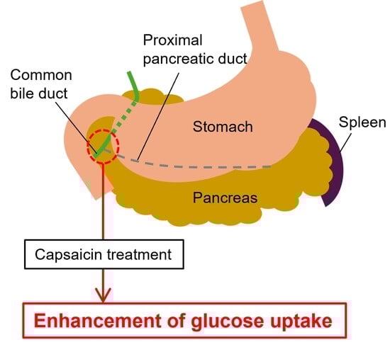
2.2. Determination of the Site of C Fiber Blockade

After the procedure described above, a hyperinsulinemic–euglycemic clamp was performed in 4 rats which were confirmed as showing stable blood glucose levels as reported elsewhere [12]. In brief, human insulin (60 pmol/kg/min; Eli Lilly Japan, Kobe, Japan) and 20% glucose were continuously infused into the jugular vein via a syringe pump. This allowed us to obtain the glucose infusion rate (GIR) required to maintain the baseline blood glucose for 30 min.
Immediately thereafter, gauze immersed in a 5% solution of capsaicin (Fujifilm Wako Pure Chemical, Osaka, Japan) dissolved in olive oil (w/v) was placed on the PP to block the C fibers [13]. The gauze covered the entire PP (n = 1) or half of the PP (n = 1), or was placed only at the junction of the PP and common bile (CB) ducts (PP-CB group; n = 2). Blood glucose levels were measured intermittently. After up to 70 min of blood glucose monitoring, the rats were sacrificed by intravenous pentobarbital overdose.

2.3. Measurement of Insulin Sensitivity Under C Fiber Blockade

Under the same conditions as above, a hyperinsulinemic–euglycemic clamp was performed in 10 rats in order to determine the individual GIRs. Then, the junction of the PP and CB ducts was treated with capsaicin (CAP group; n = 7). The control group received olive oil in place of capsaicin (CON group; n = 3). Based on the result of pilot study shown above, the rats were treated with capsaicin for 120 min. After obtaining stable GIR, the rats were sacrificed by intravenous administration of pentobarbital.

2.4. Statistical Analysis

Data are presented as means ± standard deviation (SD). Pre- and posttreatment GIRs were compared using the Wilcoxon signed-rank test. The CON and CAP groups were compared with the Mann–Whitney U test using EZR ver. 1.53 [14]. The significance level was set at p < 0.05.

3. Results

3.1. Pilot Study

First, a pilot study was performed to examine the relationship between the capsaicin dose and the blood glucose levels. No severe bleeding was observed in any case in the surgical field after the incision. The steady-state GIR was determined using the hyperinsulinemic–euglycemic clamp (target blood glucose level: 61 ± 3 mg/dL). As we have previously reported, blood glucose levels reached a steady state within 60–100 min after the start of clamp [15]. Capsaicin treatment was then performed while maintaining the GIR (5.96 ± 0.08 mg/kg/min). The rats with the entire PP or half of the PP treated with capsaicin died 10 and 70 minutes after capsaicin treatment, respectively. The blood glucose levels at death (141 and 105 mg/dL, respectively) were approximately twice as high as those recorded before the capsaicin treatment (72 and 50 mg/dL, respectively). In the PP-CB group, the blood glucose levels measured approximately 2 h after capsaicin treatment were lower than the levels recorded before the treatment (Figure 1).

3.2. Measurement of Insulin Sensitivity Under C Fiber Blockade in the Proximal Pancreatic Duct and Common Bile Duct

In the CON group (target blood glucose level: 66 ± 2 mg/dL), although the GIR slightly increased after olive oil treatment, it did not differ significantly from the pretreatment value. In contrast, in the CAP group (target blood glucose level: 79 ± 6 mg/dL), the GIR increased by approximately 17% after capsaicin treatment as compared with the pretreatment value, and the pretreatment–posttreatment difference in value was statistically significant (p < 0.05). Furthermore, while no significant difference in the baseline GIR was observed between the CAP and CON groups, after CAP treatment, the GIR was significantly higher in the CAP group as compared with the CON group (p < 0.05) (Table 1).

4. Discussion

To investigate the relationship between the pancreatic autonomic nerves and regulation of glucose uptake, we blocked the autonomic nerve fibers innervating the pancreas with capsaicin.
In the pilot study, the blood glucose elevation observed after capsaicin treatment over an extensive area of the PP was considered as having been derived from capsaicin stimulation. In contrast, localized capsaicin treatment tended to further reduce the steady-state blood glucose levels.
The results of the euglycemic clamp showed that capsaicin treatment at the junction of the PP and CB ducts promoted glucose uptake. Since the PP is innervated by vagal nerve fibers [9], the facilitation of glucose uptake by capsaicin observed in this study may have been caused by partial blockade of the vagal nerve fibers by capsaicin in the PP. It has been reported that stimulation of the vagal nerve induces insulin secretion [11]. However, since the hyperinsulinemic–euglycemic clamp strongly suppresses endogenous insulin secretion [16], the enhanced glucose uptake following C fiber blockade observed in this study may be considered a phenomenon that occurred independently of insulin secretion. On the other hand, the stimulation of pancreatic autonomic afferent fiber induces sympathetic outflow from the hypothalamus, and this system might regulate glucose metabolism independently of insulin secretion [17,18]. Therefore, the capsaicin treatment used in this study may enhance insulin sensitivity via blockade of the afferent pathway. Moreover, GIR increases observed during the euglycemic clamp are reported as being largely attributable to glucose uptake in the skeletal muscle [19]. This indicates that C fiber blockade in the PP mainly enhances glucose uptake in the skeletal muscle.
Regarding autonomic regulation of blood glucose control, it has been reported that peripheral sympathetic nervous activation may enhance insulin sensitivity [5,6] and that pancreatic vagal fiber activation may regulate blood glucose levels through inducing glucagon and insulin secretion [11,20]. However, there have been few reports on the effects of pancreatic C fiber blockade on insulin sensitivity. This study is the first to show the possibility that the pancreatic vagal nerve fibers may be involved in the regulation of insulin sensitivity.
To enhance the robustness and generalizability of our findings, expanding the sample size of experimental animals in subsequent studies will be essential. In addition, histological investigation is also necessary because the type and density of nerve fibers we blocked are unclear.
In conclusion, the findings of this study suggest that partial blockade of the pancreatic autonomic nerves may enhance insulin sensitivity. As a next step, the effectiveness of pancreatic C fiber blockade on insulin resistance must be verified using obese and/or diabetic animal models.

Author Contributions

Conceptualization, M.K.; methodology, M.K. and D.S.; formal analysis, D.S.; investigation, M.K. and D.S.; resources, M.K. and D.S.; data curation, M.K., D.S. and K.T.; writing—original draft preparation, M.K., D.S. and K.T.; writing—review and editing, F.H. and T.M.; visualization, D.S.; supervision, M.K.; project administration, M.K.; funding acquisition, M.K. All authors have read and agreed to the published version of the manuscript.

Funding

This research received no external funding.

Institutional Review Board Statement

The animal study protocol was approved by the Animal Research Committee of Yamagata University (approval no. R5077, 6 March 2023).

Informed Consent Statement

Not applicable.

Data Availability Statement

The original contributions presented in this study are included in the article. Further inquiries can be directed to the corresponding author.

Acknowledgments

We thank Aiji Noda and Nozomi Imaizumi and Natsu Nakano for their excellent technical assistance.

Conflicts of Interest

The authors declare no conflicts of interest.

Abbreviations

The following abbreviations are used in this manuscript:
CBCommon bile duct
GIRGlucose infusion rate
PPProximal pancreas

References

  1. Uysal, K.T.; Wiesbrock, S.M.; Marino, M.W.; Hotamisligil, G.S. Protection from obesity-induced insulin resistance in mice lacking TNF-α function. Nature 1997, 389, 610–614. [Google Scholar] [CrossRef] [PubMed]
  2. Maegawa, H.; Shigeta, Y.; Egawa, K.; Kobayashi, M. Impaired autophosphorylation of insulin receptors from abdominal skeletal muscles in nonobese subjects with NIDDM. Diabetes 1991, 40, 815–819. [Google Scholar] [CrossRef] [PubMed]
  3. Yki-Järvinen, H.; Daniels, M.C.; Virkamäki, A.; Mäkimattila, S.; DeFronzo, R.A.; McClain, D. Increased glutamine: Fructose-6-phosphatase amidotransferase activity in skeletal muscle of patients with NIDDM. Diabetes 1996, 45, 302–307. [Google Scholar] [CrossRef] [PubMed]
  4. Kaburagi, Y.; Satoh, S.; Tamemoto, H.; Yamamoto-Honda, R.; Tobe, K.; Veki, K.; Yamauchi, T.; Kono-Sugita, E.; Sekihara, H.; Aizawa, S.; et al. Role of insulin receptor substrate-1 and pp60 in the regulation of insulin-induced glucose transport and GLUT4 translocation in primary adipocytes. J. Biol. Chem. 1997, 272, 25839–25844. [Google Scholar] [CrossRef] [PubMed]
  5. Morton, G.J.; Muta, K.; Kaiyala, K.J.; Rojas, J.M.; Scarlett, J.M.; Matsen, M.E.; Nelson, J.T.; Acharya, N.K.; Piccinini, F.; Stefanovski, D.; et al. Evidence that the sympathetic nervous system elicits rapid, coordinated, and reciprocal adjustments of insulin secretion and insulin sensitivity during cold exposure. Diabetes 2017, 66, 823–834. [Google Scholar] [CrossRef] [PubMed]
  6. Benrick, A.; Kokosar, M.; Hu, M.; Larsson, M.; Maliqueo, M.; Marcondes, R.R.; Soligo, M.; Protto, V.; Jerlhag, E.; Sazonova, A.; et al. Autonomic nervous system activation mediates the increase in whole-body glucose uptake in response to electroacupuncture. FASEB J. 2017, 31, 3288–3297. [Google Scholar] [CrossRef] [PubMed]
  7. Sakamoto, K.; Butera, M.A.; Zhou, C.; Maurizi, G.; Chen, B.; Ling, L.; Shawkat, A.; Patlolla, L.; Thakker, K.; Calle, V.; et al. Overnutrition causes insulin resistance and metabolic disorder through increased sympathetic nervous system activity. Cell Metab. 2025, 37, 121–137. [Google Scholar] [CrossRef] [PubMed]
  8. Berthoud, H.R. The vagus nerve, food intake and obesity. Regul. Pept. 2008, 149, 15–25. [Google Scholar] [CrossRef] [PubMed]
  9. Hoornenborg, C.W.; van Dijk, T.H.; Bruggink, J.E.; van Beek, A.P.; van Dijk, G. Acute sub-diaphragmatic anterior vagus nerve stimulation increases peripheral glucose uptake in anaesthetized rats. IBRO Nurosci. Rep. 2023, 15, 50–56. [Google Scholar] [CrossRef] [PubMed]
  10. Yamamoto, J.; Imai, J.; Izumi, T.; Takahashi, H.; Kawana, Y.; Takahashi, K.; Kodama, S.; Kaneko, K.; Gao, J.; Uno, K.; et al. Neuronal signals regulate obesity induced β-cell proliferation by FoxM1 dependent mechanism. Nat. Commun. 2017, 8, 1930. [Google Scholar] [CrossRef] [PubMed]
  11. Kawana, Y.; Imai, J.; Morizawa, Y.M.; Ikoma, Y.; Kohata, M.; Komamura, H.; Sato, T.; Izumi, T.; Yamamoto, J.; Endo, A.; et al. Optogenetic stimulation of vagal nerves for enhanced glucose-stimulated insulin secretion and β cell proliferation. Nat. Biomed. Eng. 2024, 8, 808–822. [Google Scholar] [CrossRef] [PubMed]
  12. Sato, D.; Oda, K.; Kusunoki, M.; Nishina, A.; Takahashi, K.; Feng, Z.; Tsutsumi, K.; Nakamura, T. PPARγ activation alters fatty acid composition in adipose triglyceride, in addition to proliferation of small adipocytes, in insulin resistant high-fat fed rats. Eur. J. Pharmacol. 2016, 773, 71–77. [Google Scholar] [CrossRef] [PubMed]
  13. Imai, J.; Katagiri, H.; Yamada, T.; Ishigaki, Y.; Suzuki, T.; Kudo, H.; Uno, K.; Hasegawa, Y.; Gao, J.; Kaneko, K.; et al. Regulation of pancreatic β cell mass by neuronal signals from the liver. Science 2008, 322, 1250–1254. [Google Scholar] [CrossRef] [PubMed]
  14. Kanda, Y. Investigation of the freely available easy-to-use software ‘EZR’ for medical statistics. Bone Marrow Transplant. 2013, 48, 452–458. [Google Scholar] [CrossRef] [PubMed]
  15. Kusunoki, M.; Tsutsumi, K.; Sato, D.; Nakamura, A.; Habu, S.; Mori, Y.; Morishita, M.; Yonemoto, T.; Miyata, T.; Nakaya, Y.; et al. Pioglitazone-induced body weight gain is prevented by combined administration with the lipoprotein lipase activator NO-1886. Eur. J. Pharmacol. 2011, 668, 486–491. [Google Scholar] [CrossRef] [PubMed]
  16. Karakaplan, N.D.; Song, Y.; Laurenti, M.C.; Vella, A.; Jensen, M.D. Suppression of endogenous insulin secretion by euglycemic hyperinsulinemia. J. Clin. Endocrinol. Metab. 2023, 109, e596–e601. [Google Scholar] [CrossRef] [PubMed]
  17. Ito, Y.; Sun, R.; Yagimuma, H.; Taki, K.; Mizoguchi, A.; Kobayashi, T.; Sugiyama, M.; Onoue, T.; Tsunekawa, T.; Takagi, H.; et al. Protein tyrosine phosphatase 1B deficiency improves glucose homeostasis in type 1 diabetes treated with leptin. Diabetes 2022, 71, 1902–1914. [Google Scholar] [CrossRef] [PubMed]
  18. Sato, D.; Shiwaku, Y.; Nakamura, R.; Koizumi, S.; Feng, Z.; Kusunoki, M.; Nakamura, T. Characteristics of sympathetic nerve activity in the rat sciatic nerve in response to microstimulation in a sympathetic fascicle in the contralateral side. Annu. Int. Conf. IEEE Eng. Med. Biol. Soc. 2013, 2013, 6329–6332. [Google Scholar] [PubMed]
  19. DeFronzo, R.A.; Tripathy, D. Skeletal muscle insulin resistance is the primary defect in type 2 diabetes. Diabetes Care 2009, 32 (Suppl. S2), S157–S163. [Google Scholar] [CrossRef] [PubMed]
  20. Payne, S.C.; Ward, G.; MacIsaac, R.J.; Hyakumura, T.; Fallon, J.B.; Villalobos, J. Differential effects of vagus nerve stimulation strategies on glycemia and pancreatic secretions. Physiol. Rep. 2020, 8, e14479. [Google Scholar] [CrossRef] [PubMed]
Figure 1. Effect of the capsaicin treatment area of the proximal pancreas (PP) on the blood glucose levels. The PP of the rats was treated with gauze immersed in 5% capsaicin dissolved in olive oil (w/v). The whole PP, half of the PP, or the proximal pancreatic duct-common bile duct junction (PP-CB) was treated.
Obesities 05 00075 g001
Table 1. Effect of capsaicin treatment on the glucose infusion rate.
GroupnGlucose Infusion Rate (mg/kg/min)
Pre-TreatmentPost-Treatment
CON38.7 ± 1.19.6 ± 1.7
CAP712.5 ± 1.314.6 ± 1.3 †,*
Data are shown as mean ± SE.  p < 0.05 vs. pre-treatment. * p < 0.05 vs. CON group at post-treatment.
Disclaimer/Publisher’s Note: The statements, opinions and data contained in all publications are solely those of the individual author(s) and contributor(s) and not of MDPI and/or the editor(s). MDPI and/or the editor(s) disclaim responsibility for any injury to people or property resulting from any ideas, methods, instructions or products referred to in the content.

