Article

Supplementation Effects of Hibiscus sabdariffa L. Flower Aqueous Extract on Body Composition and Metabolism in Eutrophic and Obese Rats

by
João Fernando Bernardo da Costa
1,
Alana Louzada Millions Monteiro
2,
Bruna Almeida Nascimento
2,
Clarice Maia Vinagre de Oliveira
3,
Karen Pereira Coutinho
3,
Anderson Junger Teodoro
4,
Barbara Elisabeth Teixeira-Costa
4,5,* and
Mariana Sarto Figueiredo
4
1
Postgraduate Program in Nutrition Sciences, Federal Fluminense University (UFF), Rio de Janeiro 24020-140, Brazil
2
Postgraduate Program in Medicine Sciences, Federal Fluminense University (UFF), Rio de Janeiro 24020-140, Brazil
3
Department of Nutrition and Dietetics, Faculty of Nutrition, Federal Fluminense University (UFF), Rio de Janeiro 24020-140, Brazil
4
Integrated Food and Nutrition Center—CIAN, Faculty of Nutrition, Federal Fluminense University (UFF), Rio de Janeiro 24020-140, Brazil
5
Postgraduate Program in Biotechnology, Federal University of Amazonas (UFAM), Manaus 69077-000, Brazil
*
Author to whom correspondence should be addressed.
Obesities 2025, 5(4), 67; https://doi.org/10.3390/obesities5040067
Submission received: 1 August 2025 / Revised: 16 September 2025 / Accepted: 23 September 2025 / Published: 25 September 2025
(This article belongs to the Special Issue The Impact of Food Compounds on Obesity Mechanisms)

Abstract

Obesity is a chronic, multifactorial disease characterized by excess body fat and is a major risk factor for various metabolic disorders. Bioactive compounds from the diet have been recognized for their role in preventing chronic non-communicable diseases and as adjuvants in managing endocrine–metabolic dysfunctions. Hibiscus sabdariffa L. (HSL) is rich in bioactive compounds with antioxidant, antihypertensive, and antihyperlipidemic properties. This study evaluated the effects of HSL flower extract supplementation on body composition, lipid profile, and biochemical parameters in both eutrophic and high-fat diet-induced obese rats. Thirty-two Wistar rats were assigned to four groups: control, control plus HSL extract, high-fat diet, and high-fat diet plus HSL extract. The extract was administered orally at 150 mg kg−1 for thirty days. Dual-energy X-ray absorptiometry revealed that HSL supplementation significantly attenuated fat mass gain (from 98 g to 75 g) and adiposity indices (10.23 to 8.86) in obese rats without altering total body mass. Moreover, the HSL extract improved lipid profiles by reducing LDL cholesterol from 23 to 13 mg dL−1 and exhibited potential hepatoprotective effects linked with decreased ALT (40 to 26.7 U L−1) and total bilirubin (0.12 to 0.07 mg dL−1) levels. Although glucose metabolism parameters had no significant differences, a trend toward improved insulin sensitivity was observed. These results suggest that the aqueous HSL extract may exert cardioprotective, hepatoprotective, and anti-obesity effects, supporting its potential as a complementary therapeutic agent in obesity and related metabolic disorders.

1. Introduction

Sedentary behavior, genetic, environmental, socioeconomic, and social factors are known influences of energy imbalance and contribute to the accumulation of body fat [1]. The Western dietary pattern, including a high intake of saturated fats and refined carbohydrates, which are present in many fast foods and ultra-processed products, poses a significant risk for the rise in systemic metabolic alterations [2]. This contributes to the formation of inflammatory processes and, in the long term, pathological processes, such as inflammatory bowel disease, diabetes, neuropathies, and obesity [2,3]. The obesity complications are unique to each person; however, the consequences, such as cognitive damage, are common to many individuals [3]. Oxidative stress is commonly linked to obesity because it can trigger tissue damage, inflammation, extracellular matrix overproduction, activation of endoplasmic reticulum stress, and disturbance of autophagic flux [4].
It is well known that obesity is a multifactorial disease that combines individual factors and other causes linked to lifestyle factors, in which metaflammation prevails, with increased fat accumulation, reflecting a higher body mass index [1,3]. This low-grade inflammation induces important metabolic changes, such as the multimer form of adiponectin (protein hormone released by adipocytes), an increased concentration of leptin in plasma, and increased endotrophin production, leading to insulin resistance, weakening insulin function, and, consequently, prediabetes and diabetes development [3,5]. Oxidative stress and hypercholesterolemia are interconnected, primarily due to a reduced clearance of very-low-density lipoproteins (VLDLs). This impaired removal elevates the risk of developing conditions such as atherosclerosis, coronary artery disease, and non-alcoholic fatty liver disease [5].
As obesity has become a global health problem, new strategies have been studied to discover novel medicines and to combine multiple treatments, aiming to improve health and give individuals a comfortable lifestyle. Within this context, medicinal plants have been used worldwide to treat and alleviate symptoms of diverse diseases. Herbs and medicinal plants are recognized for their content of secondary metabolites and bioactive compounds that exhibit biological activities, including antioxidant effects, cancer chemoprevention, inhibition of lipid peroxidation, and stimulation of cellular antioxidant defenses [6]. Moreover, herbs can be used as coadjutant treatments for improving health conditions such as being overweight, obesity, liver disorders, hypercholesterolemia, hypertension, and cancer [7,8]. Phytotherapy is an alternative approach for the treatment or prevention of pathological states by using plants, plant parts, or their extracts [7]. These promising effects are linked to the content of bioactive substances, e.g., polyphenols, alkaloids, terpenoids, and carotenoids, with potential synergistic effects [9]. Studies have shown that the flowers and calyxes of Hibiscus sabdariffa L. contain various bioactive compounds, including ascorbic acid, β-carotene, arachidic acid, citric acid, malic acid, tartaric acid, and glycine betaine. They also show relevant contents of anthocyanins, such as cyanidin-3-rutinoside, delphinidin, delphinidin-3-glucoxyloside, delphinidin-3-monoglucoside, cyanidin-3-monoglucoside, cyanidin-3-sambubioside, and cyanidin-3,5-diglucoside, alongside other flavonol glycosides like hibiscetin-3-monoglucoside, and sabdaritrin [6,9].
Among the most widely used herbal medicines, H. sabdariffa L. stands out due to its diverse related health benefits, such as reducing blood pressure and blood lipids, as well as improving the elimination of kidney and bladder stones [8,10,11]. H. sabdariffa L., also known as roselle, is an herbal plant from the Malvacea family, native to Africa and widely cultivated in Tropical regions, such as Sudan, Eastern Taiwan, and Thailand [6,10]. The herb is widely consumed in Africa, India, and Sudan to prepare cold and hot beverages [12,13]. Reports indicate that in Nigeria, the consumption of H. sabdariffa L. can reach up to 150–180 mg kg−1 per day [6]. The plant calyces are used to color and flavor beverages, such as rum and coffee, as an aphrodisiac substance [6]. Other studies indicate that H. sabdariffa L. is commonly used in folk medicine because of its antimicrobial, antispasmodic, diuretic, antihypertensive, anti-inflammatory, hypocholesterolemic, and immune modulation properties; it is also used to treat chronic non-communicable diseases associated with obesity [6,14].
Therefore, the main objective of this study was to evaluate the effects of oral supplementation with an aqueous extract of H. sabdariffa L. flower on the body weight, lipidic markers, and biochemical parameters of eutrophic and obese rats induced by a high-fat diet.

2. Materials and Methods

2.1. Preparation of H. sabdariffa L. Aqueous Infusion and Spray-Dried Extract

H. sabdariffa L. (HSL) dried flowers (from a commercial brand) were purchased from a local market in Niteroi, Rio de Janeiro, Brazil, which can closely simulate the products that consumers typically obtain for preparing teas and infusions. The HSL flower extract was obtained in two steps: firstly, by infusion, and then by spray-drying to obtain an HSL powder extract. The HSL infusion was prepared at the Food and Dietetics Laboratory of the Fluminense Federal University (LABDI/UFF). About 10 g of whole HSL dried flowers were subjected to infusion in 1000 mL of mineral water (90 °C) for 10 min. Then, the HSL infusion extract was filtered using rapid filter paper and stored under refrigeration in an amber bottle until it was submitted to spray-drying.
The spray-dried H. sabdariffa (HSLsd) extract was prepared at the Faculty of Veterinary Medicine from the Fluminense Federal University, Niteroi, Rio de Janeiro, Brazil. The equipment used was the Spray Dryer MSD 1.0 (Labmaq do Brasil, Ribeirão Preto, SP, Brazil), with a 1.2 mm diameter atomizing nozzle, a concurrent flow regime, and a pneumatic (two-fluid) spray nozzle, programmed for a liquid passage speed of 0.5 L/h, an inlet temperature of 140 °C, and an outlet temperature of 100 °C [15]. For this process, the HSL infusion extract was loaded into the atomizer, which transformed it into millions of microparticles when in contact with the hot air inside the drying chamber. The hot air removed the water from the droplet, resulting in the dehydrated dried extract. At the end, the HSLsd extract was stored in an amber glass jar and placed in a freezer (−80 °C) until the next steps of the work and analysis.

2.2. Antioxidant Characterization of the H. sabdariffa Extract

The HSLsd extract was characterized by different in vitro antioxidant assays, such as the total phenolic content by the Folin–Ciocalteu method and the 2,2-diphenyl-1-picrylhydrazyl (DPPH) radical scavenging, ferric-reducing antioxidant potential (FRAP), and Trolox equivalent antioxidant capacity (TEAC) assays. The determination of the total phenolic compounds in the HSLsd extract was performed by the Folin–Ciocalteau method, as described by Singleton and Rossi [16]. The concentration of total phenolic compounds in the extract was expressed in mg of gallic acid g−1. The DPPH antioxidant activity was determined by measuring the DPPH radical scavenging activity according to the methodology described by Brand-Williams and Berset [17]. The FRAP method was performed according to Rufino et al. [18], and the antioxidant activity was expressed in ferrous sulfate g−1. The TEAC assay was performed according to the methodology described by Rufino et al. [18], which assesses the ability of a substance to scavenge the ABTS (2,2′-azino-bis(3-ethylbenzothiazoline-6-sulfonic acid)) radical cation (ABTS•+), a blue-green chromophore that absorbs light at 734 nm. The TEAC results were expressed in Trolox Equivalent g−1.

2.3. Experimental Design

2.3.1. Animals

The use of animals in our experimental design was approved by the Ethics Committee on Animal Care and Use of the Federal Fluminense University on 2 October 2022, filed under number 3444210721, as well as the guidelines adopted by the National Council for the Control of Animal Experimentation. All procedures followed the guidelines of the Brazilian Society of Science and Laboratory Animals and the NIH Guide for the Care and Use of Laboratory Animals (NIH Publication No. 8023, revised 1978). All experiments were conducted in accordance with the principles of the 3Rs (Reduction, Refinement, and Replacement) to minimize the number of animals used and their potential suffering. The experimental protocol and design were duly registered and approved by the Ethics Committee.
Healthy male Wistar rats (Rattus norvegicus, Wistar lineage) were selected because of their docile behavior, ease of handling, small size, rapid adaptation to laboratory environments, and short lifespan (approximately three years), which allows for the observation of multiple generations in a relatively short period. These animals are genetically homogeneous, which helps reduce interindividual variability and increases the reproducibility of results. Their physiological and behavioral characteristics are considered sufficiently similar to those of humans for use in translational research. All rats used in the experiment originated from the Laboratory Animal Center (NAL) of Fluminense Federal University (UFF) and had no prior history of experimental exposure, procedures, or interventions. All procedures performed on the animals were clearly defined in the approved experimental protocol. The development of this project did not affect animal health and welfare, as the animals were supplemented with edible plant extract, respecting the dose (low dose per animal) and the animal’s gastric capacity, considering a non-invasive procedure. The humane endpoints were established to coincide with the scheduled euthanasia of animals at the end of this study. Given that the intervention consisted of the oral administration of a natural edible compound, within the gastric capacity of the animals and at low doses, no invasive procedures were conducted. This study was terminated once the scientific objectives were fulfilled, and euthanasia was performed under anesthesia to minimize stress and pain, as previously approved by CEUA/UFF. The male Wistar (Rattus norvegicus) rats were maintained in a controlled-temperature (25 ± 2 °C) house on a 12:12 light/dark cycle. The animals had free access to food with tap water ad libitum until parturition.

2.3.2. Experimental Protocol

To evaluate the supplementation effects of the aqueous extract of HSL flowers on the body composition and metabolic profile, 32 postweaning male Wistar rats were randomized into two experimental groups: (1) a control group, which received a commercial diet (Nuvilab®, Quimtia Brasil, Colombo, PR, Brazil) (n = 16), and (2) a high-fat group, which received a high-fat diet (n = 16). The nutrient composition of the commercial and high-fat diets is presented below in Section 2.3.3. On post-natal day 90 (PN90), the control group was subdivided into the following groups: a control saline group, which received a commercial diet and gavage with saline solution (CS, n = 8), and (2) a control Hibiscus group, which received a commercial diet and gavage with the hibiscus extract at a dose of 150 mg kg−1 (CH, n = 8). The high-fat group was subdivided into (3) a high-fat saline group, which received the high-fat diet and gavage with a saline solution (HFS n = 8), and (4) a high-fat hibiscus group, which received the high-fat diet and gavage with the Hibiscus extract at a dose of 150 mg kg−1 (HFH n = 8). The Hibiscus extract at 150 mg kg−1 was chosen based on other works in the literature, which indicated that doses of 150–180 mg/kg/day are safe and do not cause histological damage to the heart and liver of the treated rats [19,20,21]. The human equivalent dose ( H E D ) was estimated using the following Equation (1) [22].
H E D   ( m g / k g ) = a n i m a l   d o s e   ( m g / k g ) × ( a n i m a l   k m / h u m a m   k m )
where k m is the correction factor ( k m ) estimated by dividing the average body weight (kg) of a species to its body surface area (m2) [22]. Considering a k m factor of 6 for rats and 37 for humans, the 150 mg/kg/day dose corresponds to approximately 24.32 mg/kg/day for a human. For a 60 kg adult, this would be equivalent to 1459.46 mg/day of Hibiscus spray-dried extract. The Hibiscus extract was administered for thirty consecutive days from PN90 to PN120 (post-natal day 120) (Figure 1), and the doses were weighed on an analytical balance and stored in Eppendorf tubes, which were kept in a freezer (−80 °C) until the moment of gavage. Prior to gavage, all doses were diluted in 1 mL commercial mineral water and homogenized in a vortex.

2.3.3. Experimental Diets

The commercial diet (Nuvilab®, Quimtia S.A, Colombo, PR, Brazil) used in the experimental design was purchased and provided by UFF, while the high-fat diet was prepared at the Experimental Nutrition Laboratory of the Faculty of Nutrition at UFF. The high-fat diet contained a mixture of the commercial diet and lard, condensed milk, powdered milk, and sugar, which was balanced according to AIN93G for micronutrients [23,24]. These ingredients were weighed, mixed in an industrial mixer, pelletized, and then dried in a ventilated oven at 50 °C until a constant weight. The nutritional composition of both the commercial and high-fat diets was determined according to AOAC [25]. The moisture content was determined through a gravimetric method in an oven at 105 °C, and weight stabilization was performed in a desiccator until a constant weight. The lipid content was determined by using the Soxhlet method based on lipid extraction with petroleum ether. The protein content was determined by the Kjeldahl method based on the food nitrogen content. The carbohydrate content was determined by a difference method. The fiber fraction was determined using a gravimetric method after hydrolysis in an acidic medium, and the mineral residue (ashes) was determined by carbonization of the previous samples by incineration at 550 °C [25].

2.3.4. Food Intake and Body Parameters

From PN49 to PN90, the body mass and food intake of all the rats were measured once a week. From PN90 to PN120, body mass and food intake were measured 2 times a week using a digital scale, model Filizola MF-6 (Filizola Balanças Industriais S/A, São Paulo, SP, Brazil). Energy intake was estimated based on the sum of calories from the content of proteins (1 g is equivalent to 4 kcal), lipids (1 g is equivalent to 9 kcal), and total carbohydrates (1 g is equivalent to 4 kcal).
At 120 days, the naso-anal length was determined in centimeters using a tape measure. The Lee index and body mass index were calculated according to the Novelli formula [25]. On PN90 and PN120, body composition was evaluated using dual-energy X-ray absorption (DXA) with a LUNAR IDXA 200368 GE (Lunar, Madison, WI, USA) densitometer, using specific software for small animals (Encore 2008 Version 12.20 GE Healthcare). Total fat mass (g), lean mass (g), and body fat percentage (%) were evaluated [25]. The adiposity index was calculated according to Nascimento’s formula [26].
On PN120, the animals were fasted for 12 h and then anesthetized (Ketamine 40 mg kg−1 + Xylazine 8 mg kg−1); blood was collected by cardiac puncture, and tissues, such as white adipose tissue compartments (retroperitoneal, peri-epididymal, and peri-mesenteric), and brown adipose tissue were weighed on analytical scales (Gehaka LTDA, AG200, São Paulo, SP, Brazil). Weight was corrected based on the ratio of body weight, according to the following Equation (2). Visceral fat mass was calculated by summing adipose tissue compartments, and the result was presented in g per body weight.
W e i g h t = t i s s u e   w e i g h t   ( g ) b o d y   w e i g h t   ( g ) × 100

2.3.5. Biochemical Parameters

For biochemical analyses, the collected blood was centrifuged at 3500 rpm for 15 min to obtain serum. Then, the albumin (g dL−1), total proteins (g dL−1), creatinine (mg dL−1), uric acid (mg dL−1), alkaline phosphatase (U L−1), total bilirubin (mg/dL), direct bilirubin (mg dL−1), aspartate aminotransferase (TGO/AST) (U L−1), alanine aminotransferase (TGP/ALT) (U L−1), and iron (mg dL−1) contents in the blood were determined. We determined the lipid profile, where total cholesterol (mg dL−1), triglycerides (mg dL−1), high-density lipoprotein (HDL) (mg dL−1), and low-density lipoprotein (LDL-mg dL−1) were calculated according to Friedewald’s formula [27], and very-low-density lipoprotein (VLDL-mg dL−1) was obtained by Tietz’s formula [28]. The biochemical analyses were conducted by colorimetric methods, with reading conducted in an automated spectrophotometer (BioClin® BS-120 Chemistry Analizer®, Shenzhen, China), using BioClin® commercial kits and specific wavelengths for each biochemical indicator. On PN120, fasting glucose was evaluated through blood from the tail circulation and measured by a glucometer (ACCU-CHEK® Advantage). Insulin was quantitatively evaluated using an ELISA kit, and the insulin resistance index (IRI) was calculated as the product of fasting insulin (µUI mL−1) × fasting glucose (mmol L−1).

2.4. Statistical Analysis

Data were analyzed using the statistical program GraphPad Prism (version 9.0.0, GraphPad Software, San Diego, CA, USA, version 9.1) and expressed as mean ± standard error of the mean. On PN90, one-way ANOVA analysis of variance was used, and the student’s t-test was applied as a post-test, with the results considered statistically significant when p < 0.05. On PN120, a one-way ANOVA analysis of variance was used, and Tukey’s test was applied as a post hoc test, with the results considered statistically significant when p < 0.05 (a 5% significance level was adopted).

3. Results

3.1. Antioxidant Characterization of the H. sabdariffa Extract

The results of the in vitro antioxidant characterization of the HSLsd extract are presented in Table 1. The extract presented a high content of phenolic substances, up to 4700 mg of gallic acid per 100 g of sample. The DPPH radical scavenging assay showed that the H. sabdariffa L. extract had up to 11,220.00 µmol of Trolox per g of sample. For the FRAP assay, the H. sabdariffa L. extract showed close to 14 µmol of ferrous sulfate per g of sample, while the TEAC assay (based on ABTS radical scavenging) presented up to 323 µmol of Trolox Equivalent per g of sample.

3.2. Food Intake and Body Parameters

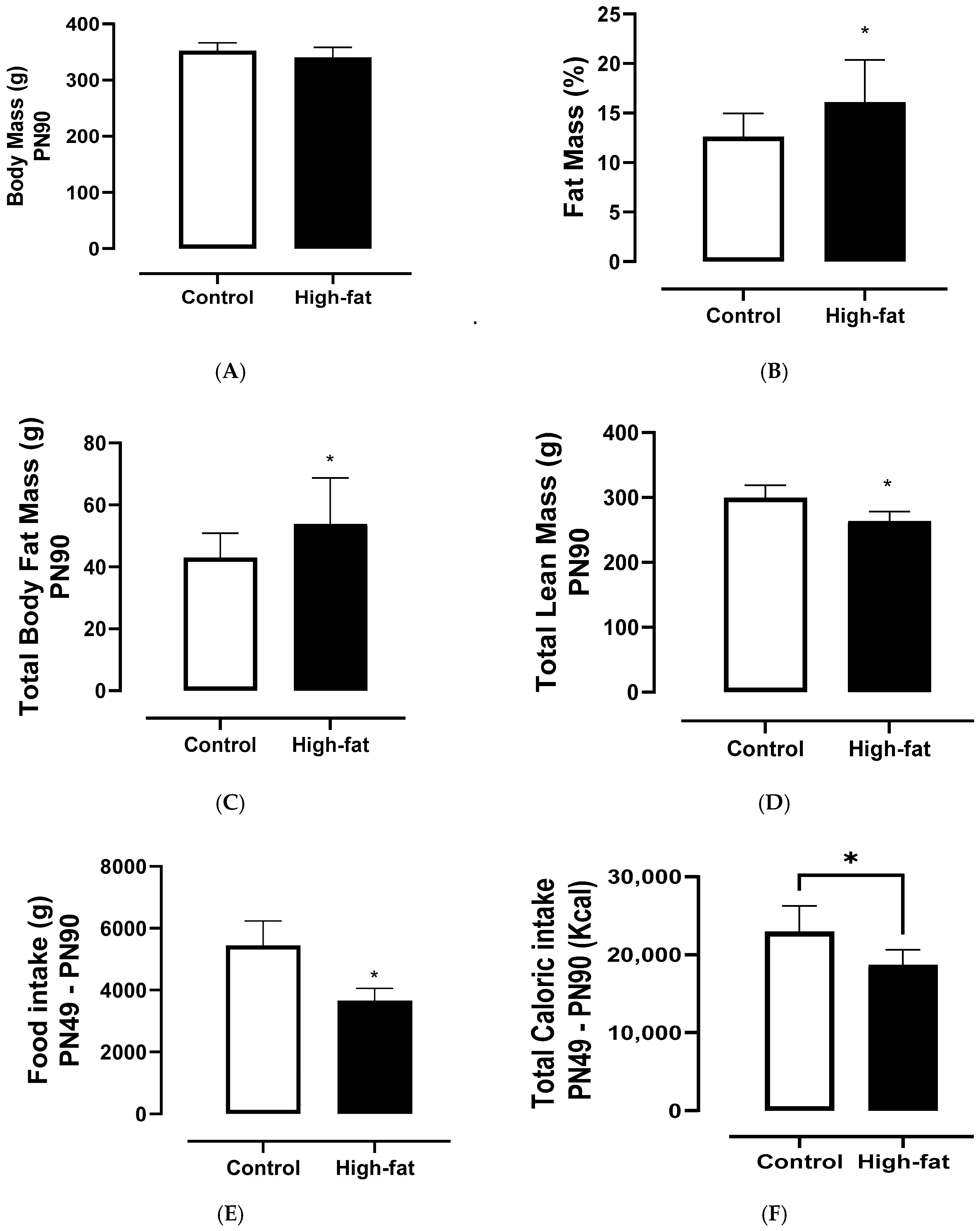
The results of the food intake and body parameters on PN90 are presented in Figure 2. The high-fat diet induced obesity in the animals in adulthood. The control group and the high-fat group had a similar body mass on PN90, as shown in Figure 2A. However, a significant increase in both fat mass (21.85%, p = 0.0073) and total body fat mass (25.16%, p = 0.0172) was observed for the high-fat group on PN90, as demonstrated in Figure 2B,C. A significant reduction in total lean mass (12.05%, p < 0.0001), food intake (32.66%, p = 0.0013), and caloric ingestion (18.63%, p = 0.0326) was noted, as shown in Figure 2D,F.
The results of the food intake and body parameters on PN120 are presented in Figure 3 and Table 2. The body mass did not change among the CS—control saline, CH—hibiscus tea, HFS—high-fat saline, and HFH—high-fat hibiscus groups after 30 days of gavage. There was a significant increase in the fat percentage of the HFS group of around 61.9% (p = 0.0027 vs. CS) and 77% (p = 0.0004 vs. CH) compared to the CS and CH groups, respectively, as shown in Figure 3B. The HFH group also presented a significant increase in fat percentage up to 47% (p = 0.0262 vs. CH) compared to the CH group (Figure 3B).
Regarding total body fat mass, it was observed that the HFS group had a significant increase of 59.3% (p = 0.0064 vs. CS) and 85.8% (p = 0.0005 vs. CH) compared to the CS and CH groups, respectively (Figure 3C). Whereas the increase in body fat mass in the HFH group reached 57.1% (p = 0.0212 vs. CH) compared to the CH group, as shown in Figure 3C.
It was noted that the lean mass was reduced among the HFS and HFH groups. The HFS group had a significant reduction of around 12.7% (p = 0.0024) and 9.2% (p = 0.0326) compared to the CS and CH groups, respectively, as can be observed in Figure 3D. The HFH group displayed a significant reduction of 11.8% (p = 0.0045) in lean mass compared to the CS group (Figure 3D). The HFS group presented a higher adiposity index (p < 0.0001 vs. CS and p < 0.0001 vs. CH), and the HFH group presented a higher adiposity index (p = 0.0014 vs. CS and p = 0.0008 vs. CH).
The total food intake showed a significant decrease of 33.43% (p < 0.0001) and 30.19% (p < 0.0001) in the HFS group compared to the CS and CH groups, respectively (Figure 3E). The total food intake also decreased significantly by around 34.3% (p < 0.0001) and 31.1% (p < 0.0001) in the HFH group compared to the CS and CH groups, respectively (Figure 3E), which led to a relevant reduction in the total calorie intake. Whereas in the CH group, a 5% reduction (p = 0.0003 vs. CS, Figure 3E) was observed in the total food intake, and the same was observed for the total calorie intake (p = 0.0003 vs. CS, Figure 3F).

3.3. Biochemical Parameters

The results of the biochemical analyses on PN120 of the serum of the animals in the CS—control saline, CH—control hibiscus, HFS—high-fat saline, and HFH—high-fat hibiscus groups are presented in Table 3. The triglyceride results ranged from 31 to 61 mg dL−1 among the groups, with no significant differences (p > 0.05), with the lowest value for the CH group and the highest value for the HFS group.
The total cholesterol results ranged from ≈37 to 46 mg dL−1 and did not show differences between the groups. The values for HDL-c and VLDL-c changed slightly among the groups, but with no significant difference (p > 0.05). The values for LDL cholesterol reduced by 35.1% (p = 0.0138 vs. CS) and 41% (p = 0.0034 vs. CS) in the HFS and HFH groups, respectively, compared to the CS group. Whereas LDL cholesterol was reduced by 38.7% (p = 0.0040 vs. CH) and 43.40% (p = 0.0008 vs. CS) in the HFS and HFH groups, respectively, compared to the CH group. No significant differences (p > 0.05) were found in the results of aspartate aminotransferase (AST), albumin, total protein, uric acid, creatinine, and iron among the groups. A significant reduction in alanine transaminase (ALT) of 34.38% (p = 0.0152) in the HFH group compared to the CS group was noted. Total bilirubin showed a reduction of 35.00% (p = 0.0106) in HFH when compared to CS. Direct bilirubin showed a reduction of 38.96% (p = 0.0009) in HFH when compared to CS.
No significant changes in the liver, pancreas, right and left adrenal glands, or brown adipose tissue were observed. However, HFS presented higher peri-mesenteric (p = 0.0133 vs. CS and p = 0.0092 vs. CH), retroperitoneal (p = 0.0049 vs. CS and p = 0.0015 vs. CH), and epididymal fat (p = 0.0155 vs. CS and p = 0.0076 vs. CH), without changes in visceral fat mass (p < 0.05, Table 3).
Regarding glucose metabolism, the studied groups did not present significant differences between them, as shown in Figure 4A. However, it was possible to observe an increase in the insulin level in the HFS group of around 74% compared to the CS group (p = 0.0049) and of 45% compared to the CH group (p = 0.0244), whereas the results of the insulin resistance index (IRI) also increased to 95.67% (p = 0.0022) and 79.65% (p = 0.0029) compared to the CS and CH groups, respectively, as presented in Figure 4B,C. However, the IRI decreased among the high-fat groups, from ≈2.2 in the HFS group to 1.7 in the HFH group.

4. Discussion

4.1. Antioxidant Characterization of the H. sabdariffa Extract

The HSLsd extract showed a relevant content of phenolic substances and antioxidant properties measured using different antioxidant assays, e.g., DPPH radical scavenging, FRAP, and TEAC. This plant is widely consumed in Central and West Africa and Southeast Asia as a cold beverage or as a hot tea, especially due to its health-related effects [14,21]. The content of phenolic compounds found in the present work, ~4712 mg gallic acid 100 g−1, agrees with the literature. Ethanolic and aqueous extracts of Hibiscus flowers were found to display a content of total phenolics ranging from 4600 ± 107 mg GAE 100 g−1 to 5400 ± 170 mg GAE 100 g−1 [29]. Phenolic substances from plants are known for their antioxidant activity by scavenging free radicals that are produced during metabolic processes [29,30]. This group of substances includes compounds such as gallic acid, syringic acid, protocatechuic acid, caffeic acid, and chlorogenic acid, as well as various flavonoids, e.g., epigallocatechin gallate, quercetin, myricetin, and kaempferol, along with numerous anthocyanins, all of which are found in H. sabdariffa flowers [11]. Suárez-Diéguez et al. [11] found up to 406 ± 7 mg GAE g−1 of phenolic substances in an aqueous–ethanolic extract of H. sabdariffa flowers (80:20) at 25 °C, in which the major compounds were myricetin, followed by apigenin and gallic acid. In the reports by Dey et al. [31], the anthocyanin content in dried H. sabdariffa calyxes was associated with reduced lactate dehydrogenase leakage, lower malondialdehyde formation, decreased serum hepatic enzymes (alanine and aspartate aminotransferase), and less oxidative liver damage [32]. Some anthocyanins found in Hibiscus extracts are cyanidin-3-sambubioside, delphinidin-3-sambubioside, delphinidin-3-glucoside, and cyanidin-3-glucoside [11]. Moreover, H. sabdariffa has been associated with diverse health benefits, such as hypoglycemic, antihypertensive, anti-inflammatory, hypocholesterolemic, immune modulator, and anticancer effects, among others [6,14].
The HSLsd extract showed relevant antioxidant properties. The antioxidant activity against the DPPH radical revealed up to 11,200 µmol of Trolox g−1, a much higher value than the findings of 134.4 ± 0.01 µmol Trolox g−1 in the study by Suárez-Diéguez et al. [11]. In another study, an aqueous extract from H. sabdariffa presented up to 97.35 ± 0.6% of DPPH inhibition [29]. Jason et al. [33] found up to 11.80 mg ml−1 as an inhibitory concentration at 50% (IC50) by the DPPH radical scavenger of an aqueous extract from H. sabdariffa powder at 200 mg ml−1. Dongmo et al. [9] found that tea formulations with higher proportions of H. sabdariffa flowers (80%) showed up to 18.4 ± 4 10−5 mol g−1 of antioxidant activity against the DPPH radical.
The results of the TEAC assay for the HSLsd extract showed a relevant antioxidant capacity, with up to 323 mg Trolox Equivalent g−1. An aqueous–ethanolic extract (80:20) of H. sabdariffa flowers in the work by Suárez-Diéguez et al. [11] showed up to 219.4 ± 0.03 µmol Trolox Equivalent g−1. In the ferric-reducing antioxidant potential (FRAP) assay, the aqueous H. sabdariffa flower extract had up to 14 µmol ferrous sulfate g−1, a lower value than the results of the study by Mak et al. [29], in which an aqueous extract presented 2349 ± 228 µmoles Fe (II) 100 g−1. Jason et al. [33] found an antioxidant capacity of around 49 mg ml−1 by the ABTS radical scavenging assay for an aqueous extract from H. sabdariffa powder at 200 mg ml−1. These results are relevant to linking the beneficial effects of H. sabdariffa to its content of polyphenols, flavonoids, and anthocyanins, which can protect against oxidative damage and anti-inflammatory action in non-communicable diseases by their cardiovascular and anti-obesogenic effects [34,35].

4.2. Food Intake and Body Parameters

In the present study, obesity was induced in the animals by offering a high-fat diet for 6 weeks, and the body change results were evaluated by dual energy X-ray absorption (DXA). DXA absorptiometry is an efficient tool for assessing body composition not only in rat models but also for anthropometric measurements in humans [36]. The high-fat diet group showed a significant increase in fat mass and total body fat mass and a decrease in lean mass and food intake, which can be linked to the increase in caloric intake related to the high-fat diet, providing more satiety for the animals. Ajiboye et al. [37] found a decreased food intake in rats fed with 100 and 200 mg kg−1 of an H. sabdariffa calyx extract. Despite having these changes, the high-fat diet group did not show variations in total body mass. These results are similar to the findings of the study by Jason et al. [33], in which they did not find any significant changes in body weight until the first 8 weeks. However, when the rats were fed with a high-fat diet for another 8 weeks, the body parameters changed significantly. Moreira et al. [38] also found an increase in body parameters after feeding rats a high-fat diet for 8 weeks.
Thus, it is well described in the literature that high-fat intake, especially with a higher content of saturated fatty acids, can alter body mass indices and induce obesity [36,39]. Jason et al. [33] suggested that the reduced food intake observed in Hibiscus extract supplementation can be related to greater leptin levels, leading to a decrease in fat accumulation in high-fat-diet-induced obese rats. A high-fat diet, along with abdominal fat accumulation, contributes to higher adipocyte size and stimulates inflammatory processes, which affect metabolism in a way that promotes predisposition to obesity. It is also associated with the development of other chronic diseases, such as diabetes mellitus, high blood pressure, and dyslipidemia [1,5,39].
On PN120, the rats fed the HSLsd extract, i.e., CH and HFH, presented a decreased body fat mass compared to CS and HFS, respectively, with significant differences for the latter group. This agrees with the results of the adiposity index (Table 2), which decreased in the CH and HFH groups, at 6.29 and 8.86, respectively, compared with the CS and HFS groups, at 6.33 and 10.23, respectively. This indicates that the rats fed with the H. sabdariffa flower extract had an attenuated body fat gain and adipose tissue weight. Similar behavior was observed by Jason et al. [33] when feeding rats with an aqueous H. sabdariffa extract at 250 and 500 mg kg−1. In their study, the adiposity index among the Hibiscus groups ranged from 4.15 to 4.33. Moreover, the relative mesenteric fat, relative retroabdominal fat, relative epididymal fat, and relative brown adipose tissue were lower in the CH and HFH groups compared with the CS and HFS groups, respectively. In another study, body weight was attenuated by the supplementation of 100 and 200 mg kg−1 of an H. sabdariffa calyx extract [37]. These results suggest that supplementation with an H. sabdariffa extract can attenuate body fat gain and other obesity indices.

4.3. Biochemical Parameters

A high-fat diet can induce dyslipidemia, biochemically presenting as altered parameters, such as total cholesterol, triglycerides, LDL cholesterol, HDL cholesterol, and others. In the present work, the levels of triglycerides, HDL-c, and VLDL-c showed similar behaviors, being lower in the groups that were supplemented with HSLsd, CH, and HFH, when compared to the CS and HFS groups, respectively (Table 3). However, only LDL-c had the greatest reduction (p < 0.05) among the Hibiscus groups, which reduced from ~24 mg dL−1 in the CH group to 13 mg dL−1 in the HFH group. Jason et al. [33] also found that rats supplemented with a Hibiscus extract had LDL-c values ranging from 185 mg dL−1 in a high-fat diet group to 137 mg dL−1 in a group fed a high-fat diet plus 500 mg kg−1 extract. Ajiboye et al. [37] also found that rats treated with 200 mg kg−1 of Hibiscus extract showed a reduction in LDL-c compared with groups fed a high-fructose diet. This effect can be associated with the content of phenolic substances in Hibiscus extracts, especially the anthocyanin concentration, since these compounds are linked to pancreatic lipase inhibition, leading to decreased levels of triglycerides and cholesterols in plasma [33,40].
Regarding the hepatic damage, the levels of AST were slightly higher in the groups supplemented with the Hibiscus extract, but no significant differences were found among them. ALT showed a relevant reduction in the Hibiscus groups, especially in the HFH group (p < 0.05). Ajiboye et al. [37] also reported changes in the levels of serum ALT and AST, from 8.31 to 3.46 nmol min−1 mg−1 and 21.64 to 11.15 nmol min−1 mg−1, respectively, in rats treated with 200 mg kg−1 of Hibiscus extract when compared to rats fed a high-fructose diet for 3 weeks. A possible hepatoprotective effect of the extract can be related to a significant reduction in total bilirubin and direct bilirubin in the high-fat Hibiscus group. Samuel et al. [41] found that aqueous Hibiscus vitifolius (Linn.) root extracts (50–2000 mg kg−1) decreased serum levels of AST, ALT, alkaline phosphatase, total bilirubin, and direct bilirubin in Wistar albino rats compared to rats that received a hepatotoxicant mixture (isoniazid at 7.5 mg kg−1, rifampicin at 10 mg kg−1 and pyrazinamide at 35 mg kg−1) to induce hepatotoxicity.
Moreover, it has been reported that the Hibiscus extract can improve lipid metabolism through the metabolic modulation of SRBP-1C (sterol regulatory element-binding protein-1c) and PPAR-y (peroxisome proliferator-activated receptor-y), but their relationship with the mechanisms involved in insulin resistance remains little explored, suggesting that this regulation also occurs by acting on the mechanisms involved in adipogenesis [42]. An eight-week study found that rats given doses of H. sabdariffa (250 mg kg−1 and 500 mg kg−1) had lower body mass, reduced food intake, improved lipid profiles, decreased inflammatory cytokines and lipid peroxidation, and changes in leptin, insulin absorption, and glucose handling. The research indicates that hibiscus suppresses lipids by downregulating the adipogenic gene [33]. Thus, based on our study, we suggest that even a lower dose of the aqueous Hibiscus extract may act by modulating some genes related to adipogenesis and glucose metabolism, such as SRBP-1C, PPAR-y, and others. However, more studies are necessary to investigate these pathways in vivo and in vitro.
Regarding the glucose parameters, no significant differences in blood glucose were observed among the four groups. However, the data on blood insulin and the insulin resistance index showed a slight reduction when comparing the high-fat saline group with the high-fat Hibiscus group. Jason et al. [33] also found high levels of insulin and the homeostasis model assessment index (HOMA-IR) when rats received a high-fat diet, similar to our findings when comparing the CS and CH groups. These authors also observed that Hibiscus supplementation at 250 mg kg−1 and 500 mg kg−1 reduced the levels of insulin and HOMA-IR. In the present study, the HFH group presented a decrease in blood glucose, insulin, and IRI compared to the HFS group. A high-fat diet reduces insulin sensitivity, which affects glucose metabolism and decreases glucose tolerance [33]. In this context, it can be suggested that the aqueous Hibiscus extract displays beneficial effects on hepatic and glycemic parameters, which are linked to the activation of the rf-2 and HO-1 pathways, increasing hepatic resilience and reducing adverse effects in diabetic individuals [33,37,41].
Mckay et al. [43] studied the effect of H. Sabdariffa L. tea on the blood pressure of 65 prehypertensive and mildly hypertensive adults, with ages ranging from 30 to 70 years old, for 6 weeks. These authors found that after the 6 weeks of supplementation, the systolic and diastolic blood pressure in the group that received the Hibiscus tea was lower than that in the placebo group [43]. Mckay et al. [43] also noted that the adults with higher systolic blood pressure had a greater response to the Hibiscus tea treatment, with no relation to age, gender, or the use of other dietetic supplements. These results indicate that the daily consumption of Hibiscus tea may be an alternative treatment for lowering blood pressure in pre- and mildly hypertensive adults. In our study, the Hibiscus extract dosage (150 mg per day) offered to the studied animals was equivalent to the consumption of one bag of tea by humans (around 1500 mg per day), which was considered a safe dose. Moreover, the serving per day in the present work had relevant effects on the adiposity indices, indicating a potential anti-obesity effect in a high-fat diet. These findings agree with the literature, which suggests that the Hibiscus extract has relevant effects on metabolic diseases, such as obesity, inflammation, diabetes, coronary artery diseases, and others. However, more studies are necessary to fully understand the effects of daily Hibiscus tea consumption on other metabolic diseases.

5. Conclusions

This study demonstrated that supplementation with an H. sabdariffa flower extract significantly improves lipid profiles, particularly by reducing LDL cholesterol levels, and exhibits potential hepatoprotective effects, as evidenced by decreased ALT and bilirubin levels. The extract can modulate lipid metabolism-related genes, such as SRBP-1C and PPAR-γ, alongside observed trends toward improved insulin sensitivity. This underscores its therapeutic potential in managing dyslipidemia and metabolic disturbances induced by a high-fat diet. Furthermore, the dual-energy X-ray absorptiometry analysis revealed that Hibiscus supplementation attenuates body fat gain and the adiposity index despite a high-fat diet, suggesting an anti-obesity effect linked to reduced fat accumulation in various adipose tissue depots. These combined effects highlight that the H. sabdariffa extract is a promising natural intervention for obesity and its associated metabolic complications. However, further studies are needed to clarify the molecular mechanisms involved and confirm these benefits in vivo and in vitro.

Author Contributions

J.F.B.d.C.: Data curation, formal analysis, investigation, methodology, software, visualization, and writing—original draft. A.L.M.M.: Data curation, formal analysis, investigation, methodology, software, visualization, and writing—original draft. B.A.N.: Data curation, formal analysis, investigation, methodology, software, visualization, and writing—original draft. C.M.V.d.O.: Data curation, formal analysis, investigation, methodology, software, visualization, and writing—original draft. K.P.C.: Data curation, formal analysis, investigation, methodology, software, visualization, and writing—original draft. A.J.T.: Data curation, formal analysis, investigation, methodology, software, visualization, and writing—original draft. B.E.T.-C.: Data curation, formal analysis, investigation, methodology, software, visualization, writing—original draft, and writing—review and editing. M.S.F.: Funding acquisition, resources, data curation, formal analysis, investigation, methodology, software, visualization, writing—original draft, and project administration. All authors have read and agreed to the published version of the manuscript.

Funding

This research received no external funding.

Institutional Review Board Statement

The animal study protocol was registered and approved by the Animal Use Ethics Committee of the Fluminense Federal University (CEUA/UFF), under protocol number 3444210721 (2 October 2022).

Informed Consent Statement

Not applicable.

Data Availability Statement

The data supporting the findings of this study are available on request from the author.

Acknowledgments

The authors acknowledge Carlos Augusto de Freitas Peregrino from the Faculty of Pharmacy of Federal Fluminense University (UFF), the Integrated Food and Nutrition Center (CIAN/UFF), the Laboratory of Food Analysis (LABAL/UFF), the Experimental Nutrition Laboratory of Fluminense Federal University (LABNE/UFF), and the Center for Research on Obesity and Comorbidities of the State University of Campinas (OCRC) for access to facilities and equipment.

Conflicts of Interest

The authors declare no conflict of interest.

References

  1. Safaei, M.; Sundararajan, E.A.; Driss, M.; Boulila, W.; Shapi’i, A. A Systematic Literature Review on Obesity: Understanding the Causes & Consequences of Obesity and Reviewing Various Machine Learning Approaches Used to Predict Obesity. Comput. Biol. Med. 2021, 136, 104754. [Google Scholar] [CrossRef]
  2. Butler, M.J.; Barrientos, R.M. The Impact of Nutrition on COVID-19 Susceptibility and Long-Term Consequences. Brain Behav. Immun. 2020, 87, 53–54. [Google Scholar] [CrossRef] [PubMed]
  3. Naomi, R.; Teoh, S.H.; Embong, H.; Balan, S.S.; Othman, F.; Bahari, H.; Yazid, M.D. The Role of Oxidative Stress and Inflammation in Obesity and Its Impact on Cognitive Impairments—A Narrative Review. Antioxidants 2023, 12, 1071. [Google Scholar] [CrossRef] [PubMed]
  4. Martínez-Martínez, E.; Cachofeiro, V. Oxidative Stress in Obesity. Antioxidants 2022, 11, 639. [Google Scholar] [CrossRef] [PubMed]
  5. Jakubiak, G.K.; Osadnik, K.; Lejawa, M.; Osadnik, T.; Goławski, M.; Lewandowski, P.; Pawlas, N. “Obesity and Insulin Resistance” Is the Component of the Metabolic Syndrome Most Strongly Associated with Oxidative Stress. Antioxidants 2022, 11, 79. [Google Scholar] [CrossRef]
  6. Maganha, E.G.; Halmenschlager, R.d.C.; Rosa, R.M.; Henriques, J.A.P.; Ramos, A.L.L.d.P.; Saffi, J. Pharmacological Evidences for the Extracts and Secondary Metabolites from Plants of the Genus Hibiscus. Food Chem. 2010, 118, 1–10. [Google Scholar] [CrossRef]
  7. Azzini, E.; Barnaba, L.; Ciarapica, D.; Polito, A. Micronutrients and Plant Food Bioactive Compounds Against Obesity Diseases. Endocr. Metab. Immune Disord. Drug Targets 2022, 23, 316–335. [Google Scholar] [CrossRef] [PubMed]
  8. Dilokthornsakul, P.; Rattanachaisit, N.; Thimkorn, P.; Pongpattanawut, S.; Dilokthornsakul, W.; Dhippayom, T. Clinical Effects of Hibiscus sabdariffa Linn. on Obesity Treatment: A Systematic Review and Meta-Analysis of Randomized Controlled Trials. Complement. Ther. Med. 2024, 84, 103063. [Google Scholar] [CrossRef]
  9. Dongmo, F.F.D.; Touohou, S.V.N.; Etame, R.M.E.; Lienou, L.L.; Koule, J.C.M.; Mbiatat, H.D.G.; Tchuenbou-Magaia, F.L.; Gouado, I. An Herbal Tea Blend of Hibiscus sabdariffa, Zingiber Officinale, and Mentha Spicata: A Potent Source of Antioxidant and Anti-Obesity Properties. Eur. J. Med. Health Res. 2024, 2, 63–74. [Google Scholar] [CrossRef]
  10. Riaz, G.; Chopra, R. A Review on Phytochemistry and Therapeutic Uses of Hibiscus sabdariffa L. Biomed. Pharmacother. 2018, 102, 575–586. [Google Scholar] [CrossRef]
  11. Suárez-Diéguez, T.; Palma-Morales, M.; Camacho Bernal, G.I.; Valdez López, E.N.; Rodríguez-Pérez, C.; Cruz-Cansino, N.d.S.; Nieto, J.A. Modulation of the Hyperglycemia Condition in Diabetic Lab Rats with Extracts of the Creole Jamaica Flower (Hibiscus sabdariffa L.) from the Morelia Region (Mexico). Antioxidants 2024, 13, 1010. [Google Scholar] [CrossRef] [PubMed]
  12. Vanissa, N.T.S.; Fabien, D.D.F.; Landry, L.L.; Ghislain, M.T.; William, D.A.; Christophe, M.K.J.; Calvin, B.Z.; De Goeithe, M.H.; Madeleine, E.E.R.; Inocent, G. Antioxidant and Antiobesogenic Properties of Aqueous Extracts of Hibiscus sabdariffa, Zingiber Officinale and Mentha Spicata in Wistar High-Fat Diet Rats. J. Food Nutr. Sci. 2022, 10, 151–164. [Google Scholar] [CrossRef]
  13. Rangel-García, C.A.; Reynoso-Camacho, R.; Pérez-Ramírez, I.F.; Morales-Luna, E.; de los Ríos, E.A.; Salgado, L.M. Serum Phospholipids Are Potential Therapeutic Targets of Aqueous Extracts of Roselle (Hibiscus sabdariffa) Against Obesity and Insulin Resistance. Int. J. Environ. Res. Public Health 2022, 19, 16538. [Google Scholar] [CrossRef] [PubMed]
  14. Ghaly, H.K.F.; Younis, F.A.A.Y.; Soliman, A.M.; El-Sabbagh, S.M. Phytochemical and Antibacterial Properties of Calyces Hibiscus sabdariffa L.: An in Vitro and in Silico Multitarget-Mediated Antibacterial Study. BMC Complement. Med. Ther. 2025, 25, 62. [Google Scholar] [CrossRef] [PubMed]
  15. Borges, L.L.; Martins, F.S.; Conceição, E.C.; Silveira, D. Optimization of the Spray-Drying Process for Developing Jabuticaba Waste Powder Employing Response Surface Methodology. J. Food Process Eng. 2015, 40, e12276. [Google Scholar] [CrossRef]
  16. Singleton, V.L.; Rossi, J.A. Colorimetry of Total Phenolics with Phosphomolybdic-Phosphotungstic Acid Reagents. Am. J. Enol. Vitic. 1965, 16, 144–158. [Google Scholar] [CrossRef]
  17. Brand-Williams, W.; Cuvelier, M.E.; Berset, C. Use of a Free Radical Method to Evaluate Antioxidant Activity. LWT-Food Sci. Technol. 1995, 28, 25–30. [Google Scholar] [CrossRef]
  18. Rufino, M.d.S.M.; Alves, R.E.; de Brito, E.S.; Pérez-Jiménez, J.; Saura-Calixto, F.; Mancini-Filho, J. Bioactive Compounds and Antioxidant Capacities of 18 Non-Traditional Tropical Fruits from Brazil. Food Chem. 2010, 121, 996–1002. [Google Scholar] [CrossRef]
  19. Akindahunsi, A.A.; Olaleye, M.T. Toxicological Investigation of Aqueous-Methanolic Extract of the Calyces of Hibiscus sabdariffa L. J. Ethnopharmacol. 2003, 89, 161–164. [Google Scholar] [CrossRef]
  20. Fakeye, T.O.; Pal, A.; Bawankule, D.U.; Yadav, N.P.; Khanuja, S.P.S. Toxic Effects of Oral Administration of Extracts of Dried Calyx of Hibiscus sabdariffa Linn. (Malvaceae). Phytother. Res. 2009, 23, 412–416. [Google Scholar] [CrossRef]
  21. Ali, B.H.; Al Wabel, N.; Blunden, G. Phytochemical, Pharmacological and Toxicological Aspects of Hibiscus sabdariffa L.: A Review. Phytother. Res. 2005, 19, 369–375. [Google Scholar] [CrossRef]
  22. Nair, A.B.; Jacob, S. A Simple Practice Guide for Dose Conversion between Animals and Human. J. Basic. Clin. Pharm. 2016, 7, 27. [Google Scholar] [CrossRef] [PubMed]
  23. Abreu, G.M.; Barros, B.F.d.B.; Teles, C.G.; Almeida, C.d.O.R.P.d.; Nascimento, B.A.; Teodoro, A.J.; Figueiredo, M.S. Early High Fat Diet Exposure Changes Body Composition, Biochemical Parameters and Redox Balance in Wistar Male Rats. Concilium 2024, 24, 450–471. [Google Scholar] [CrossRef]
  24. AOAC. Official Methods of Analysis of AOAC International, 20th ed.; Association of Official Analytical Chemists: Rockville, MD, USA, 2016. [Google Scholar]
  25. Novelli, E.L.B.; Diniz, Y.S.; Galhardi, C.M.; Ebaid, G.M.X.; Rodrigues, H.G.; Mani, F.; Fernandes, A.A.H.; Cicogna, A.C.; Novelli Filho, J.L.V.B. Anthropometrical Parameters and Markers of Obesity in Rats. Lab. Anim. 2007, 41, 111–119. [Google Scholar] [CrossRef] [PubMed]
  26. Nascimento, A.F.; Luvizotto, R.A.M.; Leopoldo, A.S.; Lima-Leopoldo, A.P.; Seiva, F.R.; Justulin, L.A.; Silva, M.D.P.; Okoshi, K.; Wang, X.D.; Cicogna, A.C. Long-Term High-Fat Diet-Induced Obesity Decreases the Cardiac Leptin Receptor Without Apparent Lipotoxicity. Life Sci. 2011, 88, 1031–1038. [Google Scholar] [CrossRef] [PubMed]
  27. Friedewald, W.T.; Levy, R.I.; Fredrickson, D.S. Estimation of the Concentration of Low-Density Lipoprotein Cholesterol in Plasma, Without Use of the Preparative Ultracentrifuge. Clin. Chem. 1972, 18, 499–502. [Google Scholar] [CrossRef] [PubMed]
  28. Tietz, N.W. Clinical Guide to Laboratory Tests, 3rd ed.; Tietz, N.W., Ed.; WB Saunders Company: Philadelphia, PA, USA, 1995. [Google Scholar]
  29. Mak, Y.W.; Chuah, L.O.; Ahmad, R.; Bhat, R. Antioxidant and Antibacterial Activities of Hibiscus (Hibiscus rosa-sinensis L.) and Cassia (Senna bicapsularis L.) Flower Extracts. J. King Saud. Univ. Sci. 2013, 25, 275–282. [Google Scholar] [CrossRef]
  30. Kumar, V.; Mahdi, F.; Khanna, A.K.; Singh, R.; Chander, R.; Saxena, J.K.; Mahdi, A.A.; Singh, R.K. Antidyslipidemic and Antioxidant Activities of Hibiscus Rosa Sinensis Root Extract in Alloxan Induced Diabetic Rats. Indian J. Clin. Biochem. 2013, 28, 46–50. [Google Scholar] [CrossRef]
  31. Dey, S.; Roy, A.; Paul, A.; Saha, N.; Tarafdar, A.; Mazumder, S. Food and Medicinal Properties of Hibiscus (Hibiscus sabdariffa & Hibiscus rosa-sinensis). Sustain. Agric. Food Environ. Res. 2023, 11, 2023. [Google Scholar] [CrossRef]
  32. Wang, C.-J.; Wang, J.-M.; Lin, W.-L.; Chu, C.-Y.; Chou, F.-P.; Tseng, T.-H. Protective Effect of Hibiscus Anthocyanins Against Tert-Butyl Hydroperoxide-Induced Hepatic Toxicity in Rats. Food Chem. Toxicol. 2000, 38, 411–416. [Google Scholar] [CrossRef] [PubMed]
  33. Janson, B.; Prasomthong, J.; Malakul, W.; Boonsong, T.; Tunsophon, S. Hibiscus sabdariffa L. Calyx Extract Prevents the Adipogenesis of 3T3-L1 Adipocytes, and Obesity-Related Insulin Resistance in High-Fat Diet-Induced Obese Rats. Biomed. Pharmacother. 2021, 138, 111438. [Google Scholar] [CrossRef] [PubMed]
  34. Herrera-Arellano, A.; Miranda-Sánchez, J.; Ávila-Castro, P.; Herrera-Álvarez, S.; Jiménez-Ferrer, J.; Zamilpa, A.; Román-Ramos, R.; Ponce-Monter, H.; Tortoriello, J. Clinical Effects Produced by a Standardized Herbal Medicinal Product of Hibiscus sabdariffa on Patients with Hypertension. A Randomized, Double-Blind, Lisinopril-Controlled Clinical Trial. Planta Med. 2006, 73, 6–12. [Google Scholar] [CrossRef]
  35. Jeffery, T.D.; Richardson, M.L. A Review of the Effectiveness of Hibiscus for Treatment of Metabolic Syndrome. J. Ethnopharmacol. 2021, 270, 113762. [Google Scholar] [CrossRef] [PubMed]
  36. Hariri, N.; Thibault, L. High-Fat Diet-Induced Obesity in Animal Models. Nutr. Res. Rev. 2010, 23, 270–299. [Google Scholar] [CrossRef]
  37. Ajiboye, T.O.; Raji, H.O.; Adeleye, A.O.; Adigun, N.S.; Giwa, O.B.; Ojewuyi, O.B.; Oladiji, A.T. Hibiscus sabdariffa Calyx Palliates Insulin Resistance, Hyperglycemia, Dyslipidemia and Oxidative Rout in Fructose-Induced Metabolic Syndrome Rats. J. Sci. Food Agric. 2016, 96, 1522–1531. [Google Scholar] [CrossRef]
  38. Moreira, M.E.d.C.; Natal, D.I.G.; Toledo, R.C.L.; Ramirez, N.M.; Ribeiro, S.M.R.; Benjamin, L.d.A.; de Oliveira, L.L.; Rodrigues, D.A.; Antônio, J.D.; Veloso, M.P.; et al. Bacupari Peel Extracts (Garcinia brasiliensis) Reduce High-Fat Diet-Induced Obesity in Rats. J. Funct. Foods 2017, 29, 143–153. [Google Scholar] [CrossRef]
  39. Coelho, D.F.; Pereira-Lancha, L.O.; Chaves, D.S.; Diwan, D.; Ferraz, R.; Campos-Ferraz, P.L.; Poortmans, J.R.; Lancha, A.H. Effect of High-Fat Diets on Body Composition, Lipid Metabolism and Insulin Sensitivity, and the Role of Exercise on These Parameters. Braz. J. Med. Biol. Res. 2011, 44, 966–972. [Google Scholar] [CrossRef] [PubMed]
  40. Lee, M.J.; Wu, Y.; Fried, S.K. Adipose Tissue Heterogeneity: Implication of Depot Differences in Adipose Tissue for Obesity Complications. Mol. Aspects Med. 2013, 34, 1–11. [Google Scholar] [CrossRef]
  41. Samuel, A.J.S.J.; Mohan, S.; Chellappan, D.K.; Kalusalingam, A.; Ariamuthu, S. Hibiscus vitifolius (Linn.) Root Extracts Shows Potent Protective Action against Anti-Tubercular Drug Induced Hepatotoxicity. J. Ethnopharmacol. 2012, 141, 396–402. [Google Scholar] [CrossRef]
  42. Villalpando-Arteaga, E.V.; Mendieta-Condado, E.; Esquivel-Solís, H.; Canales-Aguirre, A.A.; Gálvez-Gastélum, F.J.; Mateos-Díaz, J.C.; Rodríguez-González, J.A.; Márquez-Aguirre, A.L. Hibiscus sabdariffa L. Aqueous Extract Attenuates Hepatic Steatosis Through Down-Regulation of PPAR-γ and SREBP-1c in Diet-Induced Obese Mice. Food Funct. 2013, 4, 618–626. [Google Scholar] [CrossRef]
  43. McKay, D.L.; Chen, C.-Y.O.; Saltzman, E.; Blumberg, J.B. Hibiscus sabdariffa L. Tea (Tisane) Lowers Blood Pressure in Prehypertensive and Mildly Hypertensive Adults. J. Nutr. 2010, 140, 298–303. [Google Scholar] [CrossRef] [PubMed]
Figure 1. Experimental design for the oral supplementation of Wistar rats with control and high-fat diets containing a saline solution and the H. sabdariffa extract.
Obesities 05 00067 g001
Figure 2. Body composition and food intake from PN49 to PN90. Legend: (A) Body mass on PN90; (B) fat mass (%) on PN90; (C) total body fat mass (g) on PN90; (D) total lean mass on PN90; (E) food intake, PN49-90; (F) total caloric intake, PN49-PN90. control group (n = 16); high-fat group (n = 16). The results are expressed as the mean and standard deviation and considered statistically significant when p < 0.05. * vs. the control.
Obesities 05 00067 g002
Figure 3. Food intake and body composition on PN120. Legend: (A) Body mass on PN120; (B) body fat mass (%) on PN120; (C) body fat (g) on PN120; (D) total lean mass on PN120; (E) food intake (g), PN90-PN120; (F) total caloric intake, PN9090-PN120. CS—control saline group (n = 8); CH—control Hibiscus group (n = 8); HFS—high-fat saline group (n = 8); HFH—high-fat Hibiscus extract group (n = 8). The results are expressed as the mean and standard deviation and considered statistically significant when p < 0.05. * vs. CS; # vs. CH.
Obesities 05 00067 g003
Figure 4. Blood glucose, blood insulin, and IRI. Legend: IRI: insulin resistance index. (A) Blood glucose (mg/dL); (B) blood insulin (mg/mL); (C) IRI: insulin resistance index; CS—control saline group (n = 8); CH—control Hibiscus group (n = 8); HFS—high-fat saline group (n = 8); HFH—high-fat Hibiscus extract group (n = 8). The results are expressed as the mean and standard deviation and considered statistically significant when p < 0.05. * vs. CS; # vs. CH.
Obesities 05 00067 g004
Table 1. Antioxidant properties of the spray-dried H. sabdariffa L. aqueous extract.
AssaysHSLsd Extract
Total phenolic content (mg gallic acid 100 g−1)4712.33 ± 184.30
DPPH (µmol of Trolox g−1)11,220.0 ± 133.3
TEAC (µmol Trolox Equivalent g−1)322.50 ± 2.90
FRAP (µmol ferrous sulfate g−1)14.06 ± 0.20
Table 2. Body parameters on PN120.
ParametersCS (n = 8)CH (n = 8)HFS (n = 8)HFH (n = 8)p Value
Body weight (g)400.4 ± 20.0381.6 ± 16.03393.6 ± 36.12389.1 ± 17.47>0.05
Body lenght (cm)24.5 ± 0.5024.31 ± 0.4524.71 ± 0.4824.75 ± 0.46>0.05
Lee index (g/cm3)0.3 ± 0.010.29 ± 0.010.29 ± 0.010.29 ± 0.00>0.05
Body mass index (g/cm3)0.66 ± 0.040.64 ± 0.040.64 ± 0.040.63 ± 0.02>0.05
Adiposity index6.33 ± 0.416.29 ± 0.2910.23 ± 0.68 *#8.86 ± 0.35 *#<0.05
Relative mesenteric fat (g)4.39 ± 0.734.36 ± 0.706.69 ± 2.16 *#6.01 ± 1.07 *#<0.05
Relative retroabdominal fat (g)5.85 ± 1.165.43 ± 1.0810.371 ± 3.88 *#8.77 ± 1.90 *#<0.05
Relative epididymal fat (g)4.58 ± 0.614.41 ± 1.097.43 ± 3.01 *#5.85 ± 0.89<0.05
Relative visceral fat mass (g)14.84 ± 2.2814.21 ± 2.1119.87 ± 7.8719.92 ± 3.06>0.05
Relative brown adipose tissue (g)0.33 ± 0.070.32 ± 0.050.41 ± 0.110.36 ± 0.07>0.05
Legend: CS—control saline group; CH—control Hibiscus group; HFS—high-fat saline group; HFH—high-fat Hibiscus extract group. The results are expressed as the mean ± standard deviation and considered statistically significant when p < 0.05. * vs. CS; # vs. CH.
Table 3. Serum biochemical parameters on PN120.
ParametersCS (n = 8)CH (n = 8)HFS (n = 8)HFH (n = 8)p Value
Triglycerides (mg dL−1)40.43 ± 15.9731.38 ± 8.8960.86 ± 33.3443.50 ± 17.76>0.05
Total cholesterol (mg dL−1)40.29 ± 8.0145.88 ± 7.0643.00 ± 8.0836.75 ± 7.01>0.05
HDL-c (mg dL−1)16.43 ± 3.3115.88 ± 2.5316.29 ± 2.6914.63 ± 3.58>0.05
VLDL-c (mg dL−1)8.08 ± 3.196.27 ± 1.7812.17 ± 6.678.70 ± 3.55>0.05
LDL-c (mg dL−1)22.77 ± 4.0623.73 ± 4.9414.54 ± 6.13 *#13.43 ± 3.11 *#<0.05
AST (U L−1)112.10 ± 22.09111.30 ± 21.1796.71 ± 24.90107.60 ± 47.69>0.05
ALT (U L−1)40.71 ± 13.7333.13 ± 3.9432.00 ± 7.6126.71 ± 1.11 *<0.05
Alkaline phosphatase (U L−1)79.57 ± 12.8485.00 ± 17.3481.14 ± 15.9980.13 ± 18.12>0.05
Albumin (mg dL−1)3.25 ± 0.133.21 ± 0.163.40 ± 0.113.36 ± 0.12>0.05
Total proteins (mg dL−1)5.76 ± 0.275.57 ± 0.375.83 ± 0.275.73 ± 0.19>0.05
Uric acid (mg dL−1)1.57 ± 0.321.41 ± 0.361.30 ± 0.101.41 ± 0.17>0.05
Total bilirubin (mg dL−1)0.12 ± 0.020.09 ± 0.020.09 ± 0.020.07 ± 0.02 *<0.05
Direct bilirubin (mg dL−1)0.07 ± 0.010.06 ± 0.010.06 ± 0.010.04 ± 0.01 *<0.05
Creatinine (mg dL−1)0.62 ± 0.020.61 ± 0.020.70 ± 0.170.69 ± 0.11>0.05
Iron (mg dL−1)250.60 ± 52.59242.80 ± 37.21259.50 ± 31.76257.20 ± 25.11>0.05
Legend: CS—control saline group; CH—control Hibiscus group; HFS—high-fat saline group; HFH—high-fat Hibiscus extract group; HDL-c—high-density lipoprotein cholesterol; VLDL-c—very-low-density lipoprotein cholesterol; LDL-c—low-density lipoprotein cholesterol; AST—aspartate aminotransferase; ALT—alanine aminotransferase. The results are expressed as the mean ± standard deviation and considered statistically significant when p < 0.05. * vs. CS; # vs. CH.
Disclaimer/Publisher’s Note: The statements, opinions and data contained in all publications are solely those of the individual author(s) and contributor(s) and not of MDPI and/or the editor(s). MDPI and/or the editor(s) disclaim responsibility for any injury to people or property resulting from any ideas, methods, instructions or products referred to in the content.