Brief Report

Multicomponent-Type High-Intensity Interval Training Improves Vitamin D Status in Adults with Overweight/Obesity

by
Maria Protopapa
1,
Dimitrios Draganidis
2,
Alexandra Avloniti
1,
Ioannis G. Fatouros
2,
Theodoros Stampoulis
1,
Dimitrios Pantazis
1,
Dimitrios Balampanos
1,
Nikolaos Orestis Retzepis
1,
Athanasios Poulios
2,
Nikolaos Zaras
1,
Maria Bampali
3,
Ioannis Karakasiliotis
3,
George Mastorakos
4,
Theodore J. Angelopoulos
5,
Maria Michalopoulou
1,
Antonis Kambas
1,
Athanasios Z. Jamurtas
2 and
Athanasios Chatzinikolaou
1,*
1
Department of Physical Education and Sport Science, School of Physical Education, Sport Science and Occupational Therapy, Democritus University of Thrace, 69100 Komotini, Greece
2
Department of Physical Education and Sport Science, School of Physical Education, Sport Science and Dietetics, University of Thessaly, Karies, 42100 Trikala, Greece
3
Department of Medicine, Medical School, Democritus University of Thrace, 68100 Alexandroupoli, Greece
4
Unit of Endocrinology, Diabetes Mellitus and Metabolism, Aretaieion Hospital, School of Medicine, National and Kapodistrian University of Athens, 11528 Athens, Greece
5
Department of Rehabilitation and Movement Science, University of Vermont, Burlington, VT 05405, USA
*
Author to whom correspondence should be addressed.
Obesities 2025, 5(3), 51; https://doi.org/10.3390/obesities5030051
Submission received: 11 April 2025 / Revised: 16 June 2025 / Accepted: 30 June 2025 / Published: 4 July 2025

Abstract

Vitamin D deficiency is highly prevalent in individuals with overweight/obesity and this can be largely attributed to the entrapment of VitD in adipose tissue due to impaired lipolytic stimulation. Considering the well-described role of exercise in stimulating lipolysis, the present study investigated the efficacy of multicomponent-type high-intensity interval training (m-HIIT) in increasing 25-hydroxyvitamin D [25(OH)D] levels in males with overweight/obesity. Twenty middle-aged males (43.5 ± 5 years, BMI: 30.7 ± 3.3 kg/m2) participated in three weekly supervised m-HIIT sessions over a 12-week period and underwent assessments at baseline, 6, and 12 weeks. Primary outcomes were total body fat mass, android fat, hepatorenal index, and serum 25(OH)D. Participants’ daily physical activity and dietary intake habits remained unaltered throughout the 12-week training period. The m-HIIT intervention reduced fat mass (by 3% at 12 weeks), android fat (by 3.7% at 6 weeks and 4.4% at 12 weeks), and hepatorenal index (by 8% at 12 weeks). Serum 25(OH)D levels increased by ~14% (+3.21 ng/mL, p = 0.002) and ~31% (+7.24 ng/mL, p < 0.001) at 6 and 12 weeks, respectively. The elevation of 25(OH)D levels at 12 weeks was inversely related to fat mass loss (R = 0.53, p = 0.016). Plasma SGPT, SGOT, ALP, γ-GT, fetuin-A, and calcium levels remained unaltered after the 12-week training period. In conclusion, m-HIIT may be useful as a non-pharmacological intervention to increase circulating VitD levels in adults with overweight/obesity.

1. Introduction

Vitamin D (VitD) deficiency in adults is associated with various health-related complications, including musculoskeletal disorders, cancer, and diabetes mellitus [1]. Excessive body mass and obesity are associated with VitD deficiency [2], although individuals with obesity display greater concentrations of VitD in adipose tissue (AT) [3,4]. This phenomenon relates to the impaired lipolysis-mediated release of VitD from AT driven by excess adiposity [3]. Exercise training provides a powerful stimulus for the upregulation of fat metabolism and lipolysis [5,6,7], and therefore it has been proposed as a promising strategy to enhance mobilization of VitD and its metabolites from AT [4,8]. Interestingly, the recent work by Perkin et al. [9] has provided novel insights into the role of exercise training in VitD metabolism by showing that regular participation in cardiovascular-type exercise over winter ameliorates the decline of 25(OH)D in individuals with overweight. However, knowledge of how exercise training affects VitD status is limited, with research providing controversial results due to variability in exercise training load characteristics [8].
Multicomponent-type high-intensity interval training (m-HIIT) is a training modality that incorporates both resistance- and cardiovascular-based exercises, in an intermittent manner, throughout a single training session [10,11,12]). Previous research has shown significant improvements in body composition, cardiometabolic health, physical fitness, psychological status, and vitality in individuals with overweight and obesity participating in m-HIIT [10,11,12]. Two main strengths of m-HIIT are the much lower weekly time commitment and drop-out rate compared to combined training [11]. Furthermore, as opposed to the single-component, traditional HIIT mode, m-HIIT integrates cardiovascular and muscle-strengthening exercises within the same session, which is considered the best training option for adults with overweight/obesity aiming to lose body weight [13,14]. Therefore, this study aimed to investigate whether 12 weeks of m-HIIT could elicit a beneficial effect regarding serum 25(OH)D concentration in adults with overweight/obesity.

2. Materials and Methods

2.1. Participants and Study Design

The study conformed to the ethical guidelines of the Declaration of Helsinki (as revised in 2013), approved by the Ethics Committee of the Democritus University of Thrace (40042/238-18/03/2019). It was preregistered at Clinicaltrials.gov (ID NCT04098484). Based on a preliminary power analysis (performed using the GPower software 3.1., F tests, ANOVA—repeated measures, within factors, a priori; computed required sample size—given effect size f = 0.40, a err prob = 0.05, power = 0.95) a total sample size of 18 participants was required to detect statistically meaningful treatment effects among three repeated measures in a single group. Accordingly, 20 middle-aged (43.5 ± 5 years) inactive (5515 ± 1662 steps/day) males with overweight/obesity (BMI: 30.7 ± 3.3 kg/m2) provided their signed consent to participate in the study. Eligibility criteria included (i) a low physical activity level (<7500 steps/day), (ii) a BMI of 25–35 kg/m2, and (iii) the absence of musculoskeletal injuries and chronic health-related complications. Smokers and/or those receiving anti-inflammatory drugs, statins, steroids, or VitD supplements during the six months preceding the study were excluded (Supplementary Figure S1).
Before the exercise training intervention, participants visited the lab twice over the course of an initial 7-day baseline period for preliminary testing. On the first visit (Day 1), after an overnight fast, they underwent resting blood sampling and the assessment of their anthropometrics, body composition, and liver fat. In addition, they were provided with an accelerometer and dietary recall to monitor their habitual physical activity level and daily dietary intake over a 7-day period. On the second visit (Day 7), participants gave back the accelerometer and dietary recalls and provided a second, resting blood sample. Both blood samples were analyzed for the determination of serum 25(OH)D, alanine transaminase (SGPT), aspartate transaminase (SGOT), alkaline phosphatase (ALP), gamma-glutamyl transferase (γ-GT), fetuin-A, and calcium levels, and the average value for each variable was recorded as the baseline value.
After baseline testing, participants were familiarized with training procedures and contents. Thereafter, they participated in the 12-week training intervention consisting of three weekly supervised sessions (Monday–Wednesday-Friday) performed in groups of 5–8 participants indoors in Greece (Latitude 41° North), from April to June, between 5:00 p.m. and 9:00 p.m. Participants were instructed to maintain their usual dietary and physical activity habits and abstain from any other type of exercise throughout the 12-week training intervention. Baseline measurements were repeated at 6 and 12 weeks. Resting blood samples at 6 and 12 weeks were collected 4 days after the last training session to avoid the effect of the last training bout.

2.2. Multicomponent-Type High-Intensity Interval Training (m-HIIT)

The exercise training intervention consisted of small-group m-HIIT, integrating both resistance- and cardiovascular-based exercises in each session, in a circuit manner [10]. Each 445 min exercise session was performed under supervision and included a 10 min warm-up (5 min of low-intensity cardiovascular exercise and 5 min of dynamic starching and mobility exercise), a 30 min main exercise session, and a 5 min cool-down period (stretching and walking). During the main exercise session, participants performed three circuits of 8 exercises targeting the activation of major muscle groups (battle ropes, modified Olympic weightlifting, jumping jacks, planks, sit-ups combined with rowing, acceleration ladder, hip thrusts, lateral steps [10]), while being verbally encouraged to perform as many repetitions as possible in each exercise with proper execution. The work-to-rest ratio was initially set at 20:40 s (1:2 ratio) and changed to 30:30 s (1:1 ratio) when the mean heart rate dropped below the desired range of 75–90% of the maximum heart rate [11] (HRmax, calculated using the Karvonen formula [15]) and the reported rate of perceived exertion (RPE) was less than 7 [16] over two consecutive training sessions. Both the heart rate and RPE can be efficiently utilized to quantify the training load dose–response relationship for HIIT sessions, even when the work-to-rest ratio is as short as 30:30 [16,17]. A 3 min rest period was allowed between circuits. The mean and maximal heart rate were continuously monitored in each exercise session (Polar Team Solution, Polar Electro Oy, Kempele, Finland). Participants were verbally encouraged throughout the session to maintain the desired exercise intensity.

2.3. Assessment of Body Composition

Total body composition was assessed via dual-energy X-ray absorptiometry (DXA, GE Healthcare, Lunar DPX NT, Diegem, Belgium), as previously described [18]. For the estimation of liver fat content, participants underwent sonographic examination through high-resolution ultrasound (EPIC 5G, Philips, Australia) to obtain images in which both the liver and the right lobe of the liver were properly visualized. Ultrasound images were then processed by using the Image J software (1.54 g) for the calculation of hepatorenal index, according to a previous report [19].

2.4. Physical Activity Monitoring

Physical activity was objectively assessed for seven consecutive days using the ActiGraph GT3X+ accelerometers (ActiGraph, Pensacola, FL, USA) according to a previous report [18]. Briefly, accelerometers were attached to elastic belts worn around the waist, with the accelerometers placed on the right hip. Participants were instructed to wear the accelerometer throughout the day, except for when sleeping, bathing, and swimming. Wearing time validation was performed to verify a minimum of 4 valid days (i.e., ≥4 days with ≥10 wear hours/day) [18]. The sedentary time (in minutes per day), the time spent in light, moderate, vigorous, and moderate-to-vigorous activities (in minutes per day), and the total number of steps performed per day were estimated based on data obtained from three axes. The ActiLife 6.13.4 software was utilized for accelerometer and data processing.

2.5. Assessment of Dietary Intake

Participants’ daily energy, macronutrient, and micronutrient intake were assessed using 7-day food diaries [18]. They were trained by a registered dietitian on how to estimate food servings and sizes and how to record, in as much detail as possible, all foods and drinks (including water) they consumed. Food dairies were analyzed by the dietitian via the Science Fit Diet 200A software (Science Technologies, Athens, Greece).

2.6. Biochemical Analysis

Total 25(OH)D (25-OH-Vitamin D2 and D3) and fetuin-A were quantitatively measured using a commercially available solid phase enzyme-linked immunosorbent assay (ELISA) according to the manufacturer’s instructions (TECAN, IBL International GmgH, Hamburg, Germany). Serum SGPT, SGOT, ALP, γ-GT, and calcium were measured on a Clinical Chemistry Analyzer Z1145 (Zafiropoulos Diagnostica S.A., Koropi, Greece) using commercially available kits (Zafiropoulos Diagnostica S.A.) and according to the manufacturer’s instructions. All samples were analyzed in duplicates. The intraclass correlation coefficient for total 25(OH)D was 0.97 (p < 0.001; 95% confidence interval: 0.94, 0.98), and the measuring range was from 11.6 ng/mL to 98.9 ng/mL.

2.7. Statistical Analysis

Data were analyzed using one-way repeated measures ANOVA combined with a Bonferroni post hoc test when a main effect was observed (normality was verified using the Shapiro–Wilk test). To explore the relationship between the percent change of 25(OH)D and fat mass at 12 weeks, linear regression analysis was performed, followed by Pearson’s correlation analysis to estimate the strength of the relationship. Statistical significance was set at p < 0.05. Partial eta squared values were calculated to provide estimates of the magnitude of the effect size; the effect size was interpreted in terms of small (threshold value 0.01), medium (threshold value 0.06) and large (threshold value 0.014) effects [20]. All analyses were performed with SPSS 29.0 (IBM Corp., Armonk, NY, USA) statistical software. Data are presented as means ± SD.

3. Results

3.1. Internal Load During m-HIIT Sessions

The average heart rate during m-HIIT sessions ranged from 75% to 90% of HRmax throughout the training period (weeks 1–6: 140 ± 9 beats/min; weeks 7–12: 137 ± 8 beats/min). Specifically, HRmean and RPE were assessed daily and calculated weekly to monitor training load. We observed gradual declines of 8–14% and 31–55% in HRmean and RPE, respectively, between the first 5 and 6 weeks of training. Subsequently, the training program was modified, and the work-to-rest ratio was adjusted to 30:30 for weeks 5–6. A detailed presentation of participants’ internal load throughout the training intervention is provided in Supplementary Table S1.

3.2. Participants’ Dietary Intake and Physical Activity Level During the 12-Week Training Intervention

Participants’ dietary habits (Supplementary Table S2) remained unaltered throughout the intervention. No changes were noted in terms of daily dietary VitD (p = 0.152) and calcium (p = 0.309) intake at either 6 (VitD: 196 ± 96.8 IU; Calcium: 1170 ± 340.4 mg) or 12 weeks (VitD: 140.7 ± 102.1 IU; Calcium: 1166 ± 343.2 mg) compared to baseline (VitD: 153.5 ± 108.8 IU; Calcium: 1240 ± 426.3 mg). Importantly, none of the participants met the current RDA for VitD (i.e., 600 IU/day) throughout the training period.
Likewise, habitual physical activity [total step count, time spent with moderate-to-vigorous physical activities (MVPA)] remained unaltered over time (Supplementary Table S2). Sun exposure remained unaltered; a sub-analysis of participants’ physical activity between 10:00 a.m. and 3:00 p.m. [recommended time frame for optimum VitD synthesis [21]] revealed no changes (p > 0.05) in step count (baseline: 2124 ± 804 steps/day; 6 weeks: 2224 ± 847 steps/day; 12 weeks: 1941 ± 651 steps/day) and MVPA (baseline: 9.1 ± 6.2 min/day; 6 weeks: 9.6 ± 7.5 min/day; 12 weeks: 8.1 ± 5.0) over time.

3.3. Body Composition

Table 1 presents changes in participants’ body composition parameters over the course of the 12-week training intervention. Total body mass remained unchanged (p = 0.058), but fat mass was reduced from 34.6 ± 8.2 kg to 33.7 ± 7.6 kg (p = 0.010) at 12 weeks. Subgroup analysis only revealed a meaningful reduction in fat mass in the group with obesity, who lost almost 1.5 kg (p = 0.038). Android fat was decreased in the total sample from 4.29 ± 0.96 kg to 4.13 ± 0.89 kg at 6 weeks (p = 0.013) and 4.10 ± 0.92 kg at 12 weeks (p = 0.026). The group with obesity displayed higher android fat mass compared to the group with overweight at baseline (p = 0.012), 6 weeks (p = 0.027), and 12 weeks (p = 0.034). Likewise, the hepatorenal index decreased from 1.31 ± 0.15 to 1.20 ± 0.14 (−8%, p = 0.028) at 12 weeks, independent of BMI category. Lean mass increased by 0.85 ± 1.63 kg and 0.41 ± 1.68 kg at 6 and 12 weeks, respectively, without reaching statistical significance (p = 0.098).

3.4. Systemic Indices of Liver and Bone Metabolism
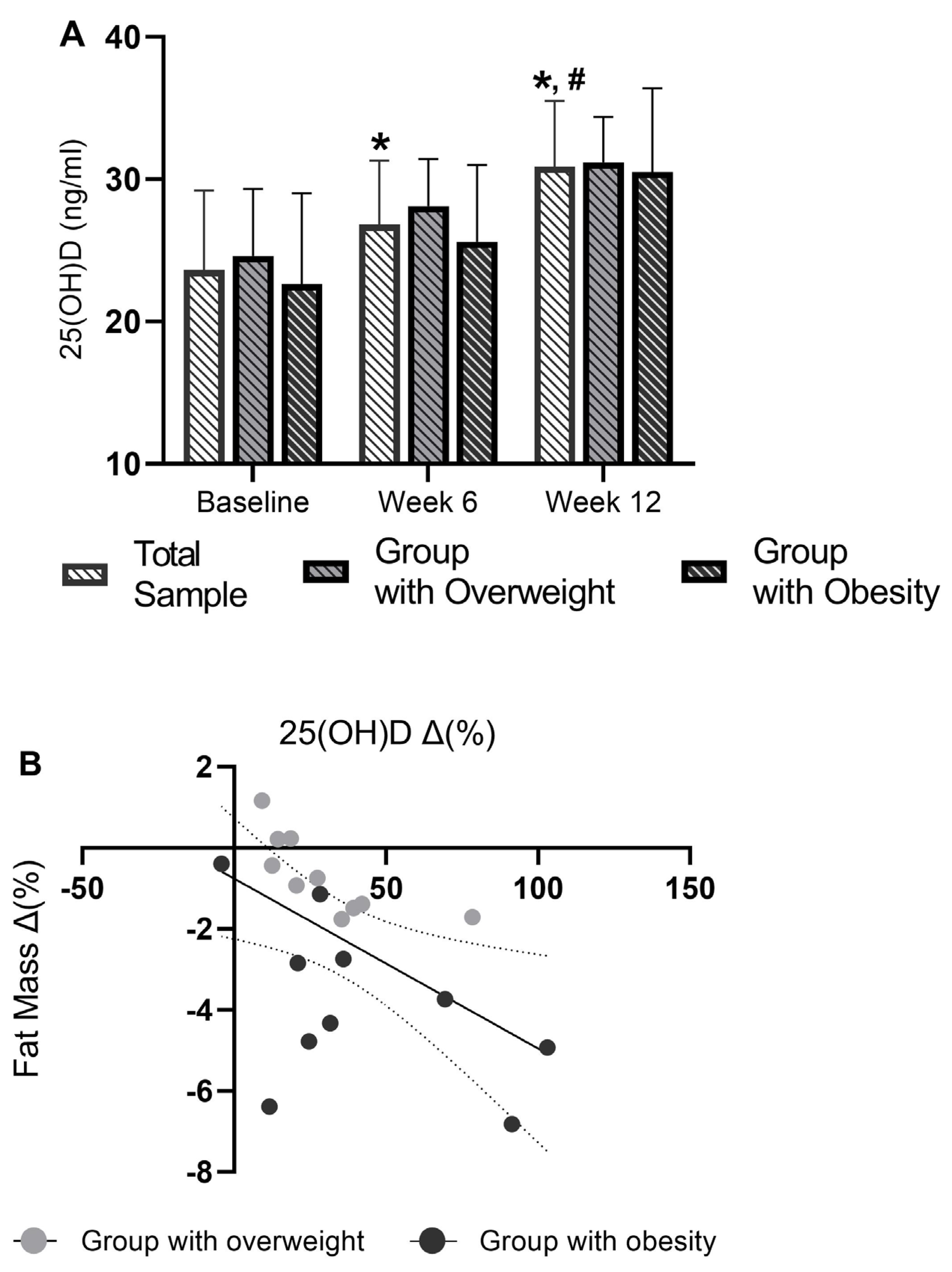
Serum indices of liver and bone metabolism are shown in Table 2. SGPT, SGPT/SGOT, γ-GT, and calcium remained constant over the 12-week training intervention. ALP and fetuin-A increased from 167.7 ± 32.1 U/L to 178.2 ± 44.1 U/L (p = 0.044) and from 24.2 ± 3.0 ng/mL to 26.6 ± 4.5 ng/mL (p = 0.031), respectively, at 6 weeks, but returned to baseline values at 12 weeks. SGOT displayed a significant rise of 10.3 U/L at 12 weeks compared to 6 weeks, but did not differ from baseline values. Subgroup analysis revealed no differences among groups (group with overweight vs. group with obesity).

3.5. Serum 25(OH)D

At baseline, out of 20 participants, 17 displayed either VitD deficiency (<20 ng/mL) or insufficiency (20–29 ng/mL) [22]. Training increased (η2 = 0.662) serum 25(OH)D by ~14% (+3.21 ng/mL, p = 0.002) and ~31% (+7.24 ng/mL, p < 0.001) at 6 and 12 weeks, respectively (Figure 1A). Serum 25(OH)D at 12 weeks increased in all participants, with 15 of them exhibiting sufficient (>30 ng/mL) concentrations and only 5 exhibiting insufficient (mean 25.5 ± 2.9 ng/mL) concentrations. Moreover, a negative linear relationship was observed between the percentage change in 25(OH)D and fat mass at 12 weeks (R = 0.53, p = 0.016) (Figure 1B).

4. Discussion

This study shows that participation in three 30 min m-HIIT sessions per week over a 3-month period can increase circulating 25(OH)D levels by 11–91% in adults with overweight/obesity and inadequate dietary VitD intake, regardless of outdoor physical activity levels. Of note, the response of serum 25(OH)D to m-HIIT was characterized by a large effect size (η2 = 0.662) and was inversely related to fat mass after the 3-month training period, suggesting that this adaptation may be mediated by the m-HIIT-induced mobilization of VitD from adipose tissue.
In individuals with overweight/obesity, the entrapment of VitD in adipose tissue due to insufficient lipolytic stimulation or impaired metabolic activity has been proposed as an underlying mechanism explaining VitD insufficiency/deficiency [4,23]. Considering the well-described role of exercise in stimulating lipolysis via improved adipocyte function and the increased activation of lipolytic enzymes [4], recent reports suggested that chronic exercise may favor the elevation of circulating 25(OH)D concentrations in individuals with overweight/obesity by inducing mobilization of VitD and its metabolites from AT [4,8]. Indeed, it was recently shown that participation in moderate-intensity cardiovascular-type exercise training over winter mitigates the decline in serum 25(OH)D levels in males and females with overweight, as compared to a control group [9]. Here, we present evidence that 12 weeks of m-HIIT may induce a clinically relevant [24] increase in circulating 25(OH)D levels (by 7.24 ng/mL or by 1.3 times) in adults with overweight/obesity and inadequate dietary VitD intake. Interestingly, the rise of 25(OH)D levels was not accompanied by changes in either participants’ dietary VitD intake or sun exposure over the 12-week training intervention. Sun exposure was estimated by monitoring participants’ habitual daily physical activity level between 10 a.m. and 3 p.m., which is the recommended sun exposure time frame for optimum VitD synthesis [21].
Furthermore, in line with the study by Perkin et al. [9], participants’ body mass remained constant over the training intervention in the present study. This might be explained by the fact that they were advised to retain their dietary habits during the training period and were not asked to strictly adhere to individualized dietary plans. However, we observed significant large reductions in total body fat mass (by 3% at 12 weeks, η2 = 0.249), android fat (by 3.7% at 6 weeks and 4.4% at 12 weeks, η2 = 0.316), and hepatorenal index (by 8% at 12 weeks, η2 = 0.175), suggesting the lipolytic effect of m-HIIT. Although lipolysis and adipocyte function were not assessed in this study, the m-HIIT protocol applied incorporates both resistance- and cardiovascular-based exercises, which have been previously shown to stimulate lipolysis in adults with overweight/obesity [5,6], into a single training session [10]. In addition, an inverse linear relationship was observed between the rise in 25(OH)D levels and the reduction in fat mass at 12 weeks. Taken together, these findings potentiate the scenario of the m-HIIT-induced mobilization of 25(OH)D from adipose tissue.
Considering the elevation of serum 25(OH)D concentration, we assessed biochemical indices of liver function in light of the crucial role of the liver in VitD metabolism. At 12 weeksm serum levels of SGPT, SGOT, SGPT/SGOT, ALP, γ-GT, and fetuin-A did not differ from their respective baseline values. Surprisingly, a significant increase in ALP and fetuin-A was noticed at 6 weeks compared to the baseline. This fluctuation might be indicative of the training-induced stimulation of bone turnover rather than altered liver function, as both ALP and fetuin-A are considered biomarkers of bone turnover rate [25,26]. Serum calcium was assessed in order to examine if the m-HIIT-induced upregulation of 25(OH)D was accompanied by enhanced calcium absorption, a crucial step for bone mineralization [25]. It is interesting that, although not statistically meaningful, a reduction of ~3% was observed in serum calcium concentration at 6 weeks. This finding makes us speculate that the m-HIIT stimulus accelerated the bone turnover rate over the first 6 weeks and that, as a response to increased calcium absorption in the intestines mediated by the elevated 25(OH)D levels, ALP and fetuin-A were increased as endocrine factors to promote the incorporation of calcium into bones [25,26].
Undoubtedly, we should acknowledge the lack of control group as a major limitation of this study, as this would allow us to take into account seasonal variation in VitD status. Seasonal variation of 25(OH)D is evident in Greece, with the lowest values observed in March (~13–20 ng/mL) and the highest in August (~29–30 ng/mL) [27,28]. In the present study, though, the rate of increment of serum 25(OH)D over the 12-week training intervention exceeded the season-dependent elevation typically seen over the same period (April to June) [27,28]. In addition, the use of m-HIIT saw our participants reach the highest 25(OH)D levels, commonly observed in August due to sun exposure in non-exercising individuals [27,28], in June. Therefore, these findings potentiate the m-HIIT-driven upregulation of ViD status, despite the absence of a control group. Furthermore, it should be noted here that data regarding the use of UV filters by the participants and questionnaires assessing sun exposure time were not collected.

5. Conclusions

In conclusion, our findings suggest that 12 weeks of m-HIIT can induce a clinically relevant elevation of circulating 25(OH)D levels in adults with overweight or obesity. This effect is inversely related to fat mass reduction. This preliminary evidence indicates that m-HIIT can favor the mobilization of VitD and its metabolites from adipose tissue. In conjunction with the findings from Perkin et al. [9], these findings confirm the efficacy of exercise in addressing VitD insufficiency/deficiency but also highlight the regulatory role of exercise mode (aerobic exercise vs. m-HIIT) and training load characteristics (a moderate intensity vs. a high intensity) in fat mass reduction and consequently VitD response.

Supplementary Materials

The following supporting information can be downloaded at: https://www.mdpi.com/article/10.3390/obesities5030051/s1, Figure S1: The CONSORT flow diagram of the study; Table S1: Quantification of participants’ internal load on a weekly basis; Table S2: Participants’ dietary intake and habitual physical activity at Baseline, 6 and 12 weeks.

Author Contributions

Conceptualization, M.P., D.D., A.A., G.M., I.G.F. and A.C.; methodology, M.P., T.S., D.P., N.O.R., D.B., A.P., N.Z., M.B. and I.K.; data curation, M.P., M.M., A.K., A.Z.J. and A.C.; writing—original draft preparation, M.P., D.D., A.A., T.J.A., I.G.F. and A.C.; writing—review and editing, all authors; project administration, A.C. and M.P.; formal analysis, A.C. and M.P. All authors have read and agreed to the published version of the manuscript.

Funding

This research received no external funding.

Institutional Review Board Statement

The study was conducted in accordance with the Declaration of Helsinki, and approved by the Institutional Research Ethics Committee of the Democritus University of Thrace (protocol code 40042/238-18 and date of approval: 18 March 2019). The study was preregistered at ClinicalTrials.gov (ID: NCT04098484).

Informed Consent Statement

Informed consent was obtained from all subjects involved in the study.

Data Availability Statement

The data that support the findings of this study are available from the corresponding author, upon reasonable request.

Acknowledgments

The authors are grateful to all participants for their contribution and commitment to this study.

Conflicts of Interest

The authors assert that the study was carried out without any commercial or financial affiliations that might be interpreted as a possible conflict of interest.

References

  1. Holick, M.F.; Chen, T.C. Vitamin D Deficiency: A Worldwide Problem with Health Consequences. Am. J. Clin. Nutr. 2008, 87, 1080S–1086S. [Google Scholar] [CrossRef] [PubMed]
  2. Pereira-Santos, M.; Costa, P.R.F.; Assis, A.M.O.; Santos, C.A.S.T.; Santos, D.B. Obesity and Vitamin D Deficiency: A Systematic Review and Meta-analysis. Obes. Rev. 2015, 16, 341–349. [Google Scholar] [CrossRef] [PubMed]
  3. Di Nisio, A.; De Toni, L.; Sabovic, I.; Rocca, M.S.; De Filippis, V.; Opocher, G.; Azzena, B.; Vettor, R.; Plebani, M.; Foresta, C. Impaired Release of Vitamin D in Dysfunctional Adipose Tissue: New Cues on Vitamin D Supplementation in Obesity. J. Clin. Endocrinol. Metab. 2017, 102, 2564–2574. [Google Scholar] [CrossRef] [PubMed]
  4. Hengist, A.; Perkin, O.; Gonzalez, J.T.; Betts, J.A.; Hewison, M.; Manolopoulos, K.N.; Jones, K.S.; Koulman, A.; Thompson, D. Mobilising Vitamin D from Adipose Tissue: The Potential Impact of Exercise. Nutr. Bull. 2019, 44, 25–35. [Google Scholar] [CrossRef]
  5. Chatzinikolaou, A.; Fatouros, I.; Petridou, A.; Jamurtas, A.; Avloniti, A.; Douroudos, I.; Mastorakos, G.; Lazaropoulou, C.; Papassotiriou, I.; Tournis, S.; et al. Adipose Tissue Lipolysis Is Upregulated in Lean and Obese Men During Acute Resistance Exercise. Diabetes Care 2008, 31, 1397–1399. [Google Scholar] [CrossRef]
  6. Petridou, A.; Chatzinikolaou, A.; Avloniti, A.; Jamurtas, A.; Loules, G.; Papassotiriou, I.; Fatouros, I.; Mougios, V. Increased Triacylglycerol Lipase Activity in Adipose Tissue of Lean and Obese Men During Endurance Exercise. J. Clin. Endocrinol. Metab. 2017, 102, 3945–3952. [Google Scholar] [CrossRef]
  7. Petridou, A.; Mougios, V. Acute Changes in Triacylglycerol Lipase Activity of Human Adipose Tissue during Exercise. J. Lipid Res. 2002, 43, 1331–1334. [Google Scholar] [CrossRef]
  8. Zhang, J.; Cao, Z.-B. Exercise: A Possibly Effective Way to Improve Vitamin D Nutritional Status. Nutrients 2022, 14, 2652. [Google Scholar] [CrossRef]
  9. Perkin, O.J.; Davies, S.E.; Hewison, M.; Jones, K.S.; Gonzalez, J.T.; Betts, J.A.; Jenkinson, C.; Lindsay, M.A.; Meadows, S.R.; Parkington, D.A.; et al. Exercise without Weight Loss Prevents Seasonal Decline in Vitamin D Metabolites: The VitaDEx Randomized Controlled Trial. Adv. Sci. 2025. [Google Scholar] [CrossRef]
  10. Batrakoulis, A.; Jamurtas, A.Z.; Draganidis, D.; Georgakouli, K.; Tsimeas, P.; Poulios, A.; Syrou, N.; Deli, C.K.; Papanikolaou, K.; Tournis, S.; et al. Hybrid Neuromuscular Training Improves Cardiometabolic Health and Alters Redox Status in Inactive Overweight and Obese Women: A Randomized Controlled Trial. Antioxidants 2021, 10, 1601. [Google Scholar] [CrossRef]
  11. Batrakoulis, A.; Jamurtas, A.Z.; Metsios, G.S.; Perivoliotis, K.; Liguori, G.; Feito, Y.; Riebe, D.; Thompson, W.R.; Angelopoulos, T.J.; Krustrup, P.; et al. Comparative Efficacy of 5 Exercise Types on Cardiometabolic Health in Overweight and Obese Adults: A Systematic Review and Network Meta-Analysis of 81 Randomized Controlled Trials. Circ. Cardiovasc. Qual. Outcomes 2022, 15, e008243. [Google Scholar] [CrossRef] [PubMed]
  12. Batrakoulis, A.; Loules, G.; Georgakouli, K.; Tsimeas, P.; Draganidis, D.; Chatzinikolaou, A.; Papanikolaou, K.; Deli, C.K.; Syrou, N.; Comoutos, N.; et al. High-intensity Interval Neuromuscular Training Promotes Exercise Behavioral Regulation, Adherence and Weight Loss in Inactive Obese Women. Eur. J. Sport. Sci. 2020, 20, 783–792. [Google Scholar] [CrossRef] [PubMed]
  13. Villareal, D.T.; Aguirre, L.; Gurney, A.B.; Waters, D.L.; Sinacore, D.R.; Colombo, E.; Armamento-Villareal, R.; Qualls, C. Aerobic or Resistance Exercise, or Both, in Dieting Obese Older Adults. N. Engl. J. Med. 2017, 376, 1943–1955. [Google Scholar] [CrossRef] [PubMed]
  14. Colleluori, G.; Aguirre, L.; Phadnis, U.; Fowler, K.; Armamento-Villareal, R.; Sun, Z.; Brunetti, L.; Hyoung Park, J.; Kaipparettu, B.A.; Putluri, N.; et al. Aerobic Plus Resistance Exercise in Obese Older Adults Improves Muscle Protein Synthesis and Preserves Myocellular Quality Despite Weight Loss. Cell Metab. 2019, 30, 261–273.e6. [Google Scholar] [CrossRef]
  15. Díaz-Buschmann, I.; Jaureguizar, K.V.; Calero, M.J.; Aquino, R.S. Programming Exercise Intensity in Patients on Beta-Blocker Treatment: The Importance of Choosing an Appropriate Method. Eur. J. Prev. Cardiol. 2014, 21, 1474–1480. [Google Scholar] [CrossRef]
  16. Coates, A.M.; Joyner, M.J.; Little, J.P.; Jones, A.M.; Gibala, M.J. A Perspective on High-Intensity Interval Training for Performance and Health. Sports Med. 2023, 53, 85–96. [Google Scholar] [CrossRef]
  17. Cipryan, L.; Laursen, P.B.; Plews, D.J. Cardiac Autonomic Response Following High-intensity Running Work-to-rest Interval Manipulation. Eur. J. Sport. Sci. 2016, 16, 808–817. [Google Scholar] [CrossRef]
  18. Draganidis, D.; Jamurtas, A.; Stampoulis, T.; Laschou, V.; Deli, C.; Georgakouli, K.; Papanikolaou, K.; Chatzinikolaou, A.; Michalopoulou, M.; Papadopoulos, C.; et al. Disparate Habitual Physical Activity and Dietary Intake Profiles of Elderly Men with Low and Elevated Systemic Inflammation. Nutrients 2018, 10, 566. [Google Scholar] [CrossRef]
  19. Marshall, R.H.; Eissa, M.; Bluth, E.I.; Gulotta, P.M.; Davis, N.K. Hepatorenal Index as an Accurate, Simple, and Effective Tool in Screening for Steatosis. Am. J. Roentgenol. 2012, 199, 997–1002. [Google Scholar] [CrossRef]
  20. Cohen, P.; Cohen, P.; West, S.G.; Aiken, L.S. Applied Multiple Regression/Correlation Analysis for the Behavioral Sciences; Psychology Press: London, UK, 2014; ISBN 9781135468255. [Google Scholar]
  21. Holick, M.F. Vitamin D Deficiency. N. Engl. J. Med. 2007, 357, 266–281. [Google Scholar] [CrossRef]
  22. Holick, M.F.; Binkley, N.C.; Bischoff-Ferrari, H.A.; Gordon, C.M.; Hanley, D.A.; Heaney, R.P.; Murad, M.H.; Weaver, C.M. Evaluation, Treatment, and Prevention of Vitamin D Deficiency: An Endocrine Society Clinical Practice Guideline. J. Clin. Endocrinol. Metab. 2011, 96, 1911–1930. [Google Scholar] [CrossRef] [PubMed]
  23. Karampela, I.; Sakelliou, A.; Vallianou, N.; Christodoulatos, G.-S.; Magkos, F.; Dalamaga, M. Vitamin D and Obesity: Current Evidence and Controversies. Curr. Obes. Rep. 2021, 10, 162–180. [Google Scholar] [CrossRef] [PubMed]
  24. Rupprecht, M.; Wagenpfeil, S.; Schöpe, J.; Vieth, R.; Vogt, T.; Reichrath, J. Meta-Analysis of European Clinical Trials Characterizing the Healthy-Adult Serum 25-Hydroxyvitamin D Response to Vitamin D Supplementation. Nutrients 2023, 15, 3986. [Google Scholar] [CrossRef] [PubMed]
  25. LeBoff, M.S.; Greenspan, S.L.; Insogna, K.L.; Lewiecki, E.M.; Saag, K.G.; Singer, A.J.; Siris, E.S. Correction to: The Clinician’s Guide to Prevention and Treatment of Osteoporosis. Osteoporos. Int. 2022, 33, 2243. [Google Scholar] [CrossRef]
  26. Gao, H.; Peng, X.; Li, N.; Gou, L.; Xu, T.; Wang, Y.; Qin, J.; Liang, H.; Ma, P.; Li, S.; et al. Emerging Role of Liver-Bone Axis in Osteoporosis. J. Orthop. Transl. 2024, 48, 217–231. [Google Scholar] [CrossRef]
  27. Katrinaki, M.; Kampa, M.; Margioris, A.; Castanas, E.; Malliaraki, N. Vitamin D Levels in a Large Mediterranean Cohort: Reconsidering Normal Cut-off Values. Hormones 2016, 15, 205–223. [Google Scholar] [CrossRef]
  28. Papadakis, G.; Keramidas, I.; Kakava, K.; Pappa, T.; Villiotou, V.; Triantafillou, E.; Drosou, A.; Tertipi, A.; Kaltzidou, V.; Pappas, A. Seasonal Variation of Serum Vitamin D among Greek Female Patients with Osteoporosis. In Vivo 2015, 29, 409–413. [Google Scholar]
Figure 1. (A) Participants’ plasma Vitamin 25(OH)D levels at baseline, 6, and 12 weeks. (B) Correlation between the percent change in Vitamin 25(OH)D levels at 12 weeks and the percent change in fat mass at 12 weeks. * indicates significant difference with baseline (p < 0.05). # indicates significant difference with week 6 (p < 0.05).
Obesities 05 00051 g001
Table 1. Participants’ body composition at baseline, 6, and 12 weeks.
BaselineWeek 6Week 12Fvaluesp ValueEffect Size
Body Mass (kg)
Total sample (n = 20)99.7 ± 13.1100.0 ± 12.999.3 ± 12.9F(2,38) = 3.067p = 0.058η2 = 0.140
Group with overweight (n = 10)88.3 ± 3.188.7 ± 3.087.8 ± 2.7F(2.36) = 0.216p = 0.807η2 = 0.012
Group with obesity (n = 10)112.2 ± 7.9 ¥111.3 ± 7.7 ¥110.7 ± 7.4 ¥
Fat mass (kg)
Total sample (n = 20)34.6 ± 8.234.1 ± 7.833.7 ± 7.6 *F(2.38) = 5.244p = 0.010η2 = 0.249
Group with overweight (n = 10)27.8 ± 2.4 27.5 ± 1.827.6 ± 2.3F(2.36) = 3.572p = 0.038η2 = 0.166
Group with obesity (n = 10)41.4 ± 5.8 ¥40.6 ± 5.5 ¥39.9 ± 5.8 *
Android fat (kg)
Total sample (n = 20)4.29 ± 0.964.13 ± 0.89 *4.10 ± 0.92 *F(2.38) = 7.823p = 0.001η2 = 0.316
Group with overweight (n = 10)3.78 ± 0.873.70 ± 0.863.68 ± 0.94F(2.36) = 2.188p = 0.127η2 = 0.108
Group with obesity (n = 10)4.81 ± 0.78 ¥4.55 ± 0.71 ¥4.53 ± 0.70 ¥
Lean mass (kg)
Total sample (n = 20)61.8 ± 5.662.6 ± 5.762.2 ± 5.9F(2.38) = 2.475p = 0.098η2 = 0.128
Group with overweight (n = 10)57.3 ± 3.058.1 ± 3.157.0 ± 3.1F(2.36) = 2.212p = 0.124η2 = 0.109
Group with obesity (n = 10)66.2 ± 3.6 ¥67.1 ± 3.6 ¥67.3 ± 2.0 ¥
Hepatorenal Index
Total sample (n = 20)1.31 ± 0.151.26 ± 0.181.20 ± 0.14 *F(2.38) = 3.948p = 0.028η2 = 0.175
Group with overweight (n = 10)1.29 ± 0.141.21 ± 0.171.16 ± 0.12F(2.36) = 0.421p = 0.660η2 = 0.023
Group with obesity (n = 10)1.33 ± 0.171.32 ± 0.171.24 ± 0.17
* indicates significant difference with Baseline (p < 0.05); ¥ indicates significant difference between subgroups (group with overweight vs. group with obesity, p < 0.05).
Table 2. Biochemical indices of liver function and bone metabolism at baseline, 6, and 12 weeks.
BaselineWeek 6Week 12F2,38p ValueEffect Size
SGPT (U/L)
Total sample (n = 20)39.9 ± 12.637.6 ± 942.4 ± 7.3F(2.38) = 2.475p = 0.098η2 = 0.116
Group with overweight (n = 10)35.5 ± 7.034.0 ± 5.438.8 ± 2.1F(2.36) = 0.12p = 0.890η2 = 0.006
Group with obesity (n = 10)44.5 ± 15.541.2 ± 10.746.0 ± 8.9
SGOT (U/L)
Total sample (n = 20)36.8 ± 11.732.9 ± 9.943.2 ± 12.6 #F(2.38) = 8.526<0.001η2 = 0.312
Group with overweight (n = 10)36.0 ± 9.331.4 ± 6.643.3 ± 11.3F(2.36) = 0.21p = 0.811η2 = 0.012
Group with obesity (n = 10)37.7 ± 14.234.6 ± 12.643.2 ± 13.8
SGPT/SGOT
Total sample (n = 20)1.12 ± 0.271.20 ± 0.381.05 ± 0.34F(2.38) = 2.033p = 0.145η2 = 0.097
Group with overweight (n = 10)1.03 ± 0.241.15 ± 0.420.97 ± 0.33F(2.36) = 0.13p = 0.879η2 = 0.007
Group with obesity (n = 10)1.22 ± 0.291.26 ± 0.351.14 ± 0.36
ALP (U/L)
Total sample (n = 20)167.6 ± 31.2178.2 ± 44 *163.8 ± 37 #F(2.38) = 6.851p = 0.003η2 = 0.304
Group with overweight (n = 10)157.1 ± 16.3167.6 ± 27.4162.2 ± 28.8F(2.36) = 3.83p = 0.031η2 = 0.176
Group with obesity (n = 10)178.2 ± 39.2188.8 ± 55.7162.2 ± 28.8
γ-GT (U/L)
Total sample (n = 20)27.9 ± 1124.5 ± 9.124.9 ± 9.5F(2.38) = 1.779p = 0.183η2 = 0.086
Group with overweight (n = 10)26.0 ± 10.822.7 ± 5.721.9 ± 7.7F(2.36) = 0.2p = 0.821η2 = 0.011
Group with obesity (n = 10)30.0 ± 11.526.4 ± 11.728.0 ± 10.4
Fetuin-A (ng/mL)
Total sample (n = 20)24.2 ± 326.6 ± 4.5 *25.7 ± 4.7F(2.38) = 3.395p = 0.044η2 = 0.156
Group with overweight (n = 10)23.7 ± 2.026.3 ± 4.926.1 ± 5.8F(2.36) = 0.65p = 0.529η2 = 0.035
Group with obesity (n = 10)24.7 ± 3.826.9 ± 4.325.2 ± 3.4
Calcium (mg/dL)
Total sample (n = 20)8.90 ± 0.718.64 ± 0.628.97 ± 0.96F(2.38) = 2.045p = 0.159η2 = 0.100
Group with overweight (n = 10)9.01 ± 0.478.72 ± 0.388.94 ± 0.5F(2.36) = 0.5p = 0.608η2 = 0.027
Group with obesity (n = 10)8.79 ± 0.98.57 ± 0.819.07 ± 1.35
* indicates a significant difference from the baseline (p < 0.05). # indicates a significant difference from week 6 (p < 0.05).
Disclaimer/Publisher’s Note: The statements, opinions and data contained in all publications are solely those of the individual author(s) and contributor(s) and not of MDPI and/or the editor(s). MDPI and/or the editor(s) disclaim responsibility for any injury to people or property resulting from any ideas, methods, instructions or products referred to in the content.

