Article

Hydrogen-Rich Water Prevents Dehydration-Induced Cellular Oxidative Stress and Cell Death in Human Skin Keratinocytes

1
Department of Pharmacology, School of Life Dentistry at Tokyo, The Nippon Dental University, Tokyo 102-8159, Japan
2
Faculty of Life Sciences, Prefectural University of Hiroshima, Hiroshima 727-0023, Japan
*
Author to whom correspondence should be addressed.
Hydrogen 2022, 3(1), 62-71; https://doi.org/10.3390/hydrogen3010005
Submission received: 21 December 2021 / Revised: 18 January 2022 / Accepted: 24 January 2022 / Published: 28 January 2022
(This article belongs to the Special Issue Feature Papers in Hydrogen)

Abstract

:
Hypohydration is linked to increased risk of a variety of diseases and can be life-threatening, especially in elderly populations. Dehydration induces cellular damage partially through the production of reactive oxygen species (ROS) in cells, tissues and organs. Hydrogen molecules are known to convert ROS to harmless water. Therefore, theoretically hydrogen-rich water (HW) might eliminate dehydration-induced ROS and reverse its harmful effects in cells. In this in vitro study, we demonstrated that air-drying for 5 min could induce ROS generation in both nucleus and cytoplasm of human keratinocytes HaCaT as quantified by CellROX® Green/Orange reagents (Thermo Fisher Scientific, Waltham, Massachusetts, U.S.), respectively. Conversely, when the air-drying time was increased to 10 and 20 min, HaCaT cells lost the ability to produce ROS. Scanning electron microscopic (SEM) images showed that 10 min air-drying could induce severe membrane damage in HaCaT cells. PrestoBlue assay showed that, when HaCaT cells were air-dried for 20 min, cell viability was decreased to 27.6% of the control cells 48 h later. However, once HaCaT cells were pretreated with HW-prepared media, dehydration-induced intracellular ROS, cell membrane damage and cell death were significantly reduced as compared with double distilled water (DDW) under the same conditions. In conclusion, our data suggested that HW can decrease dehydration-induced harmful effects in human cells partially through its antioxidant capacity.

1. Introduction

Dehydration leads to hypohydration, which occurs when an organism does not have enough water to carry out its normal functions. During water depletion, the formation of intracellular reactive oxygen species (ROS) increases. Over produced ROS result in lipid peroxidation, denaturation of proteins and nucleic acid alteration. These oxidative lesions consequently induce severe damage to cells both in vitro and in vivo [1]. Skin epidermis is the first barrier to protect the body from dehydration. This barrier function is mainly provided by keratinocytes, the major cells of epidermis [2]. However, little is known about dehydration-induced pathological changes in keratinocytes at cellular and molecular levels.
Superoxide anion (O2), hydroxyl radicals (•OH) and hydrogen peroxide (H2O2) are the well-known toxic chemical species of ROS. As a novel antioxidant, molecular hydrogen (H2) can convert the toxic ROS, hydroxyl radicals, superoxide anion, and hydrogen peroxide to harmless water (2•OH + H2 → 2H2O, O2 + 2H2 → 2H2O, H2O2 + H2 → 2H2O) [3,4,5]. Therefore, theoretically hydrogen not only eliminates dehydration-generated intracellular ROS but also supplies water to cells. In 2007, Ohsawa et al. reported that hydrogen acts as a therapeutic antioxidant by selectively reducing cytotoxic oxygen radicals in both cultured cells and a rat model of ischemia [6]. Since then, the biological activities of hydrogen gas and hydrogen-rich water (HW) have been considerably studied in human subjects, animal models and cultured cells. Results from randomized controlled clinical trials showed that daily administration of HW could improve lipid metabolism and insulin resistance in patients with type 2 diabetes [7]. In heathy adults, drinking HW could increase serum antioxidant capacity, inflammatory responses [8], enhance endurance and relieve psychometric fatigue during exercise [9]. In vivo studies showed that drinking HW could improve senescence-related neurodegenerative diseases in animal models [10,11]. In our previous in vitro studies, we have demonstrated that hydrogen-rich water (HW) could reduce oxidants (such as H2O2 and cumene hydroperoxide)-induced ROS generation and harmful effects in human fibroblasts and keratinocytes [4]. Hydrogen-generating silica material could suppress UVA-induced oxidative stress, cell death and collagen loss in three dimensional skin equivalents [5]. Moreover, hydrogen nano-bubble water showed inhibitory effects on phorbol-myristate-acetate (PMA; an inducer of endogenous superoxide anion and hydrogen peroxide)-induced inflammatory cytokines secretion in adipocytes [12]. This evidence suggests that HW can protect skin keratinocytes from dehydration-induced cellular oxidative stress and injuries. However, up to date, the effects of HW on dehydration-induced injuries in cultured cells have yet to be investigated. In the present study, we are the first to investigate protective effects of HW on dehydration-induced harmful effects to cell morphology, membrane integrity and cell viability in human keratinocytes.

2. Materials and Methods

2.1. Hydrogen-Rich Water Preparation

Hydrogen-rich water was prepared with a hydrogen water server (Gaura Mini, Gaura Inc., Tokyo, Japan). The concentrations of dissolved hydrogen (DH) in water were detected with both a polarographic diaphragm type device with dissolved hydrogen-selective electrode (DH meter, KM2100DH, Kyoei Electronics Research Institute, Saitama, Japan) according to our previous report [4]. The oxidation-reduction potential (ORP) of HW was measured by an ORP electrometer (YK-23RP-ADV, SATOTECH, Kanagawa, Japan). The measurements were performed with the manufacturers’ instructions. To confirm whether temperature change affects the characteristics of HW, we checked DH and ORP in both freshly prepared HW and boiled HW (cooling down to room temperature because the electrometers’ requirement) (Table 1).
For cell treatment, we prepared DMEM medium with HW (Table 2) and found out that the DH in HW-prepared DMEM medium was about 460 µg/L.

2.2. Cell Culture

Immortalized human skin epidermal keratinocytes HaCaT were kindly provided from Professor Norbert E. Fusenig of Deutsches Krebsforschungszentrum (Heidelberg, Germany) [13]. Cells were maintained in Dulbecco’s modified Eagle’s medium (DMEM) (Gibco) supplemented with 10% FBS, 1% Gibco® GlutaMAX™ Supplement (Thermo Fisher Scientific, Tokyo, Japan), 100 units/mL penicillin and 10 mg/mL streptomycin in a 5% CO2-atmosphere at 37 °C [14].

2.3. Drying Experiment

At 2 h after HaCaT cells being cultivated in HW-DMEM or DDW (deionized distilled water)-DMEM (Table 1), the culture medium was completely sucked up by an air pump (AP-115N, Iwaki, Tokyo, Japan) of speed as high as 13/15 L/min for 5 s. Cells were then left at a room temperature (22–24 °C) under 35–40% moisture for 5, 10 and 20 min. Regular culture media was added to the cell monolayer after drying.

2.4. Giemsa Stain

At the end of cultivation, HaCaT cells were fixed with 100% methanol and stained by Giemsa’s azur-eosin-methylene blue solution (Sigma-Aldrich, Tokyo, Japan). The samples were then observed by a phase-contrast microscope (ECLIPSE Ts2, Nikon, CO., Tokyo, Japan).

2.5. Cell Viability Assay

Cell viability in HaCaT cells were measured by the cell-permeable dye resazurin- based PrestoBlue® Assay according to the manufacturer’s protocol. At the end of cultivation, HaCaT cells were incubated for 3 h at 37 °C with fresh medium supplemented with 10% PrestoBlue® (v/v; A13261, Thermo Fisher Scientific, Waltham, MA, USA). The PrestoBlue® reduction by the cells expressed as fluorescence intensity units was measured by using a microplate reader (SH-9000Lab, HITACHI, Tokyo, Japan) with excitation/emission at 560 nm/590 nm [15].

2.6. Cellular ROS Detection

Immediately after drying, cellular ROS generation in HaCaT cells was detected with the CellROX® Orange or Green Reagent (Thermo Fisher Scientific, Waltham, MA, USA) according to the manufacturer’s recommended protocol. ROS production in cells was observed by the EVOS® FL Cell Imaging System (Thermo Fisher Scientific, Waltham, MA, USA). To quantify the cellular ROS, green and red fluorescence in 50–100 HaCaT cells from at least 10 images were analyzed with an Image J software [4].

2.7. Scanning Electron Microscope Analysis

HaCaT cells were pre-fixed for 90 min in 2% paraformaldehyde and 2.5% glutaraldehyde solution in phosphate buffer (pH 7.4). After being washed with phosphate buffer, specimens were further fixed with 1% osmium tetroxide (OsO4). Samples were then washed with distilled water and dehydrated with ascending grades of ethanol. The specimens were substituted by t-butylalcohol and freeze-dried with a vacuum evaporator. The dried samples were mounted on stubs and then coated with thin layer of gold in a magnetron ion bombarder. The preparations were examined at 5–10 kV using a scanning electron microscope (S-4000, HITACHI, Ltd., Tokyo, Japan) [16,17].

2.8. Statistical Analysis

Statistical analysis was performed according to our previous report [17]. GNU PSPP Statistical Analysis Software (version 0.8.2-gad9374) (https://www.gnu.org/software/pspp/, accessed on 12 December 2021) and EZ Analyze Excel-based tools (http://www.ezanalyze.com/, accessed on 12 December 2021) were used for analyzing data obtained from each experiment. All data, expressed as the mean ± SD were normally distributed. For multiple comparisons among groups, we performed one-way analysis of variance (ANOVA), followed by the post hoc tests (Bonferroni Correction). p < 0.05 was considered showing statistical significance. We independently repeated all experiments 3 more times.

3. Results

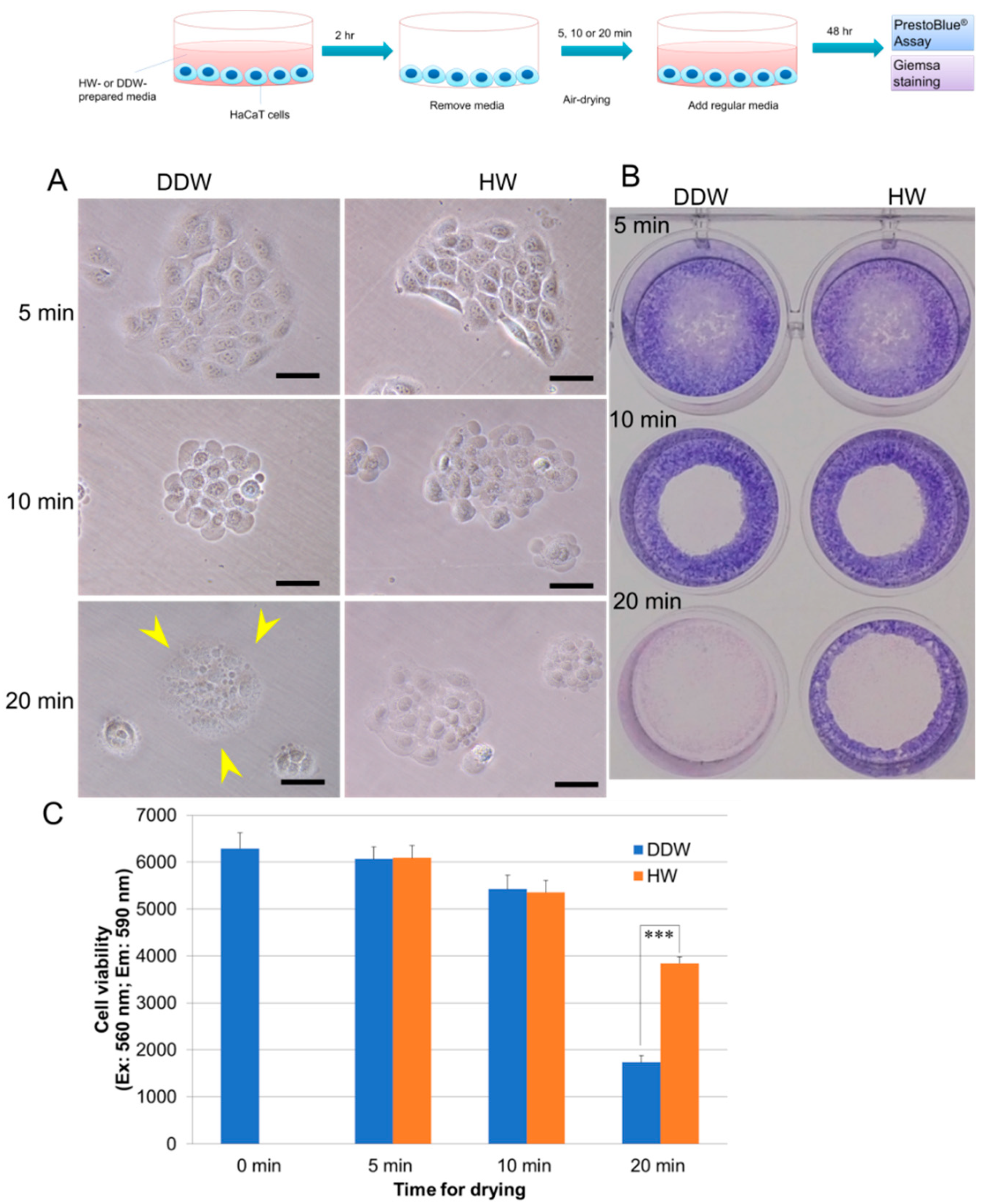
3.1. HW Prevents Dehydration-Induced Cell Death in Human Skin Keratinocytes HaCaT

To confirm whether dehydration induces cellular damage in HaCaT cells, we first tested cell viability of HaCaT cells 48 h after air-drying. Figure 1A showed that 5 min air-drying did not cause morphological changes, whereas 10 and 20 min air-drying caused membrane shrinkage in HaCaT cells. At 48 h after air-drying, Giemsa staining showed that 10 and 20 min air-drying markedly reduced cell density around the center of the culture well, especially the 20 min air-drying (Figure 1B). PrestoBlue assay showed that at 48 h after air-drying, cell viabilities of 5, 10 and 20 min air-drying in DDW-pretreated cells were 96.5%, 86.3% (p = 0.001 vs. 0 min) and 27.6% (p < 0.001 vs. 0 min) of the negative control cells (0 min) (Figure 1C). However, when HaCaT cells were pretreated with HW-prepared medium for 2 h, 20 min air-drying-induced morphological changes were significantly relieved, and cell viability was markedly increased to 61.1% of control (p < 0.001). These data suggested that HW could reduce dehydration-induced cell death.

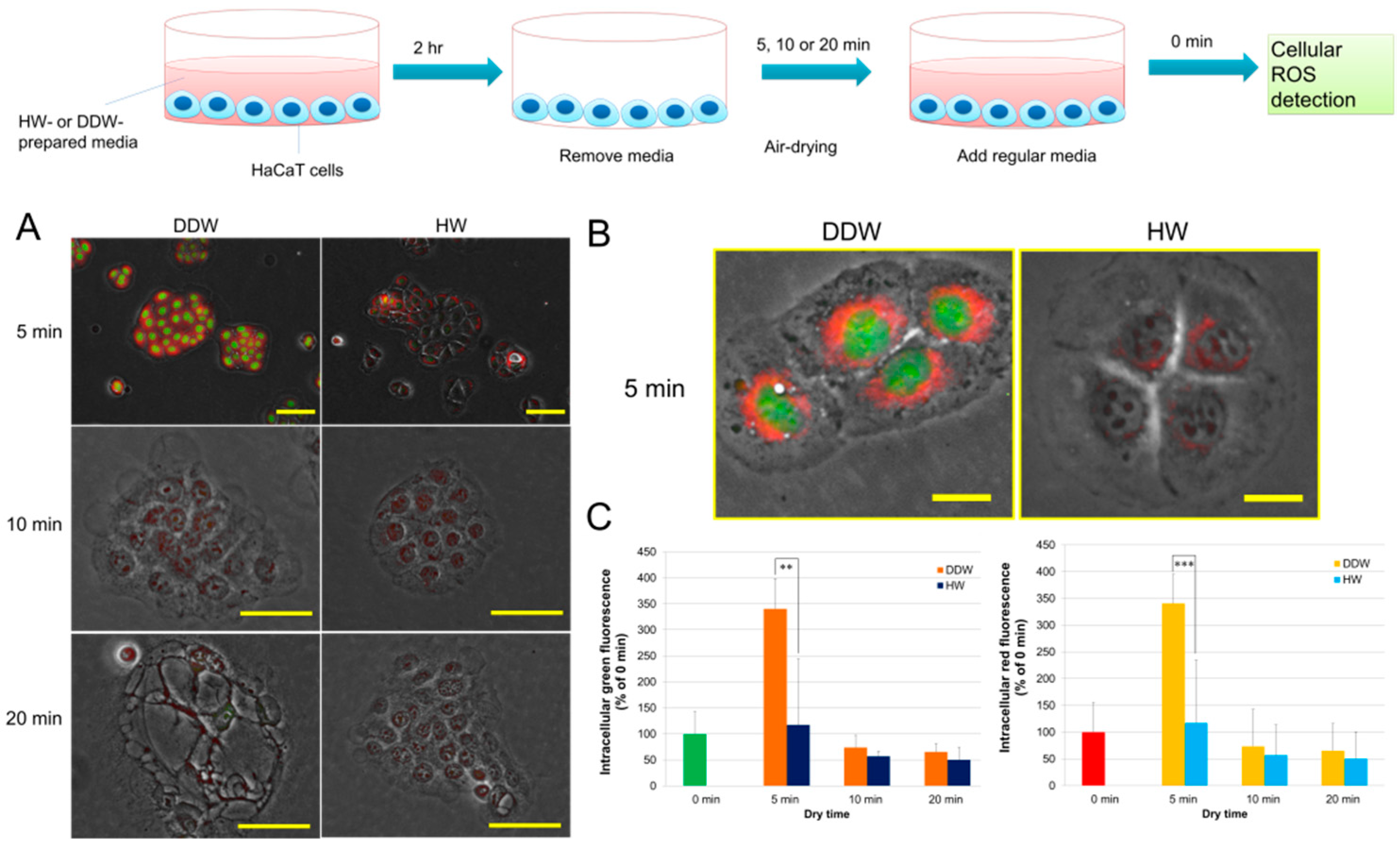
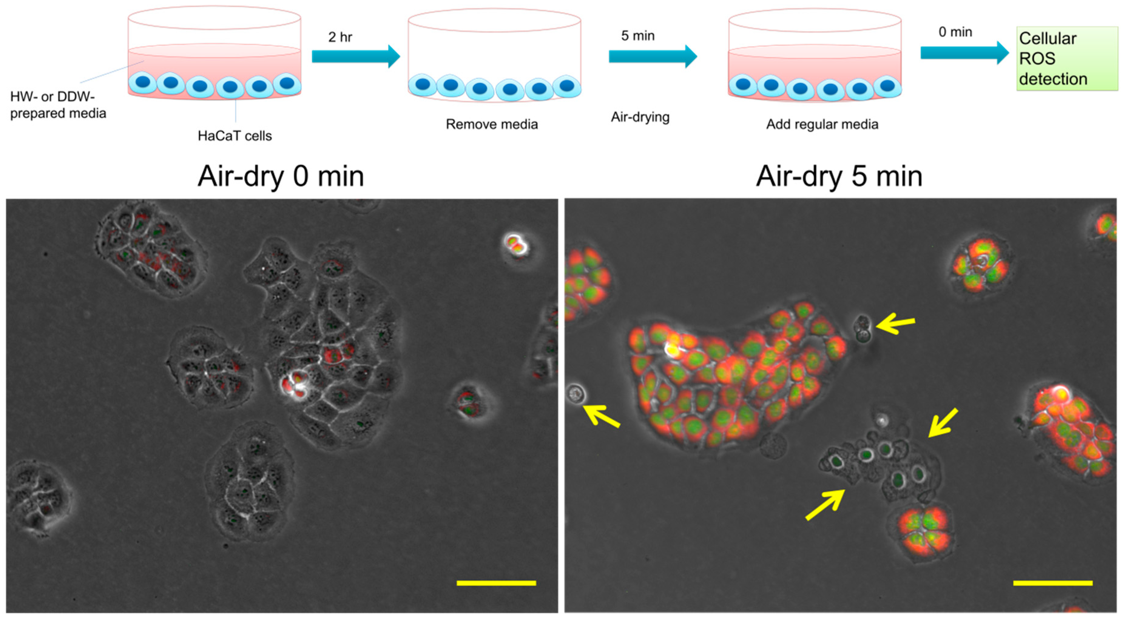
3.2. HW Diminishes 5 min Air-Drying-Induced Intracellular ROS Generation in HaCaT Cells

We further examined whether dehydration induces intracellular ROS generation. As shown in Figure 2, compared to negative control (air-dry 0 min), 5 min air-drying induced massive ROS production both in the nuclei and cytoplasm. Interestingly, when cells were seriously damaged, both nuclei and cytoplasm ROS almost vanished, suggesting that cells lost the ability to produce ROS. Figure 3A,B showed that, as compared to DDW-pretreated cells, intracellular ROS in HW-pretreated cells were more appreciably limited. Results for Image J analysis confirmed that, as compared to the negative control (0 min), in DDW-pretreated cells, the levels of nuclei ROS were 445.6% (air-dry 5 min) (p < 0.001 vs. 0 min), 53.3% (10 min) and 27.5% (20 min) (p < 0.01 vs. 0 min), and cytoplasm ROS were 340.6% (5 min) (p < 0.001 vs. 0 min), 73.6% (10 min) and 65.4% (20 min). HW-pretreatment significantly reduced 5 min air-dry-induced ROS generation to 240.8% (nuclei ROS) (p < 0.01 vs. DDW) and 117.4% (cytoplasm ROS) (p < 0.001 vs. DDW) (Figure 3C). However, 10 and 20 min air-drying reduced ROS generation in both DDW- and HW-pretreated HaCaT cells, suggesting that long time air-dry-induced severe damage deprived their ability to produce ROS.
Cellular ROS in HaCaT cells was immediately detected after air-drying with CellROX® Green and Orange dyes and observed with a fluorescence microscope. Illustration on the top represents procedures of the experiments. Typical fluorescence images of HaCaT cells display that, as compared to cells without air-drying (left panel), 5 min air-dried HaCaT cells produced intracellular ROS. Interestingly, some cells lost their ability to generate ROS because their damage was so serious (yellow arrows). Green, nuclei ROS; red, cytoplasm ROS. Scale bars = 50 µm.
Figure 3. Effects of HW on hypohydration-induced cellular oxidative stress in HaCaT keratinocytes. Cellular ROS was immediately detected as same as in Figure 2. Illustration on the top represents procedures of the experiments. (A) Typical fluorescence images of HaCaT cells. Green, nuclei ROS; Red, cytoplasm ROS. Scale bars = 20 µm. (B) Scale bars = 10 μm. (C) To quantify the cellular ROS, green and red fluorescence in HaCaT cells were analyzed with an Image J software as described in “Materials and Methods”. **, p < 0.01, ***, p < 0.01. Each bar represents the mean ± SD of three independent experiments.
Hydrogen 03 00005 g003

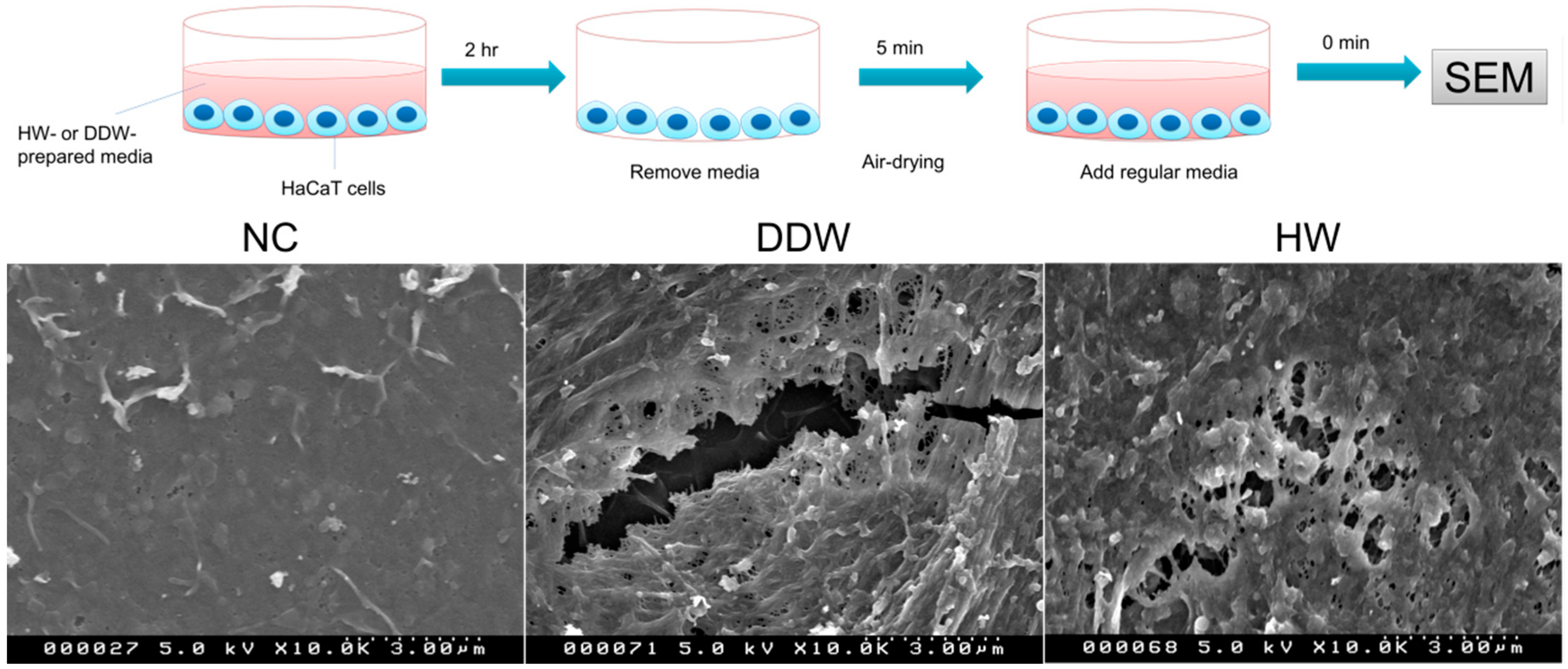
3.3. HW Reduces 10 min Air-Drying-Induced Apoptosis-like Cell Death and Membrane Damage in HaCaT Cells

We noticed that, after 10 and 20 min air-drying, some bleb-/blister-like protrusions appeared in the surface of HaCaT cells. These protrusions continuously existed on the cell surface even 48 h later (Figure 4 upper panel). SEM analysis showed that on the surface of 10 min air-dried HaCaT cells, apoptotic bodies appeared, suggesting that dehydration caused apoptosis-like death. However, in HW-pretreated cells, bleb-like protrusions and apoptotic bodies were more markedly prevented than in DDW-pretreated cells.
SEM analysis further showed that, as compared to the negative control, 5 min air-drying induced the extent membrane-fracture in DDW-pretreated cells. Nevertheless, HW-pretreatment diminished the fracture in HaCaT cells (Figure 5).
These data suggested that HW could reduce dehydration-induced apoptosis-like cell death and membrane damage in HaCaT cells.
Figure 4. Preventive effects of HW on dehydration-induced apoptosis-like cell death in HaCaT keratinocytes.
Hydrogen 03 00005 g004
HaCaT keratinocytes (1 × 10⁴ cells/well) were seeded into micro-cover glasses in a 6-well plate and treated as in Figure 1. Cells were air-dried for 10 min, and then followed by SEM as described in “Materials and Methods”. The illustration on the top represents the procedures of the experiments. The upper panel shows typical phase-contrast microscopic images (scale bars = 15 µm), and the lower panel exhibits SEM images of HaCaT cells 48 h after drying (scale bars = 3 µm), respectively.
Figure 5. Effects of HW on hypohydration-induced membrane damage in HaCaT keratinocytes.
Hydrogen 03 00005 g005
HaCaT cells were prepared as same as in Figure 4. Top panel illustrates procedures of the experiments. Typical SEM images showed the center region of cell membrane in one single HaCaT cell.

4. Discussion

Dehydration is the process of losing water, which leads to hypohydration. In the elderly, hypohydration easily occurs due to aged people having a lower percentage of body water, an impaired thirst sensitivity, less effective arginine vasopressin (AVP) and weakened ability for urinary concentration [18]. Chronic systemic hypohydration has been considered related to infection, renal calculus, stroke, venous thromboembolism, and other cardiovascular diseases [19,20,21]. The magnitude of hypohydration represents a gradual body water loss from mild = 1% to 5%, moderate = 5% to 10%, to severe ≥10% body mass deficit [22]. If a fluid deficit is greater than 5%, the patient will develop serious symptoms and organ damage. Animal studies showed that in camels, long-term dehydration could cause oxidative stress in kidney cortex and apoptosis in cortex and medulla with significantly elevated inflammatory cytokines [23]. Experimental research displayed that dehydration increases cell membrane permeability and induces variety of harmful cellular events resulting in cell dysfunction and cell death in human dermal fibroblasts and other mammalian cells [24]. Oxidative stress is one of the most deleterious events during water depletion. It has been reported that dehydration generates 10 times more ROS production in yeast cells [25] and increases cell membrane damage and membrane permeability in plants [26,27]. Dehydration also decreases the activities of endogenous antioxidants, superoxide dismutase and glutathione (GSH) in frogs [28]. However, up to now, no clinical or experimental studies showed the relationship among the magnitude of hypohydration, ROS production and related pathological changes in cells.
In the present study, we used a staggered time for air-drying HaCaT keratinocytes. Cell viability and morphological changes of HaCaT represented an increasing deterioration in an air-drying time dependent manner. This observation might represent the magnitude of hypohydration in the human body, as follows: 5 min, 10 min and 20 min air-drying to keratinocytes reflect mild, moderate and severe hypohydration, respectively. Our data showed that only 5 min air-dry could induce ROS generation in HaCaT keratinocytes, whereas longer air-dry (10 and 20 min) time suppressed ROS production. Coexistent healthier cells and severely damaged cells demonstrated that injured cells did not produce ROS (Figure 2). When the air-drying time was lengthened to 10 min, membrane disturbances and apoptotic bodies occurred in the dehydrated HaCaT cells. These harmful effects became more severe and cell viability was markedly decreased, when the air-drying time was prolonged to 20 min. As expected, HaCaT cells lost the ability to produce intracellular ROS. These observations demonstrated that ROS production is not consistent with the severity of cellular damage during dehydration. The results also suggested that severe water depletion probably injured cells through other factors other than ROS. However, once HaCaT cells were pre-treated with HW-prepared media, intracellular ROS generation, membrane disturbances and apoptosis-like cell death were significantly reduced together with a marked increase in cell viability. These data suggested that HW could protect keratinocytes from dehydration-induced harmful effects. HW probably protects cells from dehydration through the following two different mechanisms: one is by scavenging dehydration-caused ROS to relieve the cytotoxicity via four-electron reduction, resulting in prevention against apoptotic events such as caspase-3 signal transduction, mitochondrial cytochrome-c leakage and p53 upregulation, and the other one is that HW might permeate into depth of living tissues through the minimum Stokes radius and amphiphilicity of hydrogen molecules to rebalance dehydration-induced electrolyte imbalance. It has been reported that GSH plays an important role in protecting cell membranes suffering water deficiency, favoring tolerance to dehydration in yeast cells [29]. Our previous study demonstrated that HW could enhance GSH level in human fibroblasts HGF and protect HGF from H2O2-induced cell death [4]. This evidence suggests that HW might protect keratinocytes from dehydration-injuries through the elevation of GSH. Bio-chemical and molecular studies are needed.
Since only 5 min air-drying increased ROS generation in HaCaT cells, whereas 10 and 20 min air-drying decreased ROS production, perhaps in clinical practice, antioxidants (such as ascorbic acid and vitamin E) only help mild hypohydration but not moderate and severe hypohydration. More clinical or preclinical studies are needed.
The most well-known symptoms of dehydration are dry mouth and tongue. Nevertheless, during the current COVID-19 pandemic, these symptoms are easily ignored, because daily-use of masks locally moistens the nose, mouse and throat, resulting in the dangerous risk of insensitivity for systemic hypohydration. Our data presented here suggest that HW could reduce dehydration-induced cellular damage and oxidative stress. It has been reported that drinking HW (1.5 L) daily could increase serum biological antioxidant potential and reduce the inflammatory responses in healthy adults, especially in people aged over 30 [8]. Our clinical study also showed that drinking HW (1.0 L) daily could reduce urine 8-OHdG (the marker of oxidative stress) and increase the value of serum ORAC (oxygen radical absorbance capacity) in elderly healthy volunteers (data are not published). Therefore, drinking HW 1.0–1.5 L daily is recommended for reducing dehydration-induced oxidative stress or other harmful effects.
The limitations of the present study are listed as below:
I. During dehydration, functions of cardiovascular, respiratory and renal systems are all altered that might contribute to ROS production and other pathological changes. This in vitro study cannot represent these alterations nor test the effects of HW on dehydration-induced oxidative stress in a human body.
II. Hypohydration presents water-electrolyte imbalance in a human body. This study did not check the alterations of ions or related ionic channels on cellular injuries during dehydration.
III. Dehydration-caused cellular damage is not only related to ROS production but also other factors. Additionally, we did not compare the effects of HW with other antioxidants (such as ascorbic acid) on dehydration-induced oxidative stress in HaCaT cells. Further investigations are needed.

5. Conclusions

In conclusion, our data showed that mild dehydration could induce intracellular ROS production, and moderate/severe dehydration servilely injured cells. HW significantly suppressed ROS generation, cell membrane damage and apoptotic cell death in dehydrated human keratinocytes. These results suggest that HW can be used for inhibiting dehydration-induced harmful effects in human cells.

Author Contributions

Conceptualization, L.X. and N.M.; methodology, L.X. and N.M.; software, L.X.; validation, L.X. and N.M.; formal analysis, L.X.; investigation, L.X.; resources, L.X. and N.M.; data curation, L.X.; writing—original draft preparation, L.X.; writing—review and editing, L.X. and N.M.; visualization, L.X.; supervision, L.X. and N.M.; project administration, L.X. and N.M.; funding acquisition, L.X. All authors have read and agreed to the published version of the manuscript.

Funding

This research was funded in part by a Grant-in-Aid for AntiAging Scientific Research #1517 by Japanese Center for AntiAging MedSciences, Hiroshima, Japan to L.X.

Data Availability Statement

The data presented in this study are available within the article. There are no databases associated with this manuscript.

Acknowledgments

The authors appreciate Nathaniel Green’s proofreading.

Conflicts of Interest

The authors declare no conflict of interest.

Abbreviations

ROSreactive oxygen species
HWhydrogen-rich water
DDWdouble distilled water
SEMscanning electron microscope
DHdissolved hydrogen
ORPoxidation-reduction potential
DMEMDulbecco’s modified Eagle’s medium
AVParginine vasopressin

References

  1. França, M.B.; Panek, A.D.; Eleutherio, E.C. Oxidative stress and its effects during dehydration. Comp. Biochem. Physiol. Part A Mol. Integr. Physiol. 2007, 146, 621–631. [Google Scholar] [CrossRef] [PubMed]
  2. Koster, M.I. Making an epidermis. Ann. N. Y. Acad. Sci. 2009, 1170, 7–10. [Google Scholar] [CrossRef] [PubMed]
  3. Russell, G.; Nenov, A.; Kisher, H.; Hancock, J.T. Molecular Hydrogen as Medicine: An Assessment of Administration Methods. Hydrogen 2021, 2, 444–460. [Google Scholar] [CrossRef]
  4. Xiao, L.; Miwa, N. Hydrogen-rich water achieves cytoprotection from oxidative stress injury in human gingival fibroblasts in culture or 3D-tissue equivalents, and wound-healing promotion, together with ROS-scavenging and relief from glutathione diminishment. Hum. Cell 2017, 30, 72–87. [Google Scholar] [CrossRef]
  5. Xiao, L.; Mochizuki, M.; Nakahara, T.; Miwa, N. Hydrogen-Generating Silica Material Prevents UVA-ray-Induced Cellular Oxidative Stress, Cell Death, Collagen Loss and Melanogenesis in Human Cells and 3D Skin Equivalents. Antioxidants 2021, 10, 76. [Google Scholar] [CrossRef]
  6. Ohsawa, I.; Ishikawa, M.; Takahashi, K.; Watanabe, M.; Nishimaki, K.; Yamagata, K.; Katsura, K.; Katayama, Y.; Asoh, S.; Ohta, S. Hydrogen acts as a therapeutic antioxidant by selectively reducing cytotoxic oxygen radicals. Nat. Med. 2007, 13, 688–694. [Google Scholar] [CrossRef]
  7. Kajiyama, S.; Hasegawa, G.; Asano, M.; Hosoda, H.; Fukui, M.; Nakamura, N.; Kitawaki, J.; Imai, S.; Nakano, K.; Ohta, M.; et al. Supplementation of hydrogen-rich water improves lipid and glucose metabolism in patients with type 2 diabetes or impaired glucose tolerance. Nutr. Res. 2008, 28, 137–143. [Google Scholar] [CrossRef]
  8. Sim, M.; Kim, C.S.; Shon, W.J.; Lee, Y.K.; Choi, E.Y.; Shin, D.M. Hydrogen-rich water reduces inflammatory responses and prevents apoptosis of peripheral blood cells in healthy adults: A randomized, double-blind, controlled trial. Sci. Rep. 2020, 10, 12130. [Google Scholar] [CrossRef]
  9. Mikami, T.; Tano, K.; Lee, H.; Lee, H.; Park, J.; Ohta, F.; LeBaron, T.W.; Ohta, S. Drinking hydrogen water enhances endurance and relieves psychometric fatigue: A randomized, double-blind, placebo-controlled study 1. Can. J. Physiol. Pharmacol. 2019, 97, 857–862. [Google Scholar] [CrossRef]
  10. Gu, Y.; Huang, C.S.; Inoue, T.; Yamashita, T.; Ishida, T.; Kang, K.M.; Nakao, A. Drinking hydrogen water ameliorated cognitive impairment in senescence-accelerated mice. J. Clin. Biochem. Nutr. 2010, 46, 269–276. [Google Scholar] [CrossRef] [Green Version]
  11. Fujita, K.; Seike, T.; Yutsudo, N.; Ohno, M.; Yamada, H.; Yamaguchi, H.; Sakumi, K.; Yamakawa, Y.; Kido, M.A.; Takaki, A.; et al. Hydrogen in drinking water reduces dopaminergic neuronal loss in the 1-methyl-4-phenyl-1,2,3,6-tetrahydropyridine mouse model of Parkinson’s disease. PLoS ONE 2009, 4, e7247. [Google Scholar] [CrossRef] [PubMed] [Green Version]
  12. Xiao, L.; Miwa, N. Hydrogen Nano-Bubble Water Suppresses ROS Generation, Adipogenesis, and Interleukin-6 Secretion in Hydrogen-Peroxide- or PMA-Stimulated Adipocytes and Three-Dimensional Subcutaneous Adipose Equivalents. Cells 2021, 10, 626. [Google Scholar] [CrossRef] [PubMed]
  13. Boukamp, P.; Petrussevska, R.T.; Breitkreutz, D.; Hornung, J.; Markham, A.; Fusenig, N.E. Normal keratinization in a spontaneously immortalized aneuploid human keratinocyte cell line. J. Cell Biol. 1988, 106, 761–771. [Google Scholar] [CrossRef] [PubMed] [Green Version]
  14. Xiao, L.; Liao, F.; Fan, Y.; Miwa, N. Enzyme-digested Colla Corii Asini (E’jiao) accelerates wound healing and prevents ultraviolet A-induced collagen synthesis decline and wrinkle formation in three-dimensional skin equivalents. Hum. Cell 2020, 33, 1056–1067, Correction in Hum. Cell 2021, 34, 291. [Google Scholar] [CrossRef] [PubMed]
  15. Xiao, L.; Liao, F.; Ide, R.; Horie, T.; Fan, Y.; Saiki, C.; Miwa, N. Enzyme-digested Colla Corii Asini (E’jiao) prevents hydrogen peroxide-induced cell death and accelerates amyloid beta clearance in neuronal-like PC12 cells. Neural Regen. Res. 2020, 15, 2270–2272. [Google Scholar] [CrossRef] [PubMed]
  16. Xiao, L.; Aoshima, H.; Saitoh, Y.; Miwa, N. The effect of squalane-dissolved fullerene-C60 on adipogenesis-accompanied oxidative stress and macrophage activation in a preadipocyte-monocyte co-culture system. Biomaterials 2010, 31, 5976–5985. [Google Scholar] [CrossRef] [PubMed]
  17. Xiao, L.; Sakagami, H.; Miwa, N. A New Method for Testing Filtration Efficiency of Mask Materials Under Sneeze-like Pressure. In Vivo 2020, 34, 1637–1644. [Google Scholar] [CrossRef]
  18. Lavizzo-Mourey, R.J. Dehydration in the elderly: A short review. J. Natl. Med. Assoc. 1987, 79, 1033–1038. [Google Scholar]
  19. Bahouth, M.N.; Gottesman, R.F.; Szanton, S.L. Primary ‘dehydration’ and acute stroke: A systematic research review. J. Neurol. 2018, 265, 2167–2181. [Google Scholar] [CrossRef]
  20. Watso, J.C.; Farquhar, W.B. Hydration Status and Cardiovascular Function. Nutrients 2019, 11, 1866. [Google Scholar] [CrossRef] [Green Version]
  21. Manz, F. Hydration and Disease. J. Am. Coll. Nutr. 2007, 26, 541S. [Google Scholar] [CrossRef]
  22. McDermott, B.P.; Anderson, S.A.; Armstrong, L.E.; Casa, D.J.; Cheuvront, S.N.; Cooper, L.; Kenney, W.L.; O’Connor, F.G.; Roberts, W.O. National Athletic Trainers’ Association Position Statement: Fluid Replacement for the Physically Active. J. Athl. Train. 2017, 52, 877–895. [Google Scholar] [CrossRef] [Green Version]
  23. Ali, M.A.; Abu Damir, H.; Ali, O.M.; Amir, N.; Tariq, S.; Greenwood, M.P.; Lin, P.; Gillard, B.; Murphy, D.; Adem, A. The effect of long-term dehydration and subsequent rehydration on markers of inflammation, oxidative stress and apoptosis in the camel kidney. BMC Vet. Res. 2020, 16, 458. [Google Scholar] [CrossRef]
  24. Balasubramanian, S.K.; Wolkers, W.F.; Bischof, J.C. Membrane hydration correlates to cellular biophysics during freezing in mammalian cells. Biochim. Biophys. Acta 2009, 1788, 945–953. [Google Scholar] [CrossRef] [Green Version]
  25. Pereira Ede, J.; Panek, A.D.; Eleutherio, E.C. Protection against oxidation during dehydration of yeast. Cell Stress Chaperones 2003, 8, 120–124. [Google Scholar] [CrossRef] [Green Version]
  26. McKersie, B.D.; Stinson, R.H. Effect of Dehydration on Leakage and Membrane Structure in Lotus corniculatus L. Seeds. Plant Physiol. 1980, 66, 316–320. [Google Scholar] [CrossRef] [Green Version]
  27. Cruz de Carvalho, R.; Catalá, M.; Branquinho, C.; Marques da Silva, J.; Barreno, E. Dehydration rate determines the degree of membrane damage and desiccation tolerance in bryophytes. Physiol. Plant. 2017, 159, 277–289. [Google Scholar] [CrossRef]
  28. Hermes-Lima, M.; Storey, K.B. Role of antioxidant defenses in the tolerance of severe dehydration by anurans. The case of the leopard frog Rana pipiens. Mol. Cell. Biochem. 1998, 189, 79–89. [Google Scholar] [CrossRef]
  29. Espindola, A.; Gomes, D.S.; Panek, A.D.; Eleutherio, E.C. The role of glutathione in yeast dehydration tolerance. Cryobiology 2003, 47, 236–241. [Google Scholar] [CrossRef]
Figure 1. Effects of hydrogen-rich water (HW) on hypohydration-induced cell death in human skin keratinocytes HaCaT. HaCaT cells (2 × 10⁴ cells/well) were seeded into 24-well plates and cultivated for 48 h. Cells were then cultivated in DDW- or HW-prepared medium for 2 h. After the medium was removed, cells were air-dried for 5, 10 and 20 min, and then re-soaked in the regular culture medium. Cells were further cultivated for 48 h and followed by PrestoBlue® assay and Giemsa staining. Illustration on the top represents procedures of the experiments. (A) Typical phase-contrast microscopic images of HaCaT cells immediately after drying. Scale bars = 20 µm. Cell shrinkage and fragmentation were observed in 20 min dehydrated DDW-pretreated cells, as shown by yellow arrowheads. (B) Typical images of HaCaT cells stained by Giemsa solution in a 24-well plate. (C) Cell viability of HaCaT cells. ***, p < 0.001. Each bar represents the mean ± SD of three independent experiments.
Hydrogen 03 00005 g001
Figure 2. Dehydration causes intracellular ROS production in HaCaT keratinocytes.
Hydrogen 03 00005 g002
Table 1. The characteristics of HW and DDW.
DH (24 °C)DH (100 °C ⟹ 24 °C) 1ORP (24 °C)ORP (100 °C ⟹ 24 °C) 1
HW880 ± 38 ppb420 ± 25 ppb−341 ± 15 mv−160 ± 10 mv
DDW46 ± 12 ppbN/A 2571 ± 26 mVN/A 2
1 The water was boiled to 100 °C and then cooled down to 24 °C. 2 N/A: not applicable.
Table 2. HW-DMEM or DDW-DMEM preparation.
HW-DMEMDDW-DMEMFor 100 mLFinal Ratio (%)
HWDDW70 mL70
DMEM 10×DMEM 10×10 mL10
NaHCO3 0.23 MNaHCO3 0.23 M10 mL10
FBSFBS10 mL10
Publisher’s Note: MDPI stays neutral with regard to jurisdictional claims in published maps and institutional affiliations.

