Article

Site-Based Patterns of Variation in Leaf Endophytes and Ecophysiological Performance in Sweet Birch (Betula lenta L.) in the Southern Appalachian Mountains, USA: A Preliminary Study

1
Plant and Molecular Ecology Lab, Smithsonian Environmental Research Center, Edgewater, MD 21037, USA
2
Biology and Chemistry Department, Warren Wilson College, Asheville, NC 28778, USA
3
Environmental Studies Department, Warren Wilson College, Asheville, NC 28778, USA
*
Author to whom correspondence should be addressed.
Ecologies 2025, 6(2), 30; https://doi.org/10.3390/ecologies6020030
Submission received: 1 February 2025 / Revised: 13 March 2025 / Accepted: 25 March 2025 / Published: 1 April 2025

Abstract

:
Foliar endophytic fungi (FEF) live within leaves without causing visible signs of disease. FEF occur in all vascular plants, yet the exact nature of interactions between specific FEF and their hosts is not well understood. Some FEF are associated with enhanced water use efficiency, nutrient acquisition, and defense. However, others may have negative effects under high-stress conditions. We examined a series of gas exchange traits in sweet birch (Betula lenta, Fagaceae) along an elevation gradient in the Pisgah National Forest Asheville, North Carolina, USA. From these leaves, we cultured surface-sterilized samples to examine FEF frequency and diversity. FEF cultures were categorized by morphotype and identified through analysis of internal transcribed spacer (ITS) sequences. FEF colonization frequency was 100% across all sites, and we identified 68 distinct morphotypes. Genetic identification of a subset of cultures suggests highly diverse FEF communities within this study system. Leaf gas exchange traits showed significant correlations with elevation at the site level, supporting the hypothesis that water stress increases with increasing elevation. However, further research is needed to determine associations between FEF communities and elevation. These findings, especially considering the limited sample size and small spatial scale of this study, indicate that the southern Appalachians are a promising region for future studies of FEF in forest systems.

1. Introduction

Fungal endophytes are broadly defined as fungi that live within plants, between plant cells, without causing disease [1]. They occur in all land plants and are believed to make up a large proportion of all fungi on earth, and the vast majority of endophytic fungal taxa are likely as yet undescribed [2]. Increased knowledge of fungal endophytes is of interest both because of their ubiquity [3,4] and because what little we do know about them has had applications in agriculture [5,6], medicine [7,8], and ecology [9,10].
Colonization by endophytic fungi can improve nutrient acquisition and growth as well as increase drought resistance in hosts [11,12], implying a mutualistic relationship. Endophytic fungi have also been shown to increase host plant resistance to disease [1,11,13] and herbivory [10,11,14]. However, many endophytic fungi opportunistically break down senescent host tissue and harm their hosts under high-stress conditions [15,16]. Furthermore, high fungal endophyte density in leaves has been linked to decreased carbon sequestration [17], suggesting a possible cost to the symbiosis. Collectively, these observations imply that fungal host-endophyte relationships are highly context-dependent and range from mutualistic to neutral to parasitic.
The precise nature of host–endophyte interactions are likely governed by several factors that may interact with one another, including: fungal endophyte community composition [18], density of endophytic fungi within host tissue [15,19], environmental stressors [20,21], and host plant physiology [11,22].
The effects of host plant physiology on fungal endophyte colonization have been observed primarily in foliar fungal endophytes (FEF) [18,19,21,22]. Leaf defense traits have been positively correlated with FEF density [22]. Stomatal density shows a positive correlation with fungal endophyte density within leaves, possibly due to stomata providing entry points for horizontally transmitted FEF species or because higher stomatal density increases evapotranspiration rate, thereby raising interior leaf moisture and creating a more optimal environment for FEF [1,6,21]. Both photosynthetic and evapotranspiration rates have shown positive correlations with FEF density [23].
Given their influence on plant survival and fecundity, gas exchange traits have been the focus of research in plant evolutionary ecology for decades [24,25,26]. Several studies have documented variation in gas exchange physiology that is associated with abiotic stress across environmental gradients [27,28,29]. However, gas exchange traits are also strongly influenced by external environmental conditions such as light availability, ambient temperature, and humidity/rainfall [30,31], making it difficult to determine whether these correlations are due to the leaf traits themselves, or to environmental drivers of these leaf traits.
The southern Appalachian Mountains are a unique biogeographic region that harbors numerous endemic species, including trees and lichens [32,33]. An estimated 62% of the ground cover in the Appalachian Mountains is forested, with both the Dupont State Forest and Pisgah National Forest in western North Carolina falling within this region [34]. Given that FEF communities have been shown to vary across spatial scales [20,21,35], it is reasonable to predict that the distinct niches created by the mountains of western North Carolina would lead to regional variation in FEF community composition. To date, however, few studies of FEF have been conducted in the region, but see [36,37].
In the summer of 2022, we surveyed sweet birch (Betula lenta L.) trees across an elevational gradient in the southern Appalachian Mountains outside of Asheville, NC, USA. The genus Betula (Betulaceae) is well represented in southern Appalachian forests and is among most well-studied host tree genera in terms of associated FEF [15,16,20,38,39]. To our knowledge, however, this is the first exploration of FEF in B. lenta. Our goals were to (1) assess the potential for elevational variation in gas exchange traits associated with drought stress, and consequently the potential for FEF community variation across an elevational gradient; and (2) determine the FEF operational taxonomic units (OTUs) present in the region and compare FEF communities across sites. We used culture-dependent methods to isolate and identify fungal taxa. This work expands our understanding of the drivers of the distribution, abundance, diversity, and community composition of FEF in southern Appalachia and provides a framework for future research on the topic.

2. Materials and Methods
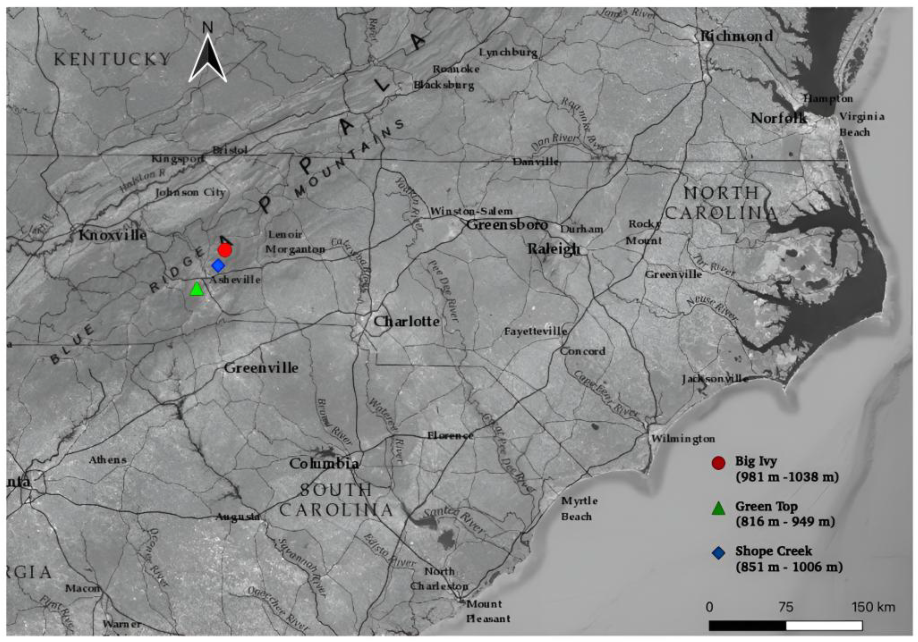
2.1. Study System

Betula lenta L. (sweet birch) is a common tree species in the Pisgah National Forest, North Carolina. The native range of the species extends from Ontario down through Mississippi, along the Atlantic coast from Maine down through Georgia, and inland from Mississippi up through Ohio [40]. Betula lenta covers a wide elevational range as well, though it is most common at elevations below 1000 m [41].
Three separate peaks within the Pisgah National Forest near Asheville, NC, were selected as study sites. At each of these sites (Shope Creek, Green Top, and Big Ivy), trees were sampled along a 222 m elevational gradient from 816 to 1038 m above sea level. To control for any effects of slope, aspect, and canopy cover on FEF colonization frequency and gas exchange physiology, we selected trees on south-facing, forested slopes dominated by deciduous hardwood trees (e.g., oaks (Quercus sp.), hickories (Carya sp.), maples (Acer sp.), with visually similar tree canopy cover (Figure 1).

2.2. Field Data Collection

Betula lenta trees with a diameter at breast height (DBH) under 3 inches were haphazardly selected per site, at roughly regular intervals along the elevational gradient. We selected young trees with leaves that were easily accessible without the use of ladders or climbing equipment. In August 2022, we haphazardly sampled three leaves per tree on ten trees at Big Ivy, eight trees at Shope Creek, and six trees Green Top for gas exchange measurements and subsequent FEF examination in the lab. The differences in sample sizes among sites was due to ambient weather conditions and equipment issues that prevented gas exchange measurements on sampling days. At each tree, we collected three leaves from different parts of the saplings that were free of visible signs of herbivory or disease, recorded elevation, GPS coordinates, and DBH.
Prior to collection, we measured each leaf’s photosynthetic rate (A, µmol CO2 m−2 s−1), transpiration rate (E, mol H2O m−2 s−1), and instantaneous water use efficiency (WUEi, A/E ∗ 10−4) using a LiCor LI-6400 Portable Photosynthesis System (LI-COR Biosciences, Lincoln, NE, USA). Instantaneous water use efficiency is an indicator of carbon gain per unit of evaporative water loss. Conditions within the LiCor 6400 were identical across all sites: light source = 6400-40 fluorometer, PARin = 1500 µmol s−1, flow = 500 µmol s−1, CO2 concentration = 400 ppm). Field sampling was conducted in August 2022, as FEF colonization is highest towards the end of the growing season [38]. Sampling was conducted between 9 A.M. and 1 P.M. under similar weather conditions, with cloud cover on collection days ranging from mostly sunny to slightly overcast.

2.3. FEF Isolation and Identification

The leaves were brought back to the lab and surface sterilized. Each leaf was dipped in 75% ethanol for 30 s, then 4% sodium hypochlorite for 1 min, and finally 75% ethanol for 15 s [42]. The leaves were then rinsed with sterile water and allowed to air dry. Samples of the rinse water were saved and cultured as a negative control to confirm removal of all surface fungi and bacteria from the leaves.
Three disks were removed from each leaf using a sterilized 5 mm diameter cork borer for a total of 216 leaf disks (24 trees × 3 leaves/tree × 3 disks/leaf). To account for within-leaf variation in endophyte frequencies in birch [39,42], one disk was taken from the tip, one from the middle, and one from the base of the leaf, from the lamina on either side of the midrib. The disks were placed individually in Petri dishes prepared with 2% malt-extract agar, sealed with Parafilm®®, and stored at room temperature in a dark cabinet to allow any endophytes present in the leaf to colonize the agar. Due to fast-growing fungal morphotypes, we recorded morphotypic variation in FEF within each plate after 72 h and attempted to obtain pure cultures of particular strains. Specifically, FEF present after 72 h—which we recognize were very likely the superior competitors given their fast growth rates—were visually categorized by morphotype using dissection microscopy. Due to budgetary constraints, three morphotypes were chosen for subculturing from each tree in the study. These were then plated on fresh media and allowed to grow out again to obtain a pure culture containing only one morphotype for subsequent molecular identification. The subcultures were visually assessed for purity using a dissection microscope.
Twenty-eight subcultures showed visually distinct individual morphotypes, with the remaining cultures containing multiple morphotypes. DNA was extracted from the 27 successful single morphotype subcultures using Qiagen DNeasy Plant Mini Kit (Qiagen, Germantown, MD, USA). Cultures with multiple morphotypes were not included in our genetic analysis due to the constraints of Sanger sequencing, which requires single source DNA template material (we recognize that attempting to subculture strains from overgrown plates may have prevented successful pure culture isolation of slower growing and less competitive strains). We used PCR to amplify the internal transcribed spacer (ITS) regions 1 and 2, using the ITS-1/ITS-2 primer pair (ITS1F: 5′-CTTGGTCATTTAGAGGAAGTAA-3′, ITS2R: 5′-GCTGCGTTCTTCATCGATGC-3′) [43,44]. PCR conditions were those described by Caruso et al. [45]. Single reaction volumes were 18 µL containing 11 µL 2x Promega GoTaq Green Master Mix, 1 µL each of 10 µM ITS1-F and ITS2R primers, and 5 µL of template DNA. Thermal cycler settings were as follows: initial denaturing 94 °C for 10 min, 35 cycles of 94 °C for 30 s, 54.4 °C for 30 s, 72 °C for 2 min, and a final 10 min extension at 72 °C. Once complete, samples were held at 4 °C until electrophoresis could be used to test for PCR success. We included a negative control (lacking fungal DNA, to control for contamination) and a known positive control (DNA extracted from the common white button mushroom Agaricus bisporus). PCR products were verified for amplification success on 2% agarose gels stained with ethidium bromide. Successful amplicons were sent to North Carolina State University Genomic Sciences Laboratory (Raleigh, NC, USA) for Sanger sequencing.
Geneious Prime (v. 2021.2) was used to trim and analyze the sequences. Post-trimmed sequences were compared with known sequences in GenBank (https://www.ncbi.nlm.nih.gov/genbank/; accessed on 20 February 2024) using BLAST (v. 2.14.0). We used the following criteria to assign an operational taxonomic unit (OTU) to each sample: pairwise identity > 96%, query coverage > 50%, and post-trimmed sequence length >200 bp. Three samples did not meet these criteria, leaving 25 samples. To assign a proposed ecological guild to each OTU, we reviewed the literature. Fungi that were reported in the literature as not causing signs of visible distress in host plants were considered endophytes (E). Fungi that caused harm to host plants were classified as pathogens (P) and those that reported as benefitting from the presence of decaying plant tissues were classified as saprotrophs (S).

2.4. Statistical Analysis

Linear regressions were used to examine the relationship between elevation as an independent variable, mean photosynthetic rate per tree (n = 3 leaves) and mean WUEi per tree, respectively. The mean transpiration rates per tree were not normally distributed. We, therefore, used a Spearman Rank Test to examine the relationship between transpiration rates and elevation.
To compare gas exchange rates among sites, we used nested ANOVAs to assess the effects of Site, Tree(Site), and Leaf(Tree(Site)) on photosynthetic rate, transpiration rate, and WUEi, respectively. Transpiration and WUEi were log10-transformed to meet ANOVA’s assumption of normality. When the nested ANOVAs yielded significant effects (α = 0.05), post-hoc Tukey HSD tests were used to identify differences among sites. All statistical analyses were performed in R (version 4.2.1) and RStudio, version 1.3.1073. The analyses were performed in base R with support from the tidyr, readr, and dplyr packages; the visualizations were generated using the ggplot2 package.

3. Results

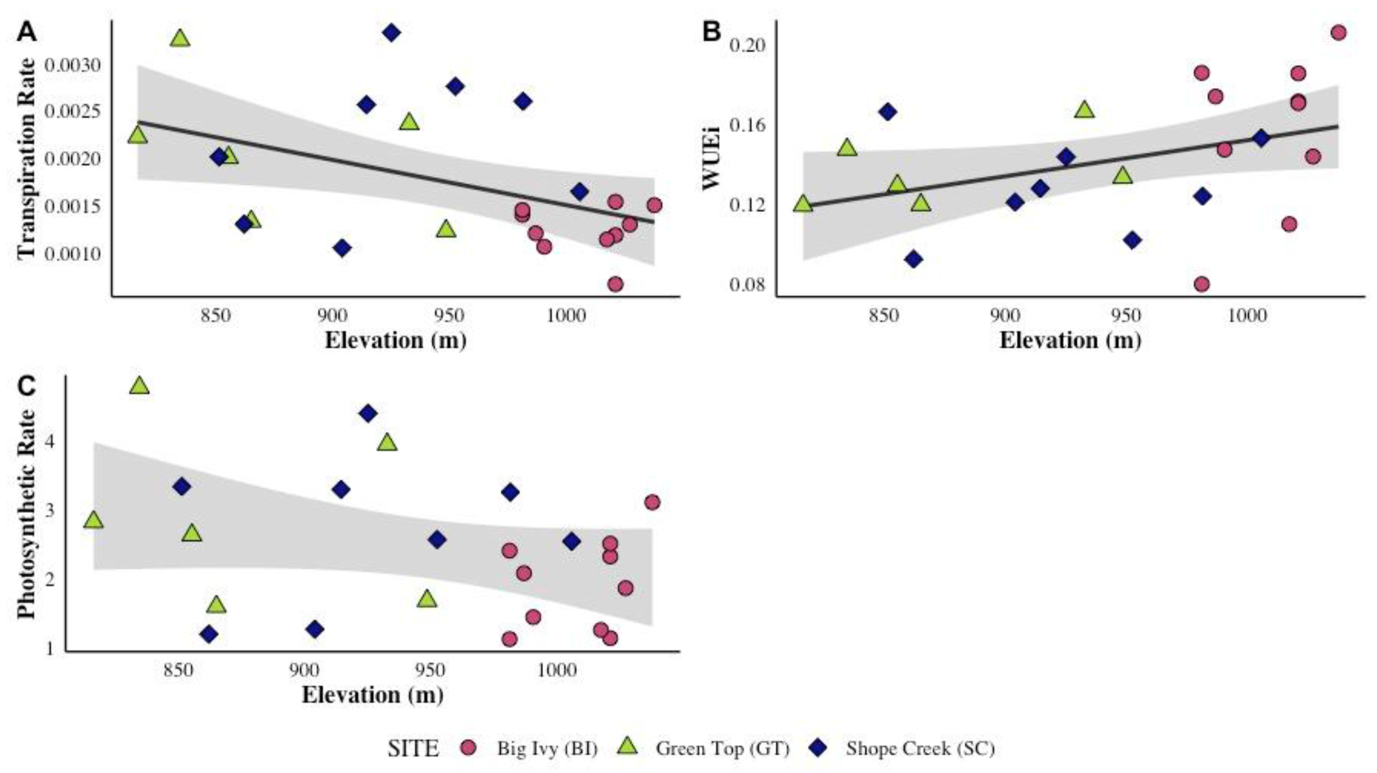
3.1. Gas Exchange Rates and Elevation

Rates of transpiration decreased with increasing elevation (Spearman Rank Test, ρ = −0.4444, S = 3322.1, P = 0.0296, Figure 2A). By contrast, WUEi tended to increase with increasing elevation (Linear Regression, adjusted R2 = 0.1187, t = 2.024, P = 0.0552, Figure 2B). This relationship, however, was not statistically significant. The photosynthetic rates were not significantly associated with elevation (Linear Regression, adjusted R2 = 0.0573, t = −1.549, P = 0.1357, Figure 2C).

3.2. Gas Exchange Rates: Site-Level Variation

Gas exchange rates varied among the three sites. Both photosynthetic rate and transpiration rate were higher at Green Top than at Big Ivy. Both Shope Creek (851–1006 m) and Green Top had significantly higher mean transpiration rates than Big Ivy but did not differ from one another (Figure 3A). Neither Green Top nor Big Ivy differed significantly from Shope Creek in terms of photosynthetic rate (Figure 3C). WUEi appeared to vary among sites (Figure 3B), but this difference was not statistically significant (Nested ANOVA, F = 3.166, P = 0.0506, Table 1).
We found some variation among trees within sites in gas exchange rates. The transpiration rates varied among trees within sites (Table 1). However, we observed no differences among trees in mean photosynthetic rate or WUEi (Table 1).

3.3. FEF Colonization and Morphotypes

Most of the initial cultures (212 out of 216) showed fungal colonization within 72 h. The remaining four plates were colonized within 10 days, making FEF frequency 100% across all sites. We noted that disks taken from the same tree, even from the same leaf, often gave rise to very different looking fungal communities, with each plate containing multiple different morphotypes (Figure 4). A total of 68 distinct FEF morphotypes were observed across all three sites (See Table S1). Negative control cultures remained clear for the duration of the experiment.

3.4. Fungal OTUs Encountered

We identified a total of 17 distinct fungal OTUs from the 25 samples that were sequenced. Of these, 24 were in the Phylum Ascomycota, with one being a member of the Basidiomycota. Most of the cultures were known endophytes with less than half of the species’ present being pathogenic and a small fraction not fitting neatly into one specific guild (Figure 5 and Figure 6, Table 2).
In several instances, morphotypes and OTUs did not correspond to one another. Sequence analyses revealed that several nearly identical morphotypes were different OTUs. For example, Samples 18 and 24 showed the same morphotype (streaky white) but were classified as different OTUs based on sequence data: Amphirosellinia fushanensis (Xylariaceae) and Rosellinia corticium (Xylariaceae). In other instances, the same taxa (e.g., Nemania diffusa, Rosellinia corticium, Colletotrichum truncatum) were associated with multiple morphotypes (Table 2, Table S1).

4. Discussion

Gas exchange rates are considered functional traits because of their direct effects on plant growth, survival, and reproduction. In recent years, plant–microbe mutualisms have been proposed to also be plant functional traits, given their tendency to vary across spatial and temporal scales and their impacts on plant performance [64]. This study investigated functional trait variation and foliar endophytic fungi in a common tree species (B. lenta) and detected evidence of variation in gas exchange physiology among sites and across an elevational gradient.
The decrease in transpiration rates observed at higher elevations suggests that these trees were experiencing more water stress than their low-elevation counterparts. This is supported by the fact that WUEi tended to increase with increasing elevation. This relationship was not quite statistically significant (Linear Regression, adjusted R2 = 0.1187, t = 2.024, P = 0.0552, Figure 2B). These findings are consistent with the findings of studies that have manipulated drought conditions in silver birch (Betula pendula Roth), a Eurasian birch species. In B. pendula, drought stress has been associated with reduced transpiration rates, leaf mass, and leaf area [65]. Other studies investigating relationships between FEF and gas exchange rates have compared plants inoculated with endophytes with non-inoculated controls under stressed and non-stressed growing conditions (synthesized in a meta-analysis by Rho and Kim [23]). In some instances, rates of transpiration and WUEi increased in response to endophyte inoculation, but only under conditions of environmental stress. It should be noted, however, that the studies included in this meta-analysis were conducted in controlled settings where both endophyte colonization and environmental stressors could be effectively manipulated.
The gas exchange rates we observed in unmanipulated field populations indicate that B. lenta trees experience water stress, but that the intensity of that stress varies among sites and across elevations. Interestingly, photosynthetic rates were not correlated with elevation and differed between only two of the three sites (Green Top and Big Ivy, Figure 2). This contrasts with the results of a previous study reporting significant declines in photosynthetic rates with increasing elevation on Mount Mansfield, Vermont, USA, in mountain paper birch (Betula papyrifera var. cordifolia (Regel) Fern. [27]). That study, however, surveyed trees across a much wider elevational range (610 m) than this study (~222 m) and used light curves to estimate maximum photosynthetic capacity (Amax) across sites. Incorporating additional sites situated at lower elevations and considering additional physiological metrics (e.g., light curves, stable isotope ratios [66] would yield deeper insights into the environmental factors that mediate FEF-host plant interactions.
The relationships between functional traits and elevation are especially interesting when considered together with the variation in FEF functional guilds among sites. Our highest elevation site (Big Ivy) appeared to harbor the most variable FEF community (Figure 5 and Figure 6). Other studies have also documented associations between elevation and FEF richness. For example, consistent with our findings, a study of FEF in Betula ermanii found that FEF diversity and richness increased with elevation in the Changbai Mountains, China [20]. By contrast, in a study of elevation and FEF in Metrosideros polymorpha in Hawaii, Zimmerman and Vitousek [67] found the highest FEF richness at their lowest elevation sites. Studies like these show that FEF communities are likely affected by environmental variables associated with elevation, but the nature of these effects are highly context dependent. Environmental factors may influence the nature of FEF-plant interactions, which in turn could affect host plant physiology, stress tolerance, and susceptibility to herbivores and pathogens, indirectly driving ecological processes at the community level [68]. An additional factor that may have contributed to the differences in transpiration rate, WUEi, and FEF OTUs observed between Big Ivy and the other sites (Figure 3 and Figure 6) is the presence of many adult yellow birch trees (B. alleghaniensis Britton) at the Big Ivy site. Both host plant-identity and the composition of co-occurring plant species at a given site are significant drivers of FEF community composition [21]. Either or both of these factors may influence FEF community structure at Big Ivy. Despite obvious differences in the adult trees of each species, B. alleghaniensis and B. lenta appear very similar at the sapling stage and it is possible some of our trees were misidentified.
While the patterns of FEF community variation reported here are intriguing, it is important to note that we likely characterized only a small proportion of the FEF diversity present at each site. While FEF colonization was 100% across all samples, we analyzed ITS sequences from <10% of the leaf disks. In addition, most of the 216 leaf disks we cultured harbored multiple morphotypes (Figure 4). Consequently, we cannot draw robust conclusions about the true relationships among elevation, site, and FEF taxonomic diversity.
This study does provide evidence that B. lenta hosts both endophytes and known plant pathogens, with Nemania diffusa having the highest prevalence (Table 2, Figure 5). Even though endophytic fungi are considered “asymptomatic” within host plants, other studies have isolated known pathogens from seemingly healthy leaves. Given that B. lenta leaves had no visible signs of distress, it is possible that the pathogens were either dormant at the time of collection or were present in such small amounts that foliar damage was not observable with the naked eye. Some fungal taxa that we encountered also have been associated with multiple ecological guilds. For example, Colletotrichum fioriniae has been shown to be a leaf endophyte and is also one of the species responsible for bitter rot disease in fruits and vegetables. Martin and Peter [60] found that endophytic C. fioriniae isolates from apple trees were mutualists within leaves, but pathogenic on the fruits. This example illustrates the importance of understanding the host plant when studying the potential ecological roles of fungal endophytes. The degree to which the species documented here act as endophytes or pathogens in B. lenta, as well as how environmental variables may affect the nature of these relationships, is unknown.
Directions for future research include using DNA metabarcoding to characterize FEF communities in B. lenta. Using a culture-independent method to identify FEF OTUs would facilitate the detection of culture-resistant taxa and would provide a far more comprehensive view of FEF communities within individual leaves than the culture-dependent methodology used here [69]. In a study of endophytic pyrophilus fungi within bryophytes and club mosses in the Great Smoky Mountains National Park (~77 km from the Green Top site), Raudabaugh et al. [36] documented nearly 10 times more OTUs using NGS metabarcoding than with culture-dependent methods. Given these findings, it is reasonable to predict that future studies using a metabarcoding approach would dramatically enhance our understanding of FEF communities in B. lenta.
Understanding FEF communities is important, given that microbial symbionts can have wide ranging, indirect effects on ecosystem properties [70,71]. Here, we report variation in drought stress encountered by sweet birch trees and variation in FEF communities across an elevational gradient. While it is not possible to draw definitive conclusions about FEF community composition, this work provides preliminary evidence of FEF taxonomic variation among sites. Other studies have linked microbial community variation to community and ecosystem attributes [68,72]. For example, Fahey et al., (in revision, Journal of Ecology) has found that foliar fungal endophytes can mediate the tree diversity-productivity relationship, as well as leaf chemistry and herbivore damage, in a long-term deciduous tree diversity experiment in Maryland, USA. However, understanding the relationships between FEF biodiversity and community and ecosystem processes in the southern Appalachian Mountains will require additional and more precise analyses of leaf microbiomes in the region.
Fungal endophytes occur in all vascular plants, influence host-plant fitness, and play a variety of crucial roles in terrestrial ecosystems across the globe. However, much remains to be understood about the factors that influence FEF abundance, distribution, and their relationship with host plants. The southern Appalachian Mountains have a long and distinctive geological and ecological history and are home to many rare and endemic fungal partnerships, such as lichens and ectomycorrhizae. To our knowledge, however, this is the first study to explore the region’s fungal endophytes in tandem with potential environmental stressors. Here, we provide a preliminary look at patterns of water stress across an elevational gradient in B. lenta and at some of the FEF taxa present in this species. These taxa likely represent a very small fraction of the true FEF community. Collectively, our findings suggest that FEFs and the ecological factors driving FEF community structure in sweet birch represent a rich arena for further investigation.

Supplementary Materials

The following supporting information can be downloaded at https://www.mdpi.com/article/10.3390/ecologies6020030/s1. Table S1: Summary table listing all 68 FEF morphotypes observed in this study.

Author Contributions

Conceptualization, G.A.D. and A.A.H.; methodology, G.A.D. and A.A.H.; software, G.A.D., G.C.Z., and A.A.H.; validation, G.A.D., G.C.Z., and A.A.H.; formal analysis, G.A.D., G.C.Z., and A.A.H.; investigation, G.A.D., G.C.Z., and A.A.H.; resources, A.A.H.; data curation, G.A.D. and G.C.Z.; writing—original draft preparation, G.A.D. and G.Z; writing—review and editing, G.A.D., G.C.Z., E.A.G., and A.A.H.; visualization, G.A.D., G.C.Z., E.A.G., and A.A.H.; supervision, A.A.H.; project administration, A.A.H.; funding acquisition, G.A.D., G.C.Z., and A.A.H. All authors have read and agreed to the published version of the manuscript.

Funding

The Dr. Alan W. Haney Fund for Undergraduate Research in the Natural Sciences provided financial support for this research. The Warren Wilson College (WWC) Work Program supported the undergraduate research students who assisted with laboratory genetics work.

Institutional Review Board Statement

Not applicable.

Informed Consent Statement

Not applicable.

Data Availability Statement

The data and code used to analyze gas exchange rates are available on GitHub (https://github.com/aahove/BELE-FEF, created on 12 March 2024). The raw data supporting the conclusions of this article will be made available by the authors upon request.

Acknowledgments

We thank Aaron Saenger for his assistance with fieldwork, as well as technical and moral support. Mark Brenner, Liesl Erb, and Olya Milenkaya provided feedback in the early stages of this research. Jill Arnold provided logistical support. We are also grateful for the support of the WWC Genetics Research Crew with lab work: Bassam Shawamreh, Emmanuella Afrane, Paddington Mbumbgwa, Emza Shackelford-Whitten, Jordan Goodyear, Otto Crouch, and Bailey Spencer. We thank the three anonymous reviewers who provided helpful comments on this manuscript.

Conflicts of Interest

The authors declare no conflicts of interest.

References

  1. Schulz, B.; Boyle, C. The Endophytic Continuum. Mycol. Res. 2005, 109, 661–686. [Google Scholar]
  2. Porras-Alfaro, A.; Bayman, P. Hidden Fungi, Emergent Properties: Endophytes and Microbiomes. Annu. Rev. Phytopathol. 2011, 49, 291–315. [Google Scholar] [CrossRef]
  3. Rodriguez, R.J.; White, J.F., Jr.; Arnold, A.E.; Redman, R.S. Fungal Endophytes: Diversity and Functional Roles. New Phytol. 2009, 182, 314–330. [Google Scholar] [CrossRef]
  4. Harrison, J.G.; Griffin, E.A. The Diversity and Distribution of Endophytes across Biomes, Plant Phylogeny and Host Tissues: How Far Have We Come and Where Do We Go from Here? Environ. Microbiol. 2020, 22, 2107–2123. [Google Scholar] [CrossRef]
  5. Tian, B.; Xie, J.; Fu, Y.; Cheng, J.; Li, B.; Chen, T.; Zhao, Y.; Gao, Z.; Yang, P.; Barbetti, M.J.; et al. A Cosmopolitan Fungal Pathogen of Dicots Adopts an Endophytic Lifestyle on Cereal Crops and Protects Them from Major Fungal Diseases. ISME J. 2020, 14, 3120–3135. [Google Scholar] [CrossRef]
  6. Arnold, A.E.; Mejía, L.C.; Kyllo, D.; Rojas, E.I.; Maynard, Z.; Robbins, N.; Herre, E.A. Fungal Endophytes Limit Pathogen Damage in a Tropical Tree. Proc. Natl. Acad. Sci. USA 2003, 100, 15649–15654. [Google Scholar] [CrossRef]
  7. Gouda, S.; Das, G.; Sen, S.K.; Shin, H.-S.; Patra, J.K. Endophytes: A Treasure House of Bioactive Compounds of Medicinal Importance. Front. Microbiol. 2016, 7, 1538. [Google Scholar] [CrossRef]
  8. Raihan, T.; Azad, A.K.; Ahmed, J.; Shepon, M.R.; Dey, P.; Chowdhury, N.; Aunkor, T.H.; Ali, H.; Suhani, S. Extracellular Metabolites of Endophytic Fungi from Azadirachta Indica Inhibit Multidrug-Resistant Bacteria and Phytopathogens. Future Microbiol. 2021, 16, 557–576. [Google Scholar] [CrossRef]
  9. Bowman, E.A.; Arnold, A.E. Distributions of Ectomycorrhizal and Foliar Endophytic Fungal Communities Associated with Pinus ponderosa along a Spatially Constrained Elevation Gradient. Am. J. Bot. 2018, 105, 687–699. [Google Scholar] [CrossRef]
  10. Rolón, B.A.; Arnold, A.E.; Juliá, M.S.; Van Bael, S.A. Evaluating Endophyte-rich Leaves and Leaf Functional Traits for Protection of Tropical Trees against Natural Enemies. Funct. Ecol. 2024, 39, 371–389. [Google Scholar] [CrossRef]
  11. Redman, R.S.; Dunigan, D.D.; Rodriguez, R.J. Fungal Symbiosis from Mutualism to Parasitism: Who Controls the Outcome, Host or Invader? New Phytol. 2001, 151, 705–716. [Google Scholar] [CrossRef] [PubMed]
  12. Hernandez, F.S.; Villarreal, R.M.; Torres, V.R.; Gallou, A. Monosporic Inoculation of Economically Important Horticultural Species with Native Endomycorrhizae under Greenhouse Conditions. Agronomy 2019, 9, 130. [Google Scholar] [CrossRef]
  13. Nashat, L.H.; Haleem, R.A.; Ali, S.H. Molecular Identification and Antimicrobial Potential of Endophytic Fungi against Some Grapevine Pathogens. PLoS ONE 2024, 19, e0309041. [Google Scholar] [CrossRef]
  14. Van Bael, S.A.; Valencia, M.C.; Rojas, E.I.; Gómez, N.; Windsor, D.M.; Herre, E.A. Effects of Foliar Endophytic Fungi on the Preference and Performance of the Leaf Beetle Chelymorpha alternans in Panama. Biotropica 2009, 41, 221–225. [Google Scholar] [CrossRef]
  15. Sieber, T.N. Endophytic Fungi in Forest Trees: Are They Mutualists? Fungal Biol. Rev. 2007, 21, 75–89. [Google Scholar] [CrossRef]
  16. Cline, L.C.; Schilling, J.S.; Menke, J.; Groenhof, E.; Kennedy, P.G. Ecological and Functional Effects of Fungal Endophytes on Wood Decomposition. Funct. Ecol. 2018, 32, 181–191. [Google Scholar] [CrossRef]
  17. Pinto, L.S.R.C.; Azevedo, J.L.; Pereira, J.O.; Vieira, M.L.C.; Labate, C.A. Symptomless Infection of Banana and Maize by Endophytic Fungi Impairs Photosynthetic Efficiency. New Phytol. 2000, 147, 609–615. [Google Scholar] [CrossRef]
  18. Gazis, R.; Chaverri, P. Wild Trees in the Amazon Basin Harbor a Great Diversity of Beneficial Endosymbiotic Fungi: Is This Evidence of Protective Mutualism? Fungal Ecol. 2015, 17, 18–29. [Google Scholar] [CrossRef]
  19. González-Teuber, M. The Defensive Role of Foliar Endophytic Fungi for a South American Tree. AoB Plants 2016, 8, plw050. [Google Scholar] [CrossRef]
  20. Yang, T.; Weisenhorn, P.; Gilbert, J.A.; Ni, Y.; Sun, R.; Shi, Y.; Chu, H. Carbon Constrains Fungal Endophyte Assemblages along the Timberline. Environ. Microbiol. 2016, 18, 2455–2469. [Google Scholar] [CrossRef]
  21. Darcy, J.L.; Swift, S.O.I.; Cobian, G.M.; Zahn, G.L.; Perry, B.A.; Amend, A.S. Fungal Communities Living within Leaves of Native Hawaiian Dicots Are Structured by Landscape-Scale Variables as Well as by Host Plants. Mol. Ecol. 2020, 29, 3102–3115. [Google Scholar] [CrossRef]
  22. González-Teuber, M.; Vilo, C.; Guevara-Araya, M.J.; Salgado-Luarte, C.; Gianoli, E. Leaf Resistance Traits Influence Endophytic Fungi Colonization and Community Composition in a South American Temperate Rainforest. J. Ecol. 2020, 108, 1019–1029. [Google Scholar] [CrossRef]
  23. Rho, H.; Kim, S.-H. Endophyte Effects on Photosynthesis and Water Use of Plant Hosts: A Meta-Analysis. In Functional Importance of the Plant Microbiome; Doty, S.L., Ed.; Springer International Publishing: Cham, Switzerland, 2017; pp. 43–69. ISBN 978-3-319-65896-4. [Google Scholar]
  24. Ackerly, D.D.; Dudley, S.A.; Sultan, S.E.; Schmitt, J.; Coleman, J.S.; Linder, C.R.; Sandquist, D.R.; Geber, M.A.; Evans, A.S.; Dawson, T.E. The Evolution of Plant Ecophysiological Traits: Recent Advances and Future Directions: New Research Addresses Natural Selection, Genetic Constraints, and the Adaptive Evolution of Plant Ecophysiological Traits. Bioscience 2000, 50, 979–995. [Google Scholar]
  25. Reich, P.B.; Wright, I.J.; Cavender-Bares, J.; Craine, J.M.; Oleksyn, J.; Westoby, M.; Walters, M.B. The Evolution of Plant Functional Variation: Traits, Spectra, and Strategies. Int. J. Plant Sci. 2003, 164, S143–S164. [Google Scholar] [CrossRef]
  26. Geber, M.A.; Griffen, L.R. Inheritance and Natural Selection on Functional Traits. Int. J. Plant Sci. 2003, 164, S21–S42. [Google Scholar] [CrossRef]
  27. Richardson, A.D.; Berlyn, G.P. Spectral Reflectance and Photosynthetic Properties of Betula papyrifera (Betulaceae) Leaves along an Elevational Gradient on Mt. Mansfield, Vermont, USA. Am. J. Bot. 2002, 89, 88–94. [Google Scholar] [CrossRef]
  28. Ramirez, H.-L.; Ivey, C.T.; Wright, J.W.; MacDonald, B.W.; Sork, V.L. Variation in Leaf Shape in a Quercus Lobata Common Garden: Tests for Adaptation to Climate and Physiological Consequences. Madroño 2020, 67, 77–84. [Google Scholar]
  29. Ramírez-Valiente, J.A.; López, R.; Hipp, A.L.; Aranda, I. Correlated Evolution of Morphology, Gas Exchange, Growth Rates and Hydraulics as a Response to Precipitation and Temperature Regimes in Oaks (Quercus). New Phytol. 2020, 227, 794–809. [Google Scholar] [CrossRef]
  30. Song, B.; Niu, S.; Wan, S. Precipitation Regulates Plant Gas Exchange and Its Long-Term Response to Climate Change in a Temperate Grassland. J. Plant Ecol. 2016, 9, 531–541. [Google Scholar]
  31. Mazer, S.J.; Dudley, L.S.; Hove, A.A.; Emms, S.K.; Verhoeven, A.S. Physiological Performance in Clarkia Sister Taxa with Contrasting Mating Systems: Do Early-Flowering Autogamous Taxa Avoid Water Stress Relative to Their Pollinator-Dependent Counterparts? Int. J. Plant Sci. 2010, 171, 1029–1047. [Google Scholar] [CrossRef]
  32. Allen, J.L.; Lendemer, J.C. Climate Change Impacts on Endemic, High-Elevation Lichens in a Biodiversity Hotspot. Biodivers. Conserv. 2016, 25, 555–568. [Google Scholar] [CrossRef]
  33. Cox, J.L.; McKinney, M.L.; Fitzpatrick, B.M.; Leppanen, C.; Nichols, R.J. Coniferous Conservation Supporting a Plethora of Plethodontids: Implications of Conserving Eastern Hemlock (Tsuga canadensis) on Southern Appalachian Montane Salamanders. For. Ecol. Manag. 2022, 508, 120010. [Google Scholar] [CrossRef]
  34. Landscape Partnership Land Use in the Appalachians. Available online: https://www.landscapepartnership.org/cooperative/our-plan/section-1/land-use-in-the-appalachians (accessed on 31 January 2025).
  35. Zhang, R.; Zhou, X.-L.; Yang, L.; Long, B.; Shen, S.-K. Composition and Assembly of the Endophytic Fungal Community of Alpine Rhododendron Hosts along Elevation Gradients. Phytobiomes J. 2024, 8, 236–247. [Google Scholar] [CrossRef]
  36. Raudabaugh, D.B.; Matheny, P.B.; Hughes, K.W.; Iturriaga, T.; Sargent, M.; Miller, A.N. Where Are They Hiding? Testing the Body Snatchers Hypothesis in Pyrophilous Fungi. Fungal Ecol. 2020, 43, 100870. [Google Scholar]
  37. Rutto, L.K.; Ren, S.; Wynn, H.C.; Chamberlain, J.L. Soil and Microbe Interactions in Two Populations of Appalachian Black Cohosh (Actaea racemosa L.). J. Torrey Bot. Soc. 2021, 148, 117–131. [Google Scholar]
  38. Ahlholm, J.U.; Helander, M.; Henriksson, J.; Metzler, M.; Saikkonen, K. Environmental Conditions and Host Genotype Direct Genetic Diversity of Venturia Ditricha, a Fungal Endophyte of Birch Trees. Evolution 2002, 56, 1566–1573. [Google Scholar]
  39. Helander, M.; Neuvonen, S.; Sieber, T.; Petrini, O. Simulated Acid Rain Affects Birch Leaf Endophyte Populations. Microb. Ecol. 1993, 26, 227–234. [Google Scholar]
  40. United States Department of Agriculture Natural Resources Conservation Service. Betula lenta L. 2025. Available online: https://plants.usda.gov/plant-profile/BELE (accessed on 1 March 2023).
  41. Rossell, I.M.; Eggleston, H. Elevational Distribution of Temperate Lianas along Trails in Pisgah National Forest. Southeast. Nat. 2017, 16, 443–450. [Google Scholar] [CrossRef]
  42. Helander, M.; Wäli, P.; Kuuluvainen, T.; Saikkonen, K. Birch Leaf Endophytes in Managed and Natural Boreal Forests. Can. J. For. Res. 2006, 36, 3239–3245. [Google Scholar]
  43. White, T.J.; Bruns, T.D.; Lee, S.B.; Taylor, J.W. Amplification and Direct Sequencing of Fungal Ribosomal RNA Genes for Phylogenetics. In PCR Protocols: A Guide to Methods and Applications; Academic Press, Inc.: Cambridge, MA, USA, 1990; pp. 315–322. ISBN 978-0-12-372180-8. [Google Scholar]
  44. Han, J.; Zhu, Y.; Chen, X.; Liao, B.; Yao, H.; Song, J.; Chen, S.; Meng, F. The Short ITS2 Sequence Serves as an Efficient Taxonomic Sequence Tag in Comparison with the Full-Length ITS. BioMed Res. Int. 2013, 2013, 741476. [Google Scholar] [CrossRef]
  45. Caruso, K.E.; Horton, J.L.; Hove, A.A. Assessing the Effect of Eastern Hemlock (Tsuga canadensis) Decline from Hemlock Woolly Adelgid (Adelges tsugae) Infestation on Ectomycorrhizal Colonization and Growth of Red Oak (Quercus rubra) Seedlings. Am. Midl. Nat. 2021, 186, 16–34. [Google Scholar] [CrossRef]
  46. Rodriguez, R.; Redman, R. More than 400 Million Years of Evolution and Some Plants Still Can’t Make It on Their Own: Plant Stress Tolerance via Fungal Symbiosis. J. Exp. Bot. 2008, 59, 1109–1114. [Google Scholar] [CrossRef]
  47. Oberhofer, M.; Malfent, F.; Zehl, M.; Urban, E.; Wackerlig, J.; Reznicek, G.; Vignolle, G.A.; Rückert, C.; Busche, T.; Wibberg, D.; et al. Biosynthetic Potential of the Endophytic Fungus Helotiales Sp. BL73 Revealed via Compound Identification and Genome Mining. Appl. Environ. Microbiol. 2022, 88, e02510-21. [Google Scholar] [CrossRef]
  48. Balasuriya, A.; Adikaram, N.K.B. Some Spatial, Temporal and Spatio-Temporal Considerations of Wood Decay of Tea (Camellia sinensis), Caused by Nemania diffusa (Syn. Hypoxylon vestitum). Crop Prot. 2009, 28, 273–279. [Google Scholar] [CrossRef]
  49. Okane, I.; Nonaka, K.; Kurihara, Y.; Abe, J.P.; Yamaoka, Y. A New Species of Leptobacillim, L. symbioticum, Isolated from Mites and Sori of Soybean Rust. Mycoscience 2020, 61, 165–171. [Google Scholar] [CrossRef]
  50. Troncoso-Rojas, R.; Tiznado-Hernández, M.E. Alternaria alternata (Black Rot, Black Spot). Postharvest Decay Control. Strateg. 2014, 147–187. [Google Scholar] [CrossRef]
  51. Li, T.; Im, J.; Lee, J. Genetic Diversity of Epicoccum nigrum and Its Effects on Fusarium graminearum. Mycobiology 2022, 50, 457–466. [Google Scholar] [CrossRef]
  52. Darapanit, A.; Boonyuen, N.; Leesutthiphonchai, W.; Nuankaew, S.; Piasai, O. Identification, Pathogenicity and Effects of Plant Extracts on Neopestalotiopsis and Pseudopestalotiopsis Causing Fruit Diseases. Sci. Rep. 2021, 11, 22606. [Google Scholar] [CrossRef]
  53. Wittstein, K.; Cordsmeier, A.; Lambert, C.; Wendt, L.; Sir, E.B.; Weber, J.; Wurzler, N.; Petrini, L.E.; Stadler, M. Identification of Rosellinia Species as Producers of Cyclodepsipeptide PF1022 A and Resurrection of the Genus Dematophora as Inferred from Polythetic Taxonomy. Stud. Mycol. 2020, 96, 1–16. [Google Scholar] [CrossRef]
  54. Toghueo, R.M.K.; Boyom, F.F. Endophytic Penicillium Species and Their Agricultural, Biotechnological, and Pharmaceutical Applications. 3 Biotech 2020, 10, 107. [Google Scholar] [CrossRef]
  55. Langer, G.J.; Bußkamp, J. Fungi Associated with Woody Tissues of European Beech and Their Impact on Tree Health. Front. Microbiol. 2021, 12, 702467. [Google Scholar] [CrossRef]
  56. do Carmo de Souza, É.S.; do Vale, H.M.M.; de Cássia Pereira Carvalho, R.; de Oliveira Soares, W.R.; Miller, R.N.G.; Dianese, G.C. Infection by Uromyces Euphorbiae: A Trigger for the Sporulation of Endophytic Colletotrichum truncatum on the Common Host Euphorbia Hirta. Mycol. Prog. 2017, 16, 941–946. [Google Scholar] [CrossRef]
  57. Shahriar, S.A.; Husna, A.; Paul, T.T.; Eaty, M.N.K.; Quamruzzaman, M.; Siddique, A.B.; Rahim, M.A.; Ahmmed, A.N.F.; Uddain, J.; Siddiquee, S. Colletotrichum Truncatum Causing Anthracnose of Tomato (Solanum lycopersicum L.) in Malaysia. Microorganisms 2023, 11, 226. [Google Scholar] [CrossRef] [PubMed]
  58. Macías-Rubalcava, M.L.; Hernández-Bautista, B.E.; Oropeza, F.; Duarte, G.; González, M.C.; Glenn, A.E.; Hanlin, R.T.; Anaya, A.L. Allelochemical Effects of Volatile Compounds and Organic Extracts from Muscodor Yucatanensis, a Tropical Endophytic Fungus from Bursera Simaruba. J. Chem. Ecol. 2010, 36, 1122–1131. [Google Scholar] [CrossRef]
  59. Ju, Y.-M.; Rogers, J.D.; Hsieh, H.-M.; Vasilyeva, L. Amphirosellinia Gen. Nov. and a New Species of Entoleuca. Mycologia 2004, 96, 1393–1402. [Google Scholar]
  60. Martin, P.L.; Peter, K.A. Quantification of Colletotrichum fioriniae in Orchards and Deciduous Forests Indicates It Is Primarily a Leaf Endophyte. Phytopathology®® 2021, 111, 333–344. [Google Scholar] [CrossRef]
  61. Gauchan, D.P.; Vélëz, H.; Acharya, A.; Östman, J.R.; Lundén, K.; Elfstrand, M.; García-Gil, M.R. Annulohypoxylon Sp. Strain MUS1, an Endophytic Fungus Isolated from Taxus wallichiana Zucc., Produces Taxol and Other Bioactive Metabolites. 3 Biotech 2021, 11, 152. [Google Scholar] [CrossRef]
  62. U’Ren, J.M.; Miadlikowska, J.; Zimmerman, N.B.; Lutzoni, F.; Stajich, J.E.; Arnold, A.E. Contributions of North American Endophytes to the Phylogeny, Ecology, and Taxonomy of Xylariaceae (Sordariomycetes, Ascomycota). Mol. Phylogenet. Evol. 2016, 98, 210–232. [Google Scholar] [CrossRef]
  63. Oses, R.; Valenzuela, S.; Freer, J.; Sanfuentes, E.; Rodríguez, J. Fungal Endophytes of Healthy Chilean Trees and Their Possible Role in Early Wood Decay. Fungal Divers. 2008, 33, 77–86. [Google Scholar]
  64. Maherali, H. Mutualism as a Plant Functional Trait: Linking Variation in the Mycorrhizal Symbiosis to Climatic Tolerance, Geographic Range, and Population Dynamics. Int. J. Plant Sci. 2020, 181, 9–19. [Google Scholar] [CrossRef]
  65. Oksanen, E. Birch as a Model Species for the Acclimation and Adaptation of Northern Forest Ecosystem to Changing Environment. Front. For. Glob. Change 2021, 4, 682512. [Google Scholar]
  66. Dawson, T.E.; Mambelli, S.; Plamboeck, A.H.; Templer, P.H.; Tu, K.P. Stable Isotopes in Plant Ecology. Annu. Rev. Ecol. Syst. 2002, 33, 507–559. [Google Scholar] [CrossRef]
  67. Zimmerman, N.B.; Vitousek, P.M. Fungal Endophyte Communities Reflect Environmental Structuring across a Hawaiian Landscape. Proc. Natl. Acad. Sci. USA 2012, 109, 13022–13027. [Google Scholar] [CrossRef] [PubMed]
  68. Jia, Q.; Qu, J.; Mu, H.; Sun, H.; Wu, C. Foliar Endophytic Fungi: Diversity in Species and Functions in Forest Ecosystems. Symbiosis 2020, 80, 103–132. [Google Scholar]
  69. Kress, W.J.; García-Robledo, C.; Uriarte, M.; Erickson, D.L. DNA Barcodes for Ecology, Evolution, and Conservation. Trends Ecol. Evol. 2015, 30, 25–35. [Google Scholar] [CrossRef]
  70. Terhonen, E.; Blumenstein, K.; Kovalchuk, A.; Asiegbu, F.O. Forest Tree Microbiomes and Associated Fungal Endophytes: Functional Roles and Impact on Forest Health. Forests 2019, 10, 42. [Google Scholar] [CrossRef]
  71. Griffin, E.A.; Carson, W.P. Tree Endophytes: Cryptic Drivers of Tropical Forest Diversity. In Endophytes of Forest Trees; Pirttilä, A.M., Frank, A.C., Eds.; Forestry Sciences; Springer International Publishing: Cham, Switzerland, 2018; Volume 86, pp. 63–103. ISBN 978-3-319-89832-2. [Google Scholar]
  72. Sáez-Sandino, T.; García-Palacios, P.; Maestre, F.T.; Plaza, C.; Guirado, E.; Singh, B.K.; Wang, J.; Cano-Díaz, C.; Eisenhauer, N.; Gallardo, A. The Soil Microbiome Governs the Response of Microbial Respiration to Warming across the Globe. Nat. Clim. Change 2023, 13, 1382–1387. [Google Scholar]
Figure 1. Map of the study area showing the three study sites in the Pisgah National Forest outside of Asheville, NC. Map generated using QGIS version 3.16.3-Hannover by G. Dougherty.
Ecologies 06 00030 g001
Figure 2. Scatterplots depicting the relationships between (A) transpiration rate (mol H2O m−2 s−1), (B) WUEi, and (C) photosynthetic rate (µmol CO2 m−2 s−1) with elevation (n = 24 trees across sites). The grey region represents a 95% confidence interval around the line of best fit (we did not include a trendline in C because the relationship was insignificant).
Ecologies 06 00030 g002
Figure 3. Box plots showing distributions of (A) transpiration rate, (B) WUEi, and (C) photosynthetic rate at all three sites. Boxes that differ by a letter indicate that trait means differ significantly from one another (Tukey HSD Test, p < 0.05).
Ecologies 06 00030 g003
Figure 4. Example of the typical morphotype diversity present in one tree. In this photograph, each row represents one leaf from a single tree. Each Petri dish contains one leaf disk and the fungi that emerged during culturing. We observed multiple morphotypes in most of the Petri dishes.
Ecologies 06 00030 g004
Figure 5. Taxonomic composition of sequenced fungi observed by study site. The bars represent the frequency of each operational taxonomic unit based on Sanger sequencing of the fungal ITS1 region.
Ecologies 06 00030 g005
Figure 6. Relative abundance (%) of various fungal ecological guilds associated with sequenced fungi from each study site. The bars represent the frequency of each ecological guild based on Sanger sequencing of the fungal ITS1 region and subsequent review of the literature.
Ecologies 06 00030 g006
Table 1. Results of nested ANOVAs for gas exchange rates: log10 (Photosynthesis), log10 (Transpiration), and instantaneous water use efficiency (WUEi). The p-values associated with statistically significant model effects (α = 0.05) are indicated in bold.
PhotosynthesisTranspirationWUEi
EffectdfSSMSFp-ValueSSMSFp-ValueSSMSFp-Value
Site21.5510.77563.5720.03533.161.5812.1584.81 × 10−50.012270.0061363.1660.0506
Tree10.020.02030.0940.76080.0080.00850.0650.79960.00040.0003970.2050.6527
Leaf number10.0190.01920.0880.76750.0290.02880.2210.64010.001750.0017530.9040.3461
Tree (site)21.1850.59262.7290.07481.3050.65235.0190.01020.000490.0002440.1260.8818
Tree (leaf number)10.1110.11080.510.47830.2030.20321.5630.21690.000570.0005710.2950.5896
Leaf number (tree, site)20.3260.16310.7510.47690.0920.04590.3530.70390.005480.0027411.4140.2524
Residuals5111.0730.2171 6.6280.13 0.098830.001938
Table 2. Fungal OTUs identified across the three sites. Sites: BI = Big Ivy, SC = Shope Creek, GT = Green Top. All OTUs are in phylum Ascomycota, except for Sample 26, which is in the Basidiomycota. In the Proposed Ecological Guild column, E = endophyte, P = pathogen, EN = Entomopathogen (associated with disease in insects), S = Saprotroph. Sample 18 was designated “unspecified” because we could not determine its ecological guild based on the current literature.
Sample No.SiteFamilyOTUProposed Ecological GuildMorphotypeReference
1BIGlomerellaceaeColletotrichum sp.E, PMT42: streaky black[46]
2BILeotiomycetesHelotiales sp.EMT58: white billows[47]
3BIXylariaceaeNemania diffusaPMT54: wacky brown moss[48]
4BICordycipitaceaeLeptobacillium symbioticumENMT19: fuzzy white pile[49]
5BIPleosporaceaeAlternaria alternataP, SMT22: gray starburst[50]
6BIDidymellaceaeEpicoccum nigrumE, PMT10: clear yellow w/orange nerves[51]
7BIXylariaceaeNemania diffusaPMT25: off-white dust pile[48]
8BIXylariaceaeNemania diffusaPMT67: white veins w/snail-shaped fruit[48]
9BISporocadaceaeNeopestalotiopsis sp.PMT51: streaky yellowish cloud ring w/black fruit[52]
10BIGlomerellaceaeColletotrichum sp.E, PMT37: orange/yellow billows[46]
12BIXylariaceaeRosellinia corticiumEMT45: streaky gray[53]
13BIAspergillaceaePenicillium sp.EMT64: white starbursts w/gray fuzzy centers[54]
14BIHypoxylaceaeJackrogersella cohaerensE, SMT13: fragrant white mesh[55]
15BIGlomerellaceaeColletotrichum truncatumE, PMT40: soot sprites[56,57]
16BIXylariaceaeNemania diffusaPMT66: white veins[48]
17SCXylariaceaeMuscodor yucatanensisEMT23: gray-green ring[58]
18SCXylariaceaeAmphirosellinia fushanensisunspecifiedMT48: streaky white[59]
20SCGlomerellaceaeColletotrichum truncatumE, PMT5: black blobs[56,57]
21SCXylariaceaeNemania diffusaPMT41: speckled black[48]
22SCGlomerellaceaeColletotrichum fioriniaeE, PMT45: streaky gray[60]
23GTHypoxylaceaeAnnulohypoxylon sp.EMT27: opaque brown w/ring[61]
24GTXylariaceaeRosellinia corticiumEMT48: streaky white[53]
25GTXylariaceaeWhalleya microplacaEMT28: opaque mat[62]
26GTPhanerochaetaceaeBjerkandera adustaE, SMT62: white smelly fuzz[63]
27GTHypoxylaceaeAnnulohypoxylon sp.EMT13: fragrant white mesh[61]
Disclaimer/Publisher’s Note: The statements, opinions and data contained in all publications are solely those of the individual author(s) and contributor(s) and not of MDPI and/or the editor(s). MDPI and/or the editor(s) disclaim responsibility for any injury to people or property resulting from any ideas, methods, instructions or products referred to in the content.

