Article

Ozonized Oleic Acid as a New Viticultural Treatment? Study of the Effect of LIQUENSO® Oxygenate on the Carpoplane Microbial Community and Wine Microorganisms Combining Metabarcoding and In Vitro Assays

by
Lea Franziska Stahl
1,2,
Manon Edo
3,
Timon Nonnenmacher
4,
Daniela Reif
3,
Friederike Rex
3,
Pascal Wegmann-Herr
3,
Andreas Kortekamp
5,
Jochen Fischer-Schuch
6,
Eckhard Thines
2,6 and
Maren Scharfenberger-Schmeer
1,3,*
1
Faculty of Applied Logistics and Polymer Sciences, University of Applied Sciences Kaiserslautern, 67659 Kaiserslautern, Germany
2
Microbiology and Wine Research, Institute for Molecular Physiology, Johannes-Gutenberg University Mainz, 55128 Mainz, Germany
3
Institute for Viticulture and Oenology, State Education and Research Center of Viticulture and Horticulture and Rural Development (DLR) Rheinpfalz, 67435 Neustadt, Germany
4
Anseros Klaus-Nonnenmacher GmbH, 72070 Tübingen, Germany
5
Institute for Plant Protection, State Education and Research Center of Viticulture and Horticulture and Rural Development (DLR) Rheinpfalz, 67435 Neustadt, Germany
6
IBWF gGmbH, Institute for Biotechnology and Drug Research, 55128 Mainz, Germany
*
Author to whom correspondence should be addressed.
Ecologies 2022, 3(3), 292-307; https://doi.org/10.3390/ecologies3030023
Submission received: 12 May 2022 / Revised: 29 June 2022 / Accepted: 25 July 2022 / Published: 29 July 2022
(This article belongs to the Special Issue Feature Papers of Ecologies 2022)

Abstract

:
In this study, an amplicon metagenomic approach was used to determine the effect of repeated treatments with ozonized oleic acid on the microbial community of grapevine carpoplane. Differences in community composition of treated vineyards were compared to non-treated and conventionally treated samples regarding the prokaryotic and eukaryotic microbiome at two developmental stages (BBCH 83, BBCH 87). The results showed effects both on occurrence and on abundance of microorganisms and the community assembly. Wine-relevant genera such as Acetobacter and members of the former genus Lactobacillus could be identified as part of the natural microbiota. The impact of the new viticultural treatment on these organisms was assessed in liquid culture-based microtiter assays. Therefore, we investigated an array of two acetic acid bacteria (AAB), four lactic acid bacteria (LAB) and nine saccharomyces and non-saccharomyces yeasts. Brettanomyces bruxellensis, Saccharomyces cerevisiae, Pediococcus sp. and Acetobacter aceti revealed the highest sensitivities against ozonized oleic acid (LIQUENSO® Oxygenat). Culture growth of these organisms was significantly reduced at an ozonide concentration of 0.25% (v/v), which corresponded to a quarter of the concentration used in the vineyard. The metabarcoding approach in combination with complementary in vitro assays allow new insights into treatment effects on the community and species scale.

1. Introduction

Grapevine epiphytic microorganisms can be neutral, beneficial or pathogenic to the plant [1]. Interactions depend on many biotic, abiotic and anthropogenic factors determining grapevine microbial communities and their structure [2,3]. Biotic factors include intrinsic biological properties such as the grapevine variety, the stage of ripening [1] and the dissemination of microorganisms by insects and birds [4]. Abiotic factors are temperature, humidity, UV radiation, oxydo-reduction potential, pH and soil composition [2,5]. The use of agrochemicals is one of the major anthropogenic determinants of the grape microbial community [3,6]. Nevertheless, resistance development of plant pathogenic organisms is threatening viticulture [7,8,9]. Thus, research must be conducted concerning new antimicrobial active compounds in plant pest management. A promising field of development is concerned with ozonized plant oils. The broad effective spectrum of these compounds is based on a rather unspecific mode of action conducted by trioxolanes, peroxides and aldehydes [10,11]. The efficiency against human pathogenic yeasts and bacteria has been proven in various publications in the past two decades [10,12,13]. Ozonized sunflower oil has been shown to be effective against cucumber powdery mildew caused by Podosphaera xanthii [14].
A complete disinfection of the grape berries is yet not desirable due to the beneficial effects of certain microorganisms on plant health and vinification [15]. They are determining the microbial terroir and influencing sensory quality of the wine [16]. Yeasts and Gram-positive lactic acid bacteria (LAB) are important drivers of the alcoholic and malolactic fermentations, as reviewed by Capozzi et al. (2021) [17]. The effect of these organisms during fermentation depends on interspecific interactions [18,19,20], the fermentation temperature [20] and wine chemical parameters such as pH nutrient and oxygen availability [21,22,23,24,25]. Non-Saccharomyces yeasts are often referred to in the context of incomplete or sluggish fermentations and undesirable off-flavors [21,26]. Nevertheless, positive effects of non-Saccharomyces yeasts, e.g., Torulaspora delbruckii, Metschnikowia pulcherrima, Schizosaccharomyces pombe, Hanseniaspora spp. and Zygosaccharomyces bailii, as well as members of the genus Pichia, have been reported, as summarized by Vicente et al. (2021) [22]. On the other hand, the negative potential of non-Saccharomyces yeasts and LAB in terms of wine quality shows in the form of diverse off-flavors [25]. Mousy off-flavors caused by Brettanomyces bruxellensis, Lentilactobacillus hilgardii, Levilactobacillus brevis, Lactiplantibacillus plantarum and Oenococcus oeni [27,28] are just one example. The effect of LAB on the wine quality depends on the species- and strain-specific enzymatic activities and their mechanism of glucose catabolism [17,29]. LAB reduce wine acidity by converting L-malic acid to L-lactic acid, thereby providing microbiological stabilization of the wine [17,29]. Esterase and glycosidase activities of some LAB add beneficial complexity to the wine flavor [17,24,30]. Likewise, LAB can reduce wine quality by the formation of acetic acid or the production of biogenic amines and carcinogenic ethyl carbamate [17,31]. Acetic acid bacteria (AAB) are Gram-negative obligate aerobic bacteria within the family of Acetobacteraceae [32,33]. Some AABs are desirable in the production of foods, beverages and biotechnological applications [33,34,35], yet they have detrimental potential in the vinification process [32]. This is due to their ability of metabolizing ethanol into acetic acid, acetaldehyde, ethyl acetate and dihydroxyacetone [36].
To determine to what extent treatment with ozonized oleic acid and the grape ripening influence the grapevine health and vinification, studies of the microbial biota of wine berries appear to be important. Nevertheless, few studies have been conducted in Germany on grape microbial diversity and its response to these factors [37].
The aim of the present study was to elucidate the influence of a new agrochemical treatment based on ozonized oleic acid (OT) on the microbiota of the grape carpoplane and wine-relevant microorganisms. The OT was compared to non-treated (NT) and conventionally treated (CT) samples. Microbial community composition of grape carpoplane was investigated in a metabarcoding analysis of bacterial 16S ribosomal RNA (rRNA) and fungal internal transcribed spacer (ITS) regions of the ribosomal DNA (rDNA). Bacterial 16S rRNA gene [38] and fungal ITS regions of the ribosomal rDNA [39] are broadly used as targets for amplicon sequencing. They provide inter- and intraspecific highly conserved regions [2]. By the number of “reads” associated with each Operational Taxonomic Unit (OTU) assigned to a species, the NGS method delivers semi-quantitative information on the taxa abundance in the sample. Thus, metabarcoding offers high-resolution information on microbial community richness and structure [40].

2. Experimental Section

2.1. Ozonides

Ozone was generated by passing oxygen from a bottle (99.5%) through an ozone generator COM-AD-08 (Anseros Klaus Nonnenmacher GmbH, Tübingen Germany) with a constant flow rate of 100 L/h. The ozone concentration was measured by an ozone analyzer GM-OEM-6000 from Anseros. The generated ozone was bubbled into 250 mL of oleic acid (Sigma-Aldrich, 65–88%, St. Louis, MO, USA) in a 500 mL glass reactor with a concentration of 130 g/m3 for four hours. During ozonation, ozone and oleic acid were mixed by vigorous stirring on a high-speed laboratory stirrer from IKA (EUROSTAR 20 high-speed control). The reactor was tempered at 12 °C during the reaction time. At the end of the reaction, the product (ozonized oleic acid, C250/10 V1) was obtained as a highly viscous colorless liquid with a peroxide value of 1800. Peroxide values were determined by measuring the amount of iodine via titration with sodium thiosulphate solution (volumetric standard solution, Carl Roth). The ozonized oleic acid was provided by Anseros, Tübingen. Anseros provided a system (Oxygenat System (OXY400)) which enabled the production of a homogeneous spray solution with the desired concentration.

2.2. Agrochemical Treatments

The samples used for the analysis were collected in parcels of one vineyard of Vitis vinifera L. cv. Portugieser in Neustadt (Weinstraße), Rhineland-Palatinate, Germany. Within the vineyard, parcels chosen in a randomized block design samples received weekly treatments with 0.8% [v/v] ozonized oleic acid (OT), a conventional treatment (CT) or no treatment (NT) from developmental stage BBCH 13 to BBCH 83. All samples with their corresponding information (date of treatment, fungicide, date of sampling) are listed in Table S1.

2.3. Wine Grape Sampling

Intact wine berries with stalks were collected separately with disinfected scissors and gloves. One hundred berries per sample were counted directly into 100 mL of a sterile aqueous solution of 0.9% [w/v] NaCl + 0.1% [v/v] Tween 80 and incubated for 24 h, at 28 °C and 90 rpm. Then, 50 mL of this “washing solution” was used to perform the NGS sequencing (conducted by the Advanced Identification Method GmbH (AIM), Leipzig, Germany). Sampling was realized two times: grapes of the category Portugieser I were collected on 30 July 2019 (BBCH 83), and Portugieser II samples on 12 August 2019 (BBCH 87).

2.4. 96-Well Microtiter Assays

The 96-well in vitro assays for analysis of the efficacy spectrum of ozonized oleic acid were performed following a modified protocol of the IBWF gGmbH (Institute for Biotechnology and Drug Research gGmbH, Mainz, Germany) published, e.g., in Petit et al. (2021) [41]. For further information on fungal and bacterial strains and experimental conditions, see Table S2. All yeast strains were cultivated in YPD liquid medium (Carl Roth, Karlsruhe, Germany). MRS liquid medium (Carl Roth, Karlsruhe, Germany) was used for in vitro cultivation of lactic acid bacteria. YPM liquid medium (yeast extract 0.5% [w/v], peptone 0.3% [w/v], n-mannitol 2.5% [w/v] [42]) was used for in vitro cultivation of acetic acid bacteria. Then, 96-well microtiter plates were filled with 100 µL of liquid culture (OD600 = 0.1) and 100 µL of liquid medium supplemented with oleic acid or ozonized oleic acid at a final concentration of 0.25% [v/v], 0.4% [v/v], 0.8% [v/v], 1.6% [v/v], 2.5% [v/v] or 5% [v/v]. Samples consisting of 100 µL of liquid culture and 100 µL of the corresponding liquid medium with or without antibiotics (yeast: Hygromycin 2.5 µg/µL final concentration, Cycloheximide 2.5 µg/µL final concentration; Delvocid 100 µg/mL [43] bacteria: Streptomycin 2.5 µg/µL, Cefuroxime 2.5 µg/µL; Chloramphenicol 0.03 µg/µL) served as controls. Data were derived from at least three sample replicates. All microtiter plates were incubated at 27 °C under constant shaking.

2.5. DNA Extraction and NGS Sequencing

DNA extraction and NGS sequencing were executed by AIM (Advanced Identification Methods, Leipzig). The DNEasy Plant Tissue Kit (Qiagen, Hilden, Germany) and 5 µL of extracted genomic DNA were used, along with Plant TAQ (Bioline, Luckenwalde, Germany) and High-Throughput Sequencing (HTS)-adapted mini-barcode primers: 341f-16s/341r-16s specific for the hypervariable region V3-V4 of the 16S rRNA gene [38]. ITS3_KYO2_NGS/ITS4_KYO3_NGS primers targeted the internal transcribed spacer (ITS) region of the nuclear ribosomal repeat, 5.8S rRNA-ITS2 [39]. High-Throughput Sequencing was performed on an Illumina MiSeq (Illumina Inc., San Diego, CA, USA). Reads were pre-processed and sequences clustered to Operational Taxonomic Units (OTUs) with a 97% identity threshold. Phylum, class, order, family, genus, and species information were assigned to each OTU using the data library GenBank [44].

2.6. Analysis of the NGS Data

Taxonomic data were prepared by AIM in the form of OTU tables and Krona charts. Krona charts were used to determine the percentage share of species in each individual sample presented as mean and standard deviations in sample triplicates. Python scripts were used to transform the OTU tables into analyzable data documents, delivering information about all different species present in each triplicate, their associated read-counts and relative abundance. For all triplicates, subsampling was performed to facilitate reliable comparisons between triplicates of the same size [45]. For triplicates of the same category (e.g., Portugieser I-ITS sequences), the triplicate with the lowest number of reads served to define subsample size. To analyze the alpha diversity, species richness and OTU richness, Shannon, Evenness and Inverse Simpson indices were calculated, and beta diversity analysis was performed using Sørensen and Jaccard similarity indices, as described in publications on metagenomics analyses [46,47]. All indices were calculated by means of the species relative abundance regarding the share of reads compared to the total reads of the associated triplicate. Normality was assessed according to the Shapiro–Wilk test. In case of a validated normality, an ANOVA test was performed. In case of a non-validated Shapiro–Wilk normality test, non-parametric distribution of data was analyzed by Conover–Iman multiple pair-wise comparisons. Principal Component Analyses for mean and individual correlations were performed with Spearman correlation matrices for non-normally distributed datasets (p < 0.05) on the 16S and ITS2 species relative abundances (in reads) normalized to the total number of reads per sample (Addinsoft (2021). XLSTAT statistical and data analysis solution. New York, NY, USA).

3. Results

3.1. Microecosystem Effects of Ozonized Oleic Acid Treatment in the Vineyard

Accumulation curves (Figures S1 and S2) showed that the sequencing depth was saturated for all samples. A suitable overall OTU coverage was achieved, where the majority of species was detected in each sample.
Regarding the 16S sequencing results, ozonide-treated (OT) Portugieser samples were associated with a lower OTU and species richness and α-diversity than non-treated (NT) and conventionally treated (CT) samples. The 16S and ITS2 OTU and species richness values revealed a decrease between samples collected one day after the last agrochemical treatment (Portugieser I) compared to samples taken two weeks after the last treatment (Portugieser II) (Table 1). In contrast to the overall decrease in species and OTU richness, Portugieser II-16S sequences revealed an increase in α-diversity in Shannon (H), Inverse Simpson (1-D) and Evenness (J) indices. This applies especially to OT samples. The analysis of richness and α-diversity of Portugieser I-ITS2 revealed only slight differences between treatments. CT samples of Portugieser II-ITS2 showed a reduced α-diversity compared to NT-Portugieser II and OT-Portugieser II, as well as compared to CT-Portugieser I.
ß-diversity was analyzed by means of Sørensen and Jaccard similarity indices (Table 2). For Portugieser I-16S sequences, the higher Sørensen and Jaccard indices between NT/CT compared to NT/OT or CT/OT indicated the distinctness of OT samples referred to in the carpoplane microbial communities. This trend was further confirmed by the lower number of species shared between NT/OT (Portugieser I:39) and between CT/OT (Portugieser I: 38) compared to NT/CT (Portugieser I: 72). While samples of the category NT/OT and CT/OT showed a moderate positive correlation with rs = 0.455 (p < 0.05) and rs = 0.439 (p < 0.05), respectively, a strong positive correlation could be demonstrated for samples of the category NT/CT (rs = 0.854, p < 0.05) [48]. The differences between NT/OT and CT/OT are less pronounced in Portugieser II-16S samples. In this category, a strong positive correlation [48] could be detected for the samples NT/CT (rs = 0.820; p < 0.05) and CT/OT (rs = 0.813; p < 0.05), while NT/OT showed a moderate positive correlation [48] of rs = 0.677 (p < 0.05; Figure S3). For Portugieser II samples, the number of species shared between NT/OT (Portugieser II: 24) and CT/OT (Portugieser II: 24) is notably lower than the number of species shared between NT/CT (Portugieser II: 29). In accordance with the results from the analysis of α-diversity, this difference is more prevalent in Portugieser I than in Portugieser II. Interestingly, no species were exclusively present in OT samples of Portugieser I-16S, and only four species exclusive to OT-Portugieser II-16S could be detected. By contrast, CT and NT samples harbored 42 and 36 exclusively detected species for Portugieser I, and 8 and 10 exclusively detected species for Portugieser II, respectively.
Analyzing ITS2 sequencing results, differences between the three treatments and Portugieser I and II were less notable compared to 16S results. Higher ß-diversity indices between NT/OT samples compared to NT/CT and CT/OT highlighted a strong similarity of OT to NT and the distinctiveness of CT samples. These results were reflected in the slightly increased numbers of shared species between NT and OT (Portugieser I: 48, Portugieser II: 30) compared to NT and CT (Portugieser I: 46, Portugieser II: 25) or CT and OT (Portugieser I: 42, Portugieser II: 23). The higher Spearman correlation between Portugieser I NT/OT (rs = 0.937; p < 0.05) compared to Portugieser I NT/CT (rs = 0.887; p < 0.05) and Portugieser I CT/OT (rs = 0.928; p < 0.05), as well as Portugieser II NT/OT (rs = 0.798; p < 0.05) compared to Portugieser II NT/CT (rs = 0.646; p < 0.05) and Portugieser II CT/OT (rs = 0.515; p < 0.05; Figure S4), further support this hypothesis.

3.2. Community Structure

For 16S sequences of Portugieser I and II, only slight variations in the phyla relative abundances between NT, CT and OT could be detected (Figure S5). The overall composition of the bacterial community seems to be widely unaffected by the treatments. Within the 16S sequences, Proteobacteria represented the phylum with the highest abundance at 89.9% ± 11.4% of total bacterial reads in Portugieser I samples and 63.6% ± 13.3% of total bacterial reads in Portugieser II samples. The dominance of Proteobacteria is largely due to the prevalence of Pantoea genus, which had an average share of over 70% in Portugieser I samples divided into 71.0% ± 18.5%, 72.3% ± 33.2% and 85.3% ± 16.8% for samples of the category NT, CT and OT, respectively. The contribution of Pantoea spp. to samples of Portugieser II was 28.3% ± 31.8% (NT), 33% ± 27.2% (CT) and 54.7% ± 21.5% (OT), respectively (Table S3). Due to the highly variable contribution of Pantoea spp. to the individual samples of the triplicates depicted by the high standard deviations, no statistical differences could be detected within samples of the category Portugieser I or II (according to ANOVA analysis with Fischer’s LSD post-hoc procedure, p > 0.05). Some of the bacterial species relevant to the vinification process could be identified in the 16S sequencing results. Among them, genera such as Acetobacter or Lactobacillus were found to be present on grape carpoplane with RA below 0.2%. Pseudomonas syringae was among the five most abundant species in samples of category I, associated with RA ranging from 2.3% ± 1.5% (OT) and 3.3% ± 1.5% (NT) to 3.5% ± 3.2% (CT). In category II, Pseudomonas syringae was still under the ten most abundant species, with RA at or below 0.4% in all treatments.
With regard to fungal communities (ITS sequences), Ascomycota represented the most abundant phylum, accounting for over 80% of all samples of Portugieser I and II, with the exception of category II CT, where Ascomycota accounted for 32.3% ± 28.4%. The conventionally treated Portugieser II samples exhibited a high prevalence of Basidiomycota in contrast to NT and OT (Table S3).

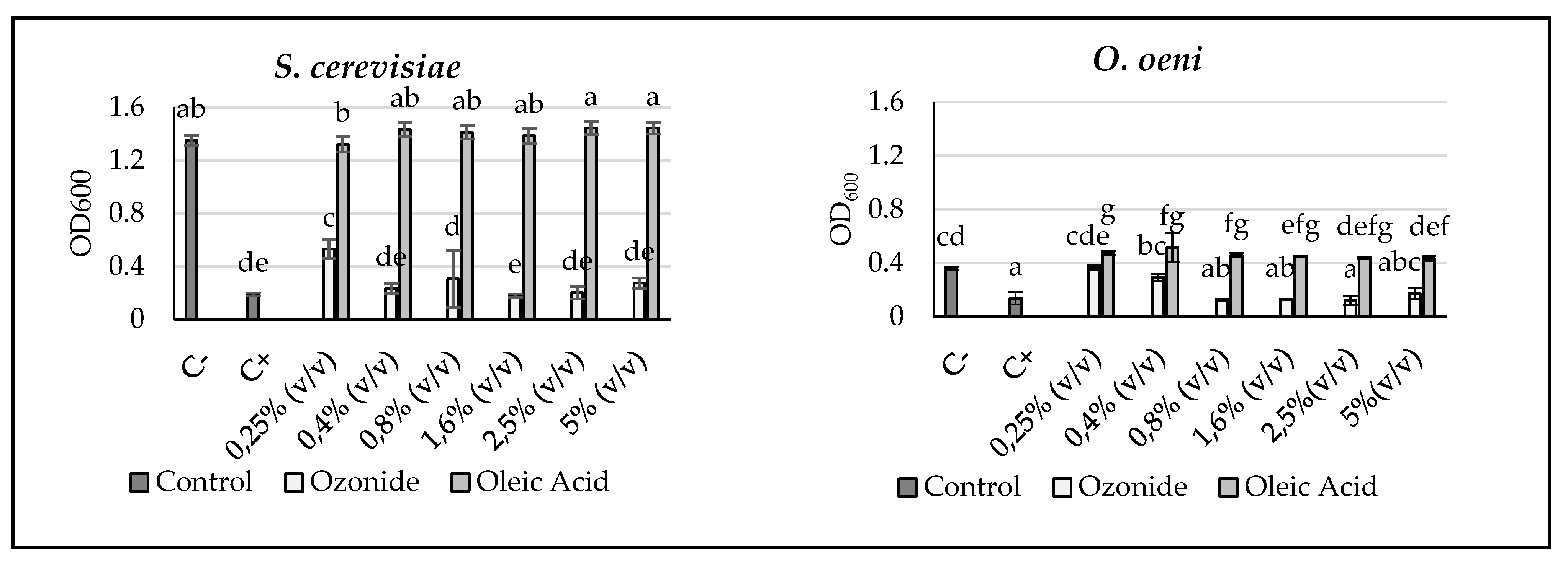
3.3. Efficiency Analyses against Yeasts and Bacteria Relevant to the Vinification Process

Since many of the wine-associated saccharomyces and non-saccharomyces yeasts and bacteria could not be detected in the ITS or 16S samples, in vitro analyses were performed to elucidate the effect of ozonide treatment. The impact on the growth of liquid cultures supplemented with increasing concentrations of ozonized oleic acid (ozonide) or oleic acid, respectively were compared regarding their optical density at λ = 600 nm (OD600) (Figure 1). An effect of the oleic acid itself should be excluded. For some organisms such as O. oeni, and L. plantarum and Gluconobacter oxydans, low concentrations of oleic acid led to a statistically significant increased optical density compared to the negative control (C−) (Table S4). With oleic acid supplementation, Pediococcus sp. showed the clearest reduction in OD600 compared to control samples. It remained stable between 86.4 ± 8.9% and 85.4 ± 9.1% at concentrations of 0.4% (v/v) to 2.5% (v/v) oleic acid and decreased to 18.7 ± 2.7% at a concentration of 5% (v/v) oleic acid (Table S4). Yet, the OD600 of Pediococcus sp. was always lower for ozonide supplementation compared to the same concentration of oleic acid, indicating a higher sensitivity towards the ozonized compound (Table 3). The ozonized oleic acid preparation applied in the vineyard had a concentration of 0.8% (v/v). In the liquid culture-based efficacy analyses, ozonide supplementations up to this concentration had no significant effect on the OD600 of L. brevis, Candida zeylanoides and Pichia fermentans. Nevertheless, culture densities of C. zeylanoides and P. fermentans showed a concentration-dependent decrease at ozonide concentrations below 1.6% (v/v). In contrast, low concentrations of ozonide seemed to favor the growth of L. brevis liquid cultures (Table 3). Acetobacter aceti, Pediococcus sp., S. cerevisiae and B. bruxellensis showed the highest sensitivity towards the ozonized oleic acid with an OD600 below 50% at the lowest ozonide concentration of 0.25% (v/v). Negative relative optical densities, as in the case of S. cerevisiae treated with 1.6% (v/v) ozonide (Table 3, Figure 1), resulted from the normalization of the OD600 to C− (100%) and C+ (0%), as described in Petit et al. (2021) [41]. This normalization was conducted to simplify comparison of the sensitivities towards the treatment between the organisms. Negative values did not deviate statistically significant from the C+ in any case.

4. Discussion

Microbial communities on wine grapes are dynamic and they change significantly over a short period of time [40,49]. The developmental stage of the grape has been reported to play a major role in the assembly of the bacterial and yeast communities on the grape carpoplane [49,50,51,52]. The frequently described increase in microbial populations and diversity from veraison to harvest time is presumably due to the increased availability of hosting surfaces and nutrients [49,50,51,52,53]. These are results of the more elastic and permeable berry skins [49,50,51,52,53]. Thus, sampling can only provide an overview of the species existing within the population at the exact moment of sampling. Comparisons between the different sampling times should be treated with caution due to the diverse and complex environmental influences. In this study, samples were taken at the developmental stages BBCH 83 (Portugieser I), which represents veraison, and BBCH 87 (Portugieser II), at which berries are soft and evenly colored but not quite ripe. In compliance with a study published by Ding et al. (2021) [54], the highest bacterial species richness and OTU richness were detected in samples of category I. This also applied to the comparison of eukaryotic species richness detected in samples of Portugieser I-ITS2 compared to the corresponding samples of category II (Table 1). It stands in clear contrast to the findings of Abdullabekova et al. (2020) [55], who found the highest number of yeast species at physiological ripeness of grape observed by a direct plating approach. The decrease in species richness during ripening appears contradictory since the samples of category II were harvested later and with a longer interval between last treatment and sampling. Since all types of samples in category II showed a comparable decrease in the richness values no matter which treatment they obtained, this trend is likely to be due to changed grape surface or environmental conditions. The high relative humidity and the decreasing average and maximum temperatures before the second sampling may have contributed to these differences (Figure S6). These factors have been demonstrated to be important drivers of the microbial community assembly [49,56,57] and diversity influencing wine aroma profiles [57]. This confirms the results of Bokulich et al. (2014) [56], who found highly significant relations of net precipitation, relative humidity and maximum temperatures alongside with the average temperature to the grape must microbial communities. Conveniently with this, Ding et al. (2021) [54] hypothesized that observed changes in fungal and bacterial abundance might be due to rainfall events during grape ripening. The Evenness, Shannon (H) and Inverse Simpson (1-D) indices were higher in 16S samples of category II compared to category I (Table 1). These indices take into account not only the number of species but also the evenness of their relative abundances. Increasing β-diversity of all 16S samples shows that differences between microbial populations on grapes of NT, CT and OT were reduced just two weeks after the last treatment (Table 2). This finding was further supported by an overall increase in the correlation of Portugieser II samples compared to Portugieser I. The higher similarity of samples in terms of Sørensen and Jaccard similarity indices represents a diminishing of the dominance of few species in the population in favor of a more even distribution of relative abundances. It is reflected by the dominance of the genus Pantoea and Pseudomonas syringae decreasing from samples of category I to category II. Nevertheless, conclusions should be drawn carefully since members of the genus Pantoea may have both positive and negative effects on grapevine health depending on environmental conditions [58]. Members of the Gram-negative genus Pantoea within the family of Erwiniaceae [59] are frequently referred to as biocontrol agents against fungal infection in a diverse array of crop plants [59,60,61,62]. They possess an epiphytic, endophytic or rhizospheric lifestyle [59,61,63]. Among others, Gasser et al. (2012) and Magnin-Robert et al. (2013) demonstrated a successful reduction in grape infection by Botrytis cinerea in vineyards treated with Pantoea ananatis [62] and Pantoea agglomerans, respectively [61]. Biocontrol activity was assumed to be carried out by a combination of the increase in plant defense mechanisms [61,62], spatial obstruction of the pathogen by micro-colonies distributed over the plant surface [62] and antibiotic active compounds [59,60]. Apart from these beneficial effects, Boiu-Sicuia et al. (2020) identified P. agglomerans as one of four bacterial species causing crown gall tumors in young Romanian vineyards [58]. P. syringae is a biotrophic [64], Gram-negative, rod-shaped bacterium [64,65] within the family of Pseudomonadaceae [65]. The P. syringae species complex is subdivided into more than 50 pathovars based on physiological and taxonomic traits as well as infection symptoms and host range, as summarized by Gerin et al. (2019) [66]. Typical symptoms of grapevine infections with P. syringae pv. Syringae are bacterial cankers [66], bacterial leaf spots (BLS) and bacterial inflorescence rot (BIR) [64,67]. Whitelaw-Weckert et al. (2011) were able to demonstrate that the necrotic areas on leaves and flowers of grapevine promoted sporulation of the previously symptomless infection with B. cinerea [64]. Thus, the reduced prevalence of P. syringae in samples of category II compared to category I could be beneficial to the health of the vine and berries in terms of B. cinerea infection. On the other hand, the reduced dominance of members of the genus Pantoea might have an adverse effect on plant health due to reduced biocontrol capacities. Further research would be necessary to clarify the effects of these findings. The sampling method applied in this study included only visibly intact berries. This should prevent a masking effect of phytopathogenic fungi present on only few heavily infected berries towards the overall microbiotic community. The exclusion was carried out because of the disproportionately higher biomass of phytopathogenic fungi on these individual berries. Thus, the data presented in this study are suitable to draw conclusions on the microbiotic community but do not support presumptions on the disease incidence or severity of fungal pathogens.
The overall species and OTU richness revealed minor differences between NT and CT (Table 1). In contrast, OT species richness and OTU richness were markedly decreased in all samples, with the exception of ITS2-Portugieser II. These differences between NT, CT and OT can be explained by the effective spectrum of the treatments. An impact of diverse chemical fungicides on bacterial non-target organisms has been reported in different crops depending on the active substance of the applied formulations and environmental conditions [68,69,70,71]. Many of the conventional fungicides have a site-specific mode of action. They attack a specific target of the fungal metabolism [9]. In contrast, ozonides possess a broad effective spectrum, as their effect is based on unspecific oxidation of all accessible surfaces [12,72]. This unspecific efficacy of ozonides against bacteria and fungi is confirmed by the higher β-diversity indices between bacterial sequences of NT/CT compared to NT/OT and CT/OT (Table 2). In contrast, ITS2 sequencing results revealed higher dissimilarities between NT/CT and CT/OT than between NT/OT in samples of category II (Table 2), presumably caused by the broader efficacy of the conventional fungicides against fungal organisms. These findings were supported by a stronger correlation of NT/OT compared to NT/CT and CT/OT. Consistent with this observation, the comparison of ß-diversities of category I and category II revealed a slight drop in β-diversity between CT/OT, whereas β-diversity of NT/CT and NT/OT revealed a minor increase. This might be due to a more persistent efficacy of CT compared to OT. Given the increasing number of fungicide-resistant phytopathogenic fungi [7,8,9] and the reported fungicidal activity of ozonized plant oils [13,14], they still remain an interesting field of research. Ozonized plant oils might be a sustainable alternative to fully synthetic agrochemicals since they are produced from fully renewable raw material [73]. Tropospheric oleic acid arises, e.g., from marine aerosols and direct forest emissions and has been proven to possess a short half-life of several minutes to few hours due to rapid loss to ozonolysis, as reviewed by Zahardis and Petrucci (2007) [74].
No fermentative yeast species could be detected in both categories of samples. Lactic acid bacteria necessary for malolactic fermentation and acetic acid bacteria were found to be present at low RA, represented by Acetobacter and the genus formerly known as Lactobacillus [17,75], respectively. Others such as O. oeni were absent in the samples. This is presumably due to the premature sampling of the berries, since these species were found to be present mainly on fully mature berries [37,40,52,55]. Consistently, O. oeni could not be identified in earlier metagenomic studies [40]. In German and other vineyards, S. cerevisiae could be detected rarely and in case of presence in low abundance [15,37]. To our knowledge, the application of organic ozonides in viticultural field experiments has not yet been reported by other research groups. Therefore, little is known on their possible effects on these oenologically and economically relevant microorganisms. Nevertheless chemical, medical and pharmacological publications reported a broad efficacy of different organic ozonides and ozonized plant oils against bacteria, filamentous fungi and yeasts [10,12,76,77]. Among them are members of the genera Pseudomonas, Bacillus [78] and various ascomycetes of the genus Candida [13,76,77]. To elucidate possible effects of the OT on grape and must microbiota, the efficacy of the ozonized oleic acid was tested against an array of two acetic acid bacteria (AAB), four lactic acid bacteria (LAB) and nine yeast species (Table 3). The AAB G. oxydans and A. aceti were chosen since they are among the most common bacterial spoilage organisms of wine [79,80]. Members of the three most abundantly found LAB genera in musts and wine, Oenococcus, Pediococcus and the genus formerly known as Lactobacillus [17,75], were selected to exemplify the efficacy of the new ozonide treatment on LAB. Publications from the medical context, such as the studies of Sechi et al. (2001) [12] and de Almeida Kogawa et al. (2015) [10], report antimicrobial activities of ozonized plant oils against Gram-positive and Gram-negative bacteria. Although these publications were based on clinical strains of human pathogenic bacteria which do not belong to the native microbiota of grapes, this general observation does apply to the examined Gram-positive (L. brevis, L. plantarum, O. oeni, Pediococcus sp.) and Gram-negative species (A. aceti, G. oxydans) (Table 3). All bacterial strains showed a significant reduction of OD600 at an ozonide concentration of 1.6% (v/v) or below. Sechi et al. (2001) [12] as well as de Almeida Kogawa et al. (2015) [10] achieved relatively high Minimal Inhibitory Concentrations (MIC) of ozonized sunflower oil against all tested bacterial strains in the range of mg/mL. de Almeida Kogawa et al. (2015) [10] concluded that the high MIC compared to antibiotics are due to the chemical composition of ozonized plant oils representing not only the active compound itself but a complex matrix of substances containing antimicrobial active oxygen species. Together with the low toxicity against vertebrates [[81] cited in [12]], Sechi et al. (2001) [12] concluded that the antimicrobial activity is based on the action against multiple cellular targets rather than due to a generalized toxicity or inhibition of specific metabolic steps. This assumption is further supported by the observation that prolonged incubation of S. aureus in ozonized sunflower oil (OLEOZON®) results in an increase in cytoplasmic membrane permeability towards K+-Ions and cytoplasm leakage [82]. A similar mode of action was described for aqueous and gaseous ozone achieved by non-selective oxidation of exposed cellular structures and subsequent penetration and oxidation of the cell interior causing cell death [72]. The efficacy of aqueous ozone was shown to be highly dependent on two factors: the ability of the microorganisms to form biofilms, and the culture density [83]. This is due to the increase in oxidizable organic matter correlated with an increase in these two factors. In the study conducted by Guzzon et al. (2013) [83], cell death of O. oeni, members of the genera Lactobacillus and Pediococcus as well as B. bruxellensis and S. pombe were achieved at a relatively low ozone concentration of 1 mg/L at 105 CFU/mL, revealing a high sensitivity. In the same study, cell death of S. cerevisiae, P. fermentans, H. uvarum and G. oxydans was achieved at a medium-high concentration of 2.5 mg/L, whereas cell death of M. pulcherrima was not achieved under the tested conditions [83]. In accordance with their results, Pediococcus sp. and B. bruxellensis revealed the highest ozonide sensitivities, followed by H. uvarum and O. oeni (Table 3). In our experiments, L. brevis, Candida zeylanoides, S. pombe and M. pulcherrima were the least sensitive towards the ozonide treatment. Nevertheless, differences from the observations of Guzzon et al. (2013) [83], such as the high ozonide sensitivity of S. cerevisiae and the ozonide tolerance of S. pombe, could be detected. In this context, it should be considered that the results of Guzzon et al. (2013) [83] are only partially transferable to this study. Both the methodology and the investigated active substance differ significantly. The antimicrobial activities of aqueous ozone and ozonized oleic acid rely at least partially on active oxygen species. In the presence of protic solvents such as water, the trioxolane structure of organic ozonides decompose into an array of organic compounds [74,84,85,86]. α-acylalkyl hydroperoxides and secondary ozonides result from the reaction [74,84,85,86]. Hydroperoxides and aldehydes originate from a subsequent decomposition of these energetically unstable products [87,88]. In the case of ozonized oleic acid, the decomposition reaction results in 1-nonanal, 9-oxononanoic acid, azelaic acid and nonanoic acid [74,89]. Further studies would be necessary to unravel the contribution of these and further compounds of the ozonized oleic acid to its antimicrobial activity. Despite the premature sampling of the grape berries, we were able to provide evidence that the new OT might be suitable to significantly reduce the growth of various microorganisms adverse to the vinification process. It is conceivable that late OT treatments just before harvest could help reduce microbiological spoilage of musts caused by microorganisms from the vineyard. Further research is necessary to create a broader data basis on the effects of the new OT on wine sensory characteristics and food safety, especially at late treatments.

5. Conclusions

In this study, we demonstrated the highly dynamic behavior of microbial communities on the example of the new OT at and after the last fungicide treatment of the season. Species and OTU richness and abundance gave indications of the strong dependence of the grape carpoplane microbiota on environmental conditions and the developmental stage of the berries. The effects of the new OT on the fungal community of the grape carpoplane were less clear than the conventional fungicide treatment. Bacterial β-diversity of OT/NT was reduced compared to CT/NT, unveiling a stronger effect of the OT on these communities. The effect of the in vitro ozonide treatment varied between the bacterial and fungal strains. Acetobacter aceti, Pediococcus sp. and S. cerevisiae showed the highest sensitivity towards the ozonized oleic acid, with culture densities below 50% at the lowest ozonide concentration of 0.25% (v/v). To gain deeper insight into the effect of the new OT on the dynamics of the grape carpoplane microbial communities, it would be beneficial to perform additional sampling covering the whole time span from berry set until harvest.

6. Patents

ANSEROS Klaus Nonnenmacher GmbH Tübingen (GE) is an original German producer of organic ozonides based on unsaturated plant oils and ozone gas, which are used together with water for the formation of LIQUENSO® Oxygenat, a water-based liquid ready to spray on to the plants and fruits (communities). The method and device for the formation and supply of the LIQUENSO® Oxygenat (OT) are patented in Europe (EP 3 478 072 B1). LIQUENSO® is a registered trademark.

Supplementary Materials

The following are available online at https://www.mdpi.com/article/10.3390/ecologies3030023/s1, Figure S1: Rarefaction curves obtained from 16S sequencing. Portugieser carpoplane sample triplicates of category I NT (A), CT (C) and OT (E) and category II NT (B), CT (D) and OT (F). Figure S2: Rarefaction curves obtained from ITS2 sequencing. Portugieser carpoplane sample triplicates of category I NT (A), CT (C) and OT (E) and category II NT (B), CT (D) and OT (F). Figure S3: Principal Component Analysis of Portugieser I-16S (top) and Portugieser II-16S (bottom). The correlation circles represent Spearman’s rank correlations (p < 0.05) of individual samples. Sample types are color-coded: NT = blue, OT = red, CT = black. Figure S4: Principal Component Analysis of Portugieser I-ITS2 (top) and Portugieser II-ITS2 (bottom). The correlation circles represent Spearman’s rank correlations (p < 0.05) of individual samples. Sample types are color-coded: NT = blue, OT = red, CT = black. Figure S5: Relative abundance of phyla in Portugieser I and Portugieser II samples expressed in % of the bacterial 16S reads (A, B) and % of the fungal ITS2 reads (C, D). Values are derived from sample triplicates after merging and subsampling. Figure S6: Weather data obtained from the weather station Neustadt (Weinstraße, Rhineland-Palatinate, Germany). The source of the daily measures of maximum, average and minimum temperatures (A), and precipitation and relative humidity (B) is the government platform Agrarmeteorologie Rheinland-Pfalz (www.Wetter.RLP.de (accessed on 29 March 2022)). Table S1: Schedule of phytosanitary treatments in the vineyard of the grape variety Vitis vinifera L. cv. Portugieser carried out in the season from 29 April 2019 to 10 September 2019. The applied amount and concentration of conventional fungicides followed the printed recommendation of the DLR Rheinpfalz (Pflanzenschutz 2019). Ozonide was applied at a concentration of 0.8% (v/v). The vineyard is located in Neustadt (Weinstraße), Germany (49°22′28.2″ N 8°11′28.3″ E). Table S2: Organisms used for 96-well microtiter-based efficacy assays and the incubation time to final photometric measurement of OD600. Table S3: Species share of the individual samples in one triplicate of the same category expressed as percent of bacterial organisms (Proteobacteria, Pantoea spp., Acetobacter spp., Lactobacillus spp. and Pseudomonas syringae) or fungal organisms (Ascomycota, Basidiomycota). Table S4: In vitro efficacy analysis of non-ozonized oleic acid. All values and standard deviations are expressed in % related to the corresponding negative controls (Table 3). Values significantly deviating from the corresponding C− (p < 0.05) are shaded.

Author Contributions

Conceptualization: A.K., F.R., M.S.-S., L.F.S., P.W.-H. and T.N.; Methodology: DNA Metabarcoding: F.R., M.E. and L.F.S.; In vitro efficacy analyses: F.R., J.F.-S., L.F.S. and E.T.; Validation: DNA Metabarcoding: M.E.; In vitro efficacy analyses: L.F.S.; Formal Analysis: DNA Metabarcoding: M.E.; PCA: L.F.S.; In vitro efficacy analyses: L.F.S.; Investigation: Experimental procedures of the ozonide treatment, berry sampling, sample preparation and shipping were carried out by L.F.S. and D.R.; In vitro efficacy analyses were carried out by L.F.S. with support from J.F.-S.; Resources: DNA Metabarcoding: DLR Rheinpfalz, Wine Campus Neustadt; In vitro efficacy analyses: IBWF; Data Curation: DNA Metabarcoding: M.E.; In vitro efficacy analyses: L.F.S. Writing—Original Draft Preparation: Abstract and Introduction: M.E. and L.F.S.; Experimental section—Ozonide preparation: T.N.; DNA Metabarcoding: Sections from the Master Thesis of M.E. were consensually adapted and processed by L.F.S.; In vitro efficacy analyses: L.F.S. Writing—Review and Editing: L.F.S., M.E., T.N., D.R., F.R., P.W.-H., A.K., J.F.-S., E.T. and M.S.-S.; Visualization: DNA Metabarcoding Results: M.E.; In vitro efficacy analyses and supplementary material: L.F.S.; Supervision: M.S.-S., E.T. and T.N.; Project Administration: M.S.-S. and T.N.; Funding Acquisition: A.K., F.R., M.S.-S. and P.W.-H.; T.N. (ANSEROS Tübingen). All authors have read and agreed to the published version of the manuscript.

Funding

The presented data originate from a cooperative project of the sponsor AiF with the company ANSEROS, the University of Kaiserslautern and the DLR Rheinpfalz, funded by the Central Innovation Program for SMEs (ZIM) of the German Federal Ministry for Economic Affairs and Climate Action. The cooperative project is listed under grant number ZF4062403SA7.

Institutional Review Board Statement

Not applicable.

Informed Consent Statement

Not applicable.

Data Availability Statement

Not applicable.

Acknowledgments

Our sincere thanks are due to Agnieszka Mos-Hummel for the technical support in the joint project and the production and support of the Ozonide LIQUENSO® Oxygenat on behalf of ANSEROS. We would like to express our sincere thanks to the team of the Staatsweingut mit Johannitergut for the application of the conventional treatments and the maintenance of the experimental vineyards. We thank Jens Schirmel for his kind support in the processing of diversity data, Rémi Goerlich for his help and contribution in programming and Jérôme Morinière from AIM for his metabarcoding data support. Furthermore, we thank Joachim Schmidt for his kind advice concerning application technique and general questions on fungicide formulations.

Conflicts of Interest

The authors declare no conflict of interest. The funders had no role in the design of the study; in the collection, analyses, or interpretation of data; in the writing of the manuscript, or in the decision to publish the results.

References

  1. Barata, A.; Malfeito-Ferreira, M.; Loureiro, V. The Microbial Ecology of Wine Grape Berries. Int. J. Food Microbiol. 2012, 153, 243–259. [Google Scholar] [CrossRef] [PubMed]
  2. Stefanini, I.; Cavalieri, D. Metagenomic Approaches to Investigate the Contribution of the Vineyard Environment to the Quality of Wine Fermentation: Potentials and Difficulties. Front. Microbiol. 2018, 9, 991. [Google Scholar] [CrossRef]
  3. Escribano-Viana, R.; López-Alfaro, I.; López, R.; Santamaría, P.; Gutiérrez, A.R.; González-Arenzana, L. Impact of Chemical and Biological Fungicides Applied to Grapevine on Grape Biofilm, Must, and Wine Microbial Diversity. Front. Microbiol. 2018, 9, 59. [Google Scholar] [CrossRef] [PubMed]
  4. Stefanini, I.; Dapporto, L.; Legras, J.-L.; Calabretta, A.; Paola, M.D.; Filippo, C.D.; Viola, R.; Capretti, P.; Polsinelli, M.; Turillazzi, S.; et al. Role of Social Wasps in Saccharomyces Cerevisiae Ecology and Evolution. Proc. Natl. Acad. Sci. USA 2012, 109, 13398–13403. [Google Scholar] [CrossRef] [PubMed] [Green Version]
  5. Liu, D.; Zhang, P.; Chen, D.; Howell, K. From the Vineyard to the Winery: How Microbial Ecology Drives Regional Distinctiveness of Wine. Front. Microbiol. 2019, 10, 2679. [Google Scholar] [CrossRef] [PubMed]
  6. Vitulo, N.; Lemos, W.J.F.; Calgaro, M.; Confalone, M.; Felis, G.E.; Zapparoli, G.; Nardi, T. Bark and Grape Microbiome of Vitis Vinifera: Influence of Geographic Patterns and Agronomic Management on Bacterial Diversity. Front. Microbiol. 2019, 9, 3203. [Google Scholar] [CrossRef] [Green Version]
  7. Chen, W.-J.; Delmotte, F.; Cervera, S.R.; Douence, L.; Greif, C.; Corio-Costet, M.-F. At Least Two Origins of Fungicide Resistance in Grapevine Downy Mildew Populations. Appl. Environ. Microbiol. 2007, 73, 5162–5172. [Google Scholar] [CrossRef] [Green Version]
  8. Leroch, M.; Kretschmer, M.; Hahn, M. Fungicide Resistance Phenotypes of Botrytis Cinerea Isolates from Commercial Vineyards in South West Germany. J. Phytopathol. 2011, 159, 63–65. [Google Scholar] [CrossRef]
  9. Delmas, C.E.L.; Dussert, Y.; Delière, L.; Couture, C.; Mazet, I.D.; Richart Cervera, S.; Delmotte, F. Soft Selective Sweeps in Fungicide Resistance Evolution: Recurrent Mutations without Fitness Costs in Grapevine Downy Mildew. Mol. Ecol. 2017, 26, 1936–1951. [Google Scholar] [CrossRef]
  10. de Almeida Kogawa, N.R.; de Arruda, E.J.; Micheletti, A.C.; Matos, M.D.F.C.; de Oliveira, L.C.S.; de Lima, D.P.; Carvalho, N.C.P.; de Oliveira, P.D.; de Castro Cunha, M.; Ojeda, M. Synthesis, Characterization, Thermal Behavior, and Biological Activity of Ozonides from Vegetable Oils. RSC Adv. 2015, 5, 65427–65436. [Google Scholar] [CrossRef]
  11. Günaydın, Y.; Sevim, H.; Tanyolaç, D.; Gürpınar, Ö.A. Ozonated Olive Oil with a High Peroxide Value for Topical Applications: In-Vitro Cytotoxicity Analysis with L929 Cells. Ozone Sci. Eng. 2018, 40, 37–43. [Google Scholar] [CrossRef]
  12. Sechi, L.A.; Lezcano, I.; Nunez, N.; Espim, M.; Duprè, I.; Pinna, A.; Molicotti, P.; Fadda, G.; Zanetti, S. Antibacterial Activity of Ozonized Sunflower Oil (Oleozon). J. Appl. Microbiol. 2001, 90, 279–284. [Google Scholar] [CrossRef] [Green Version]
  13. Geweely, N.S.I. Antifungal Activity of Ozonized Olive Oil (Oleozone). Int. J. Agri. Biol. 2006, 8, 671–678. [Google Scholar]
  14. Ma, Y.; Ke, Y.; Zhu, H.; Li, B.; Li, B.; Zhang, F.; Li, Y. Oleozon: A Novel Control Strategy against Powdery Mildew in Cucumber. J. Phytopathol. 2017, 165, 841–847. [Google Scholar] [CrossRef]
  15. Agarbati, A.; Canonico, L.; Mancabelli, L.; Milani, C.; Ventura, M.; Ciani, M.; Comitini, F. The Influence of Fungicide Treatments on Mycobiota of Grapes and Its Evolution during Fermentation Evaluated by Metagenomic and Culture-Dependent Methods. Microorganisms 2019, 7, 114. [Google Scholar] [CrossRef] [Green Version]
  16. Gilbert, J.A.; van der Lelie, D.; Zarraonaindia, I. Microbial Terroir for Wine Grapes. Proc. Natl. Acad. Sci. USA 2014, 111, 5–6. [Google Scholar] [CrossRef] [Green Version]
  17. Capozzi, V.; Tufariello, M.; De Simone, N.; Fragasso, M.; Grieco, F. Biodiversity of Oenological Lactic Acid Bacteria: Species-and Strain-Dependent plus/Minus Effects on Wine Quality and Safety. Fermentation 2021, 7, 24. [Google Scholar] [CrossRef]
  18. Strickland, M.T.; Schopp, L.M.; Edwards, C.G.; Osborne, J.P. Impact of Pediococcus spp. on Pinot Noir Wine Quality and Growth of Brettanomyces. Am. J. Enol. Vitic. 2016, 67, 188–198. [Google Scholar] [CrossRef]
  19. Wade, M.E.; Strickland, M.T.; Osborne, J.P.; Edwards, C.G. Role of Pediococcus in Winemaking. Aust. J. Grape Wine Res. 2019, 25, 7–24. [Google Scholar] [CrossRef] [Green Version]
  20. Impact of Different Temperature Profiles on Simultaneous Yeast and Bacteria Fermentation|SpringerLink. Available online: https://link.springer.com/article/10.1186/s13213-020-01565-w (accessed on 23 June 2022).
  21. Medina, K.; Boido, E.; Dellacassa, E.; Carrau, F. Growth of Non-Saccharomyces Yeasts Affects Nutrient Availability for Saccharomyces Cerevisiae during Wine Fermentation. Int. J. Food Microbiol. 2012, 157, 245–250. [Google Scholar] [CrossRef]
  22. Vicente Sánchez, J.; Calderón, F.; Santos de la Sen, A.; Marquina Díaz, D.; Benito, S. High Potential of Pichia Kluyveri and Other Pichia Species in Wine Technology. Int. J. Mol. Sci. 2021, 22, 1196. [Google Scholar] [CrossRef] [PubMed]
  23. Snowdon, E.M.; Bowyer, M.C.; Grbin, P.R.; Bowyer, P.K. Mousy Off-Flavor: A Review. J. Agric. Food Chem. 2006, 54, 6465–6474. [Google Scholar] [CrossRef] [PubMed]
  24. Costello, P.J.; Siebert, T.E.; Solomon, M.R.; Bartowsky, E.J. Synthesis of Fruity Ethyl Esters by Acyl Coenzyme A: Alcohol Acyltransferase and Reverse Esterase Activities in Oenococcus Oeni and Lactobacillus Plantarum. J. Appl. Microbiol. 2013, 114, 797–806. [Google Scholar] [CrossRef] [PubMed]
  25. Morata, A.; Loira, I.; González, C.; Escott, C. Non-Saccharomyces as Biotools to Control the Production of Off-Flavors in Wines. Molecules 2021, 26, 4571. [Google Scholar] [CrossRef]
  26. Comitini, F.; Gobbi, M.; Domizio, P.; Romani, C.; Lencioni, L.; Mannazzu, I.; Ciani, M. Selected Non-Saccharomyces Wine Yeasts in Controlled Multistarter Fermentations with Saccharomyces Cerevisiae. Food Microbiol. 2011, 28, 873–882. [Google Scholar] [CrossRef]
  27. Costello, P.J.; Lee, T.H.; Henschke, P. Ability of Lactic Acid Bacteria to Produce N-Heterocycles Causing Mousy off-Flavour in Wine. Aust. J. Grape Wine Res. 2001, 7, 160–167. [Google Scholar] [CrossRef]
  28. Romano, A.; Perello, M.C.; de Revel, G.; Lonvaud-Funel, A. Growth and Volatile Compound Production by Brettanomyces/Dekkera Bruxellensis in Red Wine. J. Appl. Microbiol. 2008, 104, 1577–1585. [Google Scholar] [CrossRef]
  29. Virdis, C.; Sumby, K.; Bartowsky, E.; Jiranek, V. Lactic Acid Bacteria in Wine: Technological Advances and Evaluation of Their Functional Role. Front. Microbiol. 2021, 11, 612118. [Google Scholar] [CrossRef]
  30. Pérez-Martín, F.; Seseña, S.; Izquierdo, P.M.; Martín, R.; Palop, M.L. Screening for Glycosidase Activities of Lactic Acid Bacteria as a Biotechnological Tool in Oenology. World J. Microbiol. Biotechnol. 2012, 28, 1423–1432. [Google Scholar] [CrossRef]
  31. Benito, S. The Management of Compounds That Influence Human Health in Modern Winemaking from an HACCP Point of View. Fermentation 2019, 5, 33. [Google Scholar] [CrossRef] [Green Version]
  32. Mamlouk, D.; Gullo, M. Acetic Acid Bacteria: Physiology and Carbon Sources Oxidation. Indian J. Microbiol. 2013, 53, 377–384. [Google Scholar] [CrossRef] [Green Version]
  33. Lynch, K.M.; Zannini, E.; Wilkinson, S.; Daenen, L.; Arendt, E.K. Physiology of Acetic Acid Bacteria and Their Role in Vinegar and Fermented Beverages. Compr. Rev. Food Sci. Food Saf. 2019, 18, 587–625. [Google Scholar] [CrossRef] [Green Version]
  34. Shinjoh, M.; Toyama, H. Industrial Application of Acetic Acid Bacteria (Vitamin C and Others). In Acetic Acid Bacteria: Ecology and Physiology; Matsushita, K., Toyama, H., Tonouchi, N., Okamoto-Kainuma, A., Eds.; Springer: Japan, Tokyo, 2016; pp. 321–338. ISBN 978-4-431-55933-7. [Google Scholar]
  35. Liu, L.; Chen, Y.; Yu, S.; Chen, J.; Zhou, J. Simultaneous Transformation of Five Vectors in Gluconobacter Oxydans. Plasmid 2021, 117, 102588. [Google Scholar] [CrossRef]
  36. Soares-Santos, V.; Pardo, I.; Ferrer, S. Cells-QPCR as a Direct Quantitative PCR Method to Avoid Microbial DNA Extractions in Grape Musts and Wines. Int. J. Food Microbiol. 2017, 261, 25–34. [Google Scholar] [CrossRef]
  37. Brysch-Herzberg, M.; Seidel, M. Yeast Diversity on Grapes in Two German Wine Growing Regions. Int. J. Food Microbiol. 2015, 214, 137–144. [Google Scholar] [CrossRef]
  38. Thijs, S.; Op De Beeck, M.; Beckers, B.; Truyens, S.; Stevens, V.; Van Hamme, J.D.; Weyens, N.; Vangronsveld, J. Comparative Evaluation of Four Bacteria-Specific Primer Pairs for 16S RRNA Gene Surveys. Front. Microbiol. 2017, 8, 494. [Google Scholar] [CrossRef]
  39. Toju, H.; Tanabe, A.S.; Yamamoto, S.; Sato, H. High-Coverage ITS Primers for the DNA-Based Identification of Ascomycetes and Basidiomycetes in Environmental Samples. PLoS ONE 2012, 7, e40863. [Google Scholar] [CrossRef] [Green Version]
  40. Pinto, C.; Pinho, D.; Sousa, S.; Pinheiro, M.; Egas, C.; Gomes, A.C. Unravelling the Diversity of Grapevine Microbiome. PLoS ONE 2014, 9, e85622. [Google Scholar] [CrossRef] [Green Version]
  41. Petit, B.; Mitaine-Offer, A.-C.; Fischer, J.; Schüffler, A.; Delaude, C.; Miyamoto, T.; Tanaka, C.; Thines, E.; Lacaille-Dubois, M.-A. Anti-Phytopathogen Terpenoid Glycosides from the Root Bark of Chytranthus Macrobotrys and Radlkofera Calodendron. Phytochemistry 2021, 188, 112797. [Google Scholar] [CrossRef]
  42. Xu, Y.; Xie, M.; Xue, J.; Xiang, L.; Li, Y.; Xiao, J.; Xiao, G.; Wang, H.-L. EGCG Ameliorates Neuronal and Behavioral Defects by Remodeling Gut Microbiota and TotM Expression in Drosophila Models of Parkinson’s Disease. FASEB J. 2020, 34, 5931–5950. [Google Scholar] [CrossRef] [Green Version]
  43. Ghosh, S.; Bagheri, B.; Morgan, H.H.; Divol, B.; Setati, M.E. Assessment of Wine Microbial Diversity Using ARISA and Cultivation-Based Methods. Ann. Microbiol. 2015, 65, 1833–1840. [Google Scholar] [CrossRef]
  44. Benson, D.A.; Cavanaugh, M.; Clark, K.; Karsch-Mizrachi, I.; Lipman, D.J.; Ostell, J.; Sayers, E.W. GenBank. Nucleic Acids Res. 2013, 41, D36–D42. [Google Scholar] [CrossRef] [PubMed] [Green Version]
  45. Nemergut, D.R.; Schmidt, S.K.; Fukami, T.; O’Neill, S.P.; Bilinski, T.M.; Stanish, L.F.; Knelman, J.E.; Darcy, J.L.; Lynch, R.C.; Wickey, P. Patterns and Processes of Microbial Community Assembly. Microbiol. Mol. Biol. Rev. 2013, 77, 342–356. [Google Scholar] [CrossRef] [PubMed] [Green Version]
  46. Finotello, F.; Mastrorilli, E.; Di Camillo, B. Measuring the Diversity of the Human Microbiota with Targeted Next-Generation Sequencing. Brief. Bioinform. 2018, 19, 679–692. [Google Scholar] [CrossRef]
  47. Hill, T.C.; Walsh, K.A.; Harris, J.A.; Moffett, B.F. Using Ecological Diversity Measures with Bacterial Communities. FEMS Microbiol. Ecol. 2003, 43, 1–11. [Google Scholar] [CrossRef]
  48. User’s Guide to Correlation Coefficients|Elsevier Enhanced Reader. Available online: https://reader.elsevier.com/reader/sd/pii/S2452247318302164?token=F352ED4A7123774FDFAC340E8319705D27C1CD7A79AC08AE39A025621711EC86BFED8F2367A02DB9758D6215D239487B&originRegion=eu-west-1&originCreation=20220621145200 (accessed on 21 June 2022).
  49. Liu, D.; Howell, K. Community Succession of the Grapevine Fungal Microbiome in the Annual Growth Cycle. Environ. Microbiol. 2021, 23, 1842–1857. [Google Scholar] [CrossRef]
  50. Moubasher, A.-A.H.; Abdel-Sater, M.A.; Soliman, Z. Biodiversity of Filamentous and Yeast Fungi in Citrus and Grape Fruits and Juices in Assiut Area, Egypt. J. Microbiol. Biotechnol. Food Sci. 2021, 2021, 353–365. [Google Scholar] [CrossRef]
  51. Garijo, P.; López, R.; Santamaría, P.; Ocón, E.; Olarte, C.; Sanz, S.; Gutiérrez, A.R. Presence of Enological Microorganisms in the Grapes and the Air of a Vineyard during the Ripening Period. Eur. Food Res. Technol. 2011, 233, 359–365. [Google Scholar] [CrossRef]
  52. Renouf, V.; Claisse, O.; Lonvaud-Funel, A. Understanding the Microbial Ecosystem on the Grape Berry Surface through Numeration and Identification of Yeast and Bacteria. Aust. J. Grape Wine Res. 2005, 11, 316–327. [Google Scholar] [CrossRef]
  53. Martins, G.; Vallance, J.; Mercier, A.; Albertin, W.; Stamatopoulos, P.; Rey, P.; Lonvaud, A.; Masneuf-Pomarède, I. Influence of the Farming System on the Epiphytic Yeasts and Yeast-like Fungi Colonizing Grape Berries during the Ripening Process. Int. J. Food Microbiol. 2014, 177, 21–28. [Google Scholar] [CrossRef]
  54. Ding, Y.; Wei, R.; Wang, L.; Yang, C.; Li, H.; Wang, H. Diversity and Dynamics of Microbial Ecosystem on Berry Surface during the Ripening of Ecolly (Vitis Vinifera L.) Grape in Wuhai, China. World J. Microbiol. Biotechnol. 2021, 37, 214. [Google Scholar] [CrossRef]
  55. Abdullabekova, D.A.; Magomedova, E.S.; Aliverdieva, D.A.; Kachalkin, A.V. Yeast Communities of Vineyards in Dagestan: Ecological, Taxonomic, and Genetic Characteristics. Biol. Bull. 2020, 47, 344–351. [Google Scholar] [CrossRef]
  56. Bokulich, N.A.; Thorngate, J.H.; Richardson, P.M.; Mills, D.A. Microbial Biogeography of Wine Grapes Is Conditioned by Cultivar, Vintage, and Climate. Proc. Natl. Acad. Sci. USA 2014, 111, E139–E148. [Google Scholar] [CrossRef] [Green Version]
  57. Liu, D.; Chen, Q.; Zhang, P.; Chen, D.; Howell, K.S. The Fungal Microbiome Is an Important Component of Vineyard Ecosystems and Correlates with Regional Distinctiveness of Wine. mSphere 2020, 5, e00534-20. [Google Scholar] [CrossRef]
  58. Boiu-Sicuia, O.-A.; Dinu, S.; Barbu, L. Physiological Profile of Some Pathogenic Bacteria Associated with Grapevine Crown Gall. Sci. Pap.-Ser. B Hortic. 2020, 64, 230–237. [Google Scholar]
  59. Jiang, L.; Jeong, J.C.; Lee, J.-S.; Park, J.M.; Yang, J.-W.; Lee, M.H.; Choi, S.H.; Kim, C.Y.; Kim, D.-H.; Kim, S.W.; et al. Potential of Pantoea Dispersa as an Effective Biocontrol Agent for Black Rot in Sweet Potato. Sci. Rep. 2019, 9, 16354. [Google Scholar] [CrossRef] [Green Version]
  60. Smits, T.H.M.; Duffy, B.; Blom, J.; Ishimaru, C.A.; Stockwell, V.O. Pantocin A, a Peptide-Derived Antibiotic Involved in Biological Control by Plant-Associated Pantoea Species. Arch. Microbiol. 2019, 201, 713–722. [Google Scholar] [CrossRef]
  61. Magnin-Robert, M.; Quantinet, D.; Couderchet, M.; Aziz, A.; Trotel-Aziz, P. Differential Induction of Grapevine Resistance and Defense Reactions against Botrytis Cinerea by Bacterial Mixtures in Vineyards. BioControl 2013, 58, 117–131. [Google Scholar] [CrossRef]
  62. Gasser, F.; Schildberger, B.; Berg, G. Biocontrol of Botrytis Cinerea by Successful Introduction of Pantoea Ananatis in the Grapevine Phyllosphere. Int. J. Wine Res. 2012, 4, 53. [Google Scholar] [CrossRef] [Green Version]
  63. Diversity of Bacterial Endophytes in 3 and 15 Year-Old Grapevines of Vitis Vinifera Cv. Corvina and Their Potential for Plant Growth Promotion and Phytopathogen Control|Elsevier Enhanced Reader. Available online: https://reader.elsevier.com/reader/sd/pii/S0944501315300318?token=6FE35BF072795D0CCD3B247C99B924899D2DE0E2FA11E3C63D920B060ADE84803E9A4DA8A6ED42BB33856BD929AC76FE&originRegion=eu-west-1&originCreation=20220620121031 (accessed on 20 June 2022).
  64. Whitelaw-Weckert, M.A.; Whitelaw, E.S.; Rogiers, S.Y.; Quirk, L.; Clark, A.C.; Huang, C.X. Bacterial Inflorescence Rot of Grapevine Caused by Pseudomonas Syringae Pv. Syringae. Plant Pathol. 2011, 60, 325–337. [Google Scholar] [CrossRef]
  65. Lipps, S.M.; Samac, D.A. Pseudomonas Viridiflava: An Internal Outsider of the Pseudomonas Syringae Species Complex. Mol. Plant Pathol. 2022, 23, 3–15. [Google Scholar] [CrossRef]
  66. Gerin, D.; Cariddi, C.; de Miccolis Angelini, R.M.; Rotolo, C.; Dongiovanni, C.; Faretra, F.; Pollastro, S. First Report of Pseudomonas Grapevine Bunch Rot Caused by Pseudomonas Syringae Pv. Syringae. Plant Dis. 2019, 103, 1954–1960. [Google Scholar] [CrossRef]
  67. Hall, S.J.; Dry, I.B.; Gopurenko, D.; Whitelaw-Weckert, M.A. Pseudomonas Syringae Pv. Syringae from Cool Climate Australian Grapevine Vineyards: New Phylogroup PG02f Associated with Bacterial Inflorescence Rot. Plant Pathol. 2019, 68, 312–322. [Google Scholar] [CrossRef]
  68. Marinho, M.D.C.; Diogo, B.S.; Lage, O.M.; Antunes, S.C. Ecotoxicological Evaluation of Fungicides Used in Viticulture in Non-Target Organisms. Environ. Sci. Pollut. Res. 2020, 27, 43958–43969. [Google Scholar] [CrossRef]
  69. Perazzolli, M.; Antonielli, L.; Storari, M.; Puopolo, G.; Pancher, M.; Giovannini, O.; Pindo, M.; Pertot, I. Resilience of the Natural Phyllosphere Microbiota of the Grapevine to Chemical and Biological Pesticides. Appl. Environ. Microbiol. 2014, 80, 3585–3596. [Google Scholar] [CrossRef] [Green Version]
  70. Puglisi, E.; Vasileiadis, S.; Demiris, K.; Bassi, D.; Karpouzas, D.G.; Capri, E.; Cocconcelli, P.S.; Trevisan, M. Impact of Fungicides on the Diversity and Function of Non-Target Ammonia-Oxidizing Microorganisms Residing in a Litter Soil Cover. Microb. Ecol. 2012, 64, 692–701. [Google Scholar] [CrossRef]
  71. Katsoula, A.; Vasileiadis, S.; Sapountzi, M.; Karpouzas, D.G. The Response of Soil and Phyllosphere Microbial Communities to Repeated Application of the Fungicide Iprodione: Accelerated Biodegradation or Toxicity? FEMS Microbiol. Ecol. 2020, 96, fiaa056. [Google Scholar] [CrossRef]
  72. Anand, S.K. A Comparative Analysis of Antimicrobial Property of Wine and Ozone with Calcium Hydroxide and Chlorhexidine. J. Clin. Diagn. Res. 2015, 9, ZC04-6. [Google Scholar] [CrossRef] [PubMed]
  73. Kadhum, A.A.H.; Wasmi, B.A.; Mohamad, A.B.; Al-Amiery, A.A.; Takriff, M.S. Preparation, Characterization, and Theoretical Studies of Azelaic Acid Derived from Oleic Acid by Use of a Novel Ozonolysis Method. Res. Chem. Intermed. 2012, 38, 659–668. [Google Scholar] [CrossRef]
  74. Zahardis, J.; Petrucci, G.A. The Oleic Acid-Ozone Heterogeneous Reaction System: Products, Kinetics, Secondary Chemistry, and Atmospheric Implications of a Model System—A Review. Atmos. Chem. Phys. 2007, 7, 1238–1240, 1244–1245, 1263–1265. [Google Scholar] [CrossRef] [Green Version]
  75. Zheng, J.; Wittouck, S.; Salvetti, E.; Franz, C.M.A.P.; Harris, H.M.B.; Mattarelli, P.; O’Toole, P.W.; Pot, B.; Vandamme, P.; Walter, J.; et al. A Taxonomic Note on the Genus Lactobacillus: Description of 23 Novel Genera, Emended Description of the Genus Lactobacillus Beijerinck 1901, and Union of Lactobacillaceae and Leuconostocaceae. Available online: https://era.library.ualberta.ca/items/b4736051-ee09-49ec-8146-8f05975db42c (accessed on 8 February 2022).
  76. Guerrer, L.V.; Cunha, K.C.; Nogueira, M.C.L.; Cardoso, C.C.; Soares, M.M.C.N.; Almeida, M.T.G. “In Vitro” Antifungal Activity of Ozonized Sunflower Oil on Yeasts from Onychomycosis. Braz. J. Microbiol. 2012, 43, 1315–1318. [Google Scholar] [CrossRef] [Green Version]
  77. Monzillo, V.; Lallitto, F.; Russo, A.; Poggio, C.; Scribante, A.; Arciola, C.R.; Bertuccio, F.R.; Colombo, M. Ozonized Gel Against Four Candida Species: A Pilot Study and Clinical Perspectives. Materials 2020, 13, 1731. [Google Scholar] [CrossRef] [Green Version]
  78. Díaz, M.F.; Hernández, R.; Martínez, G.; Vidal, G.; Gómez, M.; Fernández, H.; Garcés, R. Comparative Study of Ozonized Olive Oil and Ozonized Sunflower Oil. J. Braz. Chem. Soc. 2006, 17, 403–407. [Google Scholar] [CrossRef]
  79. Torija, M.J.; Mateo, E.; Guillamón, J.M.; Mas, A. Identification and Quantification of Acetic Acid Bacteria in Wine and Vinegar by TaqMan–MGB Probes. Food Microbiol. 2010, 27, 257–265. [Google Scholar] [CrossRef]
  80. Kántor, A.; Kačániová, M.; Petrová, J.; Medo, J.; Hleba, L.; Rovná, K. Application of Rt-Pcr for Acetobacter Species Detection in Red Wine. J. Microbiol. Biotechnol. Food Sci. 2021, 2021, 231–234. [Google Scholar]
  81. Sanchez, G.M.; Fernandez, O.S.L.; Rodriguez, C. Toxicidad Aguda Dermica Del Aceite Ozonizado ‘Oleozon’ En Ratas y Conejos Rol de Los Radicales Libres CENIC. Cienc. BioloÁgicas 1997, 28, 35. [Google Scholar]
  82. Curtiellas, V.; Ledea, O.; Rodríguez, S.; Ancheta, O.; Echevarría, M.; Sánchez, E.; Fernández, I. El OLEOZON® Sobre La Viabilidad, La Permeabilidad Celular y La Ultraestructura de Staphylococcus Aureus. Rev. CENIC Cienc. Biol. 2008, 39, 128–131. [Google Scholar]
  83. Guzzon, R.; Nardin, T.; Micheletti, O.; Nicolini, G.; Larcher, R. Antimicrobial Activity of Ozone. Effectiveness against the Main Wine Spoilage Microorganisms and Evaluation of Impact on Simple Phenols in Wine. Aust. J. Grape Wine Res. 2013, 19, 180–188. [Google Scholar] [CrossRef]
  84. Criegee, R. Mechanismus der Ozonolyse. Angew. Chem. 1975, 87, 765–771. [Google Scholar] [CrossRef]
  85. Bailey, P.S. The Reactions of Ozone with Organic Compounds. Chem. Rev. 1958, 58, 925–1010. [Google Scholar] [CrossRef]
  86. Ziemann, P.J. Aerosol Products, Mechanisms, and Kinetics of Heterogeneous Reactions of Ozone with Oleic Acid in Pure and Mixed Particles. Faraday Discuss. 2005, 130, 469–490. [Google Scholar] [CrossRef]
  87. Zhou, Z.; Abbatt, J.P. Formation of Gas-Phase Hydrogen Peroxide via Multiphase Ozonolysis of Unsaturated Lipids. Environ. Sci. Technol. Lett. 2020, 8, 114–120. [Google Scholar] [CrossRef]
  88. Zhou, Z.; Zhou, S.; Abbatt, J.P.D. Kinetics and Condensed-Phase Products in Multiphase Ozonolysis of an Unsaturated Triglyceride. Environ. Sci. Technol. 2019, 53, 12467–12475. [Google Scholar] [CrossRef]
  89. Woden, B.; Skoda, M.W.A.; Milsom, A.; Gubb, C.; Maestro, A.; Tellam, J.; Pfrang, C. Ozonolysis of Fatty Acid Monolayers at the Air–Water Interface: Organic Films May Persist at the Surface of Atmospheric Aerosols. Atmos. Chem. Phys. 2021, 21, 1325–1340. [Google Scholar] [CrossRef]
Figure 1. Efficacy of ozonized oleic acid is concentration-dependent and varies between different organisms. The OD600 of S. cerevisiae liquid cultures one day after inoculation and O. oeni liquid cultures six days after inoculation are depicted as mean values and standard deviations with n ≥ 3. Letters indicate results of ANOVA analysis followed by Fisher’s LSD post-hoc procedure (left) and Kruskal–Wallis two-sided non-parametric test, followed by Conover–Iman post-hoc procedure with Bonferroni adjustment for multiple pairwise comparisons (right) with p < 0.05.
Ecologies 03 00023 g001
Table 1. Species richness, α-diversity indices obtained from 16S and ITS sequencing of carpoplane microbiota. Samples derived from Portugieser berries 1 day (Portugieser I) or 2 weeks (Portugieser II) after the last agrochemical treatment. Non-treated (NT), conventionally treated (CT) and ozonide-treated (OT) samples were compared.
Treatment Richness α-Diversity Indices
OTUSpeciesShannon
(H)
Evenness (J)Inverse Simpson
(1-D)
Portugieser I-16S NT1311131.450.310.50
CT1271181.510.320.52
OT50430.760.200.33
Portugieser II-16S NT43401.980.540.81
CT41382.040.560.82
OT32291.360.400.62
Portugieser I-ITS2 NT94831.730.390.76
CT94801.830.420.76
OT74661.810.430.77
Portugieser II-ITS2 NT43431.900.500.77
CT44411.310.350.57
OT43411.790.480.76
Table 2. ß-diversity indices obtained from 16S and ITS sequencing of carpoplane microbiota. Samples derived from Portugieser berries 1 day (Portugieser I) or 2 weeks (Portugieser II) after the last agrochemical treatment. Non-treated (NT), conventionally treated (CT) and ozonide-treated (OT) samples were compared.
Comparison of Treatments ß-Diversity Indices
Sørensen Similarity Index (β1)Jaccard Similarity Index (β2)
Portugieser I-16S NT/CT0.620.45
NT/OT0.500.33
CT/OT0.470.31
Portugieser II-16S NT/CT0.740.59
NT/OT0.700.53
CT/OT0.720.56
Portugieser I-ITS2 NT/CT0.560.39
NT/OT0.640.48
CT/OT0.590.42
Portugieser II-ITS2 NT/CT0.600.42
NT/OT0.710.56
CT/OT0.560.39
Table 3. In vitro efficacy analysis of ozonized oleic acid. Values significantly deviating from the corresponding C− (p < 0.05) are shaded.
0.25% (v/v) Ozonide0.4% (v/v) Ozonide0.8% (v/v) Ozonide1.6% (v/v) Ozonide2.5% (v/v) Ozonide5% (v/v) OzonideC−C+
Acetobacter aceti19.9 ± 6.63.6 ± 5.23.4 ± 6.5−0.4 ± 3.16.8 ± 5.82.6 ± 2.8100.0 ± 3.20.0 ± 5.1
Gluconobacter oxydans108.8 ± 25.649.3 ± 32.35.6 ± 3.0−9.7 ± 1.6−10.5 ± 4.5−7.9 ± 0.2100.0 ± 17.00.0 ± 8.8
Levilactobacillus brevis125.1 ± 13.8116.2 ± 2.6111.1 ± 4.556.4 ± 1.676.5 ± 3.035.4 ± 2.3100.0 ± 9.10.0 ± 2.5
Lactiplantibacillus plantarum102.3 ± 6.489.1 ± 8.380.1 ± 2.949.8 ± 4.461.0 ± 1.940.4 ± 1.7100.0 ± 4.50.0 ± 1.7
Oenococcus oeni102.1 ±7.2972.4 ± 10.04.3± 2.75.5± 1.63.0 ± 13.523.4 ± 16.7100.0 ± 4.20.0 ± 3.5
Pediococcus sp.32.7 ± 6.024.3 ± 19.83.0 ± 9.32.7 ± 15.4−2.2 ± 5.5−3.0 ± 3.2100.0 ± 6.40.0 ± 3.6
Brettanomyces bruxellensis26.1 ± 17.427.4 ± 4.022.6 ± 6.22.5 ± 3.56.1 ± 15.411.0 ± 3.7100.0 ± 13.80.0 ± 2.3
Candida zeylanoides99.8 ± 6.773.1 ± 2.577.4 ± 10.273.9 ± 14.253.1 ± 17.721.8 ± 9.8100.0 ± 27.70.0 ± 3.5
Hanseniaspora uvarum85.7 ± 0.646.8 ± 14.25.9 ± 1.98.4 ± 3.48.4 ± 22.45.0 ± 3.0100.0 ± 7.60.0 ± 5.9
Metschnikowia pulcherrima56.0 ± 14.074.6 ± 8.967.3 ± 7.672.3 ± 6.839.6 ± 19.728.2 ± 22.1100.0 ± 3.30.0 ± 1.5
Pichia fermentans96.3 ± 10.691.2 ± 0.883.6 ± 0.181.1 ± 2.525.0 ± 26.37.7 ± 11.6100.0 ± 2.80.0 ± 0.6
Saccharomyces cerevisiae29.4 ± 6.13.8 ± 3.110.0 ± 18.5−0.9 ± 1.21.1 ± 4.17.3 ± 3.4100.0 ± 3.10.0 ± 1.0
Schizosaccharomyces pombe88.1 ± 2.479.0 ± 2.374.7 ± 2.067.5 ± 3.731.9 ± 26.024.0 ± 33.4100.0 ± 2.90.0 ± 3.6
Torulaspora delbruckii84.4 ± 5.286.4 ± 8.193.4 ± 4.65.5 ± 5.74.9 ± 1.415.3 ± 4.2100.0 ± 2.20.0 ± 1.1
Zygosaccharomyces bailii85.1 ± 16.184.3 ± 4.879.3 ± 8.566.2 ± 2.013.2 ± 6.740.1 ± 7.7100.0 ± 43.40.0 ± 1.2
Publisher’s Note: MDPI stays neutral with regard to jurisdictional claims in published maps and institutional affiliations.

