Review

The Influence of Magnetic Fields, Including the Planetary Magnetic Field, on Complex Life Forms: How Do Biological Systems Function in This Field and in Electromagnetic Fields?

by
David A. Hart
Department of Surgery, Faculty of Kinesiology, and McCaig Institute for Bone & Joint Health, University of Calgary, Calgary, AB T2N 4N1, Canada
Biophysica 2024, 4(1), 1-21; https://doi.org/10.3390/biophysica4010001
Submission received: 20 November 2023 / Revised: 20 December 2023 / Accepted: 24 December 2023 / Published: 2 January 2024

Abstract

:
Life on Earth evolved to accommodate the biochemical and biophysical boundary conditions of the planet millions of years ago. The former includes nutrients, water, and the ability to synthesize other needed chemicals. The latter includes the 1 g gravity of the planet, radiation, and the geomagnetic field (GMF) of the planet. How complex life forms have accommodated the GMF is not known in detail, considering that Homo sapiens evolved a neurological system, a neuromuscular system, and a cardiovascular system that developed electromagnetic fields as part of their functioning. Therefore, all of these could be impacted by magnetic fields. In addition, many proteins and physiologic processes utilize iron ions, which exhibit magnetic properties. Thus, complex organisms, such as humans, generate magnetic fields, contain significant quantities of iron ions, and respond to exogenous static and electromagnetic fields. Given the current body of literature, it remains somewhat unclear if Homo sapiens use exogenous magnetic fields to regulate function and what can happen if the boundary condition of the GMF no longer exerts an effect. Proposed deep space flights to destinations such as Mars will provide some insights, as space flight could not have been anticipated by evolution. The results of such space flight “experiments” will provide new insights into the role of magnetic fields on human functioning. This review will discuss the literature regarding the involvement of magnetic fields in various normal and disturbed processes in humans while on Earth and then further discuss potential outcomes when the GMF is no longer present to impact host systems, as well as the limitations in the current knowledge. The GMF has been present throughout evolution, but many details of its role in human functioning remain to be elucidated, and how humans have adapted to such fields in order to develop and retain function remains to be elucidated. Why this understudied area has not received the attention required to elucidate the critical information remains a conundrum for both health professionals and those embarking on space flight. However, proposed deep space flights to destinations such as Mars may provide the environments to test and assess the potential roles of magnetic fields in human functioning.

1. Introduction

1.1. Purpose

Early life on Earth evolved in the contexts of temperature, available elements, and molecules (including water), as well as the biophysical boundary conditions of the planet. The latter include gravity (1 g), exogenous and endogenous radiation from particles from beyond Earth and radioactive elements on Earth, and magnetic fields due to the planet’s magnetic field and local concentrations of molecules such as iron. Thus, the evolution of life within the boundary conditions of Earth for millions of years, even with fluctuations and variations due to solar system cycles and solar events, could not have anticipated life as we know it today with the advent of electromagnetic fields from modern devices and going beyond Earth’s boundary conditions via space flight.
The altered conditions of Low Earth Orbit (LEO) space flight have already exposed astronauts to dramatically decreased gravity and an elevated risk of exposure to radiation, and spending time in such conditions has led to several biological responses, particularly due to the loss of the 1 g gravity. However, the influence of the increased risk for radiation exposure has not yet been documented in detail, likely in part due to the relatively small number of individuals who have spent time at LEO.
It should be noted that one boundary condition of Earth that has not yet been exceeded by space flight is that of the geomagnetic field (GMF) of Earth. For readers who are not familiar with many of the characteristics of the GMF, they can refer to the following websites for general overviews on the topic (http://www.geomag.bgs.ac.uk/education/earthmag.html; http://space.com/earths-magnetic-field-explained; https://www.britannica.com/science/geomagnetic-field/Source-of-the-field; accessed 26 October 2023). At LEO, the geomagnetic field is still evident, and perhaps even at the distance of the moon, some of its influence could still be evident but greatly diminished (discussed in [1,2,3]). Thus far, only a small number of astronauts have ventured to the moon, and they stayed for a relatively short time.
Thus, humans have not ventured completely beyond the geomagnetic field of Earth. Since some of the central systems of humans (i.e., cardiovascular, muscular, and neural systems) generate and respond to magnetic fields, this boundary condition likely deserves more study as humans plan to travel to Mars and exist for the first time completely beyond the geomagnetic field of Earth. Thus, this article attempts to discuss the various aspects of magnetic fields on life, the potential involvement of magnetic fields on cellular and integrated biological processes (Figure 1), and how the lack of this boundary condition for a protracted time frame could lead to a loss of function. As depicted in Figure 1, some systems that generate EMF during their function could be susceptible to exogenous magnetic fields, as well as those that store iron-containing proteins (i.e., ferromagnetic ions). Thus, we know that we live in magnetic fields (i.e., the geomagnetic field and local concentrations of magnetic elements), but it is mostly a silent boundary condition and, as yet, does not ascribe to functions in our daily lives. However, as humans venture beyond its influence, we likely need to be prepared to understand the potential consequences and anticipate solutions to such consequences, as humans did not evolve for space travel. Furthermore, if there is an impact of static and electromagnetic fields on the functioning of complex life forms, such as humans, it is not known whether specific phases of the life cycle may be more influenced than others, such as during development or senescence (Figure 2).
The intent of this review is to discuss the influence of exogenous static and electromagnetic fields on complex organisms, including humans, discuss some of the mechanisms involved, identify gaps in our knowledge regarding how the geomagnetic field may have shaped evolutionary choices to accommodate this boundary condition, and how opportunities associated with planned deep space missions well beyond the influence of the geomagnetic field of Earth may provide opportunities to better understand the influence of both this static field and electromagnetic fields on human functions. As the latter could not have been anticipated by evolutionary considerations, this is a unique opportunity that should be prepared for prior to undertaking such flights.

1.2. Background

Life on Earth evolved slowly from simple single-cell entities that could eventually reproduce themselves using information storage molecules such as DNA, initially as prokaryotes, and eventually as eukaryotes with subcellular organelles including mitochondria, such cells adapted to a variety of environmental conditions (i.e., deep ocean vents, fresh water, cold to hot water). Interestingly, most life consists primarily of water.
In addition to securing the ability to form a plasma membrane and the machinery to perform essential functions, primitive cells needed to develop these abilities within the context of the physical and biophysical boundary conditions of Earth. These include background radiation and radiation from space that was not deflected by the geomagnetic field of the planet, the 1 g gravity of the planet, and the actual geomagnetic field plus any local magnetic influences. Thus, successful early life must have evolved mechanisms to negate such influences or evolved adaptations to embrace their influences.
As life likely evolved initially in oceans, lakes, or other water environments, perhaps initially boundary conditions such as the 1 g gravity were sensed, but further adaptations were required when complex life emerged onto land and mobility and navigation were advantageous. However, the influence of the geomagnetic field would have been felt in early life forms, either with regard to incorporating elements such as iron ions into essential processes or potentially in other as yet unknown defined manners. The commitment to an information-containing molecule such as DNA being central to reproduction fidelity is also an interesting choice as it can be sensitive to radiation damage and induction of mutations. However, early in evolution, this sensitivity could have been used advantageously to use mutations to adapt to a changing biological and physical environment. Mutations could arise from a lack of fidelity in copying the DNA and/or radiation-induced effects. As life becomes more complex and multi-cellular with differentiated functions, it is also likely that methods to minimize such influences would develop to decrease the likelihood of developing adverse situations that would compromise organism integrity. These latter could include tumor suppressor genes, DNA repair mechanisms, and controlled cell death.
While the 1 g gravity of Earth would have been felt by organisms and resulted in settling to the bottom of a lake or near a vent in the ocean, organisms could have resisted this effect via the movement of the water or by attaching themselves to something in shallow water and nutrients could have come to them via water movement. The real impact of the 1 g gravity environment would likely have been felt when multi-cellular organisms emerged to live on land, and the advantage of mobility and navigation against ground reaction forces required the evolution of new adaptations. Thus, the development of legs for quadrupedal movement and legs and arms for bipedal mobility required the evolution of effective adaptations and integration with visual or other sensors. Also of interest in this regard are species that lived on land and developed legs but then returned to the marine environment (i.e., whales and other marine mammals). The cardiovascular system of complex lifeforms living on land also required adaptations to function in the 1 g environment. As all tissues except perhaps articular cartilage are vascularized and innervated, these adaptations would affect all organ systems.
As evolution could not have predicted space flight and exposure to microgravity, it is interesting that the atrophy of mechanically loaded tissues such as bone and muscles is rapidly evident after leaving Earth and living at LEO (discussed in [3]). In addition, cardiovascular effects are also very evident after exposure to microgravity. Interestingly, even prolonged bedrest on Earth leads to loss of bone and muscle and cardiovascular changes, so such tissues have evolved a “use it or lose it” paradigm even when still under the influence of a 1 g gravity environment on Earth (discussed in [3,4]).
Another major boundary condition, the geomagnetic field, certainly “protects” life forms from the negative influence of solar radiation (discussed in [5]). Many forms of exogenous radiation originating from the sun or other cosmic sources (i.e., pulsars, black holes, supernovas] can be deflected by this magnetic field (discussed in [5]), thus protecting the DNA from mutational events or resulting in cell death. For example, it has been reported to enhance radiation resistance by promoting DNA repair processes in cells [6]. From reading the literature, this attribute is the main influence of the geomagnetic field on life. The real question then becomes, “is it the only role?” and if so, why and how did life forms develop systems that use electrical signaling with concomitant magnetic field generation within a powerful magnetic field? Such an environment may have led to the development of a bioelectric code early in the evolution of simple and then more complex life forms [7], and such a code would also have an electromagnetic component. Thus, such a system may respond to exogenous magnetic fields as well [8]. It would be intuitive to conclude that commitment to such systems, such as the brain and neural systems, as well as the heart and cardiovascular system, could have evolved approaches to either negate the geomagnetic field or embrace it. Furthermore, as the distribution of elements on Earth that could lead to local magnetic fields is not uniform (i.e., large deposits of iron-containing hematite and taconite non-uniformly concentrated in various locals), the evolved adaptations to the geomagnetic field must account for the exposure to such local concentrations or suffer the consequences.

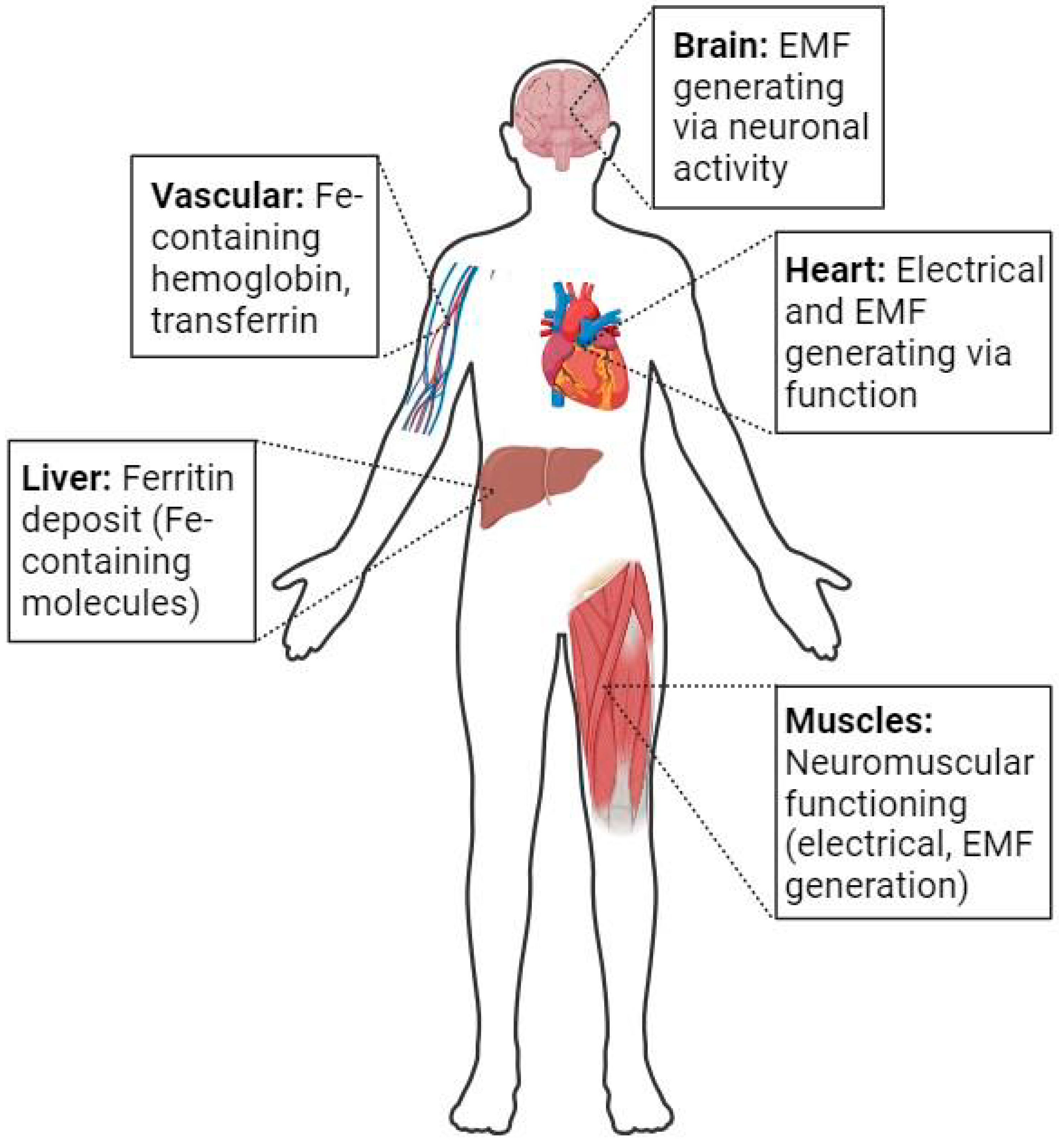
2. Iron-Containing Molecules, Magnetic Fields, and Evolution to Complex Lifeforms

As lifeforms became more complex within the boundary conditions of Earth, they likely had options for including specific elements into molecules performing a variety of functions. Bacteria depend on iron for growth (reviewed in [9]) and have complex interactions in this regard during infection [10] and in the host-gut microbiota relationship [11]. Therefore, prokaryotes evolved the use of iron ions for essential processes early, and this continued to evolve when they interacted with more complex eukaryotes. Thus, even in a strong geomagnetic field + background magnetic fields from iron deposits, life forms incorporated Fe ion-containing processes into biological systems that grew in complexity.

2.1. Incorporation of Ferro-Magnetic Ions in Essential Systems and Molecules

It is interesting that Homo sapiens “inherited” several iron ion-containing molecules via evolution [12], molecules that are essential for several important processes. These include hemoglobin (reviewed in [13,14]), cytochrome P450 enzymes [15,16,17], transferrin [18], ferritin [19], lactoferrin [20] and others [21,22,23,24].
The regulation of these iron ion-containing molecules is tightly controlled as their dysregulation may be harmful to human health [12,25,26,27,28,29,30], as heme also appears to have toxic properties [31]. Further evidence for the tight regulation of iron ions during pregnancy [32], exercise [33], and hypoxia [34] indicate that this ion is very important biologically. Thus, humans contain a considerable amount of iron ions in their bodies normally, with an unequal distribution among organs. Therefore, processes involving such Fe-containing molecules could be influenced by both static and electromagnetic fields. However, it remains to be determined whether the level of magnetism exerted by the geomagnetic field can influence the functioning of Fe-containing proteins in complex species. Species that use the geomagnetic field for navigation can sense such fields (discussed below), so future research will need to address these issues in detail.
However, much of the existing literature has focused on the biochemistry of iron ions, and the influence of static or electromagnetic fields on the function of Fe ion-containing molecules has not been the focus. Interestingly, exposure of mice [35] to static magnetic fields or rabbits to electromagnetic fields [36] led to detectable alterations in iron metabolism. Given the extensive use of Fe-containing molecules and their associated processes and the expansion of exposure to electromagnetic fields in everyday life, more extensive investigation of the relationship(s) between these aspects of Fe should be a research emphasis going forward, perhaps with a specific focus on neurodevelopment and maturation [37].
Finally, there can also be detectable alterations to iron metabolism in disease processes such as Alzheimer’s disease [38,39,40] and neurodegenerative conditions [41,42,43,44]. Whether these are the cause or effect of the diseases is not well defined, but with such local alterations in Fe ion levels, there may also be alterations to endogenous magnetic field patterns as detected by imaging modalities [45,46,47,48].

2.2. Magnetic Fields and Navigation

Numerous reports using a variety of species, particularly those that migrate, indicate that many of these species use specific brain centers to respond to magnetic fields to facilitate migration. These include birds [49,50,51], dolphins [52], fish [53], turtles [54], and some land animals [55]. Whether humans have a similar center to facilitate navigation is still under investigation (discussed in [56,57]). Thus, in this context, several species have evolved mechanisms to recognize and use the geomagnetic field to their advantage. Thus, the evolution of such migration/navigation facilitation centers must be integrated into other neural systems. How exactly this is accomplished remains to be determined for humans, but some genes have been implicated [58]. The genes involved in a fish include magr (a magnetoreceptor) and cry (a cryptochrome) [58]. These genes are orthologs of the human Cry2 and Magr genes [58]. In another fish, disruption of magnetic orientation behavior using pulsed magnetic fields led to the identification of several potentially relevant genes, including the gene frim, which is a ferritin-binding protein [59]. The involvement of cryptochromes in navigation has been implicated in several species (discussed in [60]), but they may also have other functions (discussed in [61]).
Furthermore, how such systems focus on the GMF and filter out background Fe concentrations or the influence of electromagnetic fields is not defined currently. However, the presence of such directional centers in the brain and the incorporation of their use to direct movement does indicate that evolutionarily, lifeforms recognized the GMF and used it for some purposes.

2.3. Magnetic Fields and Cognition

One of the distinguishing features of Homo sapiens is their cognition, memory, reasoning, and integration of significant aspects of the brain to formulate abstract thinking. How the current levels of these attributes developed during evolution is not known in any detail (discussed in [62,63,64,65]). However, it is likely that they developed in discrete steps rather than in some linear process. Furthermore, their development must have incorporated existing restrictions based on evolutionary choices made prior to and during the development of complex organisms. As fossil records cannot assess cognition and brain functioning, some aspects of the evolutionary tree are dependent on the interpretation of skull size and shape, as well as other activities of early hominids (artwork, burial practices, and other related activities). If, indeed, stepwise advances in cognition, memory ability, and related brain activities did occur during the evolutionary progression to Homo sapiens, then each step required effective integration into the existing paradigm. Interestingly, one is not apparently born with a fully developed set of cognition abilities, and they may continue to develop during post-natal development and maturation, as hypothesized by Piaget (reviewed in [66]). As the fully functional brain of an adult Homo sapiens expresses a pattern of electromagnetic signals as detected by MEG (reviewed in [67]); (discussed in [68]), there appears to be an intimate relationship between functional brain systems, brain biology (i.e., cell, biochemical and molecular) and magnetic fields. Whether such influences would be constant or possibly vary with the diurnal cycle and circadian rhythms [69] is unknown and will require more study.
While conventional thought would place emphasis on neural connectivity and biochemical/molecular interactions for such cognitive and memory abilities [61,70,71,72], some authors have advanced theories that consciousness is embedded in the electromagnetic field of the brain [72,73,74,75,76], while others have postulated that memory is also stored in magnetic fields [77]. The theory by McFadden, called the Conscious Electromagnetic Information (CEMI) field theory, postulates that information is stored in magnetic fields and used to regulate consciousness. While this theory has severe limitations and is not accepted by many, for such a system to work, it would also depend on the integrity of the neuronal circuits to manifest some of the information stored in such fields. It would also have to account for the GMF and its variations, exogenous electromagnetic fields, and exposure of the brain to strong magnetic fields, such as those associated with MRI, to maintain integrity. However, if such a system did exist, it would likely have to be integrated with the biological system during fetal development to accommodate subsequent maturation and have built-in safeguards to resist influences of exogenous magnetic fields. Furthermore, a decline in integration due to the loss of specific neural cells or some other mechanism (i.e., loss of vascular integrity in specific areas of the brain) during the later stages of life could contribute to disease development. Thus, many aspects of this theory are controversial, and it may not have credibility until more evidence is provided. Clearly, information storage, cognition, and how information is processed is very complex [78], and thus, the actual “system” for cognition and memory may rely on multiple components, some biological and some biophysical. An additional question is, when in evolution did such a system arise and become functional?
However, aside from the apparent limitations of a magnetic field as an information storage system related to consciousness and memory discussed above and elsewhere [79], several studies have attempted to investigate the effect of exogenous fields and hypomagnetic conditions on cognition. Regarding the former, 50–60 Hz fields (reviewed in [80]), radiofrequency fields [81], and static fields (reviewed in [82]) have been investigated and proposed. In such circumstances, results are somewhat inconclusive, but exposure to static fields, such as during an MRI, has not been associated with any reported adverse effects. However, in the latter, the time of exposure is limited, while in the former situations with 50–60 HZ and radiofrequency fields, exposure can be more chronic but variable in intensity and exposure time.
The effects of hypomagnetic fields on a variety of species have been reviewed recently [2,3,83,84]. Interest in the effects of hypomagnetic fields has been stimulated both by the desire to better understand the relationship(s) between magnetic fields and life forms and also since space flight presents the exposure of humans to life beyond the GMF of Earth [2,3,83]. Binhi and Sarimov [1] have reported that there is a zero magnetic field effect on human cognitive processes. However, the effects are modest and statistically significant but limited as the responses were assessed as acute exposure. However, in actual space flight beyond the GMF of Earth, the lack of a GMF will be chronic, but the astronauts will still be exposed to the electromagnetic fields of the equipment aboard the spacecraft or in a habitat, such as on Mars. Of course, in deep space, the astronauts will also be exposed to micro or zero gravity, which may also contribute to cognitive changes (discussed in [85]). Thus, long space-flight-associated alterations in brain activities [86] may be the result of multiple modalities (gravity, magnetic fields, stress, sleep deprivation] including magnetic field alterations. However, as the magnetic field contributions are somewhat understudied (discussed in [3]), it may be prudent to better understand such magnetic field effects on astronaut cognition prior to sending them off on long-term deep space missions. Such studies would also benefit those on Earth as well.

3. Exogenous Electromagnetic Field Effects on Biological Systems

3.1. Power Lines and Cancer

For several decades, living near high-voltage power lines and associated electromagnetic fields has been raised as a risk for human health [87,88], particularly cancers [89,90,91]. While several clinical trials have determined that the incidence of certain types of cancers is not more frequent in such environments, many people fear that some cancer clusters, particularly leukemias, are associated with living near high power lines [92,93], but thus far, the data are inconclusive [94,95,96,97]. As the incidence of cases in such “clusters” is still a small percentage of people living close to such power lines, if there is an effect of EMF on cell transformation, it is not a general effect, and the impact may be due to an underlying defect in some metabolic control mechanism in a susceptible subpopulation. A potential role for cryptochromes (discussed above in Section 2.2) in cancer has also been raised by Landler and Keays [61].

3.2. Cell Phone Use

Similar to fears about living close to high voltage power lines, in recent years, a potential risk of excessive cell phone use and tumors has also emerged [98] or been implicated in cancer development [99], but again, the results are somewhat inconclusive [100]. However, some reports [101,102,103] indicate that the effects of such fields may go beyond cancers. Of particular interest is the potential link between electromagnetic field exposure and autism [103], a spectrum of conditions localized to the brain arising during development, and the possible association between cell phone use and headaches [102], as both involve the brain.
This latter point regarding non-cancer-related issues is likely relevant to both the power line issue and cell phones, as many widely used appliances in the home emit electromagnetic fields when active. The question then arises as to why there have not been more conditions/diseases linked to exposure to electromagnetic fields. The answer is not known, but clearly, humans are very heterogeneous genetically; they are exposed to a variety of environmental insults such as air pollutants, artificial chemicals, food additives, consuming alcohol or smoking, and other variables that could confound any potential associations with electromagnetic fields. Furthermore, if, via evolution, humans had inherited intrinsic mechanisms that allow adaptation or incorporation of endogenous magnetic fields into regulatory systems, one might expect the most overt influences of exogenous EMF would be early in life during development and maturation or during senescence/aging when systems start to fail. Thus, future research should perhaps focus some attention on these phases of the life cycle.
In addition, variation in the background static magnetic fields would have differentially affected populations living in different parts of the Earth where the geomagnetic field varies, as do deposits of iron-rich ores (discussed in [3]). Exposure to such variation preceded the advent of extensive EMF exposures, most of which is a recent development over the past few hundred years. Some reports [104] have implicated such variation in childhood morbidity, but this is currently conjecture and not proven to have cause and effect.

4. Uses of Magnetic Fields for Health Applications

Humans and other animals, plants, and microorganisms have been exposed to a variety of magnetic fields other than the geomagnetic field of Earth and deposits of ferro materials (discussed in [105]). In addition, magnetic fields, static or electromagnetic, have been used for decades in attempts to improve outcomes for the repair of a variety of tissues (reviewed in [106,107,108,109,110,111]). The studies used magnetic fields of varying frequencies and intensities and employed both patient and preclinical models. However, the magnetic fields used in the studies were often variable, and it is sometimes difficult to compare studies. Magnetic fields have also been used to study cells [112] and processes such as development [113].

4.1. The Musculoskeletal System (MSK)

Magnetic fields have been used to influence the healing of a variety of tissues of the MSK system. These include soft tissues such as ligaments, tendons, cartilage, and menisci, as well as bone, and the studies have spanned several decades [114].
Ligament and Tendon Healing: Using a solid core electromagnet, Frank et al. [115] reported that exposure to the field following injury to the rabbit medial collateral ligament led to improved mechanical and biological healing parameters over 6 weeks. Subsequently, Lin et al. [116] reported that 2, 10, and 50 gauss (G) pulsing electromagnetic fields (PEMFs) during early healing of surgically induced defects in the rabbit patellar ligament/tendon led to improved outcomes, with 50 G yielding the best outcomes. More recently, Xu et al. [117] used exposure to a combination of static and electromagnetic fields (combined magnetic fields, CMF) to assess patella-patella tendon healing in a rabbit model. The authors assessed a variety of biomechanical and biological parameters and found that exposure to CMF led to significantly enhanced healing properties of the tissue at 16 weeks but less so at 8 weeks post-injury. Also, in a rabbit model, Hu et al. [118] demonstrated that exposure to a CMF led to an enhanced healing of the bone-tendon interface, potentially by enhancing osteogenesis. Thus, for these soft tissues and in rabbits, exposure to magnetic fields led to enhanced healing outcomes.
Meniscal and Cartilage Healing: Recently, Wang et al. [119] reported that healing of injuries to the avascular region of the menisci of male rats could be enhanced by exposure to pulsed electromagnetic fields (PEMF) over an 8-week study period. In addition, exposure to the PEMF also prevented the progression of the injury and development of osteoarthritis in the affected joints. The authors concluded that exposure to the PEMF led to enhanced fibrocartilage production and decreased inflammation. Interestingly, studies of electromagnetic fields with patients with established osteoarthritis (OA) do not appear to have regenerative effects on tissues but may reduce pain for some patients [120,121]. Whether this effect on pain is due to an effect on inflammatory processes in OA or some other mechanism remains to be determined by future investigations.
Bone Healing: The study of exogenous magnetic fields on bone healing has had a long history (reviewed in [106,122,123,124]). Such studies have been performed on a variety of species, and investigators have used bone cells both in vivo and in vitro. In vitro studies have been used in attempts to better understand the genes influenced by PEMFs on bone cells or osteogenic precursor differentiation [125]. In guinea pigs, exposure to both static and PEMFs enhanced bone repair of mandibular osteotomies based on histology [126].
Particular emphasis on the use of magnetic fields for bone healing has been applied to promote compromised healing, such as non-unions where natural healing either is protracted or fails. However, in a recent study of PEMFs in the healing of carpal scaphoid non-unions, the PEMF did not offer any detectable benefit [127]. Similarly, EMF exposure exhibited inconclusive effects on delayed or non-union fractures of long bones in adult patients [128]. In contrast, in a small study of 29 patients, the use of a CMF protocol led to enhanced healing of a variety of non-union fractures in both male and female adults [129]. In other reviews of the literature regarding magnetic field effects on non-unions, the consensus appears to be that exposure to magnetic fields (i.e., biophysical stimulation) is an effective modality [130,131,132] and an approach that avoids some of the complications associated with surgical interventions. Whether this variation in outcomes and conclusions is due to the type of magnetic fields that are applied, when and how they are applied, or to host factors is not clear at the present time. Furthermore, it is also not clear currently how such biophysical interventions impact cells at the biochemical or molecular levels. Some reports indicate it may involve effects on iron metabolism [133], while others indicate it may be at the level of immune and inflammatory process regulation [134]. The influence could be a combination of those elements, as an emerging theme is the effect of the magnetic fields on inflammation and inflammatory processes. This conclusion would also be supported by reports indicating static magnetic fields can also influence inflammation in the liver of mice [135].
In surrogate models of bone loss in space or immobilization, namely, prolonged hindlimb elevation in rodents to remove loading of the bones, it has been reported that PEMF exposure can prevent bone loss due to hindlimb elevation in rats [136] and that exposure to a static magnetic field in addition to loading in the 1 g environment can enhance recovery of bone in mice [137]. In the rat study, the activation of the sAC/cAMP/PKA/CREB signaling pathway was involved in the prevention of bone loss [136]. In this circumstance, the rat tissues were not overtly injured but were undergoing atrophy, and thus, the results may indicate that there was an interaction between magnetism-based mechanisms and loading via gravity and the impact of ground reaction forces.

4.2. The Brain and Neurological Integrity

As discussed earlier, the functioning of the brain leads to the generation of electromagnetic fields that can be measured by techniques such as SQUID or magnetoencephalogram (MEG) (reviewed in [67,138]). Such techniques can be used for both fetal [139] and adult [140] brain assessment. As such, these techniques can be used to detect neurological issues during development and during aging when loss of neurological integrity can occur with some frequency. However, a limitation of techniques such as MEG is that the detection system does not penetrate deep into the brain.

4.2.1. Detection of Brain Injury or Diseases

Detection of mild brain injury (i.e., post-concussion syndrome) using electromagnetic approaches has been proposed [141]. MEG can also be used for localizing and characterizing epileptic events [142] and, potentially, post-traumatic stress disorders [143]. While not an “overt injury” to the brain, space flight has led to the detection of cognition-associated changes [85,144,145,146]. Some reports have questioned whether the head-down tilt bedrest, a surrogate for spaceflight, captures the true nature of spaceflight-induced cognitive changes [147]. This surrogate is performed on Earth, so the 1 g and GMF environments are still functional, but the subjects are not exposed to the ground reaction forces associated with the 1 g environment. It does, however, mimic aspects of cardiovascular changes associated with space flight (discussed in [3,148]). To effectively capture space flight-related cognitive changes, new technology for real-time assessments may be required [149].
Detection of disruptions of functional networks using MEG and fMRI have been reported in patients with dementia [150], with some characteristics associated with specific types. MEG analysis in Alzheimer’s Disease [151,152,153] has revealed abnormalities, even in early disease [154,155]. In addition, some reports have discussed the potential role of magnetic fields as risk factors for the development of neurological and neurodegenerative diseases [156,157,158].

4.2.2. Health Benefits of Magnetic Fields on the Brain

Pulsed electromagnetic fields have been reported to positively affect microvascular perfusion and tissue oxygenation of the healthy rat brain [159]. Furthermore, the concept that electromagnetic fields could facilitate brain repair via neural stem cells has also been proposed [160] but has not yet been proven.
Interestingly, transcranial stimulation with PEMFs has been reported to positively influence depression [161,162], and potentially, transcranial magnetic stimulation may exert positive effects on post-traumatic stress disorder [163]. Furthermore, transcranial magnetic stimulation may also alleviate aspects of Alzheimer’s Disease (AD) [164]. Further studies in a mouse model of AD reported that exposure to a specific frequency of electromagnetic stimulation led to improved symptoms [165]. It should be noted that these studies were performed against a background of the GMF of Earth. Thus, improvements via exposure to a specific magnetic field appeared to “correct” disease-associated defects in the functionality of the brain. While the molecular mechanisms responsible for the improvements in patients are not known, in the mouse model [165], exposure to the 900 MHZ fields led to decreased amyloid plaque deposition in specific areas of the brain of the mice.
While many aspects of studies focused on “correcting” loss of brain integrity using exposure to magnetic fields are not well described, this is an emerging field of study. There may be significant variation in outcomes depending on the frequency or intensity of the magnetic fields used for the studies. What the implications are in this regard for psychological, cognitive, or neurodegenerative conditions remains to be determined by future research activities. Recently, Dufor et al. [166] summarized the current state regarding the use of magnetic stimulation as a therapeutic approach for the repair of the compromised brain. These authors also summarized some of the mechanistic considerations for magnetic fields on the brain and brain cells, including the potential involvement of cryptochromes, reactive oxygen species, and other molecular and cellular processes.

4.3. Magnetic Field Effects on Wound Healing

Enhancing wound healing using magnetic fields has had a long history, with cells in vitro, preclinical models, and some patient-based studies reported [167], but most studies have used rodent models of cutaneous healing or cells in vitro [168]. In some studies, authors have used diabetic animals as wound healing in such animals is often compromised.
Using cells in vitro, exposure to a variety of static magnetic fields led to changes in cell migration via the membrane and cytoskeleton [169]. Other studies have implicated low-frequency electromagnetic fields in enhancing wound healing via anti-inflammatory mechanisms [170].
Using in vivo models, Ekici et al. [171] reported that exposure to static magnetic fields led to increased mechanical strength of dermal wounds on the backs of male rats. However, other healing parameters did not appear to be affected. As the strength of the scar tissue likely relates to the organization of the extracellular matrix, this effect of the magnetic fields may represent an effect on how well the scar tissue becomes organized. Other reports have used rats with chemically induced diabetes. Cheing et al. [172] reported that PEMFs promoted early wound healing and myofibroblast proliferation in such rats and thus enhanced wound closure. Similarly, Zhao et al. [173] reported that exposure to static magnetic fields enhanced wound closure in diabetic rats with elevated wound strength. As compromised angiogenesis in diabetic rats is one variable that may lead to impaired wound healing in diabetic rats [174,175], the magnetic fields may have improved healing by alleviating such vascular issues [109]. Also, in a rat model, exposure to PEMFs enhanced the repair following the induction of a frostbite injury [176]. Exposure to the PEMFs led to improved wound strength and accelerated growth of the deep layers following injury.
Again, these effects of magnetic fields were observed on Earth in the presence of the GMF of the planet, any local EMFs from other equipment, and any input from local deposits of Fe-containing compounds.

5. Summary

Based on the above discussions, several points are evident and include the following.
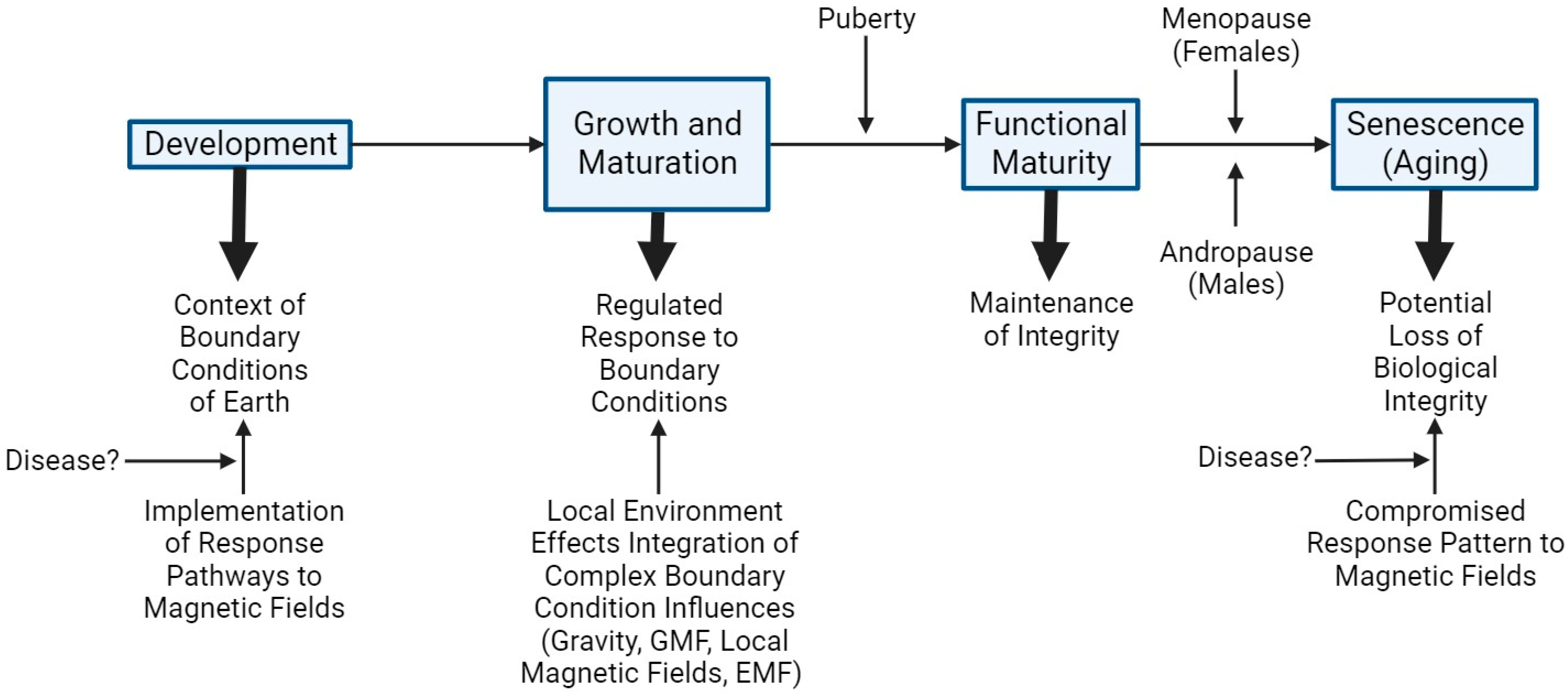
  • Evolution of complex organisms leading to Homo sapiens occurred in a complex set of boundary conditions of Earth, including both the planetary magnetic field and magnetic fields associated with local concentrations of iron.
  • Evolution led to the development and incorporation of biological systems that generate electromagnetic fields as a consequence of functioning (i.e., cardiovascular, neuromuscular, and neuronal networks) or use incorporated iron ions for function. Thus, against a background of magnetic fields, evolution led to the use of systems that generated electromagnetic fields, but such fields were not overtly impacted by the exogenous fields regarding function.
  • Many species have evolved navigation systems that use magnetic fields for guidance, and thus, they recognize that the geomagnetic field exists and can be used for directional migration. Furthermore, species that use such navigation systems can apparently filter endogenous electromagnetic fields from the geomagnetic field orientations.
  • The functioning of several biological systems can be acutely compromised or altered using exogenous magnetic fields, but such exposure does not lead to overt disruption of long-term regulation. Thus, exposure of the human body to high magnetic fields in an MRI machine (i.e., 3 Tesla), including the heart and brain, does not lead to disruption of the functioning of the brain processes or the functioning of the cardiac muscle.
  • Chronic exposure to varying levels of electromagnetic fields, likely not anticipated by evolution, does not lead to altered functioning of Homo sapiens.
  • Exposure to short-term drastically diminished geomagnetic fields via space flight to the moon did not overtly affect the functioning of astronauts.
It is difficult to integrate the points raised above, which indicate there are gaps and inconsistencies in our understanding of how Homo sapiens function within the magnetic boundary conditions of Earth. In addition to how several of the complex biological systems of Homo sapiens function within magnetic fields, how such regulatory control could contribute to disease development on Earth, and what the consequences might be if humans went beyond the influence of the GMF for prolonged periods of time remain unclear. Thus, it remains to be clarified as to how evolution adapted what are now Homo sapiens to function based on biological decisions made eons ago when single-celled and then multi-celled organisms first became reproducing species.
This situation likely requires a re-invigorated research effort to provide answers to allow for the integration of the summary points raised above. As deep space flight to destinations such as Mars, which is devoid of a geomagnetic field and where astronauts will be surrounded by equipment generating electromagnetic fields for prolonged times, there will be an opportunity to plan for relevant studies to address some of these points directly.

6. Conclusions and Suggestions Going Forward

Life, from simple single-celled organisms to Homo sapiens, has been shaped by the boundary conditions of Earth. Via evolutionary processes, commitments to specific metabolic and reproductive processes have led to the integrated functioning in the context of 1 g gravity, the temperature range, the nutrient availability, the geomagnetic field, and variation in such parameters as a result of choices made millions of years ago. Thus, magnetic field parameters do not exert their influence in isolation but in the more complex context. Until recent advances have led to the generation of significant electromagnetic fields and space flight, the opportunity to go beyond the boundary conditions, both gravity and magnetic fields, but particularly magnetic fields, potentially may have overlapping effects on some biological systems (integration of biology and biophysics).
Regarding magnetic fields, during evolution, simple cells could have either used magnetic fields in their progression to independent growth and preproduction, developed methods to negate the impact of magnetic fields or a combination of both, depending on the systems involved. As microorganisms, as well as birds and mammals, can use magnetic fields for navigation purposes, biological systems have obviously evolved to use magnetic fields to enhance survival. While not used for navigation purposes, it is of interest that many biological systems use the magnetic Fe ion as an essential component of proteins (i.e., transferrin, ferritin, lactoferrin) and cell function (i.e., hemoglobin in red blood cells for oxygen transport). Certainly, the biochemical and biological role(s) of Fe-containing molecules has been extensively studied, but whether there is also an influence of magnetic fields on function has been studied much less. This could be an area of research focus going forward.
While the effects of exogenous magnetic fields on human biology and cognition have been extensively studied within the boundary conditions of Earth, long-term space flight is presenting the opportunity and challenge of understanding the role and influence of magnetic fields on human functionality and, in particular, cognition, memory, and integrated functional demands. As space flight beyond the boundary conditions of Earth could not have been anticipated by evolutionary pressures, this excursion beyond the magnetic fields of Earth and exposure to electromagnetic fields presents a unique opportunity. It is not just deep space travel but also the opportunity to live on a planet such as Mars, which has 1/3 g gravity but little to no geomagnetic field, and live in habitats that require equipment generating EMFs. While it may be possible to optimally generate Faraday cages [177] to eliminate exogenous EMFs, one would be left with no EMF and little to no GMF, such as those used in preclinical model studies [178,179,180].
The implications of this set of “experiments” in space could have implications for those remaining on Earth. First, if there is heterogeneity in astronaut responses to such altered magnetic conditions, it would likely mean all Homo sapiens are heterogenous in this regard. Depending on the response patterns, the “outcomes” of the study of astronauts could have implications for diseases or conditions arising during fetal development that may require effective integration of biology and biophysics, particularly for the brain and cognition, behavior and memory development, and maturation. Failure to integrate could lead to or contribute to conditions such as autism. On the other end of the life spectrum, during aging, a failure of the integration system could lead to dementia and other brain conditions. Thus, deep-space flight and moving beyond the boundary conditions of Earth for an extended time could provide new information for how Homo sapiens function in a magnetic world, as well as have consequences for astronauts attempting to survive and function at long distances from the boundary conditions of Earth that shaped their evolution. Thus, magnetic influences on human health and disease may exist and have been mainly a silent partner throughout most of evolution. However, the advent of exposure to new and chronic electromagnetic fields and the opportunities provided by space flight may elaborate some of their previously undetected influences. Such further investigations may reveal commonalities of magnetic influences in all humans based on evolutionary choices made long ago, but also some individual response patterns to gain or loss of magnetic fields given the heterogeneity of humans [181] and previously documented variation in response to microgravity conditions (reviewed in [3,182]).
Thus, the answer to the question posed in the title of this review remains unanswered. Most of the evidence provided by the biologically focused literature is circumstantial, and mechanistic insights remain to be elucidated. This outcome from the analysis of the literature poses a conundrum for both biologists who are trying to understand how evolution adapted to the boundary conditions of Earth, as well as biophysicists who work at the boundary of how the physical world interfaces with the biological. While the current situation regarding the needed details is lacking, hopefully, this will stimulate new thinking and new research directions to address the issues. It is important to determine if and how biological systems such as those of humans have evolved adaptations to either negate the influence of static and electromagnetic fields into critical regulatory systems or utilize the GMF and local magnetic fields in the functioning of such systems. Finding answers is critical to understanding not only life on Earth but also the risks associated with deep space flights. Determining a mechanistic basis for how magnetic fields impact humans and other complex organisms will require research at the molecular, cellular, and physiologic levels. It may be able to build on the genetic (e.g., cryptochromes, magr, and frim) evidence generated from studies on navigation [59,60,61], the effect of magnetic fields on processes such as oxidative effects of iron (e.g., oxidoreductase activity, reactive oxygen species, mitochondrial oxidative phosphorylation) [59,60,61] (reviewed in [166]), and many of the effects of magnetic fields on physiologic processes discussed previously. Several studies [183,184,185] (reviewed in [166]) have indicated that the effect of magnetic fields on mitochondria may be central to their biological effects, and this should be one focus of future research.
Particular attention should also be given to the study of relevant intensities of the magnetic fields to allow for appropriate interpretation of the results. As the geomagnetic field of Earth is quite low (0.25–0.65 microT), and several of the magnetic fields assessed in the above discussion were considerably higher than that of the geomagnetic field, their relevance to the study of mechanisms associated with the GMF could be questioned. However, some studies using low magnetic fields and cells have identified enhanced acetylation of heat shock proteins as a potential target of exposure to the fields [186], and this may be another direction for future research. Other potential mechanistic targets have also been identified for low-frequency magnetic fields (reviewed in [166,187]), and these may also be the focus of future research. Future studies of electromagnetic fields in strengths relevant to those generated by electronic devices, household appliances, cell phones, and even electric cars may be appropriate for some studies as people on Earth are being subjected to such fields at increasing frequency.
The proposed deep space flights to destinations such as Mars, well beyond the GMF of Earth and to a planet with little to no GMF of its own, may also provide some of the missing information and lead to the development of research directions in such environments that could provide information to fill in some of the gaps in the data to move this field forward. However, in such deep space missions, the astronauts will be subjected to microgravity, increased radiation risk, prolonged living in a confined space, and an altered magnetic environment, so identifying changes that can be solely attributed to the magnetic environment may be challenging. Thus, this research effort may require both new tools to address the problem and thinking in new ways to seek out new directions for research, leading to solutions for this very important problem regarding human evolution and where it may go in the future.

Funding

This research received no external funding.

Institutional Review Board Statement

Not applicable.

Informed Consent Statement

Not applicable.

Data Availability Statement

Not applicable.

Acknowledgments

The author thanks several colleagues in the Canadian Space Health Research Network for discussions and interactions regarding space flight and associated risks. He also thanks Nicole Buckley for past discussions on the topics covered in this review and Emilie Gysel for assistance in preparing the figures in BioRender. No financial support for the preparation of this review was obtained.

Conflicts of Interest

The author attests that there were no conflicts of interest to declare regarding the preparation and publishing of this review.

References

  1. Binhi, V.N.; Sarimov, R.M. Zero magnetic field effect observed in human cognitive processes. Electromagn. Bio. Med. 2009, 28, 310–315. [Google Scholar] [CrossRef] [PubMed]
  2. Binhi, V.N.; Prato, F.S. Biological effects of the hypomagnetic field: An analytic review of experiments and theories. PLoS ONE 2017, 12, e0179340. [Google Scholar] [CrossRef] [PubMed]
  3. Hart, D.A. Homo sapiens-A species not designed for space flight: Health risks in low Earth orbit and beyond, including potential risks when traveling beyond the geomagnetic field of Earth. Life 2023, 13, 757. [Google Scholar] [CrossRef] [PubMed]
  4. Hart, D.A.; Zernicke, R.F. Optimal human functioning requires exercise across the lifespan: Mobility in a 1g environment is intrinsic to the integrity of multiple biological systems. Front. Physiol. 2020, 11, 156. [Google Scholar] [CrossRef] [PubMed]
  5. Erdmann, W.; Kmita, H.; Kosicki, J.Z.; Karzmarek, L. How the geomagnetic field influences life on Earth- an integrated approach to geomagnetobiology. Orig. Life Evol. Biosph. 2021, 51, 231–257. [Google Scholar] [CrossRef] [PubMed]
  6. Xue, X.; Ali, Y.F.; Liu, C.; Hong, Z.; Luo, W.; Nie, J.; Li, B.; Jiao, Y.; Liu, N.A. Geomagnetic shielding enhances radiation resistance by promoting DNA repair process in human bronchial epithelial cells. Int. J. Mol. Sci. 2020, 21, 9304. [Google Scholar] [CrossRef] [PubMed]
  7. Levin, M.; Martyniuk, C.J. The bioelectric code: An ancient computational medium for dynamic control of growth and form. BioSystems 2018, 164, 76–93. [Google Scholar] [CrossRef] [PubMed]
  8. Binhi, V.N.; Rubin, A.B. Theoretical concepts in magnetobiology after 40 years of research. Cells 2022, 11, 274. [Google Scholar] [CrossRef]
  9. Gonciarz, R.L.; Rensio, A.R. Emerging role of ferrous iron in bacterial growth and host-pathogen interaction: New tools for chemical (micro)biology and antibacterial therapy. Curr. Opin. Chem. Biol. 2021, 61, 170–178. [Google Scholar] [CrossRef]
  10. Baatjies, L.; Loxton, A.G.; Williams, M.J. Host and bacterial iron homeostasis, an underexplored area in tuberculosis biomarker research. Front. Immunol. 2021, 12, 742059. [Google Scholar] [CrossRef]
  11. Seyoum, Y.; Baye, K.; Humblot, C. Iron homeostasis in host and gut bacteria-a complex relationship. Gut Microbes 2021, 13, 1–19. [Google Scholar] [CrossRef]
  12. Gao, G.; Li, J.; Zhang, Y.; Chang, Y.Z. Cellular iron metabolism and regulation. Adv. Exp. Med. Biol. 2019, 1173, 21–32. [Google Scholar] [CrossRef] [PubMed]
  13. Olson, J.S. Kinetic mechanisms for O2 binding to myoglobins and hemoglobins. Mol. Aspects Med. 2022, 84, 101024. [Google Scholar] [CrossRef]
  14. Nagatomo, S.; Naga, M.; Kitagawa, T. Structural origin of cooperativity in human hemoglobin: A view from different roles of alpha and beta subunits in the alpha2beta2 tetramer. Biophys Rev. 2022, 14, 483–498. [Google Scholar] [CrossRef] [PubMed]
  15. Akhtar, M.; Wright, N. Acyl-carbon bond cleaving cytochrome P450 enzymes: CYP17A1, CYO19A1 and CYO51A1. Adv. Exp. Med. Biol. 2015, 851, 107–130. [Google Scholar] [CrossRef]
  16. Kumar, N.; Chugh, H.; Sood, D.; Singh, S.; Singh, A.; Awasthi, A.D.; Tomar, R.; Tomar, V.; Changdra, R. Biology of heme: Drug interactions and adverse drug reactions with CYO450. Curr. Top. Med. Chem. 2019, 18, 2042–2055. [Google Scholar] [CrossRef] [PubMed]
  17. Poulos, T.L.; Follmer, A.H. Updating the paradigm: Redox partner binding and conformational dynamics in cytochromes P450. Acc. Chem. Res. 2022, 55, 373–380. [Google Scholar] [CrossRef] [PubMed]
  18. Luck, A.N.; Mason, A.B. Transferrin-mediated cellular iron delivery. Curr. Top. Membr. 2012, 69, 3–35. [Google Scholar] [CrossRef]
  19. Zhang, J.; Chen, X.; Hong, J.; Tang, A.; Liu, Y.; Xie, N.; Nie, G.; Yan, X.; Liang, M. Biochemistry of mammalian ferritins in the regulation of cellular iron homeostasis and oxidative responses. Sci. China Life Sci. 2021, 64, 353–362. [Google Scholar] [CrossRef]
  20. Zhao, X.; Kruzel, M.; Aronowski, J. Lactoferrin and hematoma detoxification after intracerebral hemorrhage. Biochem. Cell Biol. 2021, 99, 97–101. [Google Scholar] [CrossRef]
  21. Meyer, O.; Gremer, L.; Ferner, R.; Dobbek, H.; Meyer-Klaucke, W.; Huber, R. The role of Se, Mo, Fe in the structure and function of carbon monoxide dehydrogenase. Biol. Chem. 2000, 38, 865–876. [Google Scholar] [CrossRef] [PubMed]
  22. Picca, A.; Saini, S.K.; Mankowski, R.T.; Kamenov, G.; Anton, S.D.; Manini, T.M.; Buford, T.W.; Wohlgemuth, S.F.; Xiao, R.; Calvani, R.; et al. Altered expression of mitoferrin and frataxin, larger labile iron pool and greater mitochondrial DNA damage in the skeletal muscle of older adults. Cells 2020, 9, 2579. [Google Scholar] [CrossRef] [PubMed]
  23. Forouzesh, D.C.; Moran, G.R. Mammalian dihydropyrimidine dehydrogenase. Ach. Biochem. Biophys. 2021, 714, 109066. [Google Scholar] [CrossRef] [PubMed]
  24. Shimada, A.; Tsukihara, T.; Yoshikawa, S. Recent progress in experimental studies on the catalytic mechanism of cytochrome c oxidase. Front. Chem. 2023, 11, 1108190. [Google Scholar] [CrossRef] [PubMed]
  25. Singh, A.; Kong, Q.; Luo, X.; Petersen, R.B.; Meyerson, H.; Singh, N. Prion protein (PrP) knockout mice show altered iron metabolism: A functional role for PrP in iron uptake and transport. PLoS ONE 2009, 4, e6115. [Google Scholar] [CrossRef] [PubMed]
  26. Alayash, A.I. Oxidation reactions of cellular and acellular hemoglobins: Implications for human health. Front. Med. Technol. 2022, 4, 1068972. [Google Scholar] [CrossRef]
  27. Pires, I.S.; Berthiaume, F.; Palmere, A.F. Engineering therapeutics to detoxify hemoglobin, heme and iron. Annu. Rev. Biomed. Eng. 2023, 25, 1–21. [Google Scholar] [CrossRef]
  28. Piperno, A.; Pelucchi, S.; Mariani, R. Hereditary hyperferritinemia. Int. J. Mol. Sci. 2023, 24, 2560. [Google Scholar] [CrossRef]
  29. Lommaert, E.; Verlinden, W.; Duysburgh, I.; Holvoet, T.; Schouten, J. Hyperferritinemia and non-HFE hemochromatosis: Differential diagnosis and workup. Acta Gastroenterol. Belg. 2023, 86, 356–359. [Google Scholar] [CrossRef]
  30. Bruno, F.; Albano, D.; Agostini, A.; Benenati, M.; Cannella, R.; Caruso, D.; Cellina, M.; Cozzi, D.; Danti, G.; De Muzio, F.; et al. Imaging of metabolic and overload disorders in tissues and organs. Jpn J. Radiol. 2023, 41, 571–595. [Google Scholar] [CrossRef]
  31. De Simone, G.; Varriccho, R.; Ruberto, T.F.; di Masi, A.; Ascenzi, P. Heme scavenging and delivery: The role of human serum albumin. Biomolecules 2023, 13, 575. [Google Scholar] [CrossRef] [PubMed]
  32. O’Brien, K.O. Maternal, fetal and placental regulation of placental iron trafficking. Placenta 2022, 125, 47–53. [Google Scholar] [CrossRef] [PubMed]
  33. Navas, F.J.; Cordova, A. Iron distribution in different tissues in rats following exercise. Biol. Trace Elem. Res. 2000, 73, 259–268. [Google Scholar] [CrossRef] [PubMed]
  34. Bishop, G.M.; Smith, M.A.; LaManna, J.C.; Wilson, A.C.; Perry, G.; Atwood, C.S. Iron homeostasis is maintained in the brain, but not the liver following mild hypoxia. Redox Rep. 2007, 12, 257–266. [Google Scholar] [CrossRef] [PubMed]
  35. Djordjevich, D.M.; De Luca, S.R.; Milovanovich, I.D.; Jankovic, S.; Stefanovic, S.; Veskovic-Moracanin, S.; Cirkovic, S.; Ilic, A.Z.; Ristic-Djurovic, J.L.; Trbovich, A.M. Hemotological parameters’ changes in mice subchronically exposed to static magnetic fields of different orientations. Ecotoxicol. Environ. Saf. 2012, 81, 98–105. [Google Scholar] [CrossRef] [PubMed]
  36. Kopani, M.; Panik, J.; Filova, B.; Bijdos, M.; Misek, J.; Kohan, M.; Jakus, J.; Povinec, P. PIXE analysis of iron in rabbit cerebellum after exposure to radiofrequency electromagnetic fields. Bratisl. Lek. Listy. 2022, 123, 864–871. [Google Scholar] [CrossRef] [PubMed]
  37. Schildroth, S.; Kordas, K.; Bauer, J.A.; Wright, R.O.; Henn, B.C. Environmental metal exposure, neurodevelopment, and the role of iron status: A review. Curr. Environ. Health Rep. 2022, 9, 758–787. [Google Scholar] [CrossRef] [PubMed]
  38. Bailey, D.K.; Kosman, D.J. Is brain iron trafficking part of the physiology of the amyloid precursor protein? J. Biol. Inorg. Chem. 2019, 24, 1171–1177. [Google Scholar] [CrossRef]
  39. Peng, Y.; Chang, X.; Lang, M. Iron homeostasis disorder and Alzheimer’s disease. Int. J. Mol. Sci. 2021, 22, 12442. [Google Scholar] [CrossRef]
  40. Onukwufor, J.O.; Dirksen, R.T.; Wojtovich, A.P. Iron dysregulation in mitochondrial dysfunction and alzheimer’s disease. Antioxidants 2022, 11, 692. [Google Scholar] [CrossRef]
  41. Rao, S.S.; Adland, P.A. Untangling tau and iron: Exploring the interaction between iron and tau in neurodegeneration. Front. Mol. Neurosci. 2018, 11, 276. [Google Scholar] [CrossRef] [PubMed]
  42. Joppe, K.; Roser, A.E.; Maass, F.; Lingor, P. The contribution of iron to protein aggregation disorders in the central nervous system. Front. Neurosci. 2019, 13, 15. [Google Scholar] [CrossRef] [PubMed]
  43. Rogers, J.T.; Cahill, C.M. Iron-responsive-like elements and neurodegenerative ferroptosis. Learn. Mem. 2020, 27, 395–413. [Google Scholar] [CrossRef] [PubMed]
  44. Mukherjee, S.; Panda, D. Contrasting effects of ferric and ferrous ions on oligomerization and droplet formation of tau: Implications in taupathies and neurodegeneration. ACS Chem. Neurosci. 2021, 12, 4393–4405. [Google Scholar] [CrossRef] [PubMed]
  45. Trojsi, F.; Sorrentino, P.; Sorrentino, G.; Tedeschi, G. Neurodegeneration of brain networks in the amyotrophic lateral sclerosis-frontotemporal lobar degeneration (ALS-FTLD) continuum: Evidence from MRI and MEG studies. CNS Spectr. 2018, 23, 378–387. [Google Scholar] [CrossRef] [PubMed]
  46. Cope, T.E.; Weil, R.S.; Duzel, E.; Dickerson, B.C.; Rowe, J.B. Advances in neuroimaging to support translational medicine in dementia. J. Neurol. Neurosurg. Psychiatry 2021, 92, 263–270. [Google Scholar] [CrossRef] [PubMed]
  47. Yang, W.; Pilozzi, A.; Huang, X. An overview of ICA/BSS-based application to Alzheimer’s brain signal processing. Biomedicines 2021, 9, 386. [Google Scholar] [CrossRef] [PubMed]
  48. Fred, A.L.; Kumar, S.N.; Haridhas, A.K.; Ghosh, S.; Bhuvana, H.P.; Sim, W.K.J.; Vimalan, V.; Givo, F.A.S.; Jousmaki, V.; Padmanabhan, P.; et al. A brief introduction to magnetoencephalography (MEG) and its clinical applications. Brain Sci. 2022, 12, 788. [Google Scholar] [CrossRef]
  49. Heyers, D.; Musielak, I.; Haase, K.; Herold, C.; Bolte, P.; Gunturkun, O.; Mouritsen, H. Morphology, biochemistry and connectivity of cluster N and the hippocampal formation in a migratory bird. Brain Struct. Funct. 2022, 227, 2731–2749. [Google Scholar] [CrossRef]
  50. Karwinkel, T.; Winklhofer, M.; Janner, L.E.; Brust, V.; Huppop, O.; Bairlein, F.; Schmaljohann, H. A magnetic pulse does not affect free-flight navigation behaviour of a medium-distance songbird migrant in spring. J. Exp. Biol. 2022, 225, jeb244473. [Google Scholar] [CrossRef]
  51. Tonelli, B.A.; Youngflesh, C.; Tingley, M.W. Geomagnetic disturbance associated with increased vagrancy in migratory landbirds. Sci. Rep. 2023, 13, 414. [Google Scholar] [CrossRef] [PubMed]
  52. Kremers, D.; Manulanda, J.L.; Hausberger, M.; Lemasson, A. Bahvoural evidence of magnetoreception in dolphins: Detection of experimental fields. Naturwissenschaften 2014, 101, 907–911. [Google Scholar] [CrossRef] [PubMed]
  53. Formicki, K.; Korzelecka-Orkisz, A.; Tanski, A. Magnetoreception in fish. J. Fish. Biol. 2019, 95, 73–91. [Google Scholar] [CrossRef] [PubMed]
  54. O’Connell, D.; Kehl, C.E.; Taylor, B.K.; Piacenza, J.; Piacenza, S.; Li, K.J.F. A computational framework for studying energetics and resource management in sea turtle migration and autonomous systems. J. Theor. Biol. 2021, 527, 110815. [Google Scholar] [CrossRef] [PubMed]
  55. Komolkin, A.V.; Kupriyanov, P.; Chudin, A.; Bojarinova, J.; Kavokin, K.; Chernetsov, N. Theoretically possible spatial accuracy of geomagnetic maps used by migrating animals. J. R. Soc. Interface 2017, 14, 20161002. [Google Scholar] [CrossRef]
  56. Chae, K.S.; Kim, S.C.; Kwon, H.J.; Kim, Y. Human magnetic sense is mediated by a light and magnetic field resonance-dependent mechanism. Sci. Rep. 2022, 12, 8997. [Google Scholar] [CrossRef]
  57. Chae, K.S.; Oh, I.T.; Lee, S.H.; Kim, S.C. Blue light-dependent human magnetoreception in geomagnetic food orientation. PLoS ONE 2019, 14, 1826. [Google Scholar] [CrossRef]
  58. Wang, Y.; Chen, J.; Zhu, F.; Hong, Y. Identification of medaka magnetoreceptor and cryptochromes. Sci. China Life Sci. 2017, 60, 271–278. [Google Scholar] [CrossRef]
  59. Fitak, R.R.; Wheeler, B.R.; Ernst, D.A.; Lohman, K.J.; Johnsen, S. Candidate genes mediating magnetoreception in rainbow trout (Oncorhynchus mykiss). Biol. Lett. 2017, 13, 20170142. [Google Scholar] [CrossRef]
  60. Liedvogel, M.; Mouritsen, H. Cryptochromes—A potential magnetoreceptor: What do we know and what do we want to know? J. R. Soc. Interface 2010, 7 (Suppl. S2), S147–S162. [Google Scholar] [CrossRef]
  61. Landler, L.; Keays, D.A. Cryptochrome: The magnetoreceptor with a sinister side? PLoS ONE 2018, 16, e3000018. [Google Scholar] [CrossRef]
  62. van Horik, J.; Emery, N.J. Evolution of cognition. Wiley Interdiscip. Rev. Cogn. Sci. 2011, 2, 621–633. [Google Scholar] [CrossRef] [PubMed]
  63. Heft, H. Evolution of human cognition. In International Encyclopedia of the Social & Behavioral Sciences, 2nd ed.; Elsevier: Amsterdam, The Netherlands, 2015; pp. 252–258. [Google Scholar]
  64. Vonk, J.; Aradhye, C. Evolution of Cognition. In Basics in Human Evolution; Muehlenbein, M.P., Ed.; Elesevier Inc.: Amsterdam, The Netherlands, 2015; Volume 3, pp. 479–491. [Google Scholar] [CrossRef]
  65. Roth, G.; Dicke, U. Origin and evolution of human cognition. Prog. Brain Res. 2019, 250, 285–316. [Google Scholar] [CrossRef] [PubMed]
  66. Ratcliff, M.J. Origins, trends and perspectives of historical epistemological research on Piaget. Integr. Psychol. Behav. Sci. 2023, 27. [Google Scholar] [CrossRef] [PubMed]
  67. Roth, B.J. Biomagnetism: The first sixty years. Sensors 2023, 23, 4218. [Google Scholar] [CrossRef] [PubMed]
  68. Ueno, S. Studies on magnetism and bioelectromagnetics for 45 years: From magnetic analog memory to human brain stimulation and imaging. Bioelectromagnetics 2012, 33, 3–33. [Google Scholar] [CrossRef]
  69. Martel, J.; Chang, S.H.; Chevalier, G.; Ojcius, D.M.; Young, J.D. Influence of electromagnetic fields on the circadian rhythm: Implications for human health and disease. Biomed. J. 2023, 46, 48–59. [Google Scholar] [CrossRef]
  70. Stock, J.B.; Zhang, S. The biochemistry of memory. Curr. Biol. 2013, 23, R741–R745. [Google Scholar] [CrossRef]
  71. Bickle, J.; Sarwich, S.S. Introduction to molecular and cellular cognition. In Mind, Cognition and Neuroscience. A Philosophical Introduction; Young, B., Jennings, C.D., Eds.; Routledge: Oxfordshire, UK, 2022; Chapter 3; pp. 32–50. [Google Scholar]
  72. McFadden, J. Consciousness: Matter or EMF? Front. Hum. Neurosci. 2023, 16, 1024934. [Google Scholar] [CrossRef]
  73. McFadden, J. The conscious electromagnetic information (cemi) field theory. The hard problem made easy? J. Conscious. Stud. 2002, 9, 45–60. [Google Scholar]
  74. McFadden, J. The cemi field theory. Gestalt information and the meaning of meaning. J. Conscious. Stud. 2013, 20, 152–182. [Google Scholar]
  75. McFadden, J. The cemi field theory. Closing the loop. J. Conscious. Stud. 2013, 20, 153–168. [Google Scholar]
  76. McFadden, J. Integrating information in the brain’s EM field: The cemi field theory of consciousness. Neurosci. Conscious. 2020, 2020, niaa016. [Google Scholar] [CrossRef] [PubMed]
  77. Banaclocha, M.A.M. Magnetic storage of information in the human cerebral cortex: A hypothesis for memory. Int. J. Neurosci. 2005, 115, 329–337. [Google Scholar] [CrossRef]
  78. Brignani, D.; Bortoletto, M.; Miniussi, C.; Maioli, C. The when and where of spatial storage in memory-guided saccades. Neuroimage 2010, 52, 1611–1620. [Google Scholar] [CrossRef]
  79. Edwards, J.C.W. EM fields and the meaning of meaning. Response to Johnjoe McFadden. J. Conscious. Stud. 2013, 20, 159–167. [Google Scholar]
  80. Crasson, M. 50-60 Hz electric and magnetic field effects on cognitive function in humans: A review. Radiat. Prot. Dosim. 2003, 106, 333–340. [Google Scholar] [CrossRef]
  81. Benke, G.; Abramson, M.J.; Zeleke, B.M.; Kaaufman, J.; Karipidis, K.; Kelsall, H.; McDonald, S.; Brzozek, C.; Feychting, M.; Brennan, S. The effect of long-term radiofrequency exposure on cognition in human observational studies: A protocol for a systematic review. Environ. Int. 2022, 159, 106972. [Google Scholar] [CrossRef]
  82. Heinrich, A.; Szostek, A.; Nees, F.; Meyer, P.; Semmler, W.; Flor, H. Effects of static magnetic fields on cognition, vital signs, and sensory perception: A meta-analysis. J. Magn. Reson. Imaging 2011, 34, 758–763. [Google Scholar] [CrossRef]
  83. Zhang, Z.; Xue, Y.; Yang, J.; Shang, P.; Yuan, X. Biological effects of hypomagnetic field: Ground-based data for space exploration. Bioelectromagnetics 2021, 42, 516–531. [Google Scholar] [CrossRef]
  84. Stahn, A.C.; Kuhn, S. Brains in space: The importance of understanding the impact of long-duration spaceflight on spatial cognition and its neural circuitry. Cogn. Process. 2021, 22 (Suppl. S1), 105–114. [Google Scholar] [CrossRef] [PubMed]
  85. Arshad, I.; Ferre, E.R. Cognition in zero gravity: Effects of non-terrestrial gravity on human behavior. Q. J. Exp. Psychol. 2023, 76, 979–994. [Google Scholar] [CrossRef] [PubMed]
  86. Salazar, A.P.; McGregor, H.R.; Hupfeld, K.E.; Beltran, N.E.; Kofman, I.S.; De Dios, Y.E.; Riascos, R.F.; Reuter-Lorenz, P.A.; Bloomberg, J.J.; Mulavara, A.P.; et al. Changing in working memory brain activity and task-based connectivity after long-duration spaceflight. Cereb. Cortex. 2023, 33, 2641–2654. [Google Scholar] [CrossRef] [PubMed]
  87. Carpenter, D.O. Human disease resulting from exposure to electromagnetic fields. Rev. Environ. Health 2013, 28, 159–172. [Google Scholar] [CrossRef] [PubMed]
  88. Lin, J.C. Carcinogenesis from chronic exposure to radio-frequency radiation. Front. Public Health 2022, 10, 1042478. [Google Scholar] [CrossRef] [PubMed]
  89. Malagoli, C.; Malavolti, M.; Wise, L.A.; Balboni, E.; Fabbi, S.; Teggi, S.; Palazzi, G.; Cellini, M.; Poli, M.; Zanichelli, P.; et al. Residential exposure to magnetic fields from high-voltage power lines and risk of childhood leukemias. Environ. Res. 2023, 232, 116320. [Google Scholar] [CrossRef] [PubMed]
  90. Auger, N.; Bilodeau-Bertrand, M.; Marcoux, S.; Kosatsky, T. Residential exposure to electromagnetic fields during pregnancy and risk for child cancer: A longitudinal cohort study. Environ. Res. 2019, 176, 108524. [Google Scholar] [CrossRef] [PubMed]
  91. Repacholi, M. Concern that “EMF” magnetic fields from power lines cause cancer. Sci. Total Environ. 2012, 426, 454–458. [Google Scholar] [CrossRef]
  92. Amoon, A.T.; Swanson, J.; Magnani, C.; Johansen, C.; Kheifets, L. Pooled analysis of recent studies of magnetic fields and childhood leukemia. Environ. Res. 2022, 204, 111993. [Google Scholar] [CrossRef]
  93. Brabant, C.; Geerinck, A.; Beaudart, C.; Tirelli, E.; Geuzaine, C.; Bruyere, O. Exposure to magnetic fields and childhood leukemia: A systematic review and meta-analysis of case-control and cohort studies. Rev. Environ. Health 2022, 38, 229–253. [Google Scholar] [CrossRef]
  94. Philips, A. Risk of cancer and exposure to power lines. Still no answers. BMJ 1994, 308, 1162–1163. [Google Scholar] [CrossRef] [PubMed]
  95. Crespi, C.M.; Swanson, J.; Vergara, X.P.; Kheifets, L. Childhood leukemia risk in the California Power Line study: Magnetic fields versus distance from power lines. Environ. Res. 2019, 171, 530–535. [Google Scholar] [CrossRef] [PubMed]
  96. Carles, C.; Esquirol, Y.; Turuban, M.; Piel, C.; Migualt, L.; Pouchieu, C.; Bouvier, G.; Fabbro-Peray, P.; Lebailly, P.; Baldi, I. Residential proximity to power lines and risk of brain tumor in the general population. Environ. Res. 2020, 185, 109473. [Google Scholar] [CrossRef]
  97. Carpenter, D.O. Extremely low frequency electromagnetic fields and cancer: How source of funding affects results. Environ. Res. 2019, 178, 108688. [Google Scholar] [CrossRef] [PubMed]
  98. Birnbaum, L.S.; Taylor, H.S.; Baldwin, H.; Ben-Ishai, P.; Davis, D. RE: Cellular telephone use and the risk of brain tumors: Update of the UK million women study. J. Natl. Cancer Inst. 2022, 114, 1551–1552. [Google Scholar] [CrossRef]
  99. Bhargav, H.; Srinivasan, T.M.; Varambally, S.; Gangadhar, B.N.; Koka, P. Effect of mobile phone-induced electromagnetic field on brain hemodynamics and human stem cell functioning: Possible mechanistic link to cancer risk and early diagnostic value of electronphotonic imaging. J. Stem Cells 2015, 10, 287–294. [Google Scholar]
  100. Nelson, N. Recent studies show cell phone use is not associated with increased cancer risk. J. Natl. Cancer Inst. 2001, 93, 170–172. [Google Scholar] [CrossRef]
  101. Jagetia, G.C. Genotoxic effects of electromagnetic field radiation from mobile phones. Environ. Res. 2022, 212, 113321. [Google Scholar] [CrossRef]
  102. Farashi, S.; Bashirian, S.; Khazaei, S.; Khazaei, M. Mobile phone electromagnetic radiation and the risk of headache: A systematic review and meta-analysis. Int. Arch. Occup. Environ. Health 2022, 95, 1587–1601. [Google Scholar] [CrossRef]
  103. Herbert, M.R.; Sage, C. Autism and EMF? Plausibility of a pathophysiological link-part 1. Pathophysiology 2013, 20, 191–209. [Google Scholar] [CrossRef]
  104. Zabroda, N.N.; Artemenko, M.V. Hygienic characteristics of the Kursk magnetic anomaly area and morbidity in the aboriginal population. Gig. Sanit. 2008, 5, 35–38. (In Russian) [Google Scholar]
  105. Wei, Y.; Wang, X. Biological effects of rotating magnetic field: A review from 1969 to 2021. Prog. Biophs. Mol. Biol. 2023, 178, 103–115. [Google Scholar] [CrossRef] [PubMed]
  106. Bassett, C.A. Beneficial effects of electromagnetic fields. J. Cell Biochem. 1993, 51, 387–393. [Google Scholar] [CrossRef] [PubMed]
  107. Trock, D.H. Electromagnetic fields and magnets. Investigational treatment for musculoskeletal disorders. Rheum. Dis. Clin. N. Am. 2000, 26, 51–62. [Google Scholar] [CrossRef] [PubMed]
  108. Perez, F.; Bandeira, J.P.; Chumbiauca, C.N.P.; Lahiri, D.K.; Morisaki, J.; Rizkalla, M. Multidimential insights into the repeated electromagnetic field stimulation and biosystem interaction in aging and age-related diseases. J. Biomed. Sci. 2022, 29, 39. [Google Scholar] [CrossRef]
  109. Mayrovitz, H.N.; Maqsood, R.; Tawakalzada, A.S. Do magnetic fields have a place in treating vascular complications in diabetes? Cureus 2022, 14, e24883. [Google Scholar] [CrossRef] [PubMed]
  110. Valone, T.F. Bioelectromagnetic healing, its history and a rationale for its use. In Proceedings of the Whole Person Healing Conference; Lumiverse Inc.: Bethesda, MD, USA, 2003; pp. 1–17. [Google Scholar]
  111. Soltani, D.; Samini, S.; Vasheghani-Farahani, A.; Shariatpanahi, S.P.; Abodolmaleki, P.; Ansari, A.M. Electromagnetic field therapy in cardiovascular diseases: A review of patents, clinically effective devices, and mechanism of therapeutic effects. Trends Cardiovasc. Med. 2023, 33, 72–78. [Google Scholar] [CrossRef] [PubMed]
  112. Zanotti, F.; Trentin, M.; Zanolla, I.; Teingo, E.; Mantarro, C.; Paola, L.D.; Tremoli, E.; ambataro, M.; Sambado, L.; Picari, M.; et al. Playing with biophysics: How a symphony of different electromagnetic fields acts to reduce the inflammation in diabetic derived cells. Int. J. Mol. Sci. 2023, 24, 1754. [Google Scholar] [CrossRef]
  113. Littman, J.; Aaron, R.K. Stimulation of chondrogenesis in a developmental model of endochondral bone formation by pulsed electromagnetic fields. Int. J. Mol. Sci. 2023, 24, 3275. [Google Scholar] [CrossRef]
  114. Haddad, J.B.; Obolensky, A.G.; Shinnick, P. The biologic effects and the therapeutic mechanism of action of electric and electromagnetic field stimulation on bone and cartilage: New findings and a review of earlier work. J. Altern. Complement Med. 2007, 13, 485–490. [Google Scholar] [CrossRef]
  115. Frank, C.; Schachar, N.; Dittrich, D.; Shrive, N.; deHaas, W.; Edwards, G. Electromagnetic stimulation of ligament healing in rabbits. Clin. Orthop. Relat. Res. 1983, 175, 263–272. [Google Scholar]
  116. Lin, Y.; Nishimura, R.; Nozaki, K.; Sasaki, N.; Kadosawa, T.; Goto, N.; Date, M.; Takeuchi, A. Effects of pulsing electromagnetic fields on the ligament healing in rabbits. J. Vet. Med. Sci. 1992, 54, 1017–1022. [Google Scholar] [CrossRef] [PubMed]
  117. Xu, D.; Zhang, T.; Qu, J.; Hu, J.; Lu, H. Enhanced patella-patella tendon healing using combined magnetic fields in a rabbit model. Am. J. Sports Med. 2014, 42, 2495–2501. [Google Scholar] [CrossRef] [PubMed]
  118. Hu, J.; Zhang, T.; Xu, D.; Qu, J.; Qin, L.; Zhou, J.; Lu, H. Combined magnetic fields accelerate bone-tendon junction injury healing through osteogenesis. Scand. J. Med. Sci. Sports 2015, 25, 398–405. [Google Scholar] [CrossRef] [PubMed]
  119. Wang, M.; Li, Y.; Feng, L.; Zhang, X.; Wang, H.; Zhang, N.; Viohl, I.; Li, G. Pulsed electromagnetic field enhances healing of a meniscal tear and mitigates posttraumatic osteoarthritis in a rat model. Am. J. Sports Med. 2022, 50, 2722–2732. [Google Scholar] [CrossRef] [PubMed]
  120. Hulme, J.; Robinson, V.; DeBie, R.; Wells, G.; Judd, M.; Tugwell, P. Electromagnetic fields for the treatment of osteoarthritis. Cochrane Database Syst. Rev. 2002, 1, CD003523. [Google Scholar] [CrossRef]
  121. Li, S.; Yu, B.; Zhou, D.; He, C.; Zhuo, Q.; Hulme, J.M. Electromagnetic fields for treating osteoarthritis. Cochrane Database Syst. Rev. 2013, 12, CD003523. [Google Scholar] [CrossRef]
  122. Cadossi, R.; Massari, L.; Racine-Avila, J.; Aaron, R.K. Pulsed electromagnetic field stimulation of bone healing and joint preservation: Cellular mechanisms of skeletal response. J. Am. Acad. Orthop. Surg. Glob. Res. Rev. 2020, 4, e1900155. [Google Scholar] [CrossRef]
  123. Zhu, F.; Liu, W.; Li, F.; Zhao, H.; Deng, X.; Wang, H.-L. Electric/magnetic intervention for bone regeneration: A systematic review and network meta-analysis. Tissue Eng. Part B Rev. 2023, 29, 217–231. [Google Scholar] [CrossRef]
  124. Ribeiro, T.P.; Flores, M.; Sadureira, S.; Zanotto, F.; Monteiro, F.; Laranjeira, M.S. Magnetic bone tissue engineering: Reviewing the effects of magnetic stimulation on bone regeneration and angiogenesis. Pharmaceutics 2023, 15, 1045. [Google Scholar] [CrossRef]
  125. Caliogna, L.; Bina, V.; Brancato, A.M.; Gastaldi, G.; Annumziata, S.; Mosconi, M.; Grassi, F.A.; Benazzo, F.; Pasta, G. The role of pemfs on bone healing: An in vitro study. Int. J. Mol. Sci. 2022, 23, 14298. [Google Scholar] [CrossRef] [PubMed]
  126. Darendeliler, M.A.; Darendeliler, A.; Sinclair, P.M. Effects of static magnetic and pulsed electromagnetic fields on bone healing. Int. J. Adult Orthodon. Orthognath. Surg. 1997, 12, 43–53. [Google Scholar] [PubMed]
  127. Pereira, A.; Diaz, J.J.H.; Saur, M.; Botero, S.S.; Facca, S.; Liverneaux, P. Carpal scaphoid non-union treatment: A retrospective trial comparing simple retrograde percutaneous screw fixation versus percutaneous screw fixation plus pulsed electromagnetic fields (Physiotim). Eur. J. Orthop. Surg. Traumatol. 2017, 27, 521–525. [Google Scholar] [CrossRef] [PubMed]
  128. Griffin, X.L.; Costa, M.L.; Parsons, N.; Smith, N. Electromagnetic field stimulation for treating delayed union or non-union of long bone fractures in adults. Cochrane Database Syst. Rev. 2011, 13, CD008471. [Google Scholar] [CrossRef] [PubMed]
  129. Sibanda, V.; Anazor, F.; Relwani, J.; Dhinsa, B.S. Outcomes of the treatment of fracture non-union using combined magnetic field bone growth stimulation: Experiences from a UK trauma unit. Cureus 2012, 14, e25100. [Google Scholar] [CrossRef] [PubMed]
  130. Assiotis, A.; Sachinis, N.P.; Chalidis, B.E. Pulsed electromagnetic fields for the treatment of tibial delayed unions and nonunions. A prospective clinical study and review of the literature. J. Orthop. Surg. Res. 2012, 7, 24. [Google Scholar] [CrossRef] [PubMed]
  131. Hannemann, P.F.W.; Mommers, E.H.H.; Schots, J.P.M.; Brink, P.R.G.; Poeze, M. The effects of low-intensity pulsed ultrasounds and pulsed electromagnetic fields bone growth stimulation in acute fractures: A systematic review and meta-analysis of randomized controlled trials. Arch. Orthop. Truam Surg. 2014, 134, 1093–1106. [Google Scholar] [CrossRef]
  132. Vicenti, G.; Bizzoca, D.; Solarino, D.; Moretti, F.; Ottaviani, G.; Simone, F.; Zavattini, G.; Maccagnano, G.; Noia, G.; Moretti, B. The role of biophysical stimulation with pemfs in fracture healing from bench to bedside. J. Biol. Regl. Homeost. Agents 2020, 34, 131–135. [Google Scholar]
  133. Lv, H.; Wang, Y.; Zhen, C.; Liu, J.; Chen, X.; Zhang, G.; Yao, W.; Guo, H.; Wei, Y.; Wang, S.; et al. A static magnetic field improves bone quality and balances the function of bone cells with regulation on iron metabolism and redox status in type 1 diabetes. FASEB J. 2023, 37, e22985. [Google Scholar] [CrossRef]
  134. Han, Y.; Yang, H.; Hua, Z.; Nie, S.; Xu, S.; Zhou, C.; Chen, F.; Li, M.; Yu, Q.; Sun, Y.; et al. Rotating magnetic field mitigates ankylosing spondylitis targeting osteocytes and chondrocytes via ameliorating immune dysfunctions. Cells 2023, 12, 972. [Google Scholar] [CrossRef]
  135. Lv, H.; Wang, Y.; Liu, J.; Zhen, C.; Zhang, X.; Liu, Y.; Lou, C.; Guo, H.; Wei, Y. Exposure to a static magnetic field attenuates hepatic damage and function abnormality in obese and diabetic mice. Biochim. Biophys. Acta Mol. Basis Dis. 2023, 1869, 166719. [Google Scholar] [CrossRef] [PubMed]
  136. Li, W.Y.; Li, X.Y.; Tian, Y.H.; Chen, X.R.; Zhou, J.; Zhu, B.Y.; Xi, H.R.; Gao, Y.H.; Xian, C.J.; Chen, K.M. Pulsed electromagnetic fields prevented the decrease of bone formation in hindlimb-suspended rats by activating sAC/cAMP/PKA/CREB signaling pathway. Bioelectromagnetics 2018, 39, 569–584. [Google Scholar] [CrossRef] [PubMed]
  137. Yang, J.; Zhou, S.; Lv, H.; Wei, M.; Fang, Y.; Shang, P. Static magnetic field of 0.2-0.4 T promotes the recovery of hindlimb unloading-induced bone loss in mice. Int. J. Radiat. Biol. 2021, 97, 746–754. [Google Scholar] [CrossRef] [PubMed]
  138. Hari, R.; Salmelin, R. Magnetocephalography: From SQUIDs to neuroscience. Neuroimage 20th anniversary special edition. Neuroimage 2012, 61, 386–396. [Google Scholar] [CrossRef]
  139. Lowery, C.L.; Govindan, R.B.; Preissl, H.; Murphy, P.; Eswaran, H. Fetal neurological assessment using noninvasive magnetocephalography. Clin. Perinatol. 2009, 36, 701–709. [Google Scholar] [CrossRef]
  140. Anninos, P.; Adamopoulos, A.; Kotini, A. MEG as a medical diagnostic tool in the Greek population. Acta Medica 2015, 58, 71–78. [Google Scholar] [CrossRef]
  141. Rizkalla, J.; Botros, D.; Alqahtani, N.; Patnala, M.; Salama, P.; Perez, F.P.; Rizkalla, M. Electromagnetic detection of mild brain injury: A novel imaging approach to post concussive syndrome. J. Biomed. Sci. Eng. 2021, 14, 347–360. [Google Scholar] [CrossRef]
  142. Burgess, R.C. Magnetoencephalography for localizing and characterizing the epileptic focus. Handb. Clin. Neurol. 2019, 160, 203–214. [Google Scholar] [CrossRef]
  143. Xiao, Y.; Chen, F.; Lei, W.; Ke, J.; Dai, Y.; Qi, R.; Lu, G.; Zhong, Y. Transcriptional signal and cell specificity of genes related to cortical structural differences of post-traumatic stress disorder. J. Psychiatr. Res. 2023, 160, 28–37. [Google Scholar] [CrossRef]
  144. Marfia, G.; Navone, S.E.; Guarnaccia, L.; Capanella, R.; Locatelli, M.; Miozzo, M.; Perelli, P.; Morte, G.D.; Catamo, L.; Tondo, P.; et al. Space flight and central nervous system: Friends or enemies: Challenges and opportunities for neuroscience and neuro-oncology. J. Neurosci. Res. 2020, 100, 1649–1663. [Google Scholar] [CrossRef]
  145. Berles, F.; Williams, R.; Berger, L.; Pike, G.B.; Lebel, C.; Iaria, G. The unresolved methodological challenge of detecting neuroplastic changes in astronauts. Life 2023, 13, 500. [Google Scholar] [CrossRef] [PubMed]
  146. Pusil, S.; Zegarra-Valdivia, J.; Cuesta, P.; Laohathai, C.; Cebolta, A.M.; Haueisen, J.; Fiedler, P.; Funke, M.; Maestu, F.; Cheron, G. Effects of spaceflight on the EEG alpha power and functional connectivity. Sci. Rep. 2023, 13, 9489. [Google Scholar] [CrossRef] [PubMed]
  147. Barkaszi, I.; Ehmann, B.; Tolgyesi, B.; Balazs, L.; Altbacker, A. Are head-down tilt bedrest studies capturing the true nature of spaceflight-induced cognitive changes? A review. Front. Physiol. 2022, 13, 1008508. [Google Scholar] [CrossRef] [PubMed]
  148. Hughson, R.L.; Robertson, A.D.; Arbeille, P.; Shoemaker, K.; Rush, J.W.E.; Fraser, K.S.; Greaves, D.K. Increased postflight carotid artery stiffness and inflight insulin resistance resulting from 6-mo spaceflight in male and female astronauts. Am. J. Physiol. Heart Circ. Physiol. 2016, 310, H628–H638. [Google Scholar] [CrossRef] [PubMed]
  149. Genik, R.J., 2nd; Green, C.C.; Graydon, F.X.; Armstrong, R.E. Cognitive avionics and watching spaceflight crews think: Generation-after-next research tools in functional neuroimaging. Aviat. Space Environ. Med. 2005, 76, B208–B212. [Google Scholar] [PubMed]
  150. Pievani, M.; de Haan, W.; Wu, T.; Seeley, W.W.; Frisoni, G.B. Functional network disruption in the degenerative dementias. Lancet Neurol. 2011, 10, 829–843. [Google Scholar] [CrossRef] [PubMed]
  151. Engels, M.M.A.; van der Flier, W.M.; Stam, C.J.; Hillebrand, A.; Scheltens, P.; van Straaten, E.C.W. Alzheimer’s disease: The state of the art in resting-state magnetoencephalography. Clin. Neurophysiol. 2017, 128, 1426–1437. [Google Scholar] [CrossRef]
  152. Dai, Z.; He, Y. Disrupted structural and functional brain connectomes in mild cognitive impairment and Alzheimer’s disease. Neurosci. Bull. 2014, 30, 217–232. [Google Scholar] [CrossRef]
  153. Lopez-Sanz, D.; Bruna, R.; de Frutos-Lucas, J.; Maestu, F. Magnetoencephalography applied to the study of Alzheimer’s disease. Prog. Mol. Biol. Transl. Sci. 2019, 165, 25–61. [Google Scholar] [CrossRef]
  154. Babiloni, C.; Blinowska, K.; Bonanni, L.; Cichocki, A.; De Haan, W.; Del Perico, C.; Dubois, B.; Escudero, J.; Fernandez, A.; Frisoni, G.; et al. What electrophysiology tells us about Alzheimer’s disease: A window into the synchronization and connectivity of brain neurons. Neurobiol. Aging 2020, 85, 58–73. [Google Scholar] [CrossRef]
  155. Maestu, F.; Fernandez, A. Role of magnetoencephalography in the early stages of Alzheimer disease. Neuroimaging Clin. N. Am. 2020, 30, 217–227. [Google Scholar] [CrossRef] [PubMed]
  156. Pritchard, C.; Silk, A.; Hansen, L. Are rises in electro-magnetic field in the human environment, interacting with multiple environmental pollutions, the tripping point for increases in neurological deaths in the Western world? Med. Hypotheses 2019, 127, 76–83. [Google Scholar] [CrossRef] [PubMed]
  157. Funk, R.H.W.; Fahnle, M. A short review on the influence of magnetic fields on neurological diseases. Front. Biosci. 2021, 13, 181–189. [Google Scholar] [CrossRef]
  158. Riancho, J.; de la Torre, J.R.S.; Paz-Fajardo, L.; Limia, C.; Santurtun, A.; Cifra, M.; Kourtidis, K.; Fdez-Arroyabe, P. The role of magnetic fields in neurodegenerative diseases. Int. J. Biometeorol. 2021, 65, 107–117. [Google Scholar] [CrossRef] [PubMed]
  159. Bragin, D.E.; Statom, G.L.; Hagberg, S.; Nemoto, E.M. Increases in microvascular perfusion and tissue oxygenation via pulsed electromagnetic fields in the healthy rat brain. J. Neurosurg. 2015, 122, 1239–1247. [Google Scholar] [CrossRef] [PubMed]
  160. Cui, M.; Ge, H.; Zhao, H.; Zou, Y.; Chen, Y.; Feng, H. Electromagnetic fields for the regulation of neural stem cells. Stem Cells Int. 2017, 2017, 9898439. [Google Scholar] [CrossRef] [PubMed]
  161. van Belkum, S.M.; Bosker, F.J.; Kortekaas, R.; Beersma, D.G.M.; Schoevers, R.A. Treatment of depression with low-strength transcranial pulsed electromagnetic fields: A mechanistic point of view. Prog. Neuropsychopharmacol. Biol. Psychiatry 2016, 71, 137–143. [Google Scholar] [CrossRef] [PubMed]
  162. Gogulski, J.; Ross, J.M.; Talbot, A.; Cline, C.C.; Donati, F.L.; Munot, S.; Kim, N.; Gibbs, C.; Bastin, N.; Yand, J.; et al. Personalized repetitive transcranial magnetic stimulation for depression. Biol. Psychiatry Cogn. Neurosci. Neuroimaging 2023, 8, 351–360. [Google Scholar] [CrossRef]
  163. Petrosino, N.J.; Cosmo, C.; Berlow, Y.A.; Zandvakilli, A.; van’t Wout-Frank, M.; Philip, N.S. Transcranial magnetic stimulation for post-traumatic stress disorder. Ther. Adv. Psychopharmacol. 2021, 11, 20451253211049921. [Google Scholar] [CrossRef]
  164. Bashir, S.; Uzair, M.; Abualait, T.; Arshad, M.; Khallaf, R.A.; Niaz, A.; Thani, Z.; Yoo, W.K.; Tunez, I.; Demirtas-Tatlidede, A.; et al. Effects of transcranial magnetic stimulation on neurobiological changes in Alzheimer’s disease. Mol. Med. Rep. 2022, 25, 109. [Google Scholar] [CrossRef]
  165. Zhi, W.; Zou, Y.; Ma, L.; He, S.; Guo, Z.; Zhao, X.; Hu, X.; Wang, L. 900 MHZ electromagnetic field exposure relieved AD-like symptoms on APP/PS1 mice: A potential non-invasive strategy for AD treatment. Biochem. Biophys. Res. Commun. 2023, 658, 97–106. [Google Scholar] [CrossRef] [PubMed]
  166. Dufor, T.; Lohof, A.M.; Sherrard, R.M. Magnetic stimulation as a therapeutic approach for brain modulation and repair: Underlying molecular and cellular mechanisms. Int. J. Mol. Sci. 2023, 24, 16456. [Google Scholar] [CrossRef] [PubMed]
  167. Gualdi, G.; Costantini, E.; Reale, M.; Americo, P. Wound repair and extremely low frequency-electromagnetic field: Insight from in vitro study and potential clinical application. Int. J. Mol. Sci. 2021, 22, 5037. [Google Scholar] [CrossRef] [PubMed]
  168. Collard, J.-F.; Hinsenkamp, M. Cellular processes involved in human epidermal cells exposed to extremely low frequency fields. Cell Signal. 2015, 27, 889–898. [Google Scholar] [CrossRef] [PubMed]
  169. Ebrahimdamavandi, S.; Mobasheri, H. Application of a static magnetic field as a complementary aid to healing in an in vitro wound model. J. Wound Care. 2019, 28, 40–52. [Google Scholar] [CrossRef]
  170. Pesce, M.; Patruno, A.; Speranza, L.; Reale, M. Extremely low frequency electromagnetic field and wound healing: Implication of cytokines as biological mediators. Eur. Cytokine Netw. 2013, 24, 1–10. [Google Scholar] [CrossRef] [PubMed]
  171. Ekici, Y.; Aydogan, C.; Balcik, C.; Haberal, N.; Kirnap, M.; Moray, G.; Haberal, M. Effect of static magnetic field on experimental dermal wound strength. Indian J. Plast. Surg. 2012, 45, 215–219. [Google Scholar] [CrossRef] [PubMed]
  172. Cheing, G.L.Y.; Li, X.; Huang, L.; Kwan, R.L.C.; Cheung, K.K. Pulsed electromagnetic fields (PEMF) promote early wound healing and myofibroblast proliferation in diabetic rats. Bioelectromagnetics 2014, 35, 161–169. [Google Scholar] [CrossRef]
  173. Zhao, J.; Li, Y.G.; Deng, K.Q.; Yun, P.; Gong, T. Therapeutic effects of static magnetic field on wound healing in diabetic rats. J. Diabetes Res. 2017, 2017, 6305370. [Google Scholar] [CrossRef]
  174. Ackermann, P.W.; Hart, D.A. Influence of comorbidities, neuropathy, vasculopathy, and diabetes on healing response quality. Adv. Wound Care 2013, 2, 410–421. [Google Scholar] [CrossRef]
  175. Ahmed, A.S.; Schizas, N.; Li, J.; Ahmed, M.; Ostenson, C.-G.; Salo, P.; Hewitt, C.; Hart, D.A.; Ackermann, P.W. Type 2 diabetes impairs tendon repair after injury in a rat model. J. Appl. Physiol. 2012, 113, 1784–1791. [Google Scholar] [CrossRef] [PubMed]
  176. Jiao, M.; Lou, L.; Jiao, L.; Hu, J.; Zhang, P.; Wang, Z.; Xu, W.; Geng, X.; Song, H. Effects of low-frequency pulsed electromagnetic fields on plateau frostbite healing in rats. Wound Repair Regen. 2016, 24, 1015–1022. [Google Scholar] [CrossRef] [PubMed]
  177. Glascott, M.W.; Brown, E.W.; Dorsey, K.; Laber, C.H.; Conley, K.; Ray, J.D.; Moores, L.C.; Netchaev, A. Selecting an optimal Faraday cage to minimize noise in electrochemical experiments. Anal. Chem. 2022, 94, 11983–11989. [Google Scholar] [CrossRef]
  178. Hansson, H.A. Purkinje nerve cell changes caused by electric fields-ultrastructural studies on long-term effects on rabbits. Med. Biol. 1981, 59, 103–110. [Google Scholar] [PubMed]
  179. Akdag, M.Z.; Dasdag, S.; Alsen, F.; Isik, B.; Yilmaz, F. Effect of ELF magnetic fields on lipid peroxidation, sperm count, p53, and trace elements. Med. Sci. Monit. 2006, 12, BR366–BR371. [Google Scholar] [PubMed]
  180. Caprani, A.; Richert, A.; Flaud, P. Experimental evidence of a potentially increased thrombo-embolic disease risk by domestic electromagnetic field exposure. Bioelectromagnetics 2004, 25, 313–315. [Google Scholar] [CrossRef] [PubMed]
  181. Hart, D.A. Influence of space environments in system physiologic and molecular integrity: Redefining the concept of human health beyond the boundary conditions of Earth. J. Biomed. Sci. Eng. 2019, 12, 400–408. [Google Scholar] [CrossRef]
  182. Hart, D.A. Human heterogeneity and survival of the species: How did it arise and being sustained?- The conundrum facing researchers. J. Biomed. Sci. Eng. 2021, 14, 212–221. [Google Scholar] [CrossRef]
  183. Waliszewski, P.; Skwarek, R.; Jeromin, L.; Minikowski, H. On the mitochondrial aspect of reactive oxygen species action in external magnetic fields. J. Photochem. Photobiol. B 1999, 52, 137–140. [Google Scholar] [CrossRef]
  184. Santini, S.J.; Cordone, V.; Falone, S.; Mijit, M.; Tatone, C.; Amicarelli, F.; Di Emido, G. Role of mitochondria in the oxidative stress induced by electromagnetic fields: Focus on reproductive systems. Oxid. Med. Cell Longev. 2018, 2018, 5076271. [Google Scholar] [CrossRef]
  185. Toda, T.; Ito, M.; Takeda, J.-I.; Masuda, A.; Mino, H.; Hattori, N.; Mohri, K.; Ohno, K. Extremely low-frequency pulses of faint magnetic field induce mitophagy to rejuvenate mitochondria. Commun. Biol. 2022, 5, 453. [Google Scholar] [CrossRef] [PubMed]
  186. Huang, Z.; Ito, M.; Zhang, S.; Toda, T.; Takeda, J.-I.; Ogi, T.; Ohno, K. Extremely low-frequency electromagnetic field induces acetylation of heat shock proteins and enhances protein folding. Ecotoxicol. Environ. Saf. 2023, 264, 115482. [Google Scholar] [CrossRef] [PubMed]
  187. Krylov, V.V.; Osipova, E.A. Molecular biological effects of weak low-frequency magnetic fields: Frequency-amplitude efficiency windows and possible mechanisms. Int. J. Mol. Sci. 2023, 24, 10989. [Google Scholar] [CrossRef] [PubMed]
Figure 1. Potential targets for magnetic fields to impact human health: Speculations. The indicated system may be uniquely sensitive to exogenous static magnetic fields and/or external EMF due to either generating EMFs during their functioning or due to the storage or use of iron-ion-containing proteins.
Biophysica 04 00001 g001
Figure 2. Could some stages of the Life Cycle be more sensitive to magnetic influences than others? During the life cycle, such as during fetal development or during the senescence of aging, they may be more sensitive to magnetic influences than others. The early time points when a potential system in relation to exogenous static magnetic fields or EMF is developing may be a time where a less-than-perfect system could arise, and during senescence, when elements of systems could fail or be compromised, leading in both cases risk for disease. At this point in time, such concepts are primarily speculations.
Biophysica 04 00001 g002
Disclaimer/Publisher’s Note: The statements, opinions and data contained in all publications are solely those of the individual author(s) and contributor(s) and not of MDPI and/or the editor(s). MDPI and/or the editor(s) disclaim responsibility for any injury to people or property resulting from any ideas, methods, instructions or products referred to in the content.

