Article

Comparative Characteristics of Biomaterials from Juvenile Dentin and Brefomatrix Using Raman Spectroscopy

by
Elena V. Timchenko
*,
Irina V. Bazhutova
,
Pavel Е. Timchenko
,
Oleg О. Frolov
and
Larisa Т. Volova
Biotechnology Center “Biotech”, Samara State Medical University, 443079 Samara, Russia
*
Author to whom correspondence should be addressed.
Biophysica 2022, 2(4), 308-314; https://doi.org/10.3390/biophysica2040028
Submission received: 7 September 2022 / Revised: 20 September 2022 / Accepted: 21 September 2022 / Published: 26 September 2022
(This article belongs to the Collection Feature Papers in Biophysics)

Abstract

:
The results of studies on the assessment of new biomaterials from juvenile teeth for further use in surgical dentistry for bone tissue repair are presented in this work. The comparative assessment of these materials and brefomatrices used in dentistry was carried out. It was shown that spectral properties of new biomaterials from juvenile dentin were similar to the spectral properties of brefomatrices from cortical tissue according to the developed discriminant model of the characteristic changes of Raman line intensities. The calculated accuracy of the discriminant model was 82.7 ± 3.2%.

1. Introduction

The medical market shows a wide variety of bone-plasty materials these days, including both biological and synthesized medical products [1]. Alveolar bone tissue features both in the upper and lower jaws [2], morphological and functional features of regenerative processes in alveolar bones, and anatomical and structural features of bone tissues in a particular clinical situation have to be considered when selecting the materials for treatment of alveolar ridge atrophy. Despite the successes in workflow for the prosthetic finishing [3] and research into the causes of inflammatory processes in dentistry [4], currently the problem of selecting the materials for increasing the bone tissue mass of the alveolar ridge for dental implantation is not fully solved [5]. The defects of the alveolar ridge in patients with chronic periodontitis and root caries in the chart can be particularly hard to correct as they are caused by chronic processes [6,7]. The biodegradable materials of biogenic nature are preferable among all materials, and those most adapted to the recipients’ organisms are allogenic materials from human tissues [8].
The quality of biomaterials is defined by complete regeneration of bone tissues and is assured by optimal conditions for regeneration processes such as no reaction of immunological rejection and biodegradation ability [9,10]. Expanding the sources of allogenic materials is an urgent task.
Brefomatrix was developed and experimentally substantiated earlier. It is actively used in surgical dentistry [11,12]. However, currently, brefomatrices are hardly produced in Russia because of the changes in Russian legislation (an Order of the Ministry of Health and Social Development of the Russian Federation No. 736 of 3 December 2007 (amended on 27 December 2011) “On approval of the list of medical indications for artificial termination of pregnancy” (Registered by the Ministry of Justice of the Russian Federation on 25 December 2007 No 10807)), according to which the termination of pregnancy must be performed strictly for medical purposes.
Biomaterials based on demineralized juvenile dentin that can be less rejected by recipients’ cells compared with brefomatrices can become an available alternative to brefomatrices.
Dentin as an autogenic material has great use potential for replacing bone defects. The similar mineral and protein composition of dentin and bones, the similar structure and the process of development of tissues [13], and the possibility of including dentin in the process of bone tissue remodeling offers us opportunities for solving the problem of insufficient mass of bone tissue for dental implantation.
Many authors emphasize that osteogenic cells produce bone matrix directly in dentin augmentat, which in turn provides stability, and consider dentin as specialized bone tissue [14,15]. Thus, in ref. [16], the material from dentin was used for bone plasty during implantation and revealed good osteoinductive properties. The authors of ref. [17] showed that dentin should be decalcified for better acceptance when preparing biomaterials from dentin. A certain number of proteins necessary for bone formation should be preserved in the process of demineralization to increase the regenerative functions of biomaterials from dentin.
Experimental studies related to the assessment of biomaterials from dentin of juvenile teeth, as analogous to brefomatrix, have not yet appeared in the literature. The assessment of biomaterials can be carried out with the use of optical methods that are rapid and non-invasive methods of control [18,19,20]. One of them is the Raman spectroscopy method used for solving biomedical tasks [21,22,23,24,25]. Therefore, the aim of this study was to use the Raman spectroscopy method for the comparative assessment of biomaterials from the dentin of juvenile teeth as analogues to brefomatrices.

2. Materials and Methods
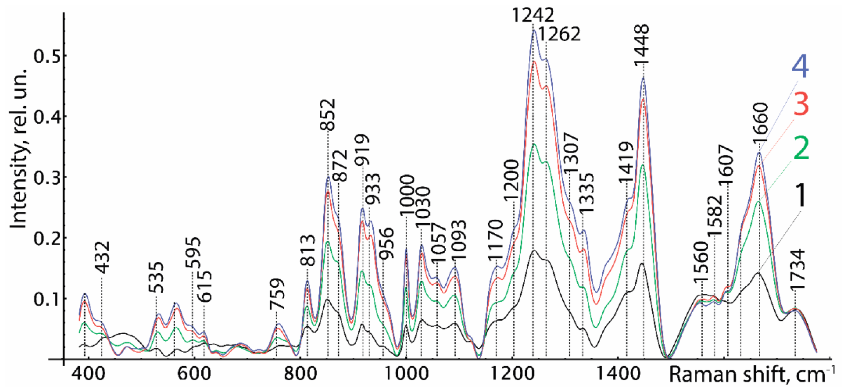
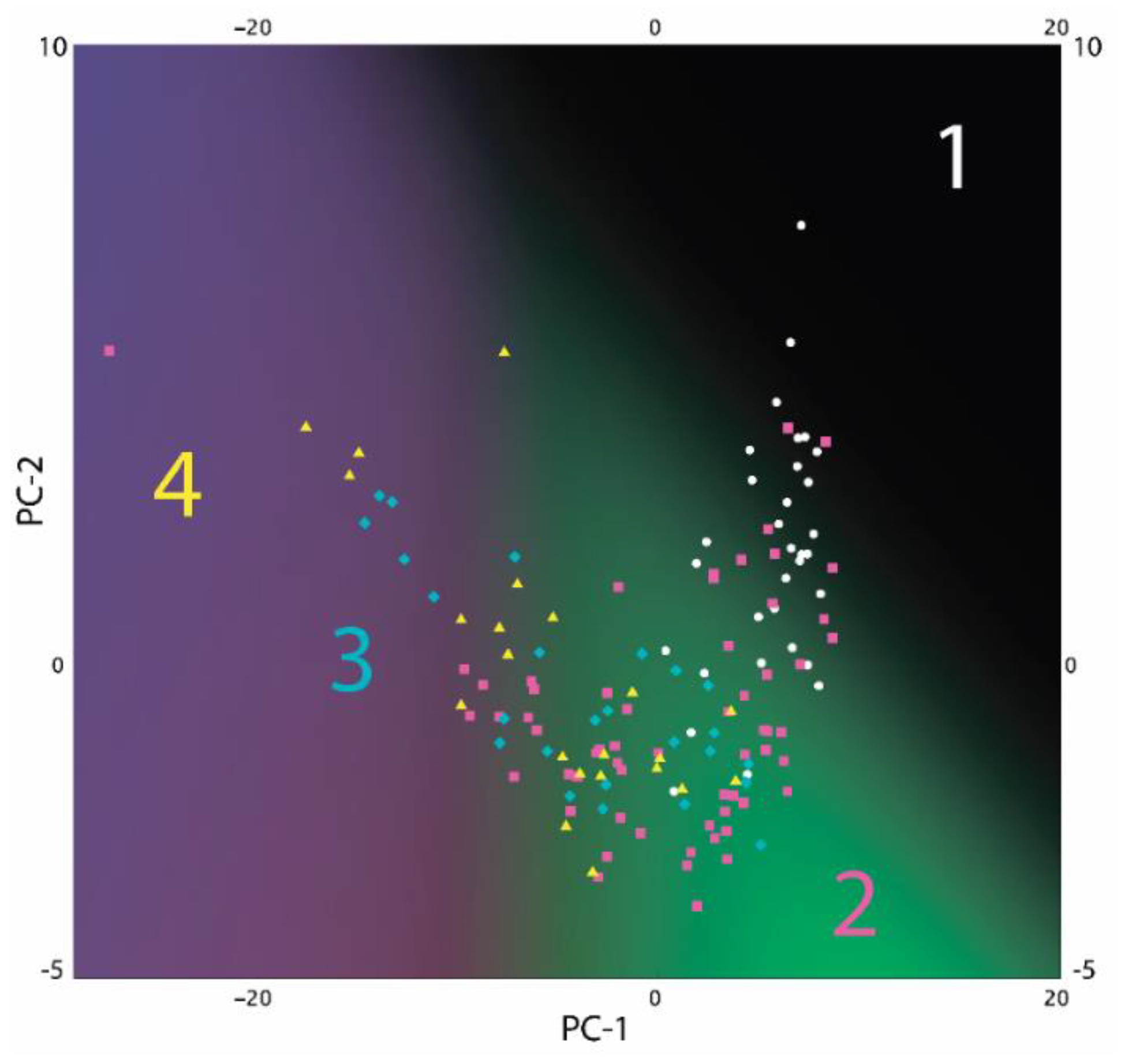
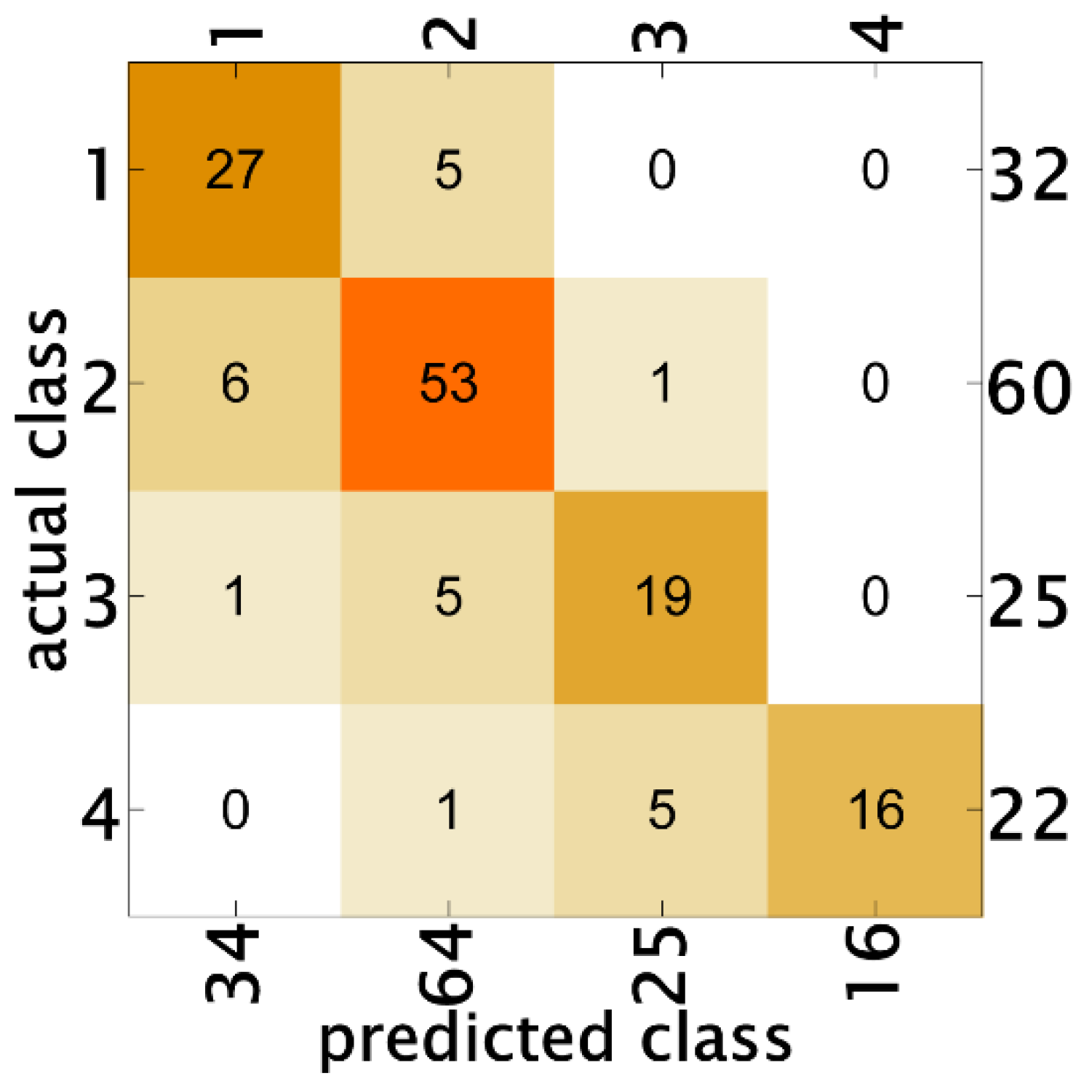
Biomaterials received from juvenile dentin were used as the materials of the study. These biomaterials were demineralized in 1.2 H and 1.8 H hydrochloric acid solution (technical specification TU-9398-001-01963143-2004). The comparison of the spectral properties of biomaterials from juvenile dentin and brefomatrix prepared using the same technology was carried out. Demineralized brefomatrices were previously used in dental practice and their efficiency was proved [12]. In our study, brefomatrices were prepared from different human tissues: spongy (the first group, 32 samples) and cortical (the second group, 32 samples) bone tissue.
The new biomaterials from juvenile dentin were divided into the following groups: demineralized in hydrochloric acid with normality of 1.2 H (the third group, 25 samples) and 1.8 H (the fourth group, 22 samples).
The study was carried out in accordance with the Declaration of Helsinki. The protocol was approved by the Ethics Committee (extract 17 August 2020 No. 210 of minutes of the meeting of the Committee on Bioethics of Samara State Medical University).
The surfaces of biomaterials at five different points using Raman spectroscopy were studied. This method was implemented using the stand including a semiconductor laser LML-785.0RB-04 (PDLD, Inc., Pennington, NJ, USA), an optical module for Raman spectroscopy (RPB 785), a spectrograph (ANDOR Sharmrock SR-303i, Belfast, UK),with an integrated digital camera (ANDOR DV-420A-OE, Belfast, UK) that was cooled to −60 °C, and a computer [26].
The use of this spectrograph provided a wavelength resolution of 0.15 nm with a low level of inherent noise. The power of the laser radiation, 200 mW, within the used exposure time (30 s) did not cause any changes to the samples. The optical probe was used for Raman spectrum registration [27].
Analysis of the Raman spectra was carried out in the range of 380−1780 cm−1 in this study.
The method of subtracting the fluorescence component of polynomial approximation with additional filtration of random noise effects was used to exclude autofluorescence from the Raman spectra. Processing and analysis of the Raman spectra were carried out using the software Wolfram Mathematica 12.2 (accessed on: https://www.wolfram.com/ accessed on 4 September 2022) [28].

3. Results

Figure 1 shows the normalized Raman spectra of the studied samples. As shown in Figure 1, The Raman spectra of all groups had spectral differences. The spectra of groups three and four had similar values of the relative intensities of the Raman lines. Figure 1 also shows that the spectra of brefomatrices from cortical bone tissue (the second group) were closer to the spectra of biomaterials prepared from juvenile dentin. This is probably due to similarity of the composition of dense cortical bone of the brefomatrix with the juvenile teeth after demineralization [29].
On average, the relative intensities of the main lines increased from the first group to the fourth group: 852–856 (proline), 873–880 (hydroxyproline), 1000, 1030 (phenylalanine), 1200–1300 (amide III), 1633–1700 (amide I). The relative intensity of the line of amide II 1539–1585 cm−1 did not change. These spectral changes were caused by the difference of organic composition of the subjects of the study.
The Raman spectra were characterized by little or no hydroxyapatite lines: ~432 (РO43−2) (Р–O Symmetric stretch) (phosphate of HA)), 584–593 (РO43−4) (Р–O Symmetric stretch)), 955–961 (РO43−1) (Р-O symmetric stretch)), ~1045 (РO43−3) (Р–O asymmetric stretch)), and ~1075 cm−1 (CO32−1) B-type substitution (C–O in-plane stretch)) that were caused by the demineralization process in the studied subjects.
To make the received Raman spectra more informative (separate the overlapping lines), a nonlinear regressive analysis of the Raman spectra was conducted, including an investigation of their spectral line decomposition.
The composition of the spectral lines was determined by automatic multi-iteration modeling of 139 Raman spectra using the software Wolfram Mathematica 12.2 with the use of machine learning methods and validated by literature analysis [30,31,32].
When modeling the spectral contours at the lines used as a template, the position x0 and the half width of the line (HWHM) dx were fixed. Only the intensity of the line in the range of 0 to the spectrum local maximum in the area of x0 was selected when modeling. HWHM was limited to the range of 1 to 16 cm−1. It allowed us to achieve high stability of the results when modeling the contour and to take into account all shifts of the Raman lines. The amplitude of the line a, which depended on the values of the independent regressors dx and x0, as defined in the initial terms of the analysis, was used as a criterion variable.
The average value of the corrected coefficient of determination for the initial result spectrum in the range of 380−1780 cm−1 of all 81 spectra was adjR2 = 0.9989.
The normalized amplitudes of the decomposed Raman lines were used for the relative quantitative analysis of the component composition. The analysis of the received data was performed with the software Wolfram Mathematica 12.2 using the method of logistic regression.
The results of the classification using the method of logistic regression in reduced two-dimensional measurements are shown in Figure 2 as a probability distribution of each measurement classified as one of the four studied groups. It can be seen that the areas of the first and second groups had insignificant intersections, but the third and fourth groups hardly had intersections with the first group and overlapped.
As a result of analysis using the method of logistic regression, we built a discriminant model of characteristic changes of intensities of the Raman lines of brefomatrix samples and the samples of biomaterials from juvenile tooth dentin. The calculated accuracy of the discriminant model was 82.7 ± 3.2%.
Figure 3 shows the decisional matrix of the model to classify the studied samples. The discriminant adequacy of the method had the values of AUC1 = 0.97, AUC2 = 0.95, AUC3 = 0.97, and AUC4 = 0.97 for every group. The precision and recall in the first group were 79% and 84%, in the second group were 83% and 88%, in the third group were 76% and 76%, and in the fourth group were 100% and 73%. Figure 3 shows that in the process of classification, group one could be incorrectly classified as group two and vice versa, and group two could be incorrectly classified as group three or four, which indicates that spectral properties of the samples from group two are similar to spectral properties of biomaterials from juvenile dentin (groups three and four). The samples from group one can be distinguished from groups three and four with high probability.
The final verification was carried out with the sample of additional 39 measurements selected randomly, which did not participate in the analysis. The model allows distinguishing brefomatrix samples from biomaterials from juvenile teeth with 100% accuracy. Four out of twenty spectra of biomaterials from juvenile teeth were wrongly classified as group two (brefomatrix from cortical bone).
Thus, the spectral properties of the new biomaterials from dentin of juvenile teeth were similar to the spectral properties of brefomatrices from cortical bone tissues. However, the used demineralization grades (1.2 and 1.8) of biomaterials from dentin of juvenile teeth were not significant in our case.

4. Discussion

The comparative assessment of new biomaterials from the dentin of juvenile teeth and brefomatrices using the Raman spectroscopy was carried out in this work.
One of the authors of this work previously carried out clinical observations with the use of brefomatrices in surgical dentistry, which led to the conclusion that surgical treatment of periodontitis using the compositions of demineralized brefomatrix and lyophilized allobone fragment powder allowed obtaining new bone segments; this was clearly visualized using an X-ray study [12].
However, the existing difficulties of obtaining donor material for further preparation of brefomatrices led us to the idea of obtaining an analogue to this material for further use in dental practice.

5. Conclusions

We found, in this work, that the spectral properties of new biomaterials from juvenile dentin are similar to the spectral properties of brefomatrices from cortical tissue according to the developed discriminant model of characteristic changes of intensities of the Raman lines. Additionally, both the demineralization grades of 1.2 H and 1.8 H can be used for preparation of these biomaterials from juvenile dentin. The calculated accuracy of the discriminant model was 82.7 ± 3.2%.
Thus, new biomaterials from juvenile dentin can be used in surgical dentistry after pre-clinical trials.

Author Contributions

Conceptualization, E.V.T., L.T.V.; methodology, P.E.T.; software, O.O.F.; vali-dation, P.E.T., E.V.T.; formal analysis, O.O.F.; investigation, P.E.T.; resources, L.T.V.; data curation, I.V.B.; writing—original draft preparation, E.V.T.; writing—review and editing, I.V.B.; visualization, O.O.F.; supervision, E.V.T.; project administration, E.V.T.; funding acquisition, L.T.V. All authors have read and agreed to the published version of the manuscript.

Funding

This research received no external funding.

Data Availability Statement

Not applicable.

Acknowledgments

The study was made with financial support from Samara State Medical University.

Conflicts of Interest

The authors declare no conflict of interest.

References

  1. Kirilova, I.A.; Sadovoy, M.A.; Podorozhnaja, V.T. Comparative characteristics of materials for bone grafting: Composition and properties. Novosib. Res. Inst. Traumatol. Orthop. Dig. 2012, 3, 72–83. [Google Scholar] [CrossRef]
  2. Smbatjan, B.S. Regeneration of Bone Tissues during Treating the Patients Using Dental Implants in Different Clinical Situations. Ph.D. Thesis, Moscow State University of Medicine and Dentistry, Moscow, Russia, 2012. [Google Scholar]
  3. Manazza, F.; La Rocca, S.; Nagni, M.; Chirico, L.; Cattoni, F. A simplified digital workflow for the prosthetic finishing of implant rehabilitations: A case report. J. Biol. Regul. Homeost. Agents 2021, 35, 87–97. [Google Scholar] [CrossRef] [PubMed]
  4. Tetè, G.; D’orto, B.; Ferrante, L.; Polizzi, E.; Cattoni, F. Role of mast cells in oral inflammation. J. Biol. Regul. Homeost. Agents 2021, 35, 65–70. [Google Scholar] [CrossRef]
  5. Trukhanova, Y.R. The Use of Osteoplastic Material «Bioimplant» for Comprehensive Treatment of Periodontal Disease. Ph.D. Thesis, Moscow State University of Medicine and Dentistry, Moscow, Russia, 2005. [Google Scholar]
  6. Xiaoru, F.; Luyuan, J.; Ping, M.; Zhipeng, F.; Songlin, W. Allogeneic Stem Cells From Deciduous Teeth in Treatment for Periodontitis in Miniature Swine. J. Periodontol. 2014, 85, 845–851. [Google Scholar] [CrossRef]
  7. Satoru, O.; Takanori, I. Application of Periodontal Ligament-Derived Multipotent Mesenchymal Stromal Cell Sheets for Periodontal Regeneration. Int. J. Mol. Sci. 2019, 20, 2796. [Google Scholar] [CrossRef]
  8. Shcheblykina, A.V.; Mishchenko, P.V.; Kumeiko, V.V. Pectin-based biocompatible degradable materials for tissue engineering. Pac. Med. J. 2013, 2, 13–17. [Google Scholar]
  9. Tansik, G.; Kilic, E.; Beter, M.; Demiralp, B.; Sendur, G.K.; Can, N.; Ozkan, H.; Ergul, E.; Guler, M.O.; Tekinay, A.B. A glycosaminoglycan mimetic peptide nanofiber gel as an osteoinductive scaffold. Biomater. Sci. 2016, 4, 1328–1339. [Google Scholar] [CrossRef]
  10. Kirichenko, V.N.; Marchenko, N.V. Experimental substantination of biological and synthetic osteoplastic materials use at surgical treatment of general periodontitis. Crime. J. Intern. Dis. 2012, 2, 106–108. [Google Scholar]
  11. Volova, L.T.; Samsonov, V.Е. The use of demineralized bone brefomatrix for plasty of different post-operative defects of the jaw. Stomatologiya [Dentistry] 1993, 73, 35–37. [Google Scholar]
  12. Bazhutova, I.V. Comparative Analysis of the Use of Osteoplastic Materials for Surgical Treatment of Periodontal Disease. Ph.D. Thesis, Federal State University “Central Research Institute of Dentistry”, Moscow, Russia, 2006. [Google Scholar]
  13. Butlerand, W.; Ritchie, H. The nature and functional significance of dentin extracellular matrix proteins. Int. J. Dev. Biol. 1995, 39, 169–179. [Google Scholar]
  14. Misch, C.E.; Dietsh, F. Bone-grafting materials in implant dentistry. Implant Dent. 1993, 2, 158–167. [Google Scholar] [CrossRef] [PubMed]
  15. Devecioglu, D.; Tözüm, T.F.; Sengün, D.; Nohutcu, R.M. Biomaterials in periodontal regenerative surgery: Effects of cryopreserved bone, commercially available coral, demineralized freeze-dried dentin, and cementum on periodontal ligament fibroblasts and osteoblasts. J. Biomater. Appl. 2004, 19, 107–120. [Google Scholar] [CrossRef]
  16. Kim, Y.K.; Kim, S.G.; Byeon, J.H.; Lee, H.J.; Um, I.U.; Lim, S.C.; Kim, S.Y. Development of a novel bone grafting material using autogenous teeth. Oral Surg. Oral Med. Oral Pathol. Oral Radiol. Endod. 2010, 109, 496–503. [Google Scholar] [CrossRef]
  17. Murata, M.; Akazawa, T.; Mitsugi, M.; Um, I.W.; Kim, K.W.; Kim, Y.K. Human Dentin as Novel Biomaterial for Bone Regeneration. Biomater.-Phys. Chem. 2011, 6, 127–140. [Google Scholar]
  18. Privalov, V.Е.; Shemanin, V.G. Optimization of differential absorption and scattering lidar for sensing molecular hydrogen in the atmosphere. Technol. Phys. 1999, 69, 65–70. [Google Scholar] [CrossRef]
  19. Sukhinina, A.V. The Method Sofoptical Spectros Copy for Dental Disease Diagnosis. Ph.D. Thesis, National Research Nuclear University MEPh, Moscow, Russia, 2014. [Google Scholar]
  20. Almahdy, A.; Downey, F.C.; Sauro, S.; Cook, R.J.; Sherriff, M.; Richards, D.; Watson, T.F.; Banerjee, A.; Festy, F. Microbiochemical analysis of carious dentine using raman and fluorescence spectroscopy. Caries Res. 2012, 46, 432–440. [Google Scholar] [CrossRef] [PubMed]
  21. Gonchukov, S.; Sukhinina, A.; Bakhmutov, D.; Minaeva, S. Raman spectroscopy of saliva as a perspective method for periodontitis diagnostics. Laser Phys. Lett. 2012, 9, 73–77. [Google Scholar] [CrossRef]
  22. Mandra, Y.V.; Ivashov, A.S.; Votjakov, S.L.; Kiseleva, D.V. Possibilities of Raman microspectrometry imaging for structural investigation of human enamel and dentin. Eksperimentalnaya Klin. Stomatol. 2011, 1, 24–28. [Google Scholar]
  23. Timchenko, E.V.; Timchenko, P.E.; Volova, L.T.; Frolov, O.O.; Zibin, M.; Bazhutova, I.V. Raman spectroscopy of changes in the tissues of teeth with periodontitis. Diagnostics 2020, 10, 876. [Google Scholar] [CrossRef] [PubMed]
  24. Ionita, I. Diagnosis of tooth decay using polarized micro-Raman confocal spectroscopy. Rom. Rep. Phys. 2009, 61, 567–574. [Google Scholar]
  25. Raghavan, M. Investigation of Mineral and Collagen Organization in Bone Using Raman Spectroscopy. Ph.D. Thesis, University of Michigan, Ann Arbor, MI, USA, 2011. [Google Scholar]
  26. Timchenko, E.V.; Timchenko, P.E.; Pisareva, E.V.; Vlasov, M.Y.; Volova, L.T.; Frolov, O.O.; Fedorova, Y.V.; Tikhomirova, G.P.; Romanova, D.A.; Daniel, M.A. Spectral Analysis of Rat Bone Tissue During Long Antiorthostostatic Hanging and at Introduction of Allogen Hydroxyapatitis. Opt. Spectrosc. 2020, 128, 989–997. [Google Scholar] [CrossRef]
  27. Timchenko, E.V.; Timchenko, P.E.; Pisareva, E.V.; Daniel, M.A.; Volova, L.T.; Fedotov, A.A.; Frolov, O.O.; Subatovich, A.N. Optical analysis of bone tissue by Raman spectroscopy in experimental osteoporosis and its correction using allogeneic hydroxyapatite. J. Opt. Technol. 2020, 87, 161–167. [Google Scholar] [CrossRef]
  28. Timchenko, P.E.; Timchenko, E.V.; Volova, L.T.; Zybin, M.A.; Frolov, O.O.; Dolgushov, G.G. Optical Assessment of Dentin Materials. Opt. Mem. Neural Netw. 2020, 29, 354–357. [Google Scholar] [CrossRef]
  29. Udaltsova, K.А. The structure of intact dentin and enamel of human juvenile teeth Mir meditsini I biologii. World Med. Biol. 2008, 4, 70–76. [Google Scholar]
  30. Movasaghi, Z.; Rehman, S.; Rehman, I. Raman Spectroscopy of Biological Tissues. Appl. Spectrosc. Rev. 2007, 42, 493–541. [Google Scholar] [CrossRef]
  31. Sukhodub, L.; Moseke, C.; Sulkio-Cleff, B.; Maleev, V.Y.; Semenov, M. Collagen–hydroxyapatite–water interactions investigated by XRD, piezogravimetry, infrared and Raman spectroscopy. J. Mol. Struct. 2004, 704, 53–58. [Google Scholar] [CrossRef]
  32. Ramakrishnaiah, R.; Rehman, G.; Basavarajappa, S.; Khuraif, A.; Durgesh, B.; Khan, A.; Rehman, I. Applications of Raman spectroscopy in dentistry: Analysis of tooth structure. Appl. Spectrosc. Rev. 2015, 50, 332–350. [Google Scholar] [CrossRef]
Figure 1. The average Raman spectra of the studied samples: 1—brefomatrix from spongy bone, 2—brefomatrix from cortical bone, 3—biomaterials from juvenile teeth with demineralization in 1.2 H, 4—biomaterials from juvenile teeth with demineralization in 1.8 H.
Biophysica 02 00028 g001
Figure 2. 2D probability distribution of each class as a function of the studied subjects: 1—brefomatrix from spongy bones, 2—brefomatrix from cortical bones, 3—biomaterials from dentin of juvenile teeth with demineralization in 1.2 H, 4—biomaterials from dentin of juvenile teeth with demineralization in 1.8 H.
Biophysica 02 00028 g002
Figure 3. Decision matrix of logistic regression: 1—brefomatrix from spongy bone, 2—brefomatrix from cortical bone, 3—biomaterials from dentin of juvenile teeth with demineralization in 1.2 H, 4—biomaterials from dentin of juvenile teeth with demineralization in 1.8 H.
Biophysica 02 00028 g003
Publisher’s Note: MDPI stays neutral with regard to jurisdictional claims in published maps and institutional affiliations.

