Case Report

Endoscopic Excision of Rare Large Maxillary Sinus Osteoma: A Case Report and Literature Review

1
Second Department of Otorhinolaryngology, Iuliu Hatieganu University of Medicine and Pharmacy, Republicii Str. 18, 400015 Cluj-Napoca, Romania
2
Fourth Department of Community Medicine, Public Health Discipline, Str. A. Iancu 31, 400083 Cluj-Napoca, Romania
*
Author to whom correspondence should be addressed.
Surgeries 2023, 4(3), 317-324; https://doi.org/10.3390/surgeries4030032
Submission received: 2 May 2023 / Revised: 20 June 2023 / Accepted: 21 June 2023 / Published: 27 June 2023

Abstract

:
Paranasal sinus osteomas are the most common benign fibro-osseous lesions that occur in the paranasal sinuses, having potential serious complications. With a prevalence ratio of 2:1 towards males, in 95% of the cases, the osteomas are situated most commonly in the frontoethmoidal region. However, osteomas of the maxillary sinus account for less than 5% of cases. The management of symptomatic paranasal sinus osteomas is surgical and is decided based on tumor size and location, patient symptoms, and experience of the surgeon. The Caldwell–Luc procedure, lateral rhinotomy, or craniotomy as external standard procedures remain mandatory if endonasal endoscopic surgery fails. In most reported cases, large tumors are excised by an external approach or in conjunction with an endoscopic technique. Endoscopic treatment of such tumors is a huge challenge for the operator. We report on a 42-year-old male patient presenting with a giant calcified mass in the right maxillary sinus. In this case, an endonasal approach was the most appropriate management even with the large size of the tumor. No recurrence at 24 months follow-up was reported.

1. Introduction

Osteomas of the paranasal sinuses are rare, slowly developing, benign, well-surrounded tumors, with a prevalence of 3% of all benign paranasal sinus tumors. The incidence in the general population is between 0.43–1%, with a peak between the fourth and sixth decades. Mostly located in the frontal sinuses or the ethmoidal region, osteomas can rarely be found in the maxillary sinuses [1]. At the beginning, symptoms are caused by tumor dimension. Headaches above the area of the lesion may appear, as well as pain, facial swelling, and persistent sinusitis not responding to medical therapy. Intracranial and ocular involvement, subsequent to osteoma extension, have to be mentioned. Periodic follow-up is suggested as treatment in asymptomatic cases. Nevertheless, specific conditions are considered indications for surgery. The surgical indications for sino-nasal osteomas are the most debated issues. In symptomatic cases with huge tumors, open, endoscopic, or combined approaches could be applied, with successful outcomes. Surgical approaches should be customized for each patient and total resection should be the operative goal [2,3,4,5,6,7]. Surgical resection of some big osteomas of the paranasal sinuses could also be performed safely and radically by endoscopic techniques with a better outcome [8].
Our research in the literature regarding the paranasal sinus osteoma morphometric standards identified mainly cases of frontoethmoidal osteomas. Schick et al. [9], reported a diameter ranging from 8 to 35 mm. A mean of 17 mm describes osteomas of a standard size; moreover, an average of 30 mm is considered very large [1]. No morphometric data on maxillary osteomas was found.
The objective of this case study is to explore the available surgical approaches and to determine the optimal surgical procedure by analyzing giant osteomas of the paranasal sinuses in the literature.

2. Case Presentation

This case report describes a rare presentation of a giant osteoma in the maxillary sinus, picked up from an incidental finding on surgical treatment based on a computed tomography (CT) scan for chronic rhinosinusitis with nasal polyps (CRSwNP). We report on a 42-year-old male patient, without past medical history, presenting with complaints of persistent headache, associated with sensibility at the level of the right maxillary sinus, chronic nasal obstruction, rhinorrhea, hyposmia, and fatigability. The symptoms started approximately 1 year ago and had a slowly progressive evolution.
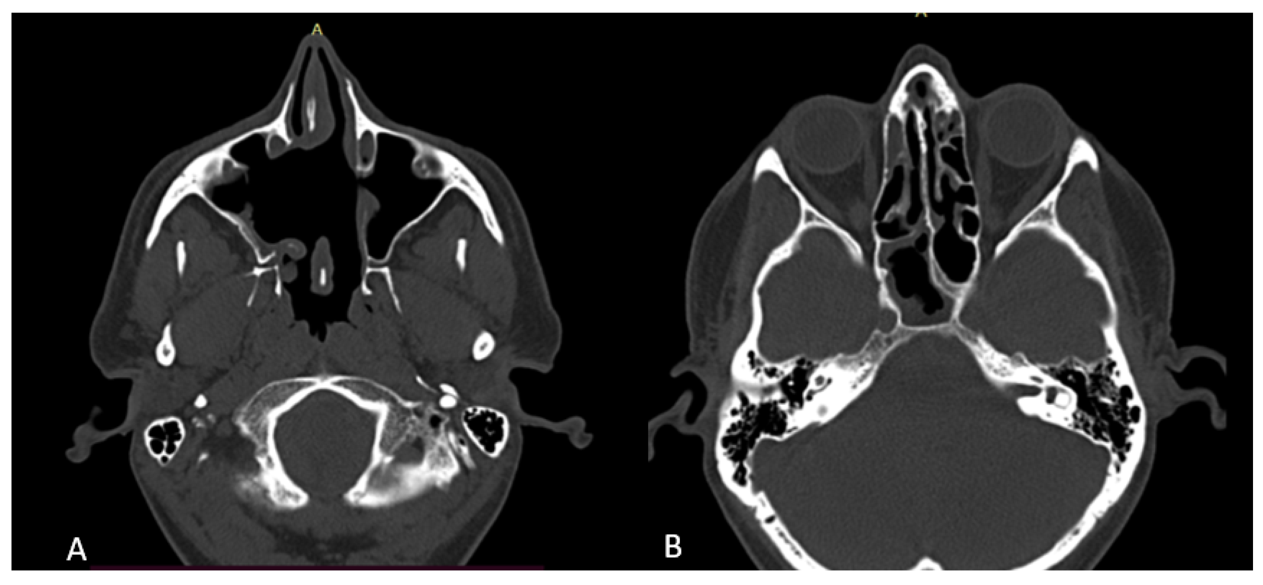
The patient underwent routine examinations with nasal endoscopy. Clinical examination showed sensibility and pain at palpation at the level of the canine fossa. Nasal endoscopy revealed the presence of nasal polyps arising from the middle meatus bilaterally, the nasal corridor being narrowed by chronic hypertrophic rhinitis and a septal deviation. Orbital and paranasal sinus computed tomography revealed a large, homogenous radiopaque mass, with bony density and well-defined margins, arising from the right maxillary sinus, measuring 3.67 cm × 3.11 mm. Bilateral osteo-meatal complexes were obliterated by nasal polyps. Sinus mucosal thickening was additionally described in all paranasal sinuses (Figure 1).
Routine blood tests and the chest X-ray was normal. The diagnosis of chronic rhinosinusitis with nasal polyps (CRSwNP) and maxillary sinus osteoma was suspected, surgical intervention was highly indicated, and different surgical methods were discussed. The surgical plan included the removal of the osteoma via an endoscopic endonasal approach under general anesthesia.
Endoscopic surgery was carried out to treat nasal polyposis, chronic rhinitis, septal deviation, and large maxillary osteoma. The pre-anesthetic assessment did not highlight any contraindications; consequently, the surgery was performed under general anesthesia with orotracheal intubation. The antibiotic prophylaxis was performed using beta-lactamases and 1000 mg iv ceftriaxone.
The nasal cavity was prepared with intranasal submucosal vasoconstriction with adrenaline 1:100,000, applied at the level of the middle turbinate and lateral nasal wall. A front-view endoscope with 0- and 45-degree angled lenses was used. The first step consisted of excision of nasal polyps, followed by uncinectomy, maxillary antrostomy, total ethmoidectomy, and sphenoidotomy on the left side. Subsequently, the same procedures were performed contralaterally, except for an extension of maxillary antrostomy to a medial endoscopic maxillectomy with uncovering of the large maxillary sinus tumor.
The osteoma was visualized as a bony mass strongly attached to the right maxillary lateral sinus wall. Because of the large size of the mass, it could not be removed through the antrostomy aperture. Consequently, the antrostomy was extended anteriorly with complete excision of the medial maxillary sinus wall and a portion of the anterior wall; thus an endoscopic Denker approach was employed, followed by piecemeal resection of the tumor. The osteoma attachment was drilled with a burr (Figure 2).
No abundant bleeding or other complications were observed during the procedure. Nonabsorbable nasal cavity dressing was applied, with packing removal in the first postoperative day. A systemic antibiotic (cefuroxime, 1 g per day for 7 days), an intravenous hemostatic agent, and pain relievers were administered. Saline nasal irrigation for the following 30-day postoperative period was recommended.
The recovery was uneventful, and the patient was discharged on day two postoperative. No other complications were mentioned, except a right nasal synechia, that was subsequently surgically treated, by radiofrequency ablation, under local anesthesia.
Histopathology analysis offers the definitive diagnosis. Our histological examination of the excised tumor revealed compact bone tissue crossed by channels of Havers, with no evidence of malignancy (Figure 3). Specific histopathological findings, consisting in a central, sharply demarcated radiolucent nidus of bony trabeculae surrounded by reactive osteosclerosis, are depicted.
Postoperative follow-up was performed at 1, 6, 12, and 24 months. A SNOT 22 questionnaire, applied at 12 and 24 months, revealed a significant improvement in quality of life (QOL) scores, from initial scores of 56 preoperatively, to 12 and 10, respectively, at 12 and 24 months. Nasal endoscopy and computed tomography performed at 24 months revealed no recurrence of the nasal polyps and right maxillary sinus osteoma (Figure 4 and Figure 5).

3. Discussion

3.1. Epidemiology, Pathogenesis, Clinical Presentation

Although osteomas are the most commonly seen benign tumors of the paranasal sinuses, they are still rare, and we are facing a lack of literature detailing the management and prognosis of craniofacial osteoid osteomas. However, they represent tumors with a slow growth rate and potential complications and have a predilection to occur in the craniofacial region [10,11,12]. Due to the rarity of giant osteomas of the paranasal sinuses, only a few case reports exist in the literature; in addition, the clinical presentation and treatment options are unclear [13]. Paranasal sinus osteomas are mostly asymptomatic, being detected accidentally on paranasal computed tomography (CT) scans. About 4% to 10% of them cause clinical symptoms related to the location, size, and growth features of the tumor, including facial deformity, swelling, facial pain, nasal discharge, nasal obstruction, headache, chronic sinusitis, orbital symptoms (proptosis, epiphora, diplopia, visual loss), and cerebral complications (dizziness, meningitis, pneumatocele, brain abscess, cerebrospinal fluid leakage) [4,5,7]. Rarely, extensive growth could determine aesthetic or functional problems that vary according to different locations [14].
Because of their asymptomatic nature, their real incidence is not certain. Paranasal sinus osteoma incidence has been reported to be around 3%, of which maxillary sinus osteomas represent only 5%. They are commonly detected in the fourth decade of life, with a male predominance [4,7]. According to Mlouka et al., in the case of a maxillary sinus osteoma, the lesion usually appears on the lateral wall of the sinus [15].
The etiology of osteomas is still controversial. Different theories regarding etiology have been proposed, including developmental, traumatic, and infectious. The developmental theory suggests the tumors could arise from rests of cartilage or osseous stem cells present in bone. The traumatic and infectious theory relies on an inflammatory process that induces bony formation. The proposed pathogenesis in the inflammatory theory is the stimulation of muco-periosteal osteoblasts by chronic inflammation, which leads to calcification and subsequent osteoma formation [16]. In the present case, this theory could be supported by the fact that the patient presented concomitant chronic rhinosinusitis. However, none of the theories adequately explains the developmental mechanism; thus, further research is needed [4,7].

3.2. Diagnosis and Radiological Features

Due to the asymptomatic progress, the osteomas are usually incidentally diagnosed by radiography or by a computed tomography scan performed for other reasons [14]. Imaging represents the diagnostic method of choice. A computed tomography (CT) scan with bone algorithm represents the gold standard, whereas magnetic resonance imaging is the optimal imaging modality for differential diagnosis, also to evaluate the extension of the disease, such as the intracranial or orbital extension [5]. Cone-beam CT is useful for assessing the relationship between osteomas and adjacent structures, and for choosing the appropriate surgical plan [14].
Although a CT scan may be used to make a preliminary diagnosis, a definitive diagnosis is obtained by histopathology analysis [1]. Histopathological osteomas are well-differentiated tumors, described as three histological types: “ivory”, “mature”, and fibrous osteoma. The first two have similar histological features, whereas fibrous osteoma is accepted as a separate entity. Osteoma, fibrous dysplasia, and ossifying fibroma form a class of benign bony abnormalities known as fibro-osseous lesions, and a differential diagnosis between them should be made. Also, the differential diagnosis should be proposed with malignant tumors, such as osteosarcoma [5,11,17].
Radiographically, osteomas appear as radiopaque lesions similar to bone cortex, and may determine bone expansion. The differential diagnosis includes several inflammatory and tumoral pathologies, but the typical craniofacial location may aid in the diagnosis [14].

3.3. Therapeutical Management

The treatment of paranasal sinus osteomas is controversial [4]. The removal of these lesions trends toward minimally invasive techniques and remains a developing topic within the field of craniofacial surgery [12]. There are two options for its management: clinical observation and surgery. Because of its slow growth rate, many authors recommend periodical clinical observation and imaging follow-up for all asymptomatic paranasal sinus osteomas [4]. According to Arslan et al. [4], there is no significant growth in asymptomatic osteomas on repeated radiological examinations, during a 28-month follow-up. Whereas indications for asymptomatic cases are still a subject of controversy in the literature, in those cases with clinical signs or symptoms, surgical treatment is the elective procedure [5]. Although clinical observation should be recommended for elderly patients and those with frontal and maxillary sinus asymptomatic osteomas, there are recommendations for removal of all ethmoid and sphenoid osteomas as soon as possible because of the potential severe complications on the visual pathways or carotid artery [4,11]. Georgalas et al. recommend observation of small asymptomatic osteomas [18].
Osteomas usually remain asymptomatic until the tumor growth reaches a certain dimension. Once the osteoma becomes symptomatic, affecting patient’s life quality, surgery may be taken into consideration. The indication is also valid in asymptomatic lesions that obliterate over 50% of the sinus volume, which grow rapidly or develop intra-orbital or intracranial extensions. Moreover, a small osteoma is technically easier to remove than a larger one, with lower complication rates. In addition, surgical treatment should be performed immediately for sphenoid sinus osteomas because of the potential severe visual and intra-cranial complications [7]. Surgical excision of symptomatic osteomas, using an appropriate surgical approach, remains necessary according to the localization of the tumor, the subsequent symptomatology, and patient age, regardless the size of the osteoma [4,11].
The Caldwell–Luc procedure [4], lateral rhinotomy or craniotomy [19], external approach, endoscopic techniques, or a conjunction of them are the available choices for surgical treatment. Dealing with large lesions, open procedures represent the gold standard, but currently, there is still a debate regarding the best treatment option. Even if the technique often depends on the localization of the osteoma, in most reported cases, large tumors were excised by combined techniques [19]. More importantly, the surgeon planning on undertaking an endoscopic approach should also be prepared for converting to an external approach. Based on the statistical analysis made by Humeniuk-Arasiewicz et al. [19], for the frontoethmoidal osteomas, the average size of tumors excised endoscopically and those removed by the external approach does not differ statistically.
Endoscopic endonasal techniques have specific advantages over the open procedures widely used in the past, so that may be considered the first-line surgical treatment of benign tumors located in the frontal region [20], and the treatment of choice for selected osteomas. Endoscopy enables closer and better visualization, excellent magnification, and good illumination of the anatomy, permitting safe anatomic dissection as well as avoiding damage to surrounding structures, which is still a huge challenge for the surgeon in the case of large osteomas. It appears to be safer, while being an equally effective means of thorough extirpation, with minimal bleeding and complications, preserving the natural endonasal drainage pathways, and reducing the traumatic impact with great esthetic results. On the other hand, endoscopic procedures require surgical training and experience [5,6,8,20,21], and even higher surgical experience in the case of pediatric patients, due to the narrow nasal cavity [1].
Castelnuovo et al. and Miman et al. discuss the rapid progress of endoscopic sinus surgery, even for large osteomas, as seen in our case. The indications for the endoscopic approach have widened recently, so that lesions which, years ago, were not considered to be suitable for endoscopic endonasal resection—even large osteomas—can be easily removed using an endoscopic procedure, without any need for an external approach. Not least, the availability of the multi-angled scopes, intranasal drills, and neuro-navigation systems, together with increasing skill in the endoscopic approach and the 2-nostril–4-hand technique, represents a further evolution that expands upon [5,8].
Consistent with the literature, the patient to whom we refer falls, in terms of age and symptoms, within the existing statistical data. The goal of complete endoscopic tumor removal was achieved, together with polypectomy, turbinectomy, septoplasty, and wide opening of all paranasal sinuses. Nasal endoscopy showed normal epithelialization 4 to 6 weeks after surgery. A right nasal fossa synechia was observed and surgically removed at 4 weeks after surgery, and there were no other complications at 6 months follow-up [7,22].
Despite meticulous surgical techniques, postoperative complications occur, so that the management of the postsurgical patient is contributory to upgrading the success of nasal endoscopy. The reported complications can be largely classified into immediate complications such as bleeding and crusting, short-term complications such as infection or synechiae development, and long-term complications such as ostial stenosis, refractory disease, or recurrent disease [23].
Regarding the follow-up after surgery, because osteoma recurrences are very rare, routine periodic postoperative surveillance with paranasal CT scanning is not justified; thus, periodic endoscopic evaluations (at 1, 3, and 6 months) should be performed [21].

4. Conclusions

Maxillary sinus osteomas are rare, slow-growing benign lesions, rarely reaching large dimensions. The management of symptomatic patients is surgical, but the decision between external and endoscopic approaches is guided by several considerations.
Our case is a rare example of a giant maxillary osteoma, which was successfully treated with an endoscopic procedure, with no postoperative complications except a thin synechia that was surgically treated 6 weeks after surgery. No other complications or recurrences were mentioned at 1, 3, 6, 12, and 24 months follow-up. Thus, this is a case of a less invasive, yet safe and effective, approach to the treatment of symptomatic giant maxillary osteomas in which the surgeon’s experience is to be of great value in achieving the total removal of the tumor without any damage to the adjacent organs.

Author Contributions

Conceptualization, A.G., I.M.I., I.M.P., B.F.C. and S.A.; methodology, A.G. and I.M.I.; software, non-applicable; validation, A.G. and S.A.; formal analysis, non-applicable; investigation, I.M.I., I.M.P. and A.G.; resources, S.A.; data curation, A.G., I.M.I. and I.M.P.; writing—original draft preparation, I.M.I., I.M.P. and B.F.C.; writing—review and editing, A.G. and S.A.; visualization, A.G., I.M.I. and S.A.; supervision, A.G. and S.A.; project administration, S.A.; funding acquisition, non-applicable. All authors have read and agreed to the published version of the manuscript.

Funding

This research received no external funding.

Institutional Review Board Statement

The study was conducted according to the guidelines of the Declaration of Helsinki and approved by the Ethics Committee of “Iuliu Hatieganu”, Medicine and Pharmacy University (protocol code AVZ33/13.12.2021).

Informed Consent Statement

Informed consent was obtained from the subject involved in the study.

Data Availability Statement

Data sharing not applicable. No new data were created or analyzed in this study. Data sharing is not applicable to this article.

Conflicts of Interest

The authors declare no conflict of interest.

References

  1. Alotaibi, N.; Hanss, J.; Benoudiba, F.; Bobin, S.; Racy, E. Endoscopic removal of large orbito-ethmoidal osteoma in pediatric patient: Case report. Int. J. Surg. Case Rep. 2013, 4, 1067–1070. [Google Scholar] [CrossRef] [Green Version]
  2. Edmond, M.; Clifton, N.; Khalil, H. A large atypical osteoma of the maxillary sinus: A report of a case and management challenges. Eur. Arch. Oto-Rhino-Laryngol. 2011, 268, 315–318. [Google Scholar] [CrossRef]
  3. Sudhoff, H.; Theegarten, D.; Luckhaupt, H. Osteom der kieferhöhle. Laryngorhinootologie 2001, 80, 275–277. [Google Scholar] [CrossRef] [PubMed]
  4. Arslan, H.H.; Tasli, H.; Cebeci, S.; Gerek, M. The management of the paranasal sinus osteomas. J. Craniofac. Surg. 2017, 28, 741–745. [Google Scholar] [CrossRef] [PubMed]
  5. Castelnuovo, P.; Valentini, V.; Giovannetti, F.; Bignami, M.; Cassoni, A.; Iannetti, G.M. Osteomas of the maxillofacial district: Endoscopic surgery versus open surgery. J. Craniofac. Surg. 2008, 19, 1446–1452. [Google Scholar] [CrossRef] [PubMed]
  6. Zhang, D.; Li, X.; Lv, L.; Yu, J.; Yang, C.; Xiong, H.; Liao, R.; Zhou, B.; Huang, X.; Liu, X.; et al. Improving the diagnosis of common parotid tumors via the combination of CT image biomarkers and clinical parameters. BMC Med. Imaging 2020, 20, 38. [Google Scholar] [CrossRef] [Green Version]
  7. Muderris, T.; Sevil, E.; Bercin, S.; Canda, B.; Kiris, M. Giant paranasal sinus osteomas: Surgical treatment options. J. Craniofac. Surg. 2014, 25, 1287–1291. [Google Scholar] [CrossRef]
  8. Miman, M.C.; Bayindir, T.; Akarcay, M.; Erdem, T.; Selimoglu, E. Endoscopic removal technique of a huge ethmoido-orbital osteoma. J. Craniofac. Surg. 2009, 20, 1403–1406. [Google Scholar] [CrossRef]
  9. Schick, B.; Steigerwald, C.; el Rahman el Tahan, A.; Draf, W. The role of endonasal surgery in the management of frontoethmoidal osteomas. Rhinology 2001, 39, 66–70. [Google Scholar]
  10. Koivunen, P.; Löppönen, H.; Fors, A.P.; Jokinen, K. The growth rate of osteomas of the paranasal sinuses. Clin. Otolaryngol. Allied Sci. 1997, 22, 111–114. [Google Scholar] [CrossRef]
  11. Eller, R.; Sillers, M. Common Fibro-osseous Lesions of the Paranasal Sinuses. Otolaryngol. Clin. N. A. 2006, 39, 585–600. [Google Scholar] [CrossRef]
  12. Langlie, J.A.; Hullfish, H.; Jabori, S.K.; Thaller, S.R. Diagnosis and Management of Craniofacial Osteomas. J. Craniofac Surg. 2023, 37253237. [Google Scholar] [CrossRef]
  13. Alkhaldi, A.S.; Alsalamah, S.; Tatwani, T. A Case of Giant Ethmoid Sinus Osteoma. Cureus 2021, 13, e18011. [Google Scholar] [CrossRef]
  14. 14. Tarsitano, A.; Ricotta, F.; Spinnato, P.; Chiesa, A.M.; Di Carlo, M.; Parmeggiani, A.; Miceli, M.; Facchini, G. Craniofacial Osteomas: From Diagnosis to Therapy. J. Clin. Med. 2021, 10, 5584. [Google Scholar] [CrossRef]
  15. Mlouka, M.; Tlili, M.; Hamrouni, A.; Selmi, R.; Khanfir, F.; Khalfi, M.S.; Ben Amor, F. Lateral maxillary sinus floor elevation in the presence of a sinus osteoma: A case report. Clin. Case Rep. 2021, 9, e04124. [Google Scholar] [CrossRef] [PubMed]
  16. Aldren, C.P.; Soames, J.V.; Birchall, J.P. Bony remodelling in an osteoma of the paranasal sinuses. J. Laryngol. Otol. 1993, 107, 633–635. [Google Scholar] [CrossRef] [PubMed]
  17. Fu, Y.S.; Perzin, K.H. Non-epithelial tumors of the nasal cavity, paranasal sinuses, and nasopharynx: A clinicopathologic study. II. Osseous and fibro-osseous lesions, including osteoma, fibrous dysplasia, ossifying fibroma, osteoblastoma, giant cell tumor, and osteosarcoma. Cancer 1974, 33, 1289–1305. [Google Scholar] [CrossRef] [PubMed]
  18. Georgalas, C.; Goudakos, J.; Fokkens, W.J. Osteoma of the Skull Base and Sinuses. Otolaryngol. Clin. N. A. 2011, 44, 875–890. [Google Scholar] [CrossRef] [PubMed]
  19. Humeniuk-Arasiewicz, M.; Stryjewska-Makuch, G.; Janik, M.A.; Kolebacz, B. Giant fronto-ethmoidal osteoma—Selection of an optimal surgical procedure. Braz. J. Otorhinolaryngol. 2018, 84, 232–239. [Google Scholar] [CrossRef]
  20. Raponi, I.; Giovannetti, F.; Cacciatore, F.; Di Carlo, D.; Valentini, V. Endoscopic Forehead Approach for Minimally Invasive Frontal Osteoma Excision. J. Craniofac. Surg. 2020, 31, e767-9. [Google Scholar] [CrossRef]
  21. Article, O. Frontoethmoidal and Intraorbital Osteomas. Arch. Otolaryngol.—Head Neck Surg. 2014, 138, 498–504. [Google Scholar]
  22. Huang, H.M.; Liu, C.M.; Lin, K.N.; Chen, H.T. Giant ethmoid osteoma with orbital extension, a nasoendoscopic approach using an intranasal drill. Laryngoscope 2001, 111, 430–432. [Google Scholar] [CrossRef] [PubMed]
  23. Tan, B.K.; Chandra, R.K. Postoperative prevention and treatment of complications after sinus surgery. Otolaryngol. Clin. North. Am. 2010, 43, 769–779. [Google Scholar] [CrossRef] [PubMed]
Figure 1. Axial and coronal orbital and paranasal non-enhanced CT scan images; (A) Axial default window; (B) Axial bone window; (C) Coronal default window; (D) Coronal bone window, showing a homogenous radiopaque mass (see black arrow), with well-defined margins, originating from the right maxillary sinus filling the sinus cavity.
Surgeries 04 00032 g001
Figure 2. Intraoperative picture. Endoscopic surgical appearance of the right maxillary osteoma, as hard whiteish mass, strongly attached to the lateral wall of the right maxillary sinus; (A) 0 grade optic; (B) 45 grade optic. Macroscopically, round, hard, ivory-white, well-circumscribed lesions attached to the underlying bone by a broad base or occasionally by a small stalk and covered by a thin layer of fibrous periosteum.
Surgeries 04 00032 g002
Figure 3. Microscopy, histopathological findings. (A) Hematoxylin–Eosin staining, ×10: small mucous glandular acini can be observed; (B) Hematoxylin–Eosin staining, ×10: tumor proliferation consisting of bone trabeculae with spongy bone appearance, irregular in shape and size arranged in a loose conjunctival stroma containing fat cells and blood vessels; (C,D) Hematoxylin–Eosin staining, ×20: the lesion is covered by respiratory mucosa with edematous chorion. Blue arrow = mucous acini, Black arrow = dense fibrous capsule, Red arrow = bony trabeculae (trabecular bone), Yellow arrow = intertrabecular loosely arranged fibrous connective tissue (“loose conjunctive stroma”).
Surgeries 04 00032 g003
Figure 4. (A,B) Postoperative axial non-enhanced CT scan images performed 24 months after surgery, showing no recurrence of the right maxillary sinus osteoma or nasal polyps.
Surgeries 04 00032 g004
Figure 5. Nasal endoscopic captures, 0 grade optic.
Surgeries 04 00032 g005
Disclaimer/Publisher’s Note: The statements, opinions and data contained in all publications are solely those of the individual author(s) and contributor(s) and not of MDPI and/or the editor(s). MDPI and/or the editor(s) disclaim responsibility for any injury to people or property resulting from any ideas, methods, instructions or products referred to in the content.