Article

Caecal Volvulus: A District General Hospital Experience and Review of the Literature

1
Department of General Surgery, William Harvey Hospital, Ashford TN24 0LZ, UK
2
Department of Radiodiagnosis and Imaging, William Harvey Hospital, Ashford TN24 0LZ, UK
*
Author to whom correspondence should be addressed.
Surgeries 2022, 3(2), 78-91; https://doi.org/10.3390/surgeries3020010
Submission received: 4 January 2022 / Revised: 8 March 2022 / Accepted: 25 March 2022 / Published: 29 March 2022

Abstract

:
Background: Caecal volvulus (CV) is an uncommon cause of large bowel obstruction with potential for complications such as ischaemia and perforation. Prompt diagnosis and treatment only will ensure better outcomes. We aim to describe our experience in the largest series of CV reported in the United Kingdom. Methods: This was a retrospective study of 16 consecutive patients diagnosed with CV between March 2017 and March 2020. Results: Out of 16 patients, 11 were female, with a median age of 64 (range 33–80) years. All patients presented with abdominal pain and vomiting. An initial diagnosis of bowel obstruction was made only in 8 patients (50%). Computed tomography (CT) scan of the abdomen and pelvis correctly diagnosed CV in fourteen (87.5%). Fifteen patients (94%) underwent surgical intervention. All but one (n = 14, 93.4%) underwent laparotomy, and one patient (6.6%) underwent successful laparoscopic intervention. Right hemicolectomy was performed in all operated patients, and fourteen patients (93.4%) had primary anastomosis. Twelve patients (80%) had one or another form of morbidity. Seven patients (47%) and three patients (20%) had grade II and III (Clavien–Dindo Classification) complications, respectively, with a median hospital stay of 10 (range 1–49) days. One patient (6.7%) managed conservatively was deemed to be futile. There was no postoperative mortality. Conclusions: CV can present with non-specific symptoms, and a definite preoperative diagnosis is only possible with the aid of CT. Open resectional procedures with primary anastomosis are the most favoured approach in management, though laparoscopic access is also feasible in fit patients.

1. Introduction

Volvulus (Latin Volvere = to roll) [1] of the large bowel is the third most common cause of large bowel obstruction worldwide [2]. Caecal volvulus (CV) is second only to sigmoid volvulus followed by transverse colon and splenic flexure volvulus. It has considerable mortality and morbidity owing to closed-loop obstruction [3,4] in addition to old age and concurrent co-morbidities. It has a multifactorial aetiology, but a highly mobile caecal segment is the most likely pre-requisite [3,5]. Patients may present with varied non-specific symptoms, and most of them do so with distension, crampy abdominal pain, and constipation. Early diagnosis with prompt treatment has led to improved morbidity and mortality. This study aims to review the clinical presentation, accuracy of diagnostic modality, management, and outcome of CV in a large district hospital. We will also discuss findings of CT abdomen and critically appraise the debate on various modalities of treatment.

2. Methodology

A retrospective study of sixteen consecutive patients diagnosed with CV between March 2017 to February 2020 was conducted. Data were gathered from patient notes and electronic record systems to determine patient demographics, presentation, management, intraoperative findings, complications, length of hospital stay and outcome. An in-depth literature review was then performed.

3. Results

Out of sixteen patients, the male to female ratio was 5:11, with a median age of 64 (range 33–80) years (Table 1). All patients presented with abdominal pain, distension, and vomiting. The majority (67%) of them had background symptoms of constipation. Only five patients (31%) had a history of previous abdominal surgery. One patient had a recent laparoscopic tubal ligation, and the other ones had resection for sigmoid volvulus four years ago. Two patients had multiple abdominal surgeries including laparoscopic nephrectomy, small bowel resection and hysterectomy. Table 1, Table 2, Table 3 and Table 4 summarise the results.
Most patients (69%) had one or multiple co-morbidities (Table 2). Fifty per cent of the patients were American Society of Anaesthesiologists (ASA) grade III or more. The use of psychotropic drugs was evident in 38% of patients.
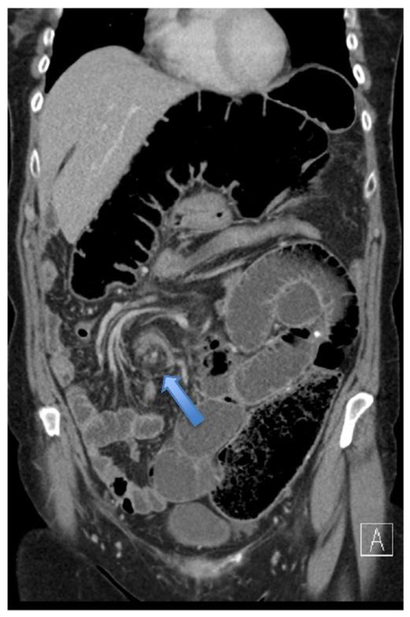
In eleven patients (68.75%), an initial abdominal radiograph was requested, of which only six patients (54%) showed a typical single dilated large bowel loop, suggestive of CV (Table 3). Five patients (46%) showed either non-specific bowel loops or features suggestive of small bowel obstruction (SBO). Overall, fifteen patients (93.8%) had a CT abdomen and pelvis (CTAP) with contrast. A radiological diagnosis of CV was made preoperatively in fourteen patients (93.3%). CTAP showed the following features to diagnose CV: Whirl sign in 12 patients (80%); double transition points in 13 patients (86.6%); split-wall sign, X-marks-the spot sign and ileocecal twist in 86.6%, 93.0% and 87% of patients, respectively. Central gas-filled appendix as an indirect supplementary sign was evident in 73% of patients. The caecum was distended to more than 10 cm in most patients (67%).
Fourteen patients (93.4%) underwent laparotomy with one (6.6%) undergoing successful laparoscopic intervention (Table 3). Eight patients (53.3%) had a viable caecum, and the rest had variable ischaemia in the twisted ileo-caecal segment. There was frank caecal perforation with localised peritonitis in one patient. All fifteen patients had right hemicolectomy; fourteen patients (93.4%) had primary ileocolonic anastomosis. One patient (6.7%) perforated and required an ileostomy and mucous fistula to be fashioned. Conservative pathway was chosen for a very frail 79-year-old lady with an ASA of IV after discussion with the patient and her next of kin. She had congestive cardiac failure, chronic obstructive pulmonary disease (COPD), diabetes mellitus and chronic kidney disease stage 3 (thus contributing to National Emergency Laparotomy Audit (NELA) mortality and morbidity of 37% and 99%, respectively).
All patients were admitted to intensive care in the immediate postoperative period. There was no mortality in operated patients (Table 4). Three of the fifteen patients (20%) were discharged without an eventful postoperative period. All three patients were ASA I and of 33, 57 and 65 years of age. Twelve patients (80%) had one or another form of morbidity. Of them, seven (47%) and three (20%) patients had those of Grade II and III (Clavien–Dindo Classification) complications, respectively. Two patients (13.3%) had an anastomotic leak; one was managed with radiological drainage and antibiotics, whereas another needed re-laparotomy, anastomosis taken down and an ileostomy with mucous fistula.
Three patients (20%) needed reoperation. Complete fascial dehiscence and evisceration were noted in one patient on day 6. Anastomotic leakage with pelvic abscess failing conservative treatment was the indication in one and a strong suspicion of mechanical obstruction in the other.
Median hospital stay was 10 (range 1–49) days. The thirty-day readmission rate was 6.25%. This was due to a patient being readmitted on the 12th day of discharge with worsening abdominal pain, fever and increased inflammatory markers. CT with oral and intravenous contrast revealed a minor anastomotic leak with an intra-abdominal abscess. It was drained under radiological guidance. The patient recovered following conservative measures.
Adenocarcinoma of the caecum was the underlying pathology in one patient (6.7%); an incidental finding of tubular adenoma and low grade appendiceal mucinous neoplasm (LAMN) was noted, respectively, in two other patients.
The median follow-up was 20 months. During this period, most of the patients (n = 10, 66.7%) had no hospital readmission or emergency department visits for abdominal complaints. Calculous cholecystitis in two patients and elective reversal of ileostomy in one patient were the reasons for readmission in the rest.
The patient with caecal adenocarcinoma died after 23 months of operation due to community-acquired pneumonia. This patient was on palliative care following liver and lung metastasis.

4. Discussion

4.1. Demography

The reported incidence of CV is 2.8 to 7.1 per million people per year worldwide [5]. Incidence varies geographically, and a ‘Volvulus Belt’ is identified for colonic volvulus, constituting areas in Africa, the Middle East, India, and Russia [2]. Similarly, a variation in the age distribution is noted with an average of 60–64 years, similar to the median age of 64 years in our study, at presentation in Western populations in contrast to younger populations in the volvulus belt. CV is also reported in the paediatric population and almost invariably associated with some or other forms of defect in rotation and attachments [6,7]. A Japanese study suggests a bimodal distribution in age [8] with a reported prevalence of CV in a young, healthy population and an average age of presentation at 33 years [9].
There are variable reports [2,4,5,6,10] of gender distribution, while some studies showed a female preponderance, which is similar to the current study [3,11].

4.2. Clinical Presentation

Clinical presentation is highly variable, ranging from chronic, intermittent episodes of mild abdominal pain that is often overlooked by patients and physicians alike, to acute presentation of severe abdominal pain, distension, and vomiting depending on the pattern, severity, and duration of intestinal obstruction [7,8,12]. The latter features resemble a more common diagnosis of small bowel obstruction. A clinical diagnosis based solely upon presenting features is almost impossible. As detailed in Table 2, the initial working diagnosis made by physicians at the emergency department is variable, and half of the time, the initial assessment was different. These non-specific symptoms at presentation can lead to a delay in diagnosis.

4.3. Aetiology

A multifactorial aetiology has been suggested [3] with anatomical factors [13,14] linked to embryologic attachments and rotation of midgut. Astray midgut rotation and mesenteric attachments, such as mesenteric commune syndrome and hypermobile caecum, constitute a definite risk for caecal volvulus, mostly in younger patients [8]. Pregnancy, prior abdominal surgery [7,13] including laparoscopic appendectomy [15] and transabdominal or retroperitoneal nephrectomy [16] has been associated with caecal volvulus. In our cohort of patients, only 32% of patients had a history of previous abdominal surgery.
CV involves an axial rotation of the caecum and ascending colon, its mesentery, and the ileum. Another variant, which is less common and has a smaller ischaemic component, is known as caecal bascule. It involves anterior and superior folding of highly mobile and distended cecum without actual axial rotation [17,18].
In older patients, the risk factors identified are colonic mass or cancer, chronic constipation, distal colon obstruction, institutionalisation, senile dementia, mental illness, and use of anti-psychotic medications [8,13]. In our series, the majority (67%) had a history of constipation and common use of laxatives before acute presentation. In the final histopathology, three of the patients (20%) in the study had neoplasia. In many, however, a definite cause was not discernible.

4.4. Radiological Investigations

4.4.1. Conventional Radiography

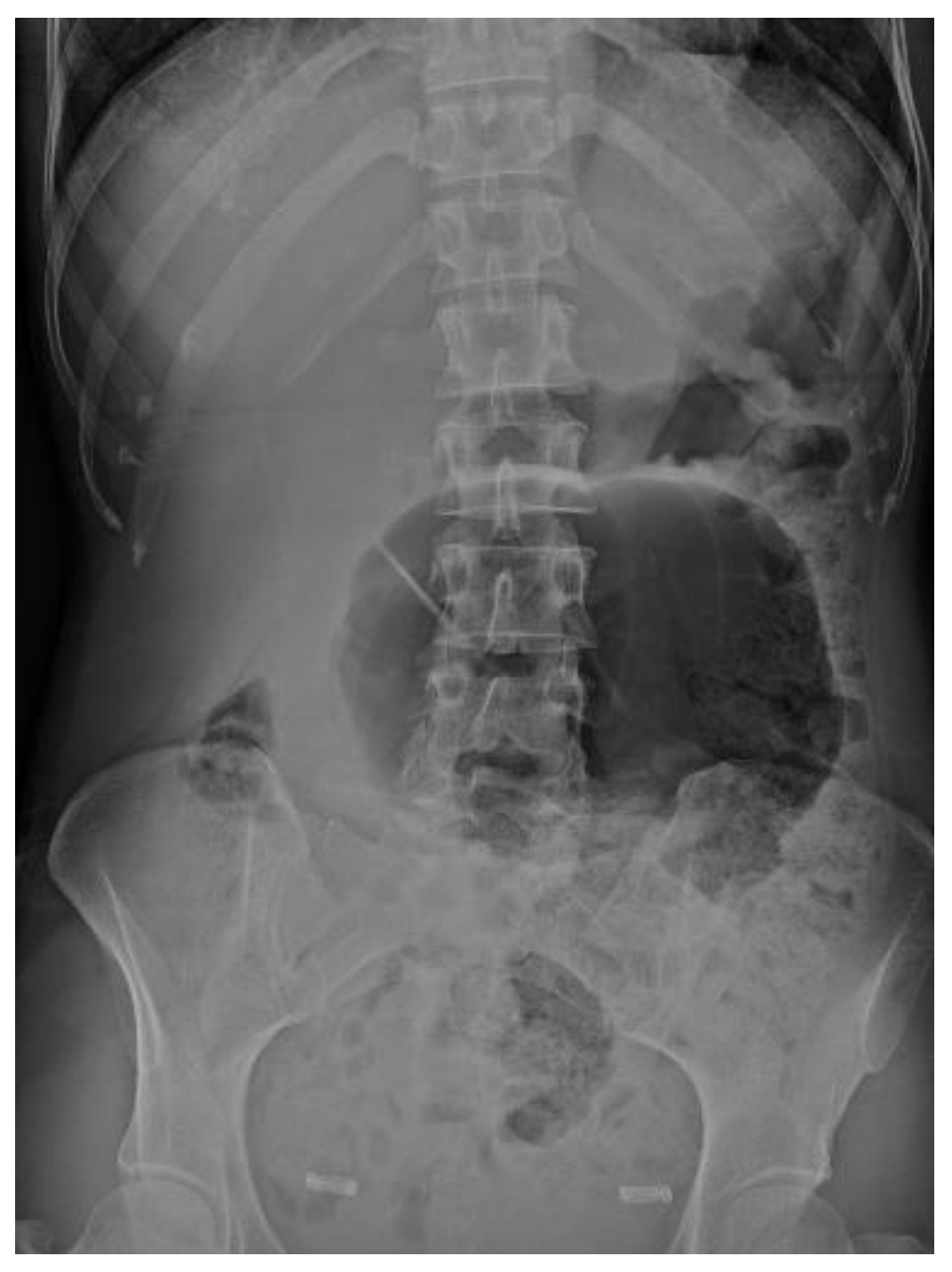
Classical features of CV in abdominal radiographs include a large bowel loop directed towards the left upper quadrant [19,20,21]. The absence of gas shadow in the distal colon and proximal small bowel dilatation are usual findings and are not specific. Similar to Rabinovici et al. [18], 45% of the abdominal radiographs performed in our series had non-specific shadows, misinterpreted as small bowel obstruction (Figure 1, Figure 2 and Figure 3 show more representative examples). Severe caecal distension, coffee-bean sign and caecal apex in the left upper quadrant are a few of the radiographic signs which have variable sensitivity and specificity to the diagnosis [6].
The sensitivity and specificity are increased with a barium enema study up to 88%, with possible demonstration of the tapering end of the volvulus site, known as the bird-beak sign [17,19]. However, it is considered tedious and risky if patients are critically ill or if there is a suspicion of gangrene or perforation [22].

4.4.2. Computerised Tomography (CT) Scan

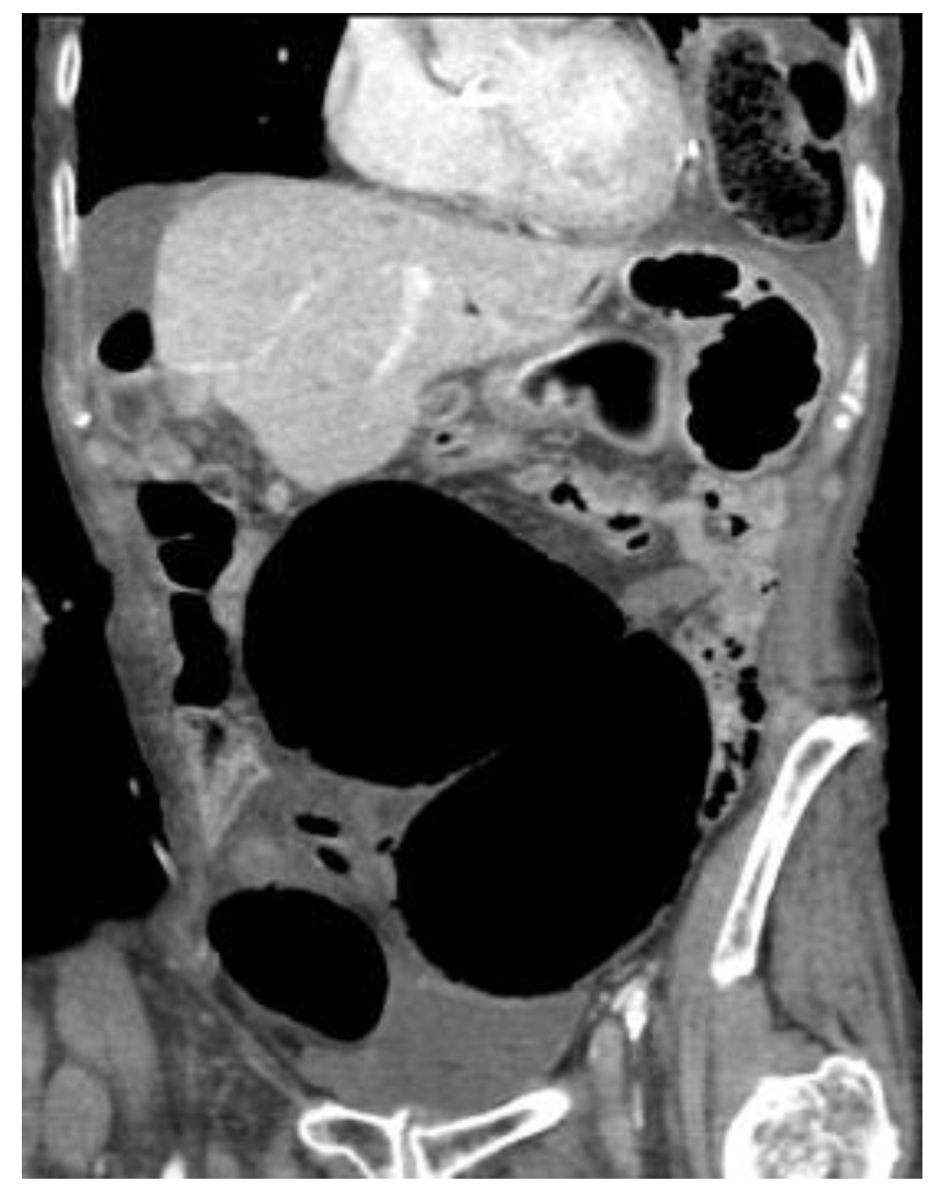
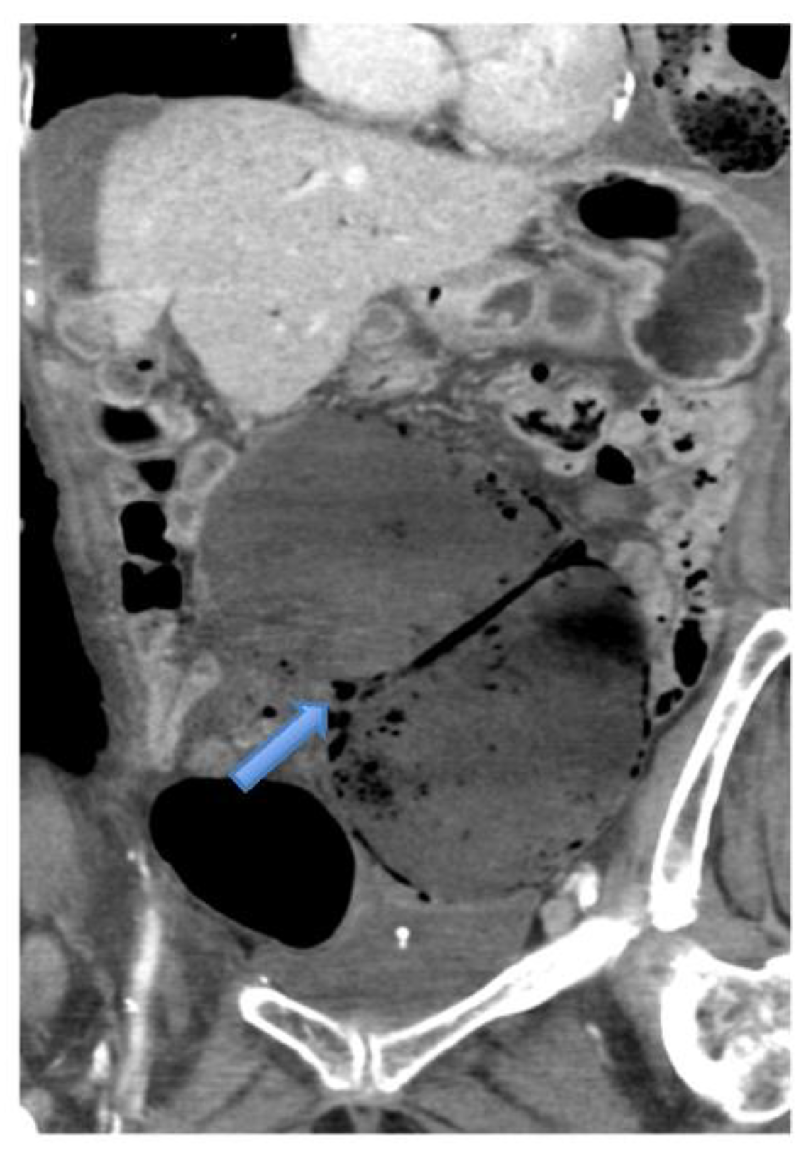
In five of our patients, the emergency physician considered a CT even before any classical radiographic examinations. It is justified because of the high sensitivity and specificity of CT which are 100% and 76.2%, respectively [23]. Our study finding of diagnostic features (Figure 4, Figure 5, Figure 6, Figure 7, Figure 8, Figure 9 and Figure 10) concurs with signs including one or two transition points, X-marks-the-spot sign, whirl sign, split-wall sign, hugely dilated caecum, dilated proximal small bowel and decompressed distal large bowel, which are all described as CT findings with variable sensitivity, specificity, and predictive values with some of them approaching near 100% [24].

4.5. Management

Only one frail patient in our series was managed conservatively. Non-surgical management such as barium enema [25], insufflation [3,6,26] and colonoscopic detorsion are fraught with low success rate and high recurrence rate [15,20]. Highly selected patients with an absence of signs of ischaemia and perforation subjected to colonoscopic detorsion resulted in a 33% success rate [27]. However, one or the other forms of urgent or semi-elective resectional procedures were later undertaken [27]. A high potential for ischaemia in volvulus warrants an earlier surgical intervention instead of a trial of colonoscopic or alternative procedures. In addition, detorsion does not rectify the pathology behind torsion [28].
Surgical procedures can be divided into non-resectional and resectional procedures accessed by either laparoscopy or laparotomy. Many choose open against laparoscopic access in anticipation of a narrow operating space secondary to a hugely dilated bowel segment. Some authors see laparoscopy as impractical [29]. Nonetheless, laparoscopic management has been increasingly reported in recent years, and when successfully managed, patients get all the benefits of minimally invasive surgery.
Detorsion without any fixation procedure has recurrence rates reaching up to 75% [15], and there are no indications to recommend.
Caecostomy is indicated in very high-risk patients with viable caecum [30] but may result in considerable high mortality [31] and morbidity owing to potential necrosis, intraperitoneal faecal leakage, or fistula formation.
Caecopexy and caeco-colopexy [27,28,32,33] involve the use of non-absorbable sutures, peritoneal flap, or mesh to fix the mobile segment of the caecum and right colon. Tissue viability and absence of thinned out distended caecum are some prerequisites. The reported success is variable with complication, mortality, and recurrence rates of 15%, 10% and 13%, respectively [20], and these are currently not favoured in comparison to resectional procedures.
Resectional procedures, as in all our operated patients, with either primary anastomosis or ileostomy and mucous fistula with delayed reversal are more favoured [16,26,28]. Resectional procedures obviate the pathology, and the chance of recurrence is theoretically none. The decision to resect is obvious in cases of frank gangrene or doubtful viability or irreversible signs of ischaemia [2,18]. A limited or standard right hemicolectomy is usually performed, with oncological resection only if a suspicious lesion is palpated [34].
A primary anastomosis can be undertaken after evaluation of the haemodynamic status and overall health of the patient. Morbidity, including anastomotic leakage following primary anastomosis, is comparable to other emergency procedures and is reported in up to 4% of patients. In contrast, we experienced anastomotic leakage in two of our patients (13%). Fortunately, one was a minor leak, which was managed conservatively.
Overall reported mortality in patients undergoing resection for caecal volvulus is between 0 to 32% [18].
However, because of recent advances and improvements in the safety of anaesthesia and critical care that patients routinely receive after the operation, the mortality is secondary to co-morbidities and continues to decrease.

5. Learning Points/Highlights

  • Variable clinical presentation in different population
  • CT scan confirms diagnosis in >90%
  • Typical Radiological Signs are described
  • Representative X-ray and CT images presented
  • Resectional procedures are favoured
  • Secondary pathology as cancer might be the cause
  • Significant morbidity expected but mortality is declining.

6. Limitations

We acknowledge that this is a retrospective review and intended to be of descriptive nature. The small-sized sample will limit drawing any generalizable conclusions. Similarly, the presentation and recommendations on the choice of investigation might vary in other populations and resource-limited settings.

7. Conclusions

Caecal volvulus is an uncommon cause of large bowel obstruction, and the most appropriate management is still debatable. Preoperative CT scan aids in definitive diagnosis. In acute settings, appropriate resuscitation followed by resectional procedures is the most favoured management. Laparoscopic access should be used preferentially wherever expertise and patient status allow. Our population is more aged, and the co-morbidities correlate with postoperative morbidity. However, judicious use of imaging ancillaries, a streamlined management pathway and a team effort in care can together decrease mortality and morbidity.

Author Contributions

A.P., A.C., S.R., J.Y.C., A.K.S. have given final approval of the version to be published. A.P.—concept and design of the study; collected, interpreted, and analysed data; performed a detailed literature review, drafted the first manuscript, and revised it following feedback. A.C.—critically reviewed and provided intellectual contributions, Revised the manuscript. S.R.—collected, interpreted, and analysed data; reviewed literature and helped finalise the manuscript. J.Y.C.—collection and interpretation of data, review of all radiological investigations and interpretation of CT images and signs; selection of typical representative images for publication, critically reviewed the manuscript and provided intellectual contributions. A.K.S.—supervised the project from design to final manuscript preparations, provided critical review, contributed to re-writing of the manuscript. All authors have read and agreed to the published version of the manuscript.

Funding

This research received no external funding.

Institutional Review Board Statement

The study was conducted in accordance with the Declaration of Helsinki, and approved by Research and Innovation Department, East Kent Hospitals University NHS Foundation Trust (R&D Ref 2022/GAP/07).

Informed Consent Statement

Patient consent was waived due to retrospective review and non-interventional nature of the study.

Data Availability Statement

The data will be made available upon request by the corresponding author after clearance from the R&I department.

Conflicts of Interest

The article has been reviewed and agreed by all the authors and declare no conflicts of interest. We received no funding for this study.

References

  1. The Merriam-Webster.com Dictionary. Merriam-Webster Inc. Available online: http://www.merriam-webster.com/dictionary/volvulus (accessed on 14 July 2020).
  2. Gingold, D.; Murrell, Z. Management of colonic volvulus. Clin. Colon Rectal Surg. 2012, 25, 236–244. [Google Scholar] [CrossRef] [PubMed] [Green Version]
  3. Ballantyne, G.H.; Brandner, M.D.; Beart, R.W., Jr.; Ilstrup, D.M. Volvulus of the colon. Incidence and mortality. Ann. Surg. 1985, 202, 83–92. [Google Scholar] [CrossRef] [PubMed]
  4. Horesh, N.; Nadler, R.; Pery, R.; Gravetz, A.; Amitai, M.M.; Rosin, D. Surgical Treatment of Caecal Volvulus. Clin. Surg. 2017, 2, 1346. [Google Scholar]
  5. Consorti, E.T.; Liu, T.H. Diagnosis and treatment of caecal volvulus. Postgrad. Med. J. 2005, 81, 772–776. [Google Scholar] [CrossRef] [PubMed]
  6. Mansoor, K.; Al Hamidi, S.; Khan, A.M.; Samujh, R. Rare case of pediatric cecal volvulus. J. Indian Assoc. Pediatr. Surg. 2009, 14, 110–112. [Google Scholar] [CrossRef] [PubMed]
  7. Shehata, A.E.; Helal, M.A.; Ibrahim, E.A.; Magdy, B.; El Seoudy, M.; Shaban, M.; Taher, H. Cecal volvulus in a child with congenital dilated cardiomyopathy: A case report. Int. J. Surg. Case Rep. 2020, 66, 30–32. [Google Scholar] [CrossRef]
  8. Katoh, T.; Shigemori, T.; Fukaya, R.; Suzuki, H. Cecal volvulus: Report of a case and review of Japanese literature. World J. Gastroenterol. WJG 2009, 15, 2547–2549. [Google Scholar] [CrossRef]
  9. Gupta, S.; Gupta, S.K. Acute caecal volvulus: Report of 22 cases and review of literature. Ital. J. Gastroenterol. 1993, 25, 380–384. [Google Scholar]
  10. Khaniya, S.; Shakya, V.C.; Koirala, R.; Pokharel, K.; Regmi, R.; Adhikary, S.; Agrawal, C.S. Caecal volvulus: A twisted tale. Trop. Doctor. 2010, 40, 244–246. [Google Scholar] [CrossRef]
  11. Neil, D.A.; Reasebeck, P.G.; Reasbeck, J.C.; Effeney, D.J. Caecal volvulus: Ten-year experience in an Australian teaching hospital. Ann. R. Coll. Surg. Engl. 1987, 69, 283–285. [Google Scholar]
  12. Madiba, T.E.; Thomson, S.R. The management of cecal volvulus. Dis. Colon Rectum 2002, 45, 264–267. [Google Scholar] [CrossRef] [PubMed]
  13. Wolfer, J.A. Volvulus of the cecum. Surg. Gynecol. Obstet. 1942, 74, 882–894. [Google Scholar]
  14. Donhauser, J.L.; Atwell, S. Volvulus of The Cecum: With a Review of One Hundred Cases in the Literature and a Report of Six New Cases. Arch. Surg. 1949, 58, 129–148. [Google Scholar] [CrossRef]
  15. McIntosh, S.A.; Ravichandran, D.; Wilmink, A.B.M.; Baker, A.; Purushotham, A.D. Cecal Volvulus Occurring after Laparoscopic Appendectomy. JSLS 2001, 5, 317–318. [Google Scholar]
  16. Caecal Volvulus Following Left-Side Laparoscopic Retroperitoneal Nephroureterectomy|BMJ Case Reports. Available online: https://casereports.bmj.com/content/12/7/e228878 (accessed on 15 July 2020).
  17. Rakinic, J. The ASCRS Textbook of Colon and Rectal Surgery, 2nd ed.; Beck, D.E., Roberts, P.L., Saclaride, T.J., Eds.; Springer: New York, NY, USA, 2011; pp. 395–406. [Google Scholar]
  18. Rabinovici, R.; Simansky, D.A.; Kaplan, O.; Mavor, E.; Manny, J. Cecal volvulus. Dis. Colon Rectum 1990, 33, 765–769. [Google Scholar] [CrossRef]
  19. Figiel, L.S.; Figiel, S.J. Volvulus of the cecum and ascending colon. Radiology 1953, 61, 496–515. [Google Scholar] [CrossRef]
  20. Young, W.S. Further radiological observations in caecal volvulus. Clin. Radiol. 1980, 31, 479–483. [Google Scholar] [CrossRef]
  21. Anderson, J.R.; Mills, J.O. Caecal volvulus: A frequently missed diagnosis? Clin. Radiol. 1984, 35, 65–69. [Google Scholar] [CrossRef]
  22. Friedman, J.D.; Odland, M.D.; Bubrick, M.P. Experience with colonic volvulus. Dis. Colon Rectum 1989, 32, 409–416. [Google Scholar] [CrossRef]
  23. Dane, B.; Hindman, N.; Johnson, E.; Rosenkrantz, A.B. Utility of CT Findings in the Diagnosis of Cecal Volvulus. AJR Am. J. Roentgenol. 2017, 209, 762–766. [Google Scholar] [CrossRef]
  24. Rosenblat, J.M.; Rozenblit, A.M.; Wolf, E.L.; DuBrow, R.A.; Den, E.I.; Levsky, J.M. Findings of Cecal Volvulus at CT. Radiology 2010, 256, 169–175. [Google Scholar] [CrossRef]
  25. O’Mara, C.S.; Wilson, T.H.; Stonesifer, G.L.; Cameron, J.L. Cecal volvulus: Analysis of 50 patients with long-term follow-up. Curr. Surg. 1980, 37, 132–136. [Google Scholar] [CrossRef]
  26. Anderson, J.R.; Welch, G.H. Acute volvulus of the right colon: An analysis of 69 patients. World J. Surg. 1986, 10, 336–342. [Google Scholar] [CrossRef] [PubMed]
  27. Renzulli, P.; Maurer, C.A.; Netzer, P.; Büchler, M.W. Preoperative colonoscopic derotation is beneficial in acute colonic volvulus. Dig. Surg. 2002, 19, 223–229. [Google Scholar] [CrossRef]
  28. Tirol, F.T. Cecocolic Torsion: Classification, Pathogenesis, and Treatment. JSLS 2005, 9, 328–334. [Google Scholar]
  29. Bauman, Z.M.; Evans, C.H. Volvulus. Surg. Clin. N. Am. 2018, 98, 973–993. [Google Scholar] [CrossRef]
  30. Ruiz-Tovar, J.; Calero García, P.; Morales Castiñeiras, V.; Martínez Molina, E. Caecal volvulus: Presentation of 18 cases and review of literature. Cir. Esp. 2009, 85, 110–113. [Google Scholar] [CrossRef]
  31. Ostergaard, E.; Halvorsen, J.F. Volvulus of the caecum. An evaluation of various surgical procedures. Acta Chir. Scand. 1990, 156, 629–631. [Google Scholar]
  32. Majeski, J. Operative therapy for cecal volvulus combining resection with colopexy. Am. J. Surg. 2005, 189, 211–213. [Google Scholar] [CrossRef]
  33. Howard, R.S.; Catto, J. Cecal volvulus: A case for non-resectional therapy. Arch. Surg. 1980, 115, 273–277. [Google Scholar] [CrossRef]
  34. Tuech, J.J.; Becouarn, G.; Cattan, F.; Arnaud, J.P. Volvulus of the right colon. Plea for right hemicolectomy. Apropos of a series of 23 cases. J. Chir. 1996, 133, 267–269. [Google Scholar]
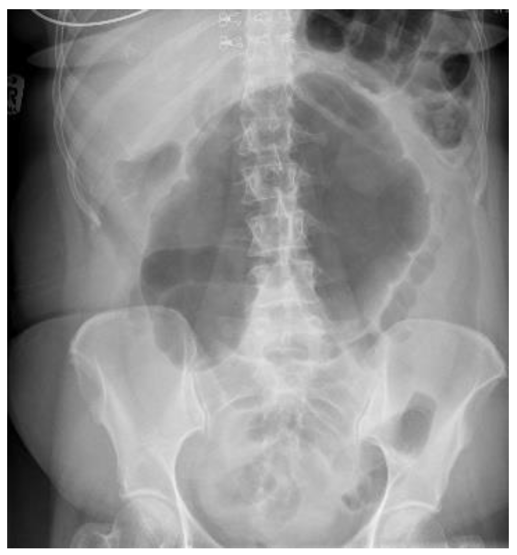
Figure 1. Abdominal radiograph again demonstrating classical features of CV. The patient was 15 days postoperative laparoscopic tubal ligation and surgical clips can be seen bilaterally in the pelvis.
Surgeries 03 00010 g001
Figure 2. Abdominal radiograph again demonstrating classical features of CV. The patient was 15 days postoperative laparoscopic tubal ligation and surgical clips can be seen bilaterally in the pelvis.
Surgeries 03 00010 g002
Figure 3. CT topogram demonstrating classical features of a caecal volvulus.
Surgeries 03 00010 g003
Figure 4. CT axial image demonstrating a grossly distended caecum with diameter > 10 cm.
Surgeries 03 00010 g004
Figure 5. CT coronal image showing a grossly dilated caecum which measures more than 10 cm in diameter.
Surgeries 03 00010 g005
Figure 6. CT coronal image demonstrating the X-marks-the-spot sign. Double transition points have been marked (hand-drawn yellow and red arrows). There is also a whirl sign (blue arrow).
Surgeries 03 00010 g006
Figure 7. CT coronal image demonstrating a central appendix sign where there is a dilated gas-filled appendix (blue arrow). The caecum and proximal small bowel loops are also dilated.
Surgeries 03 00010 g007
Figure 8. CT Axial image demonstrating the ‘split-wall sign’ (blue arrow).
Surgeries 03 00010 g008
Figure 9. CT coronal image demonstrating the split-wall sign (blue arrow) which refers to the mesenteric fat in between the bowel walls.
Surgeries 03 00010 g009
Figure 10. CT coronal image showing a typical whirl sign (blue arrow).
Surgeries 03 00010 g010
Table 1. Demographics.
VariablesNumber, (Range or Percentage)Remarks
Total Patients16
Gender, M:F5:11
Median Age, years64 (33–80)
Table 2. Clinical characteristics.
Variables Number, (Range or Percentage) Remarks
ASA Grade
I5 (31.2%)
II3 (18.8%)
III7 (43.7%)
IV1 (6.3%)
Co-morbidities Hypertension, ischaemic heart disease, diabetes mellitus, COPD, and depression.
None5 (31.2%)
Single or Multiple11 (68.8%)
Use of psychotropic drugs 6 (37.5%)
Abdomen pain, distension, and vomiting 16 (100%)
Previous abdominal surgery Nephrectomy, laparoscopic sterilisation, sigmoid colectomy, hysterectomy.
None11 (68.7%)
One3
Multiple2
History of large bowel volvulus1 (6.8%)Sigmoid resection for volvulus 4 years before this admission.
Initial diagnosis by emergency physician:
Large bowel obstruction6 (37.5%)
Small bowel obstruction2 (12.5%)
Others8 (50.0%)
Acute appendicitis2
Post-op collection1
Urinary tract infection1
Viscus perforation1
Pancreatitis1
Biliary colic1
Gastroenteritis1
Table 3. Radiological investigations.
VariablesNumber, (Range or Percentage)Remarks
Abdominal radiograph11 (68.75%)Five patients had CT as the only imaging.
Non-specific bowel loops/SBO5 (46%)
Classical single loop of large bowel6 (54%)
CT Abdomen and pelvis15One patient operated without CT.
CV not described1
CV correctly identified14 (93.3%)
Caecal diameter >10 cm10 (67%)
Whirl sign12 (80%)
Split-wall sign13 (86.6%)
X-marks-the-spot sign14 (93.3%)
Double transition point13 (86.6%)
Ileocaecal twist 13 (86.6%)
Central appendix11 (73.3%)
Table 4. Peri-operative findings.
VariablesNumber, (Range or Percentage)Remarks
Intervention
Conservative Management1 (6.25%)
Surgery15 (93.7%)
Laparoscopic1 (6.25%)
Open14 (93.3%)
Intraoperative Findings
Gangrenous Caecum1 (6.7%)
Patchy ischaemia12 (80%)
No ischaemia2
Procedure
Resection and primary anastomosis14 (93.3%)
Resection, ileostomy, and mucous fistula1
Complications
30-day mortality in conservatively managed1 (100%)
30-day mortality in operated patientsNone
Uneventful recovery3 (20%)
Morbidity12 (80%)
Postoperative complications (Clavien–Dindo)
I 5
II3
III7
IV 1
Major Morbidity
Anastomosis Leak 2
CT guided Drainage 1
Re-operation and ileostomy1
Reoperation 3
Anastomotic leak 1
Fascial Dehiscence 1
Bowel Obstruction/suspicion of ischaemia 1
Median Length of stay (days)10 (1–49)
Histopathology
Low grade appendiceal mucinous neoplasm (LAMN)1
Tubular Adenoma 1
Adenocarcinoma 1
No additional pathology12
Median follow-up20 months (6–31)
Publisher’s Note: MDPI stays neutral with regard to jurisdictional claims in published maps and institutional affiliations.