Case Report

First Successful Fertility Preservation Using Oocyte Vitrification in Patient with Autoimmune Polyendocrinopathy-Candidiasis-Ectodermal Dystrophy

1
Department of Obstetrics and Gynecology, Juntendo University Graduate School of Medicine, Tokyo 113-8421, Japan
2
Rose Ladies Clinic, Tokyo 158-0082, Japan
3
Department of Obstetrics and Gynecology, Juntendo University Faculty of Medicine, 2-1-1 Hongo, Bunkyo-ku, Tokyo 113-8421, Japan
*
Author to whom correspondence should be addressed.
Endocrines 2025, 6(3), 31; https://doi.org/10.3390/endocrines6030031
Submission received: 2 April 2025 / Revised: 11 June 2025 / Accepted: 20 June 2025 / Published: 1 July 2025

Abstract

Background/Objectives: Autoimmune polyendocrinopathy-candidiasis-ectodermal dystrophy (APECED) is a rare autoimmune disorder caused by mutations in the AIRE gene. Approximately 60% of affected females develop premature ovarian insufficiency (POI) by age 30, often most commonly due to steroidogenic autoantibodies. Although APECED is typically diagnosed in childhood, its reproductive implications are underrecognized. This study reports a case of successful fertility preservation in an adult woman with APECED and reviews the relevant literature. Methods: We describe the clinical course of a 37-year-old woman with genetically confirmed APECED who underwent ovarian stimulation for fertility preservation. A comprehensive PubMed search was also conducted to identify English-language case reports on fertility preservation in APECED-associated POI. Results: The patient experienced menarche at age 13, adrenal insufficiency at 14, and menstrual irregularities from age 18. Genetic analysis confirmed an AIRE mutation (NM_000383: exon 11: c.1400+1G>A). Given her relatively high anti-Müllerian hormone level, she opted for fertility preservation and underwent six cycles of ovarian stimulation, resulting in the cryopreservation of 17 mature oocytes. During ovarian stimulation, multiple follicular developments were observed, but serum E2 levels remained low. The literature review identified fewer than 20 reported cases addressing fertility preservation in APECED, highlighting its rarity and the lack of standardized management. Conclusions: Although APECED frequently leads to early POI due to impaired steroidogenesis, residual ovarian function may persist. Early assessment of ovarian reserve and timely fertility counseling are crucial, even in asymptomatic patients or those diagnosed in childhood. Reproductive planning should be integrated into the long-term care of women with APECED.

1. Introduction

Autoimmune polyendocrinopathy-candidiasis-ectodermal dystrophy (APECED), also known as autoimmune polyglandular syndrome type I (APS-I), is an extremely rare disorder caused by mutations in a single gene. The condition is characterized by a diverse range of clinical features, with the three major manifestations being chronic mucocutaneous candidiasis, hypoparathyroidism, and adrenal cortical insufficiency. Clinical diagnosis of APECED is typically established when at least two of these three primary features are present [1]. However, a definitive diagnosis requires genetic testing, which identifies mutations in the autoimmune regulator (AIRE) gene located on human chromosome 21 [2]. APECED is estimated to occur in fewer than 1 in 100,000 live births annually, with approximately 500 cases reported worldwide to date [3].
Several cohort studies have investigated the incidence of premature ovarian insufficiency (POI) and pregnancy outcomes in APECED, indicating that 40–70% of affected individuals develop POI [4,5]. A retrospective cohort study on 40 APECED patients aged 12 years and older (mean age: 37.3 years; range: 14.6–61.9 years; 16 patients were over 40 years old) between 1965 and 2018 found that 28 patients (70%) developed POI at a median age of 16 years (range: 11.3–36.5 years) [6].
In contrast, the incidence of POI in the general population is estimated at 1–3% [7], highlighting the significantly higher and earlier risk of POI in APECED patients. Consequently, fertility preservation should be considered promptly after diagnosis. To the best of our knowledge, there have been no reported cases of fertility preservation treatment for APECED patients. In this paper, we present the first documented case of successful fertility preservation through oocyte cryopreservation in a patient with APECED.

2. Case Presentation

A 37-year-old nulligravida woman presented with a complex medical history. She developed alopecia at age 10 and experienced menarche at 13. At age 14, she was diagnosed with heart failure and adrenal cortical insufficiency (Addison’s disease). From age 18 onward, she experienced menstrual irregularities and hot flashes. Hormonal testing at a local clinic (specific details unavailable) led to a diagnosis of ovarian dysfunction, prompting the initiation of hormone replacement therapy. After her elder sister was diagnosed with APECED at age 28, the patient underwent genetic testing at age 26. Whole-exome sequencing revealed the same pathogenic AIRE gene mutation (NM_000383: exon11: c.1400+1G>A) as her elder sister, confirming the diagnosis of APECED. Subsequent manifestations included mucocutaneous candidiasis at age 39. Having been counseled about her risk of ovarian failure by a previous physician, the unmarried patient expressed interest in oocyte cryopreservation for fertility preservation and was referred to our hospital.
An initial blood test conducted on day 3 of her menstrual cycle without estrogen replacement revealed a low estradiol (E2) level (35.2 pg/mL), relatively high gonadotropin levels (follicle-stimulating hormone [FSH] 5.9 mIU/mL; luteinizing hormone [LH] 11.3 mIU/mL), and a low testosterone level (<0.04 ng/mL), indicating an ovarian dysfunction phenotype. However, anti-Müllerian hormone (AMH) was 2.06 ng/mL, suggesting a relatively preserved ovarian reserve. The patient underwent four oocyte retrieval cycles using either a gonadotropin-releasing hormone (GnRH) agonist short protocol combined with FSH/human menopausal gonadotropin (hMG), or direct FSH/HMG stimulation. The decision to use a higher dose of FSH/HMG (300–375 IU) was based on the patient’s ovarian dysfunction phenotype and poor response. The representative changes in serum hormone levels during controlled ovarian stimulation are shown in Table 1 and Figure 1.
Interestingly, despite the development of multiple follicles, serum estrogen levels remained lower than expected, and progesterone levels increased in the latter half of the cycle without evidence of ovulation. Because serum estrogen levels failed to rise during the first two stimulation cycles despite multiple follicular developments, neither a GnRH agonist nor antagonist was used in the subsequent two cycles to suppress ovulation. Nevertheless, an endogenous LH surge did not occur, and oocyte retrieval was performed following the human chorionic gonadotropin (hCG) trigger. A total of 17 mature oocytes were successfully cryopreserved across four stimulation cycles (Table 2). The patient remains unmarried, continues hormone replacement therapy for ovarian dysfunction, and provided written informed consent for the publication of this case report.

3. Discussion

We report a case of fertility preservation through oocyte cryopreservation in a patient with APECED, which demonstrated atypical hormonal response during controlled ovarian stimulation. To the best of our knowledge, this is the first reported case of successful fertility preservation by oocyte cryopreservation in an APECED patient.
Although APECED is a monogenic disorder, it presents with a broad spectrum of clinical features beyond the classic triad of chronic mucocutaneous candidiasis, hypoparathyroidism, and adrenal cortical insufficiency. Additional manifestations include hypogonadism, alopecia, malabsorption syndrome, vitiligo, chronic hepatitis, and keratopathy [3,8]. This heterogeneous symptomatology reflects the essential role of the AIRE gene in establishing central immune tolerance. Central immune tolerance is a critical mechanism that prevents self-reactive T cells from entering the periphery, occurring in two stages within the thymus [9]. Precursor T cells originating in the bone marrow become mature in the thymus, where they develop T cell receptors. In the thymic cortex, positive selection takes place, where cortical thymic epithelial cells (cTECs) present self-major histocompatibility complex (MHC) molecules to develop as T cells. During this process, only T cells capable of recognizing self-MHC with appropriate affinity are selected to survive and migrate to the thymic medulla. In the medulla, negative selection occurs, where medullary thymic epithelial cells (mTECs) present peripheral tissue antigens (PTAs) via MHC molecules to T cells [9]. This process eliminates T cells that strongly react to self-antigens, ensuring self-tolerance. The AIRE protein plays a critical role in this process by promoting the expression of PTAs in mTECs [10]. Through this coordinated system, T cells that recognize self-MHC but do not react strongly to self-tissues are selected to form a functional and self-tolerant T cell repertoire [11,12].
Previous studies have shown that AIRE regulates the transcription of a wide array of PTAs derived from various tissues. Mutations in the AIRE gene lead to decreased or deficient expression of AIRE-dependent gene groups, resulting in some organ-derived PTAs not being translated and presented. Consequently, self-reactive T cells targeting certain organs cannot be eliminated in the thymus and are exported to the periphery, where they attack those organs and cause autoimmune diseases.
The phenotypic variability in APECED is highlighted by this case of two sisters with identical AIRE gene mutations. To date, over 140 different AIRE mutations have been identified [13], however no clear genotype–phenotype correlation has been shown in APECED. Despite having identical mutations, the sisters exhibited distinct clinical features: the elder sister presented with hypoparathyroidism, adrenal cortical insufficiency, and pure red cell aplasia, while the patient displayed adrenal cortical insufficiency, alopecia, and mucocutaneous candidiasis. Moreover, at age 33, the elder sister showed no follicular development and had an AMH level below detection (<0.01 ng/mL) despite multiple stimulation attempts. In contrast, the patient in this report had a higher AMH level (2.06 ng/mL) and preserved ovarian follicles, enabling successful oocyte cryopreservation.
This phenotypic divergence may be explained from AIRE’s complex epigenetic regulation of promiscuous gene expression in mTECs [14,15]. AIRE interacts with epigenetic and transcriptional factors, including histone modifiers, DNA-dependent protein kinase (DNA-PK), and the positive transcription elongation factor b (P-TEFb) complex [14]. These interactions modulate chromatin accessibility and antigen expression, contributing to observed phenotypic variability. APECED follows an autosomal recessive inheritance pattern [16]. In this case, the consanguineous marriage of the sisters’ parents likely resulted in homozygosity for the AIRE mutation, leading to disease manifestation.
In this case, a characteristic feature was observed during ovarian stimulation: estrogen levels did not increase despite the development of multiple follicles, and progesterone levels showed a slight rise in the latter half of the cycle without ovulation. In APECED patients with POI, autoantibodies are frequently produced against gonad-specific self-antigen proteins, including CYP17A1 and CYP11A1 [17,18,19]. It has been reported that the POI onset group (n = 28) had a higher prevalence of adrenal cortical insufficiency and ovarian antibodies compared to the non-POI onset group (n = 12), with rates of adrenal cortical insufficiency at 93% versus 58% (p = 0.017) and ovarian antibodies at 81% versus 30% (p = 0.003) [6]. The hormonal changes observed during ovarian stimulation in this case, characterized by persistently low testosterone levels, no increase in estrogen despite follicular development, and a rise in progesterone during the latter half phase, suggested the production of autoantibodies targeting CYP17A1.
Currently, the diagnostic criteria for POI often include hypogonadotropic hypogonadism and amenorrhea for more than four months [20]. In APECED, steroid production disorders caused by autoantibody production may lead to early menstrual irregularities, amenorrhea, and a hypogonadotropic hypogonadism pattern, resulting in a POI diagnosis even when there is a discrepancy with the remaining follicle count, as demonstrated in this case presentation. Therefore, in APECED cases diagnosed with POI, some patients may retain residual follicles despite fulfilling the diagnostic criteria.
A comprehensive literature search was conducted to identify all peer-reviewed English-language articles related to Autoimmune Polyendocrinopathy-Candidiasis-Ectodermal Dystrophy (APECED) and its association with premature ovarian insufficiency (POI). The search strategy followed PRISMA guidelines and utilized the PubMed database. The search was performed on 9 April 2025, and included all articles published from database inception until that date. The following Boolean search string was used: (“APECED” OR “autoimmune polyendocrinopathy-candidiasis-ectodermal dystrophy” OR “autoimmune polyglandular syndrome type 1” OR “APS-1”) AND (“premature ovarian insufficiency” OR “POI” OR “primary ovarian insufficiency” OR “ovarian failure” OR “premature menopause” OR “gonadal failure”). This search identified 35 articles. Of these, 13 articles (6 cohort studies and 7 case reports) were selected for analysis and are detailed in Table 3 and Table 4 [6,19,21,22,23,24,25,26,27,28,29,30,31].
Previous cohort studies on APECED have indicated that the prevalence of POI ranges from 41% to 84%, with reported onset between 11 and 39 years [6,19,21,22,23,24]. Regarding AIRE gene mutations, no mutation-specific associations with POI phenotype have been established. Two cohort studies have detailed pregnancy outcomes in APECED, comparing patients with and without POI [6,22]. A longitudinal follow-up study of 40 APECED patients aged 11 years and older from 1965 to 2018 reported that 70% developed POI, with a median age of onset at 16 years (range: 11.3–36.5 years) [6]. Among those who developed POI, 25% (7/28) became pregnant, with a miscarriage rate of 38% (3/8, including one person who experienced two miscarriages) [6]. However, half of the pregnancies in POI cases involved ovum donation, while no information was available about the number of patients who conceived through ovum donation. Thus, in patients using autologous eggs, the pregnancy rate in POI-complicated cases was 11% (or 14%), which was lower compared to the pregnancy rate of 33% (4/12) in non-POI-complicated cases. In POI-complicated cases, the live birth rate was the same (50%, 4/8) between groups using autologous eggs and ovum donation [6]. These findings suggest that egg quality in POI-complicated cases is not affected by APECED, resulting in comparable birth rates to non-POI-complicated cases. Another multicenter cohort study involving 321 APECED patients from Italy, Finland, Russia, Norway, and the United States found that 40% (17/43) were diagnosed with POI (median age 28 years, range: 13–39 years). Of these patients, 29% (5/17) achieved pregnancy, resulting in seven pregnancies; however, 71% (5/7) of these pregnancies were achieved through ovum donation. The pregnancy rate using autologous eggs was similar between POI-complicated cases (12%, 2/17), and non-POI-complicated cases over 16 years (17%, 38/223) [22]. The discrepancy in pregnancy rate in APECD patients with or without POI between these studies may be attributed to the limited number of successful pregnancies. In POI-complicated cases, miscarriage occurred in 43% of pregnancies (3/7). In non-POI-complicated cases, the reported pregnancy complications included miscarriage in 14% (11/76), stillbirth in 3% (2/76), preterm birth in 7% (5/76), gestational diabetes mellitus in 1% (1/76), and placental complications in 4% (3/76). Although the miscarriage rate appears higher in POI-complicated cases than in non-POI-complicated cases, it is difficult to draw definitive conclusions due to the limited number of cases.
In general, the number of primordial follicles remains around 1000 at the time of menopause, and continues to decline with age, typically becoming depleted within 5 to 10 years after menopause [32,33]. However, in the cohort study, two patients conceived naturally with autologous oocytes, 3 and 10 years after POI diagnosis, respectively [22]. These findings suggest that APECED patients with POI may retain residual ovarian follicles capable of supporting pregnancy. In our case, the patient‘s AMH level was 2.06 ng/mL, allowing for oocyte preservation. A previous report also described that among 19 APECED patients, 15 patients had AMH levels below the detection limit, while 3 patients had detectable AMH levels (0.27, 0.04 and 0.89 ng/mL) at the time of POI diagnosis [21]. These results underscore the heterogeneity of ovarian function in APECED patients with POI, highlighting the need for individualized fertility assessment and management.
Pregnancy outcomes in APECED patients are generally favorable, as shown in Table 1. A study of 43 patients documented 83 pregnancies, including five through egg donation. Of these pregnancies, 60 resulted in deliveries (72%), with 54 full-term deliveries and 6 preterm deliveries. Of the live births, the majority (53/60) were natural pregnancies, while three resulted from fertility treatments and four from ovum donation. Notably, the miscarriage rate was 17% (14 cases) [22], which is comparable to that in healthy women. During pregnancy, APECED manifestations remained largely stable. Only one case developed primary adrenal insufficiency, which led to an adrenal crisis and stillbirth [22]. However, there is a report demonstrating the increased early mortality risks due to endocrine abnormalities, tumors, and infections in APECED patients [34]. Therefore, careful monitoring and management of complications is essential both before and during pregnancy.
When considering fertility preservation in APECED patients, it is important to evaluate the genetic implications for future generations. As APECED is an autosomal recessive disorder, the impact on the next generation requires careful consideration. Children born to a parent with APECED will invariably be carriers of the mutated gene. However, the likelihood of disease development in these children is extremely low, except in cases of consanguineous marriage. The likelihood of a partner carrying the same gene mutation is very small in the general population, further reducing the risk of the child inheriting two copies of the mutated gene and developing APECED.
Clinicians should actively suspect APECED in patients with early-onset POI who also present with one or more of the three major manifestations of the disease, and recommend genetic testing accordingly. Since DOR and POI are progressive conditions, it is essential to monitor ovarian reserve over time and to initiate fertility preservation at the early onset of ovarian dysfunction. For patients with DOR or POI caused by APECED, fertility preservation should ideally be performed during periods when disease-related systemic complications are stable, and only after a thorough evaluation of the patient’s overall health condition. After oocyte cryopreservation, long-term follow-up is required until patients utilize their oocytes for IVF-ET treatment. Given that patients with APECED who are eligible for fertility preservation are often young adolescents, it is crucial to obtain informed assent from the patients as well as informed consent from the parents, following comprehensive counseling. Although current evidence suggests that long-term cryopreservation does not impair oocyte quality [35], it is generally recommended that oocytes be used within the reproductive age range, taking perinatal prognosis into consideration.

4. Conclusions

APECED patients frequently experience menstrual irregularities and amenorrhea due to impaired steroidogenesis, though some retain a sufficient ovarian follicle pool to allow for fertility preservation. The disease typically presents with mucocutaneous candidiasis in childhood, hypoparathyroidism around age 10, and adrenal cortical insufficiency during puberty, often leading to a definitive diagnosis by other specialists during childhood or adolescence. Given the significantly higher prevalence of POI in APECED patients compared to the general population, early referral to specialists in obstetrics, gynecology, and reproductive medicine is strongly recommended. This enables timely assessment of ovarian reserve and consideration of fertility preservation.

Author Contributions

Y.T.: Data curation, Formal analysis, Writing—original draft. B.I.: Resources. K.K.: Conceptualization, Funding acquisition, Methodology, Project administration, Validation, Writing—review and editing. All authors have read and agreed to the published version of the manuscript.

Funding

This research was funded by The Japan Society for the Promotion of Science (JSPS), Scientific Research B (19H03801, 23H03048), and Challenging Exploratory Research (18K19624, 23K18333) (to K.K.).

Institutional Review Board Statement

This study was conducted in accordance with the Declaration of Helsinki and approved by the Institutional Ethics Committee of Rose Ladies Clinic (Approval Code: 43043, Approval Date: 31 May 2018).

Informed Consent Statement

Written informed consent has been obtained from the patient to publish this paper.

Data Availability Statement

The datasets generated during and/or analyzed during the current study are available from the corresponding author on reasonable request.

Acknowledgments

The authors thank Aaron JW Hsueh (Stanford University School of Medicine, Stanford, CA, USA) for his critical review of the manuscript.

Conflicts of Interest

The authors declare no conflicts of interest.

Abbreviations

The following abbreviations are used in this manuscript:
APECEDAutoimmune Poly-Endocrinopathy-Candidiasis-Ectodermal Dystrophy
APS-IAutoimmune Polyglandular Syndrome Type I
AIRE geneAutoimmune Regulator Gene
POIPremature Ovarian Insufficiency
E2Estradiol
FSHFollicle-Stimulating hormone
LHLuteinizing Hormone
HRTHormone Replacement Therapy
GnRHGonadotropin-Releasing Hormone
hMGHuman Menopausal Gonadotropin
hCGHuman Chorionic Gonadotropin
PTAPeripheral Tissue Antigen
MHCMajor Histocompatibility Complex
TCRT Cell Receptor
cTECCortical Thymic Epithelial Cell
mTECMedullary Thymic Epithelial Cell
P-TEFbPositive Transcription Elongation Factor b
DNA-PKDNA-Dependent Protein Kinase

References

  1. Akirav, E.M.; Ruddle, N.H.; Herold, K.C. The Role of Aire in Human Autoimmune Disease. Nat. Rev. Endocrinol. 2011, 7, 25–33. [Google Scholar] [CrossRef] [PubMed]
  2. Nagamine, K.; Peterson, P.; Scott, H.S.; Kudoh, J.; Minoshima, S.; Heino, M.; Krohn, K.J.; Lalioti, M.D.; Mullis, P.E.; Antonarakis, S.E.; et al. Positional Cloning of the Apeced Gene. Nat. Genet. 1997, 17, 393–398. [Google Scholar] [CrossRef] [PubMed]
  3. Husebye, E.S.; Anderson, M.S.; Kämpe, O. Autoimmune Polyendocrine Syndromes. N. Engl. J. Med. 2018, 378, 1132–1141. [Google Scholar] [CrossRef] [PubMed]
  4. Szeliga, A.; Calik-Ksepka, A.; Maciejewska-Jeske, M.; Grymowicz, M.; Smolarczyk, K.; Kostrzak, A.; Smolarczyk, R.; Rudnicka, E.; Meczekalski, B. Autoimmune Diseases in Patients with Premature Ovarian Insufficiency-Our Current State of Knowledge. Int. J. Mol. Sci. 2021, 22, 2594. [Google Scholar] [CrossRef]
  5. Komorowska, B. Autoimmune Premature Ovarian Failure. Prz. Menopauzalny 2016, 15, 210–214. [Google Scholar] [CrossRef]
  6. Saari, V.; Holopainen, E.; Mäkitie, O.; Laakso, S. Pubertal Development and Premature Ovarian Insufficiency in Patients with Apeced. Eur. J. Endocrinol. 2020, 183, 513–520. [Google Scholar] [CrossRef]
  7. Li, M.; Zhu, Y.; Wei, J.; Chen, L.; Chen, S.; Lai, D. The Global Prevalence of Premature Ovarian Insufficiency: A Systematic Review and Meta-Analysis. Climacteric 2023, 26, 95–102. [Google Scholar] [CrossRef]
  8. Perheentupa, J. Autoimmune Polyendocrinopathy-Candidiasis-Ectodermal Dystrophy. J. Clin. Endocrinol. Metab. 2006, 91, 2843–2850. [Google Scholar] [CrossRef]
  9. Takaba, H.; Takayanagi, H. The Mechanisms of T Cell Selection in the Thymus. Trends Immunol. 2017, 38, 805–816. [Google Scholar] [CrossRef]
  10. Passos, G.A.; Speck-Hernandez, C.A.; Assis, A.F.; Mendes-da-Cruz, D.A. Update on Aire and Thymic Negative Selection. Immunology 2018, 153, 10–20. [Google Scholar] [CrossRef]
  11. Derbinski, J.; Gäbler, J.; Brors, B.; Tierling, S.; Jonnakuty, S.; Hergenhahn, M.; Peltonen, L.; Walter, J.; Kyewski, B. Promiscuous Gene Expression in Thymic Epithelial Cells Is Regulated at Multiple Levels. J. Exp. Med. 2005, 202, 33–45. [Google Scholar] [CrossRef] [PubMed]
  12. Mathis, D.; Benoist, C. A Decade of Aire. Nat. Rev. Immunol. 2007, 7, 645–650. [Google Scholar] [CrossRef] [PubMed]
  13. Stenson, P.D.; Mort, M.; Ball, E.V.; Chapman, M.; Evans, K.; Azevedo, L.; Hayden, M.; Heywood, S.; Millar, D.S.; Phillips, A.D.; et al. The Human Gene Mutation Database (Hgmd(®)): Optimizing Its Use in a Clinical Diagnostic or Research Setting. Hum. Genet. 2020, 139, 1197–1207. [Google Scholar] [CrossRef]
  14. Akiyama, T.; Shinzawa, M.; Qin, J.; Akiyama, N. Regulations of Gene Expression in Medullary Thymic Epithelial Cells Required for Preventing the Onset of Autoimmune Diseases. Front. Immunol. 2013, 4, 249. [Google Scholar] [CrossRef]
  15. Ucar, O.; Rattay, K. Promiscuous Gene Expression in the Thymus: A Matter of Epigenetics, Mirna, and More? Front. Immunol. 2015, 6, 93. [Google Scholar] [CrossRef]
  16. Skrabic, V.; Skrabic, I.; Skrabic, R.; Roje, B.; Simunovic, M. Clinical Characteristics in the Longitudinal Follow-up of Apeced Syndrome in Southern Croatia-Case Series. Genes 2022, 13, 558. [Google Scholar] [CrossRef]
  17. De Martino, L.; Capalbo, D.; Improda, N.; D’Elia, F.; Di Mase, R.; D’Assante, R.; D’Acunzo, I.; Pignata, C.; Salerno, M. APECED: A Paradigm of Complex Interactions between Genetic Background and Susceptibility Factors. Front. Immunol. 2013, 4, 331. [Google Scholar] [CrossRef]
  18. Capalbo, D.; Improda, N.; Esposito, A.; De Martino, L.; Barbieri, F.; Betterle, C.; Pignata, C.; Salerno, M. Autoimmune Polyendocrinopathy-Candidiasis-Ectodermal Dystrophy from the Pediatric Perspective. J. Endocrinol. Investig. 2013, 36, 903–912. [Google Scholar]
  19. Garelli, S.; Dalla Costa, M.; Sabbadin, C.; Barollo, S.; Rubin, B.; Scarpa, R.; Masiero, S.; Fierabracci, A.; Bizzarri, C.; Crinò, A.; et al. Autoimmune Polyendocrine Syndrome Type 1: An Italian Survey on 158 Patients. J. Endocrinol. Investig. 2021, 44, 2493–2510. [Google Scholar] [CrossRef]
  20. De Vos, M.; Devroey, P.; Fauser, B.C. Primary Ovarian Insufficiency. Lancet 2010, 376, 911–921. [Google Scholar] [CrossRef]
  21. Saari, V.; Laakso, S.; Tiitinen, A.; Mäkitie, O.; Holopainen, E. Endocrine Disorders and Genital Infections Impair Gynecological Health in Apeced (APS-1). Front. Endocrinol. 2021, 12, 784195. [Google Scholar] [CrossRef] [PubMed]
  22. Laakso, S.; Holopainen, E.; Betterle, C.; Saari, V.; Vogt, E.; Schmitt, M.M.; Winer, K.K.; Kareva, M.; Sabbadin, C.; Husebye, E.S.; et al. Pregnancy Outcome in Women with Apeced (APS-1): A Multicenter Study on 43 Females with 83 Pregnancies. J. Clin. Endocrinol. Metab. 2022, 107, e528–e537. [Google Scholar] [CrossRef] [PubMed]
  23. Ahonen, P.; Myllärniemi, S.; Sipilä, I.; Perheentupa, J. Clinical Variation of Autoimmune Polyendocrinopathy-Candidiasis-Ectodermal Dystrophy (Apeced) in a Series of 68 Patients. N. Engl. J. Med. 1990, 322, 1829–1836. [Google Scholar] [CrossRef]
  24. Reato, G.; Morlin, L.; Chen, S.; Furmaniak, J.; Smith, B.R.; Masiero, S.; Albergoni, M.P.; Cervato, S.; Zanchetta, R.; Betterle, C. Premature Ovarian Failure in Patients with Autoimmune Addison’s Disease: Clinical, Genetic, and Immunological Evaluation. J. Clin. Endocrinol. Metab. 2011, 96, e1255–e1261. [Google Scholar] [CrossRef] [PubMed]
  25. Pellegrino, M.; Bellacchio, E.; Dhamo, R.; Frasca, F.; Betterle, C.; Fierabracci, A. A Novel Homozygous Mutation of the Aire Gene in an Apeced Patient from Pakistan: Case Report and Review of the Literature. Front. Immunol. 2018, 9, 1835. [Google Scholar] [CrossRef]
  26. Alkhammash, S.; Saumet, J.; Genest, G. Infertility and Pregnancy in Patients with Autoimmune Polyendocrinopathy-Candidiasis-Ectodermal Dystrophy: More Than Just Primary Ovarian Failure? Am. J. Reprod. Immunol. 2019, 82, e13169. [Google Scholar] [CrossRef]
  27. Alrufaidi, A.M.; Alnashery, M.M.; Alghanimi, A.A.; Elmansor, R.E.A.; Ghazy, R.M. An Uncommon Presentation of Autoimmune Polyglandular Syndrome Type 1 (APS-1)—A Case Report. Clin. Case Rep. 2024, 12, e9015. [Google Scholar] [CrossRef]
  28. Zheng, W.B.; Li, L.J.; Zhao, D.C.; Wang, O.; Jiang, Y.; Xia, W.B.; Li, M. A Novel Variant in Aire Causing a Rare, Non-Classical Autoimmune Polyendocrine Syndrome Type 1. Mol. Med. Rep. 2020, 22, 1285–1294. [Google Scholar] [CrossRef]
  29. Ruan, J.; Wang, X.; Jiang, X.; Chen, M. Acquired Pure Red Cell Aplasia and T Cell Large Granular Lymphocytic Leukaemia in Patients with Autoimmune Polyglandular Syndrome Type 1. BMC Med. Genom. 2021, 14, 22. [Google Scholar] [CrossRef]
  30. Ward, L.; Paquette, J.; Seidman, E.; Huot, C.; Alvarez, F.; Crock, P.; Delvin, E.; Kämpe, O.; Deal, C. Severe Autoimmune Polyendocrinopathy-Candidiasis-Ectodermal Dystrophy in an Adolescent Girl with a Novel Aire Mutation: Response to Immunosuppressive Therapy. J. Clin. Endocrinol. Metab. 1999, 84, 844–852. [Google Scholar] [CrossRef]
  31. Fierabracci, A.; Lanzillotta, M.; Vorgučin, I.; Palma, A.; Katanić, D.; Betterle, C. Report of Two Siblings with Apeced in Serbia: Is There a Founder Effect of c.769C>T Aire Genotype? Ital. J. Pediatr. 2021, 47, 126. [Google Scholar] [CrossRef] [PubMed]
  32. Faddy, M.J.; Gosden, R.G.; Gougeon, A.; Richardson, S.J.; Nelson, J.F. Accelerated Disappearance of Ovarian Follicles in Mid-Life: Implications for Forecasting Menopause. Hum. Reprod. 1992, 7, 1342–1346. [Google Scholar] [CrossRef]
  33. Richardson, S.J.; Senikas, V.; Nelson, J.F. Follicular Depletion During the Menopausal Transition: Evidence for Accelerated Loss and Ultimate Exhaustion. J. Clin. Endocrinol. Metab. 1987, 65, 1231–1237. [Google Scholar] [CrossRef] [PubMed]
  34. Borchers, J.; Pukkala, E.; Mäkitie, O.; Laakso, S. Patients with Apeced Have Increased Early Mortality Due to Endocrine Causes, Malignancies and Infections. J. Clin. Endocrinol. Metab. 2020, 105, e2207–e2213. [Google Scholar] [CrossRef] [PubMed]
  35. Goldman, K.N.; Kramer, Y.; Hodes-Wertz, B.; Noyes, N.; McCaffrey, C.; Grifo, J.A. Long-Term Cryopreservation of Human Oocytes Does Not Increase Embryonic Aneuploidy. Fertil. Steril. 2015, 103, 662–668. [Google Scholar] [CrossRef] [PubMed]
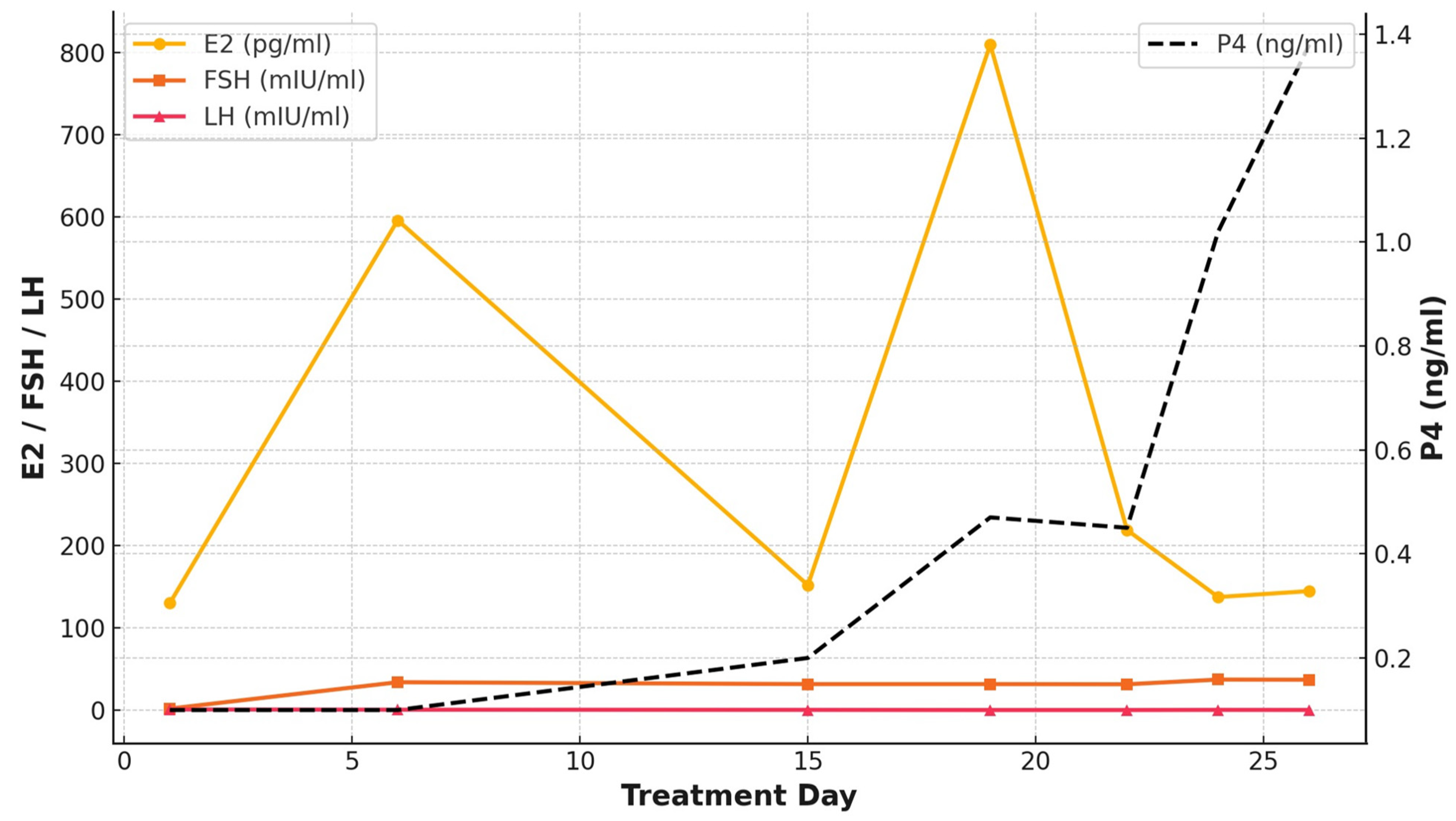
Figure 1. Hormone trends during ovarian stimulation in the 4th cycle. Serum estradiol (E2), follicle-stimulating hormone (FSH), luteinizing hormone (LH), and progesterone (P4) levels were measured throughout the treatment cycle. E2, FSH, and LH values are plotted on the left Y-axis, while P4 values are plotted on the right Y-axis. Despite the development of multiple follicles, serum E2 levels remained low, whereas P4 levels gradually increased during the luteal phase without ovulation.
Endocrines 06 00031 g001
Table 1. Changes in hormonal levels during ovarian stimulation in the 4th cycle.
Treatment Days161519222426
MedicationsEstradiol valerate (10 mg/1 A)111(-)(-)(-)(-)
HMG (IU/day)300300300375375375375
hCG (IU) 5000
Conjugated estrogen tablet (0.625 mg/day)1111111
Follicular
diameter
Right ovary [mm × (follicle number)]ND3(×3)10(×7)10(×10)12
<10(×10)
16, 14,
<10(×15)
18, 12, 10,
<10(×7)
Left ovary [mm × (follicle number)]ND3(×4)9(×4)9(×7)10
<10(×6)
11, 11,
<10(×5)
11, 11,
<10(×5)
Serum
hormonal levels
E2 (pg/mL)130596.1151.9810219137.5144.7
FSH (mIU/mL)1.93431.731.731.537.237
LH (mIU/mL)0.90.60.4<0.2<0.20.40.3
P (ng/mL)<0.1<0.10.20.470.451.021.38
Note: HMG: human menopausal gonadotropin; hCG: human chorionic gonadotropin; E2: estradiol; FSH: follicle stimulating hormone; LH: luteinizing hormone; P: progesterone; ND: not detected.
Table 2. Outcomes of oocyte retrieval in each controlled ovarian stimulation cycle.
CycleCOS ProtocolFollicular Diameter
at hCG Trigger
[mm × (Follicle Number)]
Number of Collected Oocytes (n)Number of Mature
Oocytes (n)
1GnRH agonist Short + FSH/HMG18, 15, 15, 15, 14, 13, 1276
2GnRH agonist Short + FSH/HMG16, 15, 12, 10, 10, <10(×7)32
3FSH/HMG alone18, 15, <10(×6)52
4FSH/HMG alone18, 12, 11, 11, 10, <10(×12)127
Note: COS: controlled ovarian stimulation; GnRH: gonadotropin-releasing hormone; FSH: follicle-stimulating hormone; HMG: human menopausal gonadotropin; hCG: human chorionic gonadotropin.
Table 3. Cohort studies.
Author (Year)Study DesignCohort Size (n)AIRE MutationPOI Prevalence (%)POI Onset Age (Years)AMH Level
(ng/mL)
Presence of Ovarian
Autoantibodies (%)
Ahonen et al. (1990)
[23]
Retrospective study 54N/A60%
(41/54)
No details
(range 13–30)
N/AN/A
Reato et al.
(2011)
[24]
Retrospective study 49N/A41%
(20/49)
Median 24.1
(range 14–39)
N/APOI: 84.6% (11/13)
non-POI: 44% (11/25)
(Data were available in 38 out of 49 patients.)
Saari et al. (2020)
[6]
Longitudinal follow-up study40c.769C>T/c.769C>T (75%:30/40)
c.769C>T/other (15%:6/40)
c.769C>T/x (5%:2/40)
Unknown (5%:2/40)
70%
(28/40)
Median 16.0
(range 11.3–36.5)
N/APOI: 81% (22/27)
non-POI: 30% (3/10)
(Data were available in 37 out of 40 patients.)
Saari et al. (2021)
[21]
Cross-sectional study 19c.769C>T (p.Arg257Ter) (79%:15/19)
c.932G>A (p.Cys311Tyr) (5.3%:1/19)
c.967_979del13 (p.Leu323fs) (5.3%:1/19)
c.137C>G (p.Thr46Arg) (5.3%:1/19)
c.901G>A (p.Val301Met) (5.3%:1/19)
84%
(16/19)
Median 16.5
(range 11.3–36.5)
Undetectable AMH (<0.03):
79% (15/19),
detectable AMH cases:
0.27, 0.04, and 0.89
(Data of one patient are not available in the literature.)
N/A
Garelli et al. (2021)
[19]
Retrospective study103R139X (21.3%:58/272 *)
R257X (11.8%:32/272)
W78R (11.4%:31/272)
C322fsX372 (8.8%:24/272)
T16M (6.2%:17/272)
R203X (4.0%:11/272)
A21V (2.9%:8/272)
Less frequent mutations 12.9%,
very rare 9.6%,
no mutations 11%
50%
(51/103)
Mean 22 ± 7.2 N/APOI: 85% (35/41)
non-POI: 57% (24/42)
(Data were available in 83 out of 103 patients.)
Laakso et al. (2022)
[22]
Multinational retrospective study 321 ** c.769C>T, p.(Arg257Ter) 51% (22/43 ***)
c.967_979del13, p.(Leu327fs) 21% (9/43)
Other 16% (7/43)
Unknown 12% (5/43)
40%
(17/43)
Median 28
(range 13–39)
N/AN/A
Author (Year)Pregnancy
Outcomes
Pregnancy MethodPregnancy ComplicationsLive Birth Rate
Ahonen et al.
(1990)
[23]
2 pregnancies in 2 patientsN/AN/AN/A
Reato et al.
(2011)
[24]
N/AN/AN/AN/A
Saari et al. (2020)
[6]
POI: 8 pregnancies in 7 patients

non-POI:7 pregnancies in 4 patients
POI:
Spontaneous pregnancy 50% (4/8)
Ovum donation 50% (4/8)

non-POI:
Spontaneous pregnancy 71% (5/7)
Infertility treatment 29% (2/7)
POI:
Spontaneous miscarriage 38% (3/8)
(2 from spontaneous pregnancy, and 1 from ovum donation)
Induced abortion 13% (1/8)

non-POI:
Induced abortion 29% (2/7)
(2 from spontaneous pregnancy)
POI: 50% (4/8)
(2 from spontaneous pregnancy, 2 from ovum donation)

non-POI: 71% (5/7)
(3 from spontaneous pregnancy, 2 from infertility treatment)
Saari et al. (2021)
[21]
10 pregnancies in 8 patientsSpontaneous pregnancy 50% (5/10)
Ovum donation 50% (5/10)
N/AN/A
Garelli et al. (2021)
[19]
N/AN/AN/AN/A
Laakso et al. (2022)
[22]
POI: 7 pregnancies in 5 patients

non-POI: 76 pregnancies in 38 patients

POI:
Spontaneous pregnancy 29% (2/7)
Ovum donation 71% (5/7)

non-POI:
Spontaneous pregnancy 96% (73/76)
Infertility treatment 4% (3/76)
POI:
Miscarriage 43% (3/7)

non-POI:
Miscarriage 14% (11/76)
Stillbirths 3% (2/76) ****
Preterm births 7% (5/76)
Gestational diabetes mellitus 1% (1/76)
Placental complications 4% (3/76)
POI: 57% (4/7)
non-POI: 80% (56/76)
Note: HRT: Hormone replacement therapy, N/A: not available, AMH: anti-Müllerian hormone, POI: premature ovarian insufficiency. * AIRE gene mutations were investigated in 272 alleles from 136 patients. ** Of the 321 cases examined, 240 participants were over 16 years of age. *** Data on POI prevalence were available only for pregnant patients. **** Gestational hypertension or adrenal crisis due to primary adrenal insufficiency caused the two stillbirths.
Table 4. Case reports.
Author (Year)AIRE Gene
Mutation
Age of POI
Onset (Years)
AMH Level (ng/mL)Presence of
Ovarian
Autoantibodies
Pregnancy StatusPregnancy MethodPregnancy
Complications
Live Birth
Ward et al.
(1999)
[30]
L93R13.6N/AN/AN/AN/AN/AN/A
Pellegrino et al.
(2018)
[25]
c.396G>C (p.R132S),
homozygous
14.8N/APositiveN/AN/AN/AN/A
Alkhammash et al.
(2019)
[26]
N/AN/AN/AN/A1Ovum
donation
Gestational
diabetes
Hypertension
N/A
Zheng et al.
(2020)
[28]
c.623G>T18N/AN/AN/AN/AN/AN/A
Ruan et al.
(2021)
[29]
Heterozygous mutations
c.371C>T and c.623G>T
21N/AN/AN/AN/AN/AN/A
Fierabracci et al.
(2021)
[31]
c.769C>T
(R257X)
16N/AN/AN/AN/AN/AN/A
Alrufaidi et al.
(2024)
[27]
Homozygous
pathogenic variants
(No details)
28N/APositiveN/AN/AN/AN/A
Note: N/A: not available, AMH: anti-Müllerian hormone, POI: premature ovarian insufficiency.
Disclaimer/Publisher’s Note: The statements, opinions and data contained in all publications are solely those of the individual author(s) and contributor(s) and not of MDPI and/or the editor(s). MDPI and/or the editor(s) disclaim responsibility for any injury to people or property resulting from any ideas, methods, instructions or products referred to in the content.

