Review

Precocious Puberty and Benign Variants in Female Children: Etiology, Diagnostic Challenges, and Clinical Management

1
Department of Maternal Infantile and Urological Sciences, Sapienza University of Rome, 00161 Rome, Italy
2
Molecular Genetics and Functional Genomics, Ospedale Pediatrico Bambino Gesù, IRCCS, 00146 Rome, Italy
*
Author to whom correspondence should be addressed.
These authors contributed equally to this work.
Endocrines 2025, 6(2), 29; https://doi.org/10.3390/endocrines6020029
Submission received: 3 March 2025 / Revised: 28 April 2025 / Accepted: 6 June 2025 / Published: 17 June 2025
(This article belongs to the Section Pediatric Endocrinology and Growth Disorders)

Abstract

Precocious puberty, defined as the onset of secondary sexual characteristics before age 8 in girls, presents a diagnostic challenge in distinguishing between normal variants and pathological conditions requiring intervention. Central precocious puberty (CPP) results from early activation of the hypothalamic–pituitary–gonadal axis, whereas peripheral precocious puberty (PPP) arises from excess sex steroid production independent of gonadotropins. Benign variants, including premature thelarche and premature adrenarche, require careful differentiation to prevent unnecessary treatment. This review explores the physiological mechanisms governing puberty, the epidemiological trends influencing its early onset, and the genetic and environmental factors contributing to its variability in female children. A structured diagnostic approach incorporating clinical evaluation, hormone assessments, imaging studies, and genetic insights is discussed. Management strategies vary depending on the etiology, with gonadotropin-releasing hormone analogs recommended for CPP and targeted therapies for PPP. In contrast, benign variants often necessitate observation and periodic follow-up. Given the increasing prevalence of early puberty, further research is essential to refine diagnostic thresholds and optimize treatment protocols. Early and accurate identification of precocious puberty ensures appropriate intervention, mitigating potential risks associated with early maturation, including compromised adult height and psychosocial challenges.

1. Introduction

Puberty in girls is defined as the onset of secondary sexual characteristics, typically beginning after the age of eight years. The first clinical sign of pubertal development is the appearance of breast buds, known as thelarche, followed by the growth of pubic and axillary hair, termed pubarche. These changes are accompanied by the maturation of external genitalia and an overall acceleration in growth velocity, culminating in the attainment of peak height velocity. In physiological conditions, menarche usually occurs within two to three years after the initial signs of puberty [1].
Pubertal development is orchestrated by a complex neuroendocrine axis, primarily governed by the activation of the hypothalamic–pituitary–gonadal (HPG) axis [2]. During childhood, gonadotropin-releasing hormone (GnRH) secretion from the hypothalamus remains suppressed, but as puberty approaches, pulsatile GnRH secretion resumes, stimulating the anterior pituitary to release follicle-stimulating hormone (FSH) and luteinizing hormone (LH). These gonadotropins, in turn, act on the ovaries to promote follicular development and the production of sex steroids, such as estrogen, which drive the development of secondary sexual characteristics and the maturation of reproductive organs [3].
Although early pubertal development may sometimes represent a benign variant, distinguishing these cases from pathological forms requiring intervention presents significant clinical challenges [4]. Key controversies include the evolving epidemiological trends toward earlier puberty, the appropriate thresholds for diagnosis, and the timing and criteria for initiating therapy to prevent compromised adult height and psychosocial difficulties [5]. These factors underline the complexity of managing early puberty and highlight the need for an evidence-based, individualized approach.
The timing and progression of pubertal development can vary significantly among individuals, influenced by genetic, environmental, nutritional, and psychosocial factors. While the majority of girls experience a normal pubertal trajectory, some may exhibit variations that deviate from the expected pattern [6]. These variations can range from benign, self-limiting conditions to pathological states requiring medical intervention. Among the benign variants, premature thelarche and premature adrenarche are common findings that often resolve spontaneously without progressing to true central precocious puberty (CPP). On the other hand, pathological conditions, such as CPP or peripheral precocious puberty (PPP), can lead to early epiphyseal closure, compromised adult height, and psychological distress if left untreated [7]. Distinguishing between benign variants and pathological puberty requires a comprehensive clinical assessment, including detailed history-taking, physical examination, hormonal evaluations, and radiological investigations [8]. Early and accurate diagnosis is essential to determine whether observation, further investigation, or therapeutic intervention is warranted. Moreover, understanding the psychological impact of early pubertal changes is crucial, as these can affect self-esteem, body image, and social interactions, potentially leading to long-term emotional and behavioral consequences [9].
In this review, we aim to provide an updated overview of the etiology, diagnostic challenges, and clinical management of precocious puberty and its benign variants in young girls. By clarifying the spectrum of pubertal presentations and outlining evidence-based approaches to diagnosis and treatment, we hope to support clinicians in delivering optimal care while avoiding unnecessary interventions in self-limiting conditions.

2. Physiology of Puberty

Puberty is initiated by the activation of the HPG axis, the primary regulatory system for sexual development and reproductive function. This complex neuroendocrine axis is tightly controlled by an intricate interplay of hormonal signals, genetic factors, and environmental cues [10]. While the mechanisms governing pubertal onset are increasingly understood, certain aspects remain incompletely elucidated, particularly regarding the early phases of HPG axis activation.
Before the onset of true puberty, there are two distinct phases of transient HPG axis activation which occur during fetal life and the early postnatal period. These phases are crucial for gonadal development and sexual differentiation, although their long-term implications for pubertal timing remain under investigation [11].

2.1. Fetal Activation of the HPG Axis

The first phase of HPG axis activation occurs during fetal development. By the 12th to 14th week of gestation, the fetal hypothalamic–pituitary axis becomes active, leading to elevated levels of gonadotropins in the fetal circulation [12]. Interestingly, during early gestation, FSH and LH secretion appears to be independent of hypothalamic GnRH stimulation, as evidenced by studies comparing hormone levels in anencephalic and healthy fetuses. In anencephalic fetuses, gonadotropin levels remain detectable until the 17th to 18th week of gestation, suggesting an intrinsic capacity of the fetal pituitary to secrete FSH and LH, independent of hypothalamic input [13]. As pregnancy progresses, placental hormones, particularly estrogens and progesterone, exert an inhibitory effect on the fetal hypothalamus and pituitary gland, leading to a progressive decline in gonadotropin levels during the last trimester. This suppression persists until birth, when the removal of placental influence allows for the reactivation of the HPG axis [14,15].

2.2. Postnatal Reactivation: Mini-Puberty

The second phase of HPG axis activation, known as “mini-puberty,” occurs during the early postnatal period. Within the first few days after birth, the removal of placental inhibition leads to a surge in gonadotropin secretion, stimulating the gonads to produce sex steroids. This phenomenon follows a sex-specific pattern: in girls, estradiol levels fluctuate and remain relatively elevated during the first 3 to 4 years of life [11,16,17], playing a critical role in genital development and breast tissue priming. While the clinical relevance of mini-puberty in long-term reproductive outcomes remains debated, some studies suggest that early hormonal exposure may influence future pubertal timing and gonadal function [11,18]. The transient hormonal surge during mini-puberty can also lead to the appearance of secondary sexual characteristics, such as isolated premature thelarche in girls [19], which is typically self-limiting and non-progressive.

2.3. Onset of True Puberty

True puberty begins when hypothalamic GnRH neurons increase the pulsatile secretion of GnRH, stimulating the anterior pituitary to release LH and FSH [10]. During childhood, GnRH secretion remains quiescent due to inhibitory signals from the central nervous system. As puberty approaches, this inhibition is lifted through mechanisms that are not yet fully understood, though several key factors have been identified. A critical trigger for pubertal reactivation is the Kisspeptin system. Kisspeptin, a neuropeptide produced by Kiss1 neurons, acts directly on GnRH neurons via the G-protein-coupled receptor 54 (GPR54). Mutations in the KISS1 or GPR54 genes have been shown to impair pubertal onset, highlighting their essential role [20,21,22]. Additionally, leptin, an adipocytokine secreted by adipose tissue, plays a permissive role in puberty initiation. Leptin enhances Kiss1 expression and inhibits the secretion of neuropeptide Y, an inhibitor of GnRH secretion [23,24]. These findings underscore the importance of metabolic status and body composition in determining the timing of puberty, emphasizing the need for adequate nutrition and weight control to avoid pubertal disorders [25].

2.4. Endocrine and Physiological Changes

Once the HPG axis becomes fully active, gonadotropins stimulate the gonads to produce sex steroids. In girls, LH stimulates ovarian theca cells to produce androgens, while FSH promotes granulosa cell proliferation and enhances aromatase activity, converting androgens into estradiol. Rising estradiol levels drive breast development, uterine growth, and the accumulation of adipose tissue in a typical female distribution. Estradiol also increases the expression of FSH receptors, facilitating follicular growth and ovulation. SHBG levels decline slightly during puberty, further enhancing the availability of active sex steroids [10].
Concurrently, the adrenal glands contribute to pubertal changes through adrenarche, marked by the increased production of adrenal androgens, including dehydroepiandrosterone (DHEA) and androstenedione [26,27]. These androgens are responsible for the growth of axillary and pubic hair and may also contribute to the onset of acne. Adrenarche typically precedes gonadarche and occurs independently of HPG axis activation.
In addition to sex steroids, other hormones play pivotal roles in supporting pubertal growth and development. Growth hormone and insulin-like growth factor 1 (IGF-1) promote linear growth and organ maturation [28], while thyroid hormones and cortisol contribute to overall metabolic homeostasis [29,30].

2.5. Clinical Progression and Pubertal Staging

The progression of pubertal development follows a predictable sequence, first described by Marshall and Tanner, who categorized secondary sexual characteristics into five distinct stages, known as Tanner stages. This staging system remains the gold standard for assessing pubertal progression in clinical practice [6,31].
In girls, the first sign of true puberty is breast budding (Tanner stage 2), typically occurring around 8 to 10 years of age. This is followed by a growth spurt, which usually peaks within one year of thelarche. Pubic hair development typically follows, although adrenarche and pubarche can sometimes precede thelarche. Menarche, the culmination of pubertal development, usually occurs approximately 2 to 2.5 years after the onset of breast development, when growth velocity begins to decelerate [32].

3. Disorders of Pubertal Development

Precocious puberty is broadly classified into two distinct categories: CPP and PPP. In addition to these pathological forms, there are para-physiological conditions defined as benign and non-progressive variants of puberty, such as isolated premature adrenarche and isolated premature thelarche [4]. Proper differentiation between these conditions is essential to avoid unnecessary investigations and interventions while ensuring timely management of true pathological cases [Table 1].
The increasing trend toward earlier pubertal onset in girls is attributed to a complex interplay of genetic, environmental, and behavioral factors. Among these, endocrine-disrupting chemicals (EDCs), obesity, ethnicity, and lifestyle changes play significant roles, complicating the definition of specific epidemiological patterns [33]. EDCs, such as bisphenol A, phthalates, and polychlorinated biphenyls, can interfere with hormonal signaling pathways, potentially accelerating pubertal onset. Additionally, the rise in childhood obesity has been strongly correlated with early thelarche, as adipose tissue promotes estrogen production through aromatization of androgens [33].
Longitudinal studies conducted since the 1940s have consistently documented a secular trend toward earlier thelarche [34,35,36]. However, despite this anticipatory trend in breast development, the average age of menarche has remained relatively stable in industrialized countries, resulting in a lengthened interval between thelarche and menarche [37]. This dissociation suggests that early breast development does not necessarily imply accelerated progression through all stages of puberty.
Epidemiological studies conducted in the United States have highlighted significant ethnic variations in pubertal timing. A landmark study by Herman-Giddens et al. [38] reported that the average age of thelarche is approximately 10 years in White girls and 9 years in Black girls, representing a noticeable decrease from the mean age of 11 years observed in the 1980s. Moreover, the study found that breast development occurs before the age of 8 in approximately 15% of Black girls and 5% of White girls, underscoring the influence of genetic and environmental factors on pubertal timing [38].
These findings have sparked ongoing debates regarding the appropriateness of current age cutoffs for defining precocious puberty. While some experts advocate for lowering the age threshold for pubertal onset, the prevailing consensus remains to use 8 years as the reference age for girls. This conservative approach helps prevent the underdiagnosis of true precocious puberty and ensures that girls at risk of accelerated bone maturation, reduced adult height, and psychological stress receive timely evaluation and management [39].

3.1. Gonadotropin-Dependent Precocious Puberty

CPP is characterized by the premature activation of the HPG axis, leading to the early onset of secondary sexual characteristics before the age of 8 years in girls [40]. This condition represents approximately 80% of all cases of precocious puberty, making it more prevalent than peripheral forms [41]. While the isolated appearance of secondary sexual characteristics, such as premature thelarche or adrenarche, is often benign and self-limiting, CPP is associated with true pubertal progression and a significant risk of adverse physical and psychological outcomes if left untreated.
Epidemiological data indicate a notable sex difference in CPP presentation. In girls, idiopathic CPP accounts for the vast majority of cases, whereas in boys, organic causes are more commonly identified. These include central nervous system (CNS) abnormalities, such as hypothalamic hamartomas, pituitary gonadotropin-secreting tumors, meningomyelocele, hydrocephalus, encephalitis, neonatal hypoxic-ischemic encephalopathy, and neurofibromatosis type 1 [42]. Hypothalamic hamartomas are the most common organic CNS lesions associated with CPP. These benign, congenital, non-neoplastic malformations consist of ectopic neurosecretory neurons capable of autonomous GnRH secretion, effectively acting as an accessory GnRH pulse generator [43]. CPP due to hypothalamic hamartomas can present very early, sometimes in infancy as early as 12 months of age. A hallmark feature is the presence of gelastic seizures—sudden episodes of inappropriate laughter [44]—which may precede or accompany the pubertal signs. Other brain tumors have also been associated with CPP, including astrocytomas, optic pathway gliomas, craniopharyngiomas, hypothalamic or suprasellar tumors, pineal tumors, and intracranial germ cell tumors. These tumors may directly disrupt hypothalamic inhibition of GnRH neurons or indirectly stimulate the HPG axis through mass effect or hormonal secretion [45]. Radiation therapy to the brain or spinal cord, commonly administered for pediatric malignancies, can increase the risk of CPP [46] by damaging inhibitory GABAergic neurons and altering hypothalamic regulation. Similarly, traumatic brain injuries and surgical insults to the CNS have been implicated in premature pubertal activation [47]. Infectious causes, although rare, include neuroinfections such as meningitis or encephalitis. Tuberculous meningitis, in particular, has been reported as a potential cause of hypothalamic injury leading to CPP [48].
Recent advances in genetic research have shed light on the hereditary basis of CPP. Genetic mutations have been implicated in a subset of familial and sporadic cases. Notably, loss-of-function mutations in the MKRN3 gene, a maternally imprinted and paternally expressed gene located on chromosome 15q11.2, are the most frequently identified genetic cause of familial CPP [41]. Gain-of-function mutations in the KISS1 and KISS1R (GPR54) genes have also been associated with idiopathic CPP, although they account for a very small number of cases. These mutations lead to enhanced kisspeptin signaling, promoting premature activation of GnRH neurons. Similarly, loss-of-function mutations in the DLK1 gene, which is also maternally imprinted and paternally expressed, have been linked to familial CPP [49]. Other genetic causes identified include rare mutations in the MECP2 gene, primarily associated with Rett syndrome, but which have also been implicated in isolated cases of CPP [50]. Additionally, imprinting defects at the 14q32.2 locus, as seen in Temple syndrome, can contribute to early puberty onset. Temple syndrome is characterized by growth failure, early puberty, and distinct facial features, and highlights the complexity of genetic regulation over pubertal timing [51]. Besides isolated genetic causes, certain syndromic conditions such as neurofibromatosis type 1 have been linked to CPP, primarily due to associated hypothalamic involvement [52].
A rare but noteworthy manifestation of precocious puberty involves the transition from peripheral to central forms [53]. This phenomenon is exemplified by conditions such as McCune–Albright syndrome (MAS), characterized by polyostotic fibrous dysplasia, café-au-lait macules, and autonomous ovarian or testicular cysts. In such cases, prolonged exposure to elevated sex steroid levels can lead to the loss of negative feedback mechanisms, ultimately triggering hypothalamic activation and CPP, even after appropriate therapy for the peripheral cause has been initiated [54]. This highlights the complex interplay between central and peripheral mechanisms in the pathogenesis of precocious puberty [55].
As a gonadotropin-dependent condition, CPP follows the typical physiological sequence of pubertal events, beginning with thelarche, followed by pubarche, peak height velocity, and eventually menarche in girls. Importantly, the progression of secondary sexual characteristics in CPP is consistent with the individual’s biological sex, distinguishing it from conditions associated with androgen excess or other endocrine disorders [4]. Clinically, CPP is often associated with an accelerated growth velocity and significant advancement in bone age, which can lead to early epiphyseal fusion and compromised adult height if untreated [56]. The rapid progression of puberty can also impose psychological challenges, including increased vulnerability to mood disorders, anxiety, and social difficulties, particularly when pubertal changes are not synchronized with peers [57].

3.2. Gonadotropin-Independent Precocious Puberty

PPP is a gonadotropin-independent condition characterized by the early development of secondary sexual characteristics resulting from elevated circulating steroid hormone levels. Unlike CPP, which is driven by premature activation of the HPG axis, PPP arises from direct hormonal stimulation of target tissues, bypassing gonadotropin regulation. As such, PPP is often referred to as “precocious pseudopuberty” rather than true puberty [55]. A hallmark feature of PPP is the potential discordance between the child’s biological sex and the secondary sexual characteristics expressed. Estrogen excess typically leads to feminization, while androgen excess results in virilization, regardless of the individual’s genetic sex [55]. This contrasts with CPP, where pubertal progression follows the physiological pattern of the child’s biological sex.
PPP can arise from either acquired or congenital/genetic causes, with endogenous or exogenous sources of steroid hormone excess playing central roles [53].
Acquired forms of PPP are predominantly associated with excessive production or external exposure to sex steroids. In girls, functional ovarian cysts represent the most common endogenous cause of PPP. These cysts can transiently produce significant amounts of estrogen, resulting in breast development (premature thelarche) and, in some cases, vaginal bleeding. Importantly, the pubertal signs associated with functional ovarian cysts typically regress spontaneously following cyst resolution, often without requiring medical intervention [55,58]. Ovarian tumors are another important cause of PPP in girls. Granulosa cell tumors, the most common sex cord-stromal tumors in this age group, typically lead to estrogen excess and isosexual precocious puberty. Other tumors, including Sertoli–Leydig cell tumors, Leydig cell tumors, and gonadoblastomas, can also cause hormone secretion and, depending on the predominant hormone, may present with either isosexual or contrasexual pubertal signs [59]. Additionally, rare hCG-secreting tumors can stimulate ovarian steroidogenesis indirectly by acting through the LH receptor. These tumors may arise in gonadal or extragonadal sites, and their identification requires measurement of serum and, if needed, cerebrospinal fluid beta-hCG levels [60].
Exogenous exposure to estrogenic or androgenic compounds is another well-documented cause of PPP. This exposure may result from the ingestion of hormone-containing medications, consumption of industrialized food products contaminated with EDCs, or topical absorption from products such as lavender oils, cosmetics, or transdermal medications handled by caregivers [61,62]. In such cases, discontinuation of exposure typically leads to the resolution of symptoms.
Several genetic and congenital conditions can result in gonadotropin-independent pubertal development, affecting both sexes. MAS represents one of the most well-known genetic causes of PPP. This rare mosaic disorder results from a postzygotic, gain-of-function mutation in the GNAS gene, leading to constitutive activation of Gs alpha proteins. This aberrant signaling promotes autonomous hormone production in affected endocrine tissues, including the ovaries, adrenal glands, and thyroid [63]. The classical triad of MAS includes café-au-lait macules with characteristic “coast of Maine” borders, polyostotic fibrous dysplasia of the bone, and PPP. In affected girls, MAS often presents between the ages of 2 and 6 years, with recurrent estrogen-producing ovarian cysts driving rapid breast development, growth acceleration, and intermittent vaginal bleeding [64]. As a mosaic disorder, MAS exhibits considerable clinical heterogeneity, depending on the distribution and extent of affected tissues. This variability underscores the need for individualized diagnostic and therapeutic approaches, including careful hormonal monitoring and consideration of aromatase inhibitors to reduce estrogen production [55,65].
Adrenal disorders are another major cause of PPP, particularly those associated with androgen excess. Androgen-secreting adrenal tumors, though rare, can lead to significant virilization in young girls. More commonly, congenital adrenal hyperplasia (CAH), an autosomal recessive disorder caused by enzymatic defects in adrenal steroidogenesis, results in excessive androgen production [66]. The most prevalent form, 21-hydroxylase deficiency, accounts for over 90% of CAH cases. Other enzymatic defects include 11-beta-hydroxylase deficiency, 3-beta-hydroxysteroid dehydrogenase type 2 deficiency, hexose-6-phosphate dehydrogenase deficiency, and phosphoadenosine phosphosulfate synthase type 2 deficiency [67]. Rarely, adrenal tumors can secrete both androgens and estrogens, either due to intra-adrenal aromatization of androgens or excessive peripheral aromatization. These tumors can lead to complex presentations, such as simultaneous breast development and virilization, necessitating detailed biochemical and imaging evaluations for accurate diagnosis [68].
Although uncommon, longstanding untreated hypothyroidism can also lead to PPP. This paradoxical phenomenon, known as Van Wyk and Grumbach syndrome, is characterized by the triad of juvenile hypothyroidism, isosexual precocious puberty, and multicystic enlarged ovaries [69]. It is thought to result from hormonal dysregulation, including the development of estrogen-producing ovarian cysts secondary to elevated thyroid-stimulating hormone (TSH) levels [70]. Cross-reactivity between TSH and FSH receptors has also been proposed as a contributing mechanism [71]. In such cases, thyroid hormone replacement therapy typically resolves the pubertal signs, highlighting the importance of thyroid function assessment in the differential diagnosis of PPP [72].

3.3. Benign Variants

3.3.1. Premature Thelarche

Premature thelarche is a self-limiting, non-progressive condition characterized by isolated breast tissue development before the age of eight years, without accompanying signs of true puberty, such as accelerated growth velocity, advanced bone age, or adrenarche. It is one of the most common benign variants of early pubertal development and typically occurs during two distinct age periods: the first within the initial two years of life and the second after the age of six years [73]. The first peak coincides with the physiological activation of the HPG axis during the so-called “mini-puberty,” a transient phase occurring between the first few weeks and the sixth month of life [11]. During this period, elevated gonadotropin levels, particularly FSH, stimulate ovarian activity, resulting in increased estradiol production. The heightened sensitivity of mammary tissue to circulating estrogens may lead to unilateral or bilateral breast development, typically not progressing beyond Tanner stages 2–3 [74].
Although the exact etiology of premature thelarche remains largely idiopathic, several contributing factors have been proposed. Exposure to exogenous estrogenic compounds, including phytoestrogens, EDCs, or estrogen-containing medications, has been implicated in some cases [75]. Moreover, the rising prevalence of childhood obesity has emerged as an additional risk factor, as increased adipose tissue promotes estrogen production through peripheral aromatization of androgens. This phenomenon complicates the clinical distinction between true breast development and adipomastia, particularly in overweight or obese girls [37].
The potential progression of premature thelarche to CPP remains a subject of ongoing debate [76]. While the condition is generally benign, longitudinal studies suggest that approximately 10–18% of cases may eventually progress to CPP, necessitating careful monitoring [77]. However, identifying reliable clinical or anthropometric markers predictive of progression has proven challenging, with conflicting evidence regarding the influence of age at onset. Some studies have indicated that breast development before the age of two years may be associated with a higher risk of progression to CPP, whereas others have found no significant correlation between age of onset and pubertal outcomes [74]. Therefore, regular follow-up every 3 to 6 months is recommended for girls with premature thelarche, with particular attention to the appearance of additional pubertal signs, such as pubic hair development, accelerated linear growth, or advanced bone age [75].
When premature thelarche demonstrates a stable or regressive course, it can usually be managed conservatively with periodic observation. However, in cases showing signs of progression, supplementary diagnostic evaluations are warranted. These typically include hormonal assays (basal and GnRH-stimulated gonadotropin levels), pelvic ultrasound, and bone age assessment [75]. Pelvic ultrasound is particularly valuable for evaluating ovarian volume and uterine morphology, which can help differentiate benign premature thelarche from early central activation of the HPG axis. In benign cases, ovarian volumes remain within prepubertal ranges, while CPP is typically associated with enlarged ovaries and increased uterine length [75]. Bone age assessment provides further insights, as significant advancement relative to chronological age is more indicative of CPP. A notable study by Bizzarri et al. [78] highlighted the diagnostic challenges associated with early breast development in girls under the age of three. The study found that peak LH and FSH levels following GnRH stimulation were frequently elevated in girls without clinical signs of pubertal progression, many of whom did not subsequently develop CPP. In this age group, basal LH levels and uterine diameter measurements on pelvic ultrasound were shown to be more reliable indicators for distinguishing benign premature thelarche from early-onset CPP.
Given its predominantly self-limiting nature, the initial management of premature thelarche should prioritize careful clinical follow-up rather than immediate intervention. Monitoring should include evaluations every 3 months to assess pubertal progression and every 6 months to track growth velocity [75]. Particular attention should be paid to signs of estrogenization, such as areolar hyperpigmentation, increased breast bud consistency, and the emergence of pubic hair. In cases of rapid progression, further endocrine evaluations, including serum estradiol, basal and stimulated gonadotropin levels, and imaging studies, are warranted to rule out CPP or underlying pathological conditions [79]. Early identification of progressing cases ensures timely intervention, while conservative management avoids unnecessary medicalization in self-limiting cases.

3.3.2. Premature Adrenarche

Premature adrenarche is a benign, gonadotropin-independent condition characterized by the early maturation of the adrenal glands, resulting in increased production of adrenal androgens, particularly DHEA and its sulfate (DHEA-S) [80]. This hormonal surge leads to the early appearance of secondary sexual characteristics, such as pubic and axillary hair (pubarche), body odor, and, occasionally, mild acne. Importantly, premature adrenarche occurs in the absence of other signs of true puberty, such as clitoromegaly, penile enlargement, breast development, or testicular growth [39,81]. It is typically diagnosed when these signs emerge before the age of 8 in girls, with children maintaining normal growth patterns consistent with their established percentiles on growth charts. Unlike CPP or PPP, premature adrenarche does not involve activation of the HPG axis, as reflected by prepubertal levels of FSH, LH, estradiol, and testosterone [39,81].
The prevalence varies among populations, with a higher incidence reported in Black girls, likely due to the earlier onset of pubic hair development in this group [49]. Emerging evidence also suggests an association with metabolic risk factors, including higher body mass index (BMI), insulin resistance, and dyslipidemia, underscoring the influence of nutritional and environmental factors on adrenal maturation [82].
Premature adrenarche is primarily driven by premature activation of adrenal androgen synthesis, a process typically initiated during adrenarche, which physiologically occurs around 6 to 8 years of age. This process involves increased activity of the adrenal zona reticularis, leading to elevated circulating levels of DHEA, DHEA-S, and androstenedione. These weak androgens can stimulate the development of pubic and axillary hair without triggering gonadal activation [80]. Laboratory findings typically show elevated age-adjusted DHEA-S levels, often ranging from 30 to 150 μg/dL, while cortisol, FSH, LH, estradiol, and testosterone remain within prepubertal ranges. In contrast to pathological conditions, premature adrenarche does not cause significant acceleration in growth velocity or bone maturation [39,81].
Although generally benign, it may be associated with certain metabolic and endocrine alterations. The study by DeSalvo et al. [83] found that approximately 30% of girls with premature adrenarche exhibit advanced bone age, typically up to 2 years beyond chronological age, along with a higher BMI. Despite this skeletal advancement, longitudinal studies indicate that affected children usually achieve their target adult height, provided that no underlying pathology is present.
While most cases follow a benign course, clinicians should remain vigilant for signs suggesting an alternative diagnosis, such as non-classic CAH, virilizing adrenal tumors, or gonadal disorders. Mild forms of non-classic CAH, particularly 21-hydroxylase deficiency, account for approximately 3–5% of premature adrenarche cases [67]. Early pubic hair growth accompanied by rapid linear growth, clitoral hypertrophy, or significant penile enlargement should prompt further evaluation to exclude such conditions.
The diagnosis is primarily clinical, supported by hormonal assessments and, when warranted, imaging studies. In straightforward cases with isolated pubarche and no additional pubertal signs, extensive testing is generally not required. However, in cases of rapid progression, abnormal growth patterns, or virilizing features, targeted investigations are indicated. Measurements of serum DHEA-S, androstenedione, 17-hydroxyprogesterone (17-OHP), and testosterone are useful to exclude non-classic CAH and other adrenal disorders [84]. Radiographic evaluation of the left hand and wrist can reveal advanced bone age in up to 30% of children. However, bone age advancement alone has limited reliability in predicting pathological causes and often reflects androgenic activity rather than true pathology [83]. Imaging studies, such as ultrasound or magnetic resonance imaging (MRI), are reserved for cases with rapid progression, atypical findings, or markedly elevated androgen levels, as these may suggest adrenal tumors [85].
While premature adrenarche is typically a self-limiting condition, emerging evidence suggests potential associations with long-term metabolic risk factors, including insulin resistance, polycystic ovary syndrome, and cardiovascular disease, particularly in girls with concurrent obesity [49]. Therefore, clinical follow-up should include periodic assessments of growth velocity, BMI, and pubertal progression. Management primarily involves reassurance and lifestyle counseling, particularly regarding healthy nutrition, physical activity, and weight management [86]. Hormonal treatment is rarely indicated, except when an underlying endocrine disorder is identified.

3.3.3. Premature Menarche
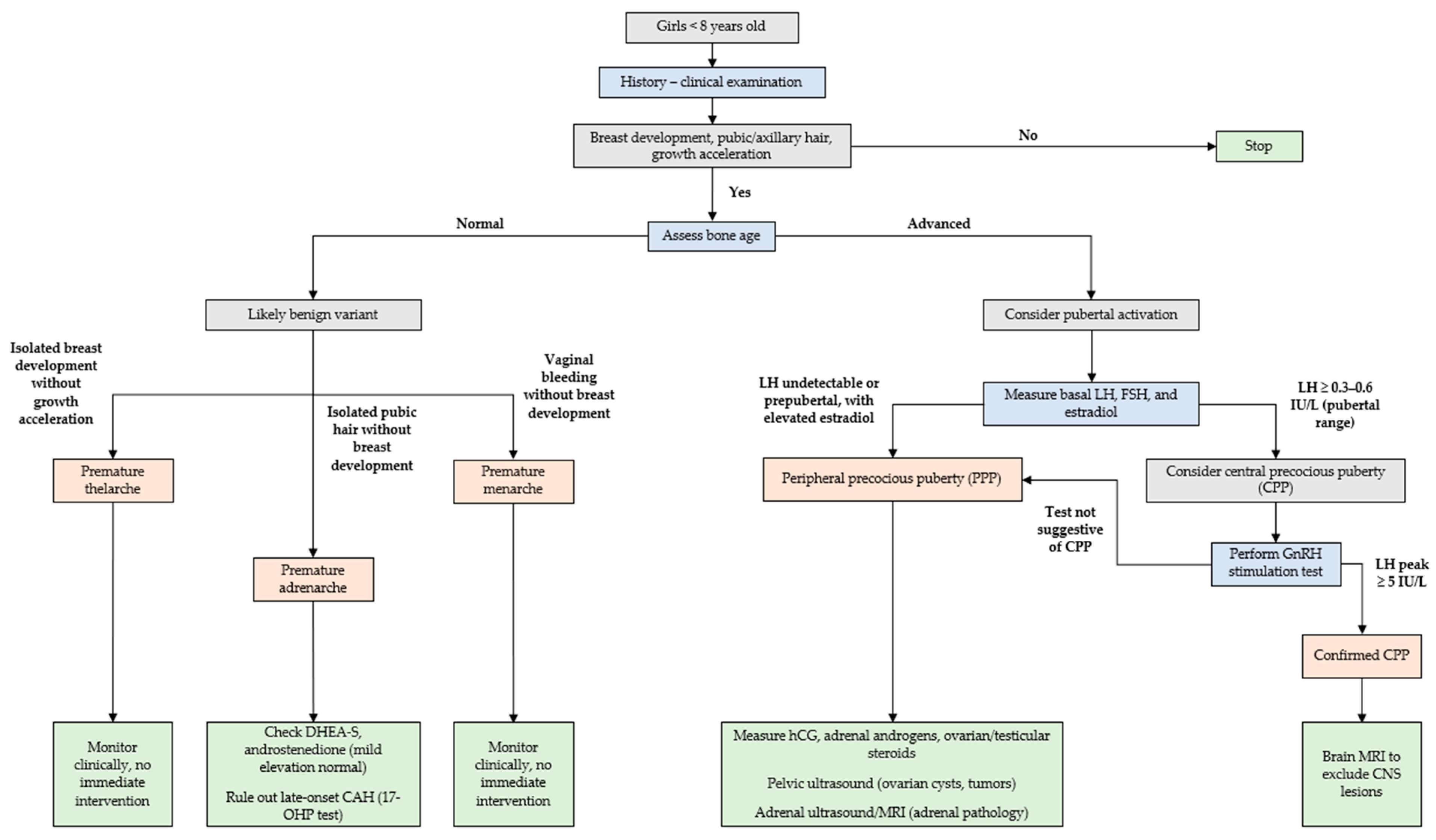
Premature menarche, defined as the occurrence of vaginal bleeding in girls younger than 8 years of age, is a rare but distressing presentation that often raises significant concern among families and healthcare providers. Although alarming, most cases are self-limiting and benign, resolving spontaneously without progression to true puberty or underlying pathology [87]. The differential diagnosis of vaginal bleeding in female children is broad and includes both physiological and pathological causes. Physiological explanations include transient estrogen stimulation, often associated with functional ovarian cysts or mini-puberty, and the transient postnatal activation of the HPG axis. Pathological causes, though rare, include trauma, sexual abuse, foreign bodies, vaginal infections, coagulation disorders, and, in rare instances, hormone-secreting tumors [39]. In a study by Nella et al. [88], involving over 1000 girls referred for suspected precocious puberty, only 24 presented with vaginal bleeding. Among these, the majority experienced a single self-limiting episode, while approximately 33% had intermittent episodes lasting up to one year. Notably, 79% of these girls exhibited no other secondary sexual characteristics, such as breast development, pubic hair growth, or growth acceleration. Hormonal evaluations, including basal and GnRH-stimulated gonadotropin levels, typically revealed prepubertal values, and imaging studies, such as pelvic ultrasound, often demonstrated the presence of transient ovarian cysts. Importantly, the bleeding resolved spontaneously in all cases, and long-term follow-up revealed that the girls subsequently achieved menarche at an age consistent with normal pubertal development.

4. Management

4.1. History and Clinical Examination

A comprehensive patient history is the cornerstone of evaluating suspected precocious puberty and distinguishing between benign variants and pathological forms [Figure 1]. This evaluation should include the patient’s age at the onset of physical changes, the progression of secondary sexual characteristics, and any exposure to sex steroids, whether endogenous or exogenous. A detailed family history should be obtained, with particular attention to familial cases of early puberty, as well as maternal menarcheal age, which is a known determinant of pubertal timing in daughters [89]. Environmental exposures should also be investigated, including possible contact with estrogen- or androgen-containing substances, such as contraceptive pills, testosterone gels, or EDCs present in cosmetics, plastics, or industrialized food products [90]. Additionally, drug use within the household, including herbal remedies with phytoestrogenic properties, should be explored [55]. CNS-related symptoms must be thoroughly assessed, as intracranial lesions can underlie CPP. Symptoms such as headaches, visual disturbances, polydipsia, polyuria, mood changes, or seizures or a history of brain trauma, CNS infections, or neonatal complications may indicate an organic etiology requiring further investigation [41,91].
Growth assessment is critical for monitoring pubertal progression. Height should be evaluated in relation to mid-parental height, calculated using standardized formulas [92]:
mid-parental height = [father’s height (cm) − 13 + mother’s height (cm)]/2 ± 8 cm.
Longitudinal growth tracking using growth charts allows for the detection of accelerated growth velocity, which is often an early indicator of CPP [93]. In CPP, growth velocity typically exceeds the 95th percentile for age, while PPP may exhibit discordant growth patterns due to isolated sex steroid excess. Pubertal staging should be conducted using the Tanner scale, with particular attention to distinguishing true breast development from lipomastia, commonly observed in overweight girls. Skin findings, such as café-au-lait macules, may suggest underlying syndromic conditions like neurofibromatosis type 1 or MAS [94].
Specific clinical features may further clarify the underlying cause of precocious puberty. For instance, adrenal tumors often present with virilization due to androgen excess, accompanied by systemic signs such as moon facies, striae, hypertension, and rapid weight gain [95]. In girls, isolated vaginal bleeding without breast development should raise suspicion for MAS, which results from a gain-of-function mutation in the GNAS gene. In MAS, PPP is driven by estrogen secretion from functional ovarian cysts, with vaginal bleeding often triggered by cyst involution and subsequent estrogen withdrawal. Additional findings, such as polyostotic fibrous dysplasia and café-au-lait macules with irregular “coast of Maine” borders, further support the diagnosis [96].
In conclusion, a meticulous history and thorough physical examination remain essential for differentiating benign variants from pathological forms of precocious puberty and guiding appropriate management.

4.2. Laboratory Tests

Laboratory investigations play a pivotal role in diagnosing and differentiating pubertal disorders. However, careful patient selection for testing is crucial to avoid unnecessary investigations, as many benign variants can be effectively managed through clinical observation alone [74]. Baseline hormonal evaluations typically include measurements of [97]:
  • Gonadotropins (LH, FSH): Baseline LH is particularly informative, with prepubertal values generally defined as <0.3–0.6 IU/L and pubertal values exceeding 1 IU/L [98,99].
  • LH/FSH ratio: This ratio tends to be higher in progressive CPP compared to benign conditions [62,63].
  • Estradiol: Prepubertal estradiol levels are typically <1 pg/mL, as measured by highly sensitive mass spectrometry assays [62,64].
  • Adrenal androgens: Elevated DHEA-S, androstenedione, and 17-OHP (and testosterone) levels may indicate premature adrenarche or non-classic CAH [59].
  • IGF-1 and inhibin B: These markers provide insights into growth patterns and gonadal function, respectively [100].
  • Thyroid function: Evaluation of TSH and free thyroxine is essential to exclude hypothyroidism, which can rarely present with pubertal changes [39].
The GnRH stimulation test remains the gold standard for diagnosing CPP. It involves administering GnRH or its analog and measuring LH levels 30–60 min post-administration. A peak LH level > 5 IU/L is considered diagnostic for CPP. However, the use of stimulation testing should be reserved for cases with ambiguous clinical or hormonal findings, as it can be invasive and resource-intensive [97,98,101].
A study by Cleemann Wang et al. [97] highlighted the limitations of relying on single hormonal markers for differentiating CPP from benign conditions, such as premature thelarche. The combination of hormonal profiles with anthropometric data, such as a growth velocity exceeding 7 cm/year and bone age advanced by more than one year [8,102], significantly enhances diagnostic accuracy, often obviating the need for stimulation testing [97].
It is important to note that hormonal cutoffs may vary depending on assay sensitivity and laboratory standards, highlighting the need for cautious interpretation of results and integration with clinical findings [103,104].

4.3. Imaging

Pelvic ultrasound is a highly sensitive and specific, non-invasive, cost-effective tool for distinguishing CPP from benign conditions, such as isolated premature thelarche or premature adrenarche. Ultrasound evaluates ovarian volumes, uterine dimensions, and the presence of ovarian follicles or cysts. In prepubertal girls, the uterus is typically small and tubular, with a length of less than 3.5 cm, and the endometrial stripe is absent or very thin. The ovaries are also small, usually with a volume of less than 1–2 mL, and may contain small, simple follicles (<9 mm), reflecting a quiescent gonadal status [105]. In contrast, in pubertal girls, the uterus undergoes significant changes. It becomes larger and assumes a pear-shaped configuration, with a body-to-cervix ratio greater than 1:1. The endometrial stripe may become visible under the influence of estrogen stimulation. Ovarian volume increases (greater than 2–3 mL), and multiple growing follicles of varying sizes are typically observed. The presence of ovarian follicles larger than 9 mm and an ovarian volume exceeding 2 mL are suggestive of activation of the hypothalamic–pituitary–gonadal axis [105]. Thus, ultrasound findings consistent with a pubertal uterus and ovaries, in conjunction with clinical and hormonal findings, support the diagnosis of central precocious puberty. Conversely, a prepubertal ultrasound appearance suggests a benign variant such as isolated premature thelarche or a non-progressive form [102,106]. A study by Badouraki et al. [107] established specific ultrasound cutoffs for girls aged 0–8 years presenting with pubertal signs. Uterine length proved to be the most reliable marker for differentiating CPP from premature thelarche, with girls affected by CPP demonstrating larger uterine dimensions, increased ovarian volumes, and a higher prevalence of ovarian microcysts or macrocysts.
Skeletal maturation is evaluated through a radiograph of the left hand and wrist using either the Greulich and Pyle method [108], which compares images to an atlas of standardized radiographs, or the Tanner–Whitehouse 3 method [109], which assigns scores to 20 distinct hand and wrist bones. Bone age is primarily influenced by sex steroids, particularly estrogens and androgens, which accelerate epiphyseal maturation. However, genetic predisposition, nutritional status, obesity, pathological conditions, and pharmacological interventions can also modulate skeletal development. Advanced bone age, defined as exceeding chronological age by ≥ 2 standard deviations, necessitates further evaluation. Repeating radiographic assessments at 6- to 12-month intervals is recommended for children demonstrating pubertal progression or increased growth velocity [110].
Brain MRI plays a crucial role in evaluating children with CPP, particularly for identifying CNS abnormalities, such as hypothalamic hamartomas, structural brain lesions, or intracranial tumors. Current guidelines recommend MRI for girls younger than six years, as organic causes are more prevalent in this age group, as well as for any child presenting with neurological symptoms, including headaches, visual disturbances, or seizures [111,112]. In asymptomatic girls aged six to eight years, routine neuroimaging remains controversial, as significant CNS abnormalities are rare, with an estimated prevalence of 2–7% [40,112]. Given the potential risks of MRI, including sedation, contrast exposure, and psychological distress from incidental findings, shared decision making with caregivers is essential [112,113]. Advances in imaging techniques and biomarker identification may further refine neuroimaging indications, reducing unnecessary procedures while ensuring timely diagnosis.
While pelvic ultrasound is crucial for distinguishing CPP from benign conditions, it also aids in evaluating premature adrenarche by assessing adrenal gland morphology. Adrenal ultrasound or MRI may be considered in cases of rapidly progressing adrenarche, significant virilization, or markedly advanced bone age to exclude adrenal hyperplasia, androgen-secreting tumors, or other adrenal pathologies [27]. MRI can detect adrenal enlargement or nodular hyperplasia, though routine imaging is not recommended in typical cases of premature adrenarche [114]. Additionally, bone age assessment plays a key role in monitoring these conditions, as premature adrenarche is usually associated with bone maturation that remains within normal limits or only slightly advanced [115].

4.4. Treatment

The treatment of precocious puberty is tailored according to the underlying cause, with distinct approaches for central and peripheral forms [116].
In CPP, treatment is primarily indicated to preserve adult height potential by preventing premature epiphyseal fusion, and to mitigate psychosocial impacts such as emotional distress, anxiety, and behavioral difficulties related to early physical maturation [4]. Gonadotropin-releasing hormone agonists (GnRHa), which suppress the HPG axis by maintaining persistently high GnRH concentrations, represent standard therapy. This desensitization of pituitary gonadotrophs halts gonadotropin secretion, effectively slowing pubertal progression, reducing growth velocity, and minimizing bone age advancement, thereby preserving adult height potential [117]. GnRHa formulations are various [118], including: monthly depot injections (e.g., leuprolide acetate, triptorelin), with a standard starting dose of 3.75 mg every 28 days in Europe [119]; 3-monthly and 6-monthly depot formulations, offering longer intervals between injections; and annual subcutaneous implants, providing sustained suppression for up to 12 months. Monitoring during treatment involves regular assessments of pubertal staging, growth velocity, bone age progression, and hormonal suppression status. Hormonal monitoring typically includes measuring LH levels after pharmacologic stimulation with GnRH or GnRH analogs [4]. Optimal suppression is indicated by a stimulated LH peak below 2.5–4.5 IU/L. Dose adjustments or alternative therapeutic strategies may be required if pubertal signs persist despite treatment or stimulated LH levels remain elevated [41,120]. Long-term outcomes for patients treated appropriately with GnRHa are generally favorable. Most treated individuals achieve adult heights within their target range based on mid-parental height. Additionally, studies indicate that psychosocial outcomes, including self-esteem and peer relationships, are improved in those receiving timely treatment [121]. Continued follow-up into adolescence is recommended to monitor final height attainment, reproductive function, and psychological well-being.
Treatment for PPP varies according to etiology. CAH requires glucocorticoids, such as hydrocortisone or prednisone, to suppress adrenal androgen production. In MAS, aromatase inhibitors, such as letrozole or anastrozole, effectively reduce estrogen production by blocking androgen conversion. Letrozole has been used off-label in selected cases of PPP associated with estrogen excess in MAS. While dosing regimens may vary, its use aims to reduce peripheral estrogen production, slow pubertal progression, and preserve adult height potential. Careful clinical monitoring, including assessment of growth velocity, pubertal progression, and bone maturation, is essential during treatment to ensure efficacy and safety [41,122]. Surgical resection is the primary intervention for ovarian or adrenal tumors, followed by endocrinological monitoring to ensure hormonal normalization [123,124]. Monitoring during and after treatment for PPP includes periodic hormonal evaluations, imaging studies if needed to assess residual or recurrent lesions, and growth monitoring to ensure normal developmental progression. Children should also be monitored for potential complications related to the underlying condition or its treatment, such as adrenal insufficiency after glucocorticoid therapy or recurrence of ovarian cysts [125]. Long-term outcomes depend on the specific etiology; in most cases where the underlying cause is effectively treated, normal pubertal development resumes, and adult height is preserved. However, continued surveillance into adolescence is advisable to identify late-onset endocrine, metabolic, or reproductive sequelae, particularly in syndromic or genetic conditions associated with PPP [125].
In conclusion, effective management of precocious puberty relies on individualized care, integrating clinical, hormonal, and imaging findings to tailor treatment strategies. While GnRHa therapy remains the cornerstone for CPP, targeted approaches for PPP, alongside regular follow-up, ensure optimal outcomes while minimizing unnecessary interventions. Further research is warranted to refine therapeutic protocols and improve long-term outcomes for affected children.

5. Conclusions

The evaluation of precocious puberty and its benign variants, such as premature thelarche and premature adrenarche, requires a systematic approach to distinguish self-limiting conditions from pathological forms requiring intervention. Accurate diagnosis relies on a detailed patient history, physical examination, and targeted hormonal evaluations. Key diagnostic markers include elevated basal or GnRH-stimulated LH levels for CPP and elevated adrenal androgens with suppressed gonadotropins for peripheral forms. Bone age assessment and pelvic ultrasound further aid in characterizing pubertal progression and differentiating benign from pathological cases.
While most cases of premature thelarche and adrenarche are self-limiting, close clinical follow-up is essential to detect signs of progression. Regular monitoring of growth velocity, pubertal staging, and hormonal profiles ensures timely identification of children at risk for accelerated puberty and associated complications. Ultimately, a structured, evidence-based approach allows for appropriate management, avoiding unnecessary interventions while ensuring optimal outcomes for young girls presenting with early pubertal signs.

Author Contributions

Conceptualization, R.P. and A.B.; methodology, R.P. and A.B.; writing—original draft preparation, R.P., A.B., L.B. and V.M.; writing—review and editing, R.P., F.T., M.N., I.P. and L.T.; supervision, R.P., M.N., I.P. and L.T. All authors have read and agreed to the published version of the manuscript.

Funding

This research received no external funding.

Institutional Review Board Statement

Not applicable.

Informed Consent Statement

Not applicable.

Data Availability Statement

Not applicable.

Conflicts of Interest

The authors declare no conflicts of interest.

Abbreviations

The following abbreviations are used in this manuscript:
17-OHP17-hydroxyprogesterone
BMIBody mass index
CAHCongenital adrenal hyperplasia
CNSCentral nervous system
CPPCentral precocious puberty
DHEADehydroepiandrosterone
DHEA-SDehydroepiandrosterone sulfate
EDCsEndocrine-disrupting chemicals
FSHFollicle-stimulating hormone
GnRHGonadotropin-releasing hormone
GnRHaGonadotropin-releasing hormone agonists
GPR54G-protein-coupled receptor 54
HPGHypothalamic–pituitary–gonadal
IGF-1Insulin-like growth factor 1
LHLuteinizing hormone
MASMcCune–Albright syndrome
MRIMagnetic resonance imaging
PPPPeripheral precocious puberty
SHBGSex hormone-binding globulin
TSHThyroid-stimulating hormone

References

  1. Guarneri, A.M.; Kamboj, M.K. Physiology of Pubertal Development in Females. Pediatr. Med. 2019, 2, 42. [Google Scholar] [CrossRef]
  2. Spaziani, M.; Tarantino, C.; Tahani, N.; Gianfrilli, D.; Sbardella, E.; Lenzi, A.; Radicioni, A.F. Hypothalamo-Pituitary Axis and Puberty. Mol. Cell. Endocrinol. 2021, 520, 111094. [Google Scholar] [CrossRef] [PubMed]
  3. Janes, L. Hypothalamic-Pituitary-Gonadal (HPG) Axis. In Encyclopedia of Personality and Individual Differences; Zeigler-Hill, V., Shackelford, T.K., Eds.; Springer International Publishing: Cham, Switzerland, 2016; pp. 1–2. ISBN 978-3-319-28099-8. [Google Scholar]
  4. Micangeli, G.; Paparella, R.; Tarani, F.; Menghi, M.; Ferraguti, G.; Carlomagno, F.; Spaziani, M.; Pucarelli, I.; Greco, A.; Fiore, M.; et al. Clinical Management and Therapy of Precocious Puberty in the Sapienza University Pediatrics Hospital of Rome, Italy. Children 2023, 10, 1672. [Google Scholar] [CrossRef] [PubMed]
  5. Kumar, M.; Mukhopadhyay, S.; Dutta, D. Challenges and Controversies in Diagnosis and Management of Gonadotropin Dependent Precocious Puberty: An Indian Perspective. Indian J. Endocrinol. Metab. 2015, 19, 228. [Google Scholar] [CrossRef]
  6. Marshall, W.A.; Tanner, J.M. Variations in Pattern of Pubertal Changes in Girls. Arch. Dis. Child. 1969, 44, 291–303. [Google Scholar] [CrossRef]
  7. Berberoğlu, M. Precocious Puberty and Normal Variant Puberty: Definition, Etiology, Diagnosis and Current Management—Review. J. Clin. Res. Pediatr. Endocrinol. 2009, 1, 164–174. [Google Scholar] [CrossRef]
  8. Varimo, T.; Huttunen, H.; Miettinen, P.J.; Kariola, L.; Hietamäki, J.; Tarkkanen, A.; Hero, M.; Raivio, T. Precocious Puberty or Premature Thelarche: Analysis of a Large Patient Series in a Single Tertiary Center with Special Emphasis on 6- to 8-Year-Old Girls. Front. Endocrinol. 2017, 8, 213. [Google Scholar] [CrossRef]
  9. Algedik, P.; Kirmizibekmez, H.; Dursun, F. Psychological Effects of Precocious Puberty on Young Girls and Their Mothers. J. Pediatr. Endocrinol. Diabetes 2025, 4, 129–134. [Google Scholar] [CrossRef]
  10. Alotaibi, M.F. Physiology of Puberty in Boys and Girls and Pathological Disorders Affecting Its Onset. J. Adolesc. 2019, 71, 63–71. [Google Scholar] [CrossRef]
  11. Kuiri-Hänninen, T.; Sankilampi, U.; Dunkel, L. Activation of the Hypothalamic-Pituitary-Gonadal Axis in Infancy: Minipuberty. Horm. Res. Paediatr. 2014, 82, 73–80. [Google Scholar] [CrossRef]
  12. Clements, J.A.; Reyes, F.I.; Winter, J.S.D.; Faiman, C. Studies on Human Sexual Development. III. Fetal Pituitary and Serum, and Amniotic Fluid Concentrations of LH, CG, and FSH. J. Clin. Endocrinol. Metab. 1976, 42, 9–19. [Google Scholar] [CrossRef]
  13. Pilavdzic, D.; Kovacs, K.; Asa, S.L. Pituitary Morphology in Anencephalic Human Fetuses. Neuroendocrinology 1997, 65, 164–172. [Google Scholar] [CrossRef]
  14. Debieve, F.; Beerlandt, S.; Hubinont, C.; Thomas, K. Gonadotropins, Prolactin, Inhibin A, Inhibin B, and Activin A in Human Fetal Serum from Midpregnancy and Term Pregnancy. J. Clin. Endocrinol. Metab. 2000, 85, 270–274. [Google Scholar] [CrossRef]
  15. Troisi, R.; Potischman, N.; Roberts, J.M.; Harger, G.; Markovic, N.; Cole, B.; Lykins, D.; Siiteri, P.; Hoover, R.N. Correlation of Serum Hormone Concentrations in Maternal and Umbilical Cord Samples. Cancer Epidemiol. Biomark. Prev. Publ. Am. Assoc. Cancer Res. Cosponsored Am. Soc. Prev. Oncol. 2003, 12, 452–456. [Google Scholar]
  16. Winter, J.S.D.; Faiman, C.; Hobson, W.C.; Prasad, A.V.; Reyes, F.I. Pituitary-Gonadal Relations in Infancy. I. Patterns of Serum Gonadotropin Concentrations from Birth to Four Years of Age in Man and Chimpanzee. J. Clin. Endocrinol. Metab. 1975, 40, 545–551. [Google Scholar] [CrossRef]
  17. Kuiri-Hänninen, T.; Kallio, S.; Seuri, R.; Tyrväinen, E.; Liakka, A.; Tapanainen, J.; Sankilampi, U.; Dunkel, L. Postnatal Developmental Changes in the Pituitary-Ovarian Axis in Preterm and Term Infant Girls. J. Clin. Endocrinol. Metab. 2011, 96, 3432–3439. [Google Scholar] [CrossRef]
  18. Reyes, F.I.; Boroditsky, R.S.; Winter, J.S.D.; Faiman, C. Studies on Human Sexual Development. II. Fetal and Maternal Serum Gonadotropin and Sex Steroid Concentrations1. J. Clin. Endocrinol. Metab. 1974, 38, 612–617. [Google Scholar] [CrossRef]
  19. Kaplowitz, P.B.; Lee, P.A. Females with Breast Development before Three Years of Age. Endocrinol. Metab. Clin. N. Am. 2024, 53, 195–201. [Google Scholar] [CrossRef]
  20. Messager, S.; Chatzidaki, E.E.; Ma, D.; Hendrick, A.G.; Zahn, D.; Dixon, J.; Thresher, R.R.; Malinge, I.; Lomet, D.; Carlton, M.B.L.; et al. Kisspeptin Directly Stimulates Gonadotropin-Releasing Hormone Release via G Protein-Coupled Receptor 54. Proc. Natl. Acad. Sci. USA 2005, 102, 1761–1766. [Google Scholar] [CrossRef]
  21. De Roux, N.; Genin, E.; Carel, J.-C.; Matsuda, F.; Chaussain, J.-L.; Milgrom, E. Hypogonadotropic Hypogonadism Due to Loss of Function of the KiSS1-Derived Peptide Receptor GPR54. Proc. Natl. Acad. Sci. USA 2003, 100, 10972–10976. [Google Scholar] [CrossRef]
  22. d’Anglemont De Tassigny, X.; Fagg, L.A.; Dixon, J.P.C.; Day, K.; Leitch, H.G.; Hendrick, A.G.; Zahn, D.; Franceschini, I.; Caraty, A.; Carlton, M.B.L.; et al. Hypogonadotropic Hypogonadism in Mice Lacking a Functional Kiss1 Gene. Proc. Natl. Acad. Sci. USA 2007, 104, 10714–10719. [Google Scholar] [CrossRef]
  23. Iwasa, T.; Matsuzaki, T.; Kinouchi, R.; Gereltsetseg, G.; Murakami, M.; Nakazawa, H.; Yasui, T.; Irahara, M. Changes in the Responsiveness of Serum Leptin and Hypothalamic Neuropeptide Y mRNA Levels to Food Deprivation in Developing Rats. Int. J. Dev. Neurosci. 2011, 29, 377–380. [Google Scholar] [CrossRef]
  24. Roemmich, J.N.; Rogol, A.D. Role of Leptin during Childhood Growth and Development. Endocrinol. Metab. Clin. N. Am. 1999, 28, 749–764. [Google Scholar] [CrossRef]
  25. Kaplowitz, P.B.; Slora, E.J.; Wasserman, R.C.; Pedlow, S.E.; Herman-Giddens, M.E. Earlier Onset of Puberty in Girls: Relation to Increased Body Mass Index and Race. Pediatrics 2001, 108, 347–353. [Google Scholar] [CrossRef]
  26. Rege, J.; Turcu, A.F.; Kasa-Vubu, J.Z.; Lerario, A.M.; Auchus, G.C.; Auchus, R.J.; Smith, J.M.; White, P.C.; Rainey, W.E. 11-Ketotestosterone Is the Dominant Circulating Bioactive Androgen During Normal and Premature Adrenarche. J. Clin. Endocrinol. Metab. 2018, 103, 4589–4598. [Google Scholar] [CrossRef]
  27. Ibáñez, L.; Dimartino-Nardi, J.; Potau, N.; Saenger, P. Premature Adrenarche--Normal Variant or Forerunner of Adult Disease? Endocr. Rev. 2000, 21, 671–696. [Google Scholar] [CrossRef]
  28. Cole, T.J.; Ahmed, M.L.; Preece, M.A.; Hindmarsh, P.; Dunger, D.B. The Relationship between Insulin-like Growth Factor 1, Sex Steroids and Timing of the Pubertal Growth Spurt. Clin. Endocrinol. 2015, 82, 862–869. [Google Scholar] [CrossRef]
  29. Blumenthal, H.; Leen-Feldner, E.W.; Badour, C.L.; Trainor, C.D.; Babson, K.A. Pubertal Maturation and Cortisol Level in Response to a Novel Social Environment among Female Adolescents. J. Adolesc. 2014, 37, 893–900. [Google Scholar] [CrossRef]
  30. Wang, Y.; He, D.; Fu, C.; Dong, X.; Jiang, F.; Su, M.; Xu, Q.; Huang, P.; Wang, N.; Chen, Y.; et al. Thyroid Function Changes and Pubertal Progress in Females: A Longitudinal Study in Iodine-Sufficient Areas of East China. Front. Endocrinol. 2021, 12, 653680. [Google Scholar] [CrossRef]
  31. Marshall, W.A.; Tanner, J.M. Variations in the Pattern of Pubertal Changes in Boys. Arch. Dis. Child. 1970, 45, 13–23. [Google Scholar] [CrossRef]
  32. Alaaraj, N.; Soliman, A.T.; De Sanctis, V.; Hamed, N.; Alyafai, F.; Ahmed, S.; Khalil, A.; Bedair, E.; Elawwa, A. Growth, Bone Maturation and Ovarian Size in Girls with Early and Fast Puberty (EFP) and Effects of Three Years Treatment with GnRH Analogue (GnRHa): Early and Fast Puberty (EFP) and GnRH Analogue (GnRHa) Treatment. Acta Biomed. Atenei Parm. 2022, 92, e2021333. [Google Scholar] [CrossRef]
  33. Cheng, T.S.; Ong, K.K.; Biro, F.M. Trends Toward Earlier Puberty Timing in Girls and Its Likely Mechanisms. J. Pediatr. Adolesc. Gynecol. 2022, 35, 527–531. [Google Scholar] [CrossRef]
  34. Eckert-Lind, C.; Busch, A.S.; Petersen, J.H.; Biro, F.M.; Butler, G.; Bräuner, E.V.; Juul, A. Worldwide Secular Trends in Age at Pubertal Onset Assessed by Breast Development Among Girls: A Systematic Review and Meta-Analysis. JAMA Pediatr. 2020, 174, e195881. [Google Scholar] [CrossRef]
  35. Euling, S.Y.; Herman-Giddens, M.E.; Lee, P.A.; Selevan, S.G.; Juul, A.; SØrensen, T.I.A.; Dunkel, L.; Himes, J.H.; Teilmann, G.; Swan, S.H. Examination of US Puberty-Timing Data from 1940 to 1994 for Secular Trends: Panel Findings. Pediatrics 2008, 121, S172–S191. [Google Scholar] [CrossRef]
  36. Biro, F.M.; Greenspan, L.C.; Galvez, M.P.; Pinney, S.M.; Teitelbaum, S.; Windham, G.C.; Deardorff, J.; Herrick, R.L.; Succop, P.A.; Hiatt, R.A.; et al. Onset of Breast Development in a Longitudinal Cohort. Pediatrics 2013, 132, 1019–1027. [Google Scholar] [CrossRef] [PubMed]
  37. Biro, F.M.; Pajak, A.; Wolff, M.S.; Pinney, S.M.; Windham, G.C.; Galvez, M.P.; Greenspan, L.C.; Kushi, L.H.; Teitelbaum, S.L. Age of Menarche in a Longitudinal US Cohort. J. Pediatr. Adolesc. Gynecol. 2018, 31, 339–345. [Google Scholar] [CrossRef]
  38. Herman-Giddens, M.E.; Slora, E.J.; Wasserman, R.C.; Bourdony, C.J.; Bhapkar, M.V.; Koch, G.G.; Hasemeier, C.M. Secondary Sexual Characteristics and Menses in Young Girls Seen in Office Practice: A Study from the Pediatric Research in Office Settings Network. Pediatrics 1997, 99, 505–512. [Google Scholar] [CrossRef]
  39. Kaplowitz, P.; Bloch, C.; the Section on Endocrinology; Sills, I.N.; Bloch, C.A.; Casella, S.J.; Gonzalez, J.L.; Lynch, J.L.; Wintergerst, K.A. Evaluation and Referral of Children with Signs of Early Puberty. Pediatrics 2016, 137, e20153732. [Google Scholar] [CrossRef]
  40. Latronico, A.C.; Brito, V.N.; Carel, J.-C. Causes, Diagnosis, and Treatment of Central Precocious Puberty. Lancet Diabetes Endocrinol. 2016, 4, 265–274. [Google Scholar] [CrossRef]
  41. Alghamdi, A. Precocious Puberty: Types, Pathogenesis and Updated Management. Cureus 2023, 15, e47485. [Google Scholar] [CrossRef]
  42. De Sanctis, V.; Corrias, A.; Rizzo, V.; Bertelloni, S.; Urso, L.; Galluzzi, F.; Pasquino, A.M.; Pozzan, G.; Guarneri, M.P.; Cisternino, M.; et al. Etiology of Central Precocious Puberty in Males: The Results of the Italian Study Group for Physiopathology of Puberty. J. Pediatr. Endocrinol. Metab. 2000, 13, 687–694. [Google Scholar] [CrossRef]
  43. Mitrica, M.; Manole, A.M.; Toma, M.; Sirbu, O.M.; Sirbu, A.M.; Munteanu, A.E. Hypothalamic Hamartomas: A Narrative Review. Biomedicines 2025, 13, 371. [Google Scholar] [CrossRef]
  44. Scholly, J.; Bartolomei, F. Gelastic Seizures and the Hypothalamic Hamartoma Syndrome: Epileptogenesis beyond the Lesion? Handb. Clin. Neurol. 2021, 182, 143–154. [Google Scholar] [CrossRef]
  45. Huynh, Q.T.V.; Ho, B.T.; Le, N.Q.K.; Trinh, T.H.; Lam, L.H.T.; Nguyen, N.T.K.; Huang, S.-Y. Pathological Brain Lesions in Girls with Central Precocious Puberty at Initial Diagnosis in Southern Vietnam. Ann. Pediatr. Endocrinol. Metab. 2022, 27, 105–112. [Google Scholar] [CrossRef]
  46. Ogilvy-Stuart, A.L.; Clayton, P.E.; Shalet, S.M. Cranial Irradiation and Early Puberty. J. Clin. Endocrinol. Metab. 1994, 78, 1282–1286. [Google Scholar] [CrossRef]
  47. Dassa, Y.; Crosnier, H.; Chevignard, M.; Viaud, M.; Personnier, C.; Flechtner, I.; Meyer, P.; Puget, S.; Boddaert, N.; Breton, S.; et al. Pituitary Deficiency and Precocious Puberty after Childhood Severe Traumatic Brain Injury: A Long-Term Follow-up Prospective Study. Eur. J. Endocrinol. 2019, 180, 281–290. [Google Scholar] [CrossRef]
  48. Chatterjee, R.; Mukherjee, S.; Anuradha, S.; Naskar, P.; Basu, D. Pituitary Hormone Involvement in Tuberculous Meningitis. Afro-Egypt. J. Infect. Endem. Dis. 2023, 13, 218–224. [Google Scholar] [CrossRef]
  49. Pagani, S.; Calcaterra, V.; Acquafredda, G.; Montalbano, C.; Bozzola, E.; Ferrara, P.; Gasparri, M.; Villani, A.; Bozzola, M. MKRN3 and KISS1R Mutations in Precocious and Early Puberty. Ital. J. Pediatr. 2020, 46, 39. [Google Scholar] [CrossRef]
  50. Canton, A.P.M.; Tinano, F.R.; Guasti, L.; Montenegro, L.R.; Ryan, F.; Shears, D.; de Melo, M.E.; Gomes, L.G.; Piana, M.P.; Brauner, R.; et al. Rare Variants in the MECP2 Gene in Girls with Central Precocious Puberty: A Translational Cohort Study. Lancet Diabetes Endocrinol. 2023, 11, 545–554. [Google Scholar] [CrossRef]
  51. Kagami, M.; Nagasaki, K.; Kosaki, R.; Horikawa, R.; Naiki, Y.; Saitoh, S.; Tajima, T.; Yorifuji, T.; Numakura, C.; Mizuno, S.; et al. Temple Syndrome: Comprehensive Molecular and Clinical Findings in 32 Japanese Patients. Genet. Med. 2017, 19, 1356–1366. [Google Scholar] [CrossRef]
  52. Katz, J.; Ratnam, S.; Listernick, R.H.; Habiby, R.L.; Gutmann, D.H. Precocious Puberty in Children with Neurofibromatosis Type 1. J. Pediatr. 2025, 278, 114440. [Google Scholar] [CrossRef] [PubMed]
  53. Bercaw-Pratt, J. Peripheral Precocious Puberty. In Female Puberty; Dietrich, J.E., Ed.; Springer: New York, NY, USA, 2014; pp. 79–88. ISBN 978-1-4939-0911-7. [Google Scholar]
  54. Schmidt, H.; Kiess, W. Central Precocious Puberty in a Girl with McCune-Albright Syndrome Responds to Treatment with GnRH Analogue. J. Pediatr. Endocrinol. Metab. 1998, 11, 77–82. [Google Scholar] [CrossRef] [PubMed]
  55. Eugster, E.A. Peripheral Precocious Puberty: Causes and Current Management. Horm. Res. Paediatr. 2009, 71, 64–67. [Google Scholar] [CrossRef]
  56. Knific, T.; Lazarevič, M.; Žibert, J.; Obolnar, N.; Aleksovska, N.; Šuput Omladič, J.; Battelino, T.; Avbelj Stefanija, M. Final Adult Height in Children with Central Precocious Puberty—A Retrospective Study. Front. Endocrinol. 2022, 13, 1008474. [Google Scholar] [CrossRef]
  57. Dorn, L.D. Psychological and Social Problems in Children with Premature Adrenarche and Precocious Puberty. In When Puberty is Precocious; Pescovitz, O.H., Walvoord, E.C., Eds.; Humana Press: Totowa, NJ, USA, 2007; pp. 309–327. ISBN 978-1-58829-742-6. [Google Scholar]
  58. Schultz, K.A.P.; Sencer, S.F.; Messinger, Y.; Neglia, J.P.; Steiner, M.E. Pediatric Ovarian Tumors: A Review of 67 Cases. Pediatr. Blood Cancer 2005, 44, 167–173. [Google Scholar] [CrossRef]
  59. Stefanovska, M.J.; Celebic, A.; Calleja-Agius, J.; Staric, K.D. Ovarian Cancer in Children and Adolescents: A Unique Clinical Challenge. Eur. J. Surg. Oncol. 2024, 51, 108785. [Google Scholar] [CrossRef]
  60. Shibata, N.; Nyuzuki, H.; Sasaki, S.; Ogawa, Y.; Okada, M.; Nagasaki, K. Peripheral Precocious Puberty in a Girl with an Intracranial hCG-Producing Tumor: Case Report and Literature Review. Endocr. J. 2021, 68, 1463–1467. [Google Scholar] [CrossRef] [PubMed]
  61. Henley, D.; Korach, K. Physiological Effects and Mechanisms of Action of Endocrine Disrupting Chemicals That Alter Estrogen Signaling. Hormones 2010, 9, 191–205. [Google Scholar] [CrossRef]
  62. Kunz, G.J.; Klein, K.O.; Clemons, R.D.; Gottschalk, M.E.; Jones, K.L. Virilization of Young Children After Topical Androgen Use by Their Parents. Pediatrics 2004, 114, 282–284. [Google Scholar] [CrossRef]
  63. Lumbroso, S.; Paris, F.; Sultan, C. European Collaborative Study Activating Gsalpha Mutations: Analysis of 113 Patients with Signs of McCune-Albright Syndrome—A European Collaborative Study. J. Clin. Endocrinol. Metab. 2004, 89, 2107–2113. [Google Scholar] [CrossRef]
  64. Javaid, M.K.; Boyce, A.; Appelman-Dijkstra, N.; Ong, J.; Defabianis, P.; Offiah, A.; Arundel, P.; Shaw, N.; Pos, V.D.; Underhil, A.; et al. Best Practice Management Guidelines for Fibrous Dysplasia/McCune-Albright Syndrome: A Consensus Statement from the FD/MAS International Consortium. Orphanet J. Rare Dis. 2019, 14, 139. [Google Scholar] [CrossRef] [PubMed]
  65. Zacharin, M. The Spectrum of McCune Albright Syndrome. Pediatr. Endocrinol. Rev. PER 2007, 4 (Suppl. S4), 412–418. [Google Scholar] [PubMed]
  66. Profeta, G.; Micangeli, G.; Tarani, F.; Paparella, R.; Ferraguti, G.; Spaziani, M.; Isidori, A.M.; Menghi, M.; Ceccanti, M.; Fiore, M.; et al. Sexual Developmental Disorders in Pediatrics. Clin. Ter. 2022, 173, 475–488. [Google Scholar] [CrossRef]
  67. Temeck, J.W.; Pang, S.; Nelson, C.; New, M.I. Genetic Defects of Steroidogenesis in Premature Pubarche*. J. Clin. Endocrinol. Metab. 1987, 64, 609–617. [Google Scholar] [CrossRef]
  68. Cavlan, D.; Bharwani, N.; Grossman, A. Androgen- and Estrogen-Secreting Adrenal Cancers. Semin. Oncol. 2010, 37, 638–648. [Google Scholar] [CrossRef] [PubMed]
  69. Pant, V.; Baral, S. Van Wyk Grumbach Syndrome with Precocious Puberty and Ovarian Cysts: Value of Thyroid Function Tests. J. Pediatr. Surg. Case Rep. 2019, 43, 32–34. [Google Scholar] [CrossRef]
  70. Gil, M.; Martino, F.; Besseghine, G.; Ortiz, M.V.R.; Rojas, S.; Rodriguez, M.E.; Arcari, A.; Freire, A. Peripheral Precocious Puberty Secondary to Severe Hypothyroidism. Arch. Argent. Pediatr. 2025, 123, e202410456. [Google Scholar] [CrossRef]
  71. Vassart, G. A Molecular Dissection of the Glycoprotein Hormone Receptors. Trends Biochem. Sci. 2004, 29, 119–126. [Google Scholar] [CrossRef]
  72. Cabrera, S.M.; DiMeglio, L.A.; Eugster, E.A. Incidence and Characteristics of Pseudoprecocious Puberty Because of Severe Primary Hypothyroidism. J. Pediatr. 2013, 162, 637–639. [Google Scholar] [CrossRef]
  73. Van Winter, J.T.; Noller, K.L.; Zimmerman, D.; Melton, L.J. Natural History of Premature Thelarche in Olmsted County, Minnesota, 1940 to 1984. J. Pediatr. 1990, 116, 278–280. [Google Scholar] [CrossRef]
  74. De Vries, L.; Guz-Mark, A.; Lazar, L.; Reches, A.; Phillip, M. Premature Thelarche: Age at Presentation Affects Clinical Course but Not Clinical Characteristics or Risk to Progress to Precocious Puberty. J. Pediatr. 2010, 156, 466–471. [Google Scholar] [CrossRef] [PubMed]
  75. Leung, A.K.C.; Lam, J.M.; Hon, K.L. Premature Thelarche: An Updated Review. Curr. Pediatr. Rev. 2024, 20, 500–509. [Google Scholar] [CrossRef] [PubMed]
  76. Pasquino, A.M.; Pucarelli, I.; Passeri, F.; Segni, M.; Mancini, M.A.; Municchi, G. Progression of Premature Thelarche to Central Precocious Puberty. J. Pediatr. 1995, 126, 11–14. [Google Scholar] [CrossRef] [PubMed]
  77. Volta, C.; Bernasconi, S.; Cisternino, M.; Buzi, F.; Ferzetti, A.; Street, M.E.; Da Milano, A.M. Isolated Premature Thelarche and Thelarche Variant: Clinical and Auxological Follow-up of 119 Girls. J. Endocrinol. Investig. 1998, 21, 180–183. [Google Scholar] [CrossRef]
  78. Bizzarri, C.; Spadoni, G.L.; Bottaro, G.; Montanari, G.; Giannone, G.; Cappa, M.; Cianfarani, S. The Response to Gonadotropin Releasing Hormone (GnRH) Stimulation Test Does Not Predict the Progression to True Precocious Puberty in Girls with Onset of Premature Thelarche in the First Three Years of Life. J. Clin. Endocrinol. Metab. 2014, 99, 433–439. [Google Scholar] [CrossRef]
  79. Khokhar, A.; Mojica, A. Premature Thelarche. Pediatr. Ann. 2018, 47, E12–E15. [Google Scholar] [CrossRef]
  80. Rosenfield, R.L. Normal and Premature Adrenarche. Endocr. Rev. 2021, 42, 783–814. [Google Scholar] [CrossRef]
  81. Williams, R.M.; Ward, C.E.; Hughes, I.A. Premature Adrenarche. Arch. Dis. Child. 2012, 97, 250–254. [Google Scholar] [CrossRef]
  82. Liimatta, J.; Utriainen, P.; Laitinen, T.; Voutilainen, R.; Jääskeläinen, J. Cardiometabolic Risk Profile Among Young Adult Females with a History of Premature Adrenarche. J. Endocr. Soc. 2019, 3, 1771–1783. [Google Scholar] [CrossRef]
  83. DeSalvo, D.J.; Mehra, R.; Vaidyanathan, P.; Kaplowitz, P.B. In Children with Premature Adrenarche, Bone Age Advancement by 2 or More Years Is Common and Generally Benign. J. Pediatr. Endocrinol. Metab. 2013, 26, 215–221. [Google Scholar] [CrossRef]
  84. Armengaud, J.-B.; Charkaluk, M.-L.; Trivin, C.; Tardy, V.; Bréart, G.; Brauner, R.; Chalumeau, M. Precocious Pubarche: Distinguishing Late-Onset Congenital Adrenal Hyperplasia from Premature Adrenarche. J. Clin. Endocrinol. Metab. 2009, 94, 2835–2840. [Google Scholar] [CrossRef]
  85. Pinto, E.M.; Zambetti, G.P.; Rodriguez-Galindo, C. Pediatric Adrenocortical Tumours. Best Pract. Res. Clin. Endocrinol. Metab. 2020, 34, 101448. [Google Scholar] [CrossRef] [PubMed]
  86. Sopher, A.B.; Jean, A.M.; Zwany, S.K.; Winston, D.M.; Pomeranz, C.B.; Bell, J.J.; McMahon, D.J.; Hassoun, A.; Fennoy, I.; Oberfield, S.E. Bone Age Advancement in Prepubertal Children with Obesity and Premature Adrenarche: Possible Potentiating Factors. Obesity 2011, 19, 1259–1264. [Google Scholar] [CrossRef]
  87. Ejaz, S.; Lane, A.; Wilson, T. Outcome of Isolated Premature Menarche: A Retrospective and Follow-Up Study. Horm. Res. Paediatr. 2015, 84, 217–222. [Google Scholar] [CrossRef] [PubMed]
  88. Nella, A.A.; Kaplowitz, P.B.; Ramnitz, M.S.; Nandagopal, R. Benign Vaginal Bleeding in 24 Prepubertal Patients: Clinical, Biochemical and Imaging Features. J. Pediatr. Endocrinol. Metab. 2014, 27, 821–825. [Google Scholar] [CrossRef] [PubMed]
  89. Pallavee, P.; Samal, R. Precocious Puberty: A Clinical Review. Int. J. Reprod. Contracept. Obstet. Gynecol. 2018, 7, 771. [Google Scholar] [CrossRef]
  90. Yang, S.; Zhang, L.; Khan, K.; Travers, J.; Huang, R.; Jovanovic, V.M.; Veeramachaneni, R.; Sakamuru, S.; Tristan, C.A.; Davis, E.E.; et al. Identification of Environmental Compounds That May Trigger Early Female Puberty by Activating Human GnRHR and KISS1R. Endocrinology 2024, 165, bqae103. [Google Scholar] [CrossRef]
  91. Aguirre, R.S.; Eugster, E.A. Central Precocious Puberty: From Genetics to Treatment. Best Pract. Res. Clin. Endocrinol. Metab. 2018, 32, 343–354. [Google Scholar] [CrossRef]
  92. Tanner, J.M.; Goldstein, H.; Whitehouse, R.H. Standards for Children’s Height at Ages 2-9 Years Allowing for Height of Parents. Arch. Dis. Child. 1970, 45, 755–762. [Google Scholar] [CrossRef]
  93. Carel, J.-C. Precocious Puberty and Statural Growth. Hum. Reprod. Update 2004, 10, 135–147. [Google Scholar] [CrossRef]
  94. Neocleous, V.; Fanis, P.; Toumba, M.; Gorka, B.; Kousiappa, I.; Tanteles, G.A.; Iasonides, M.; Nicolaides, N.C.; Christou, Y.P.; Michailidou, K.; et al. Pathogenic and Low-Frequency Variants in Children with Central Precocious Puberty. Front. Endocrinol. 2021, 12, 745048. [Google Scholar] [CrossRef] [PubMed]
  95. Yoon, J.S.; So, C.H.; Lee, H.S.; Lim, J.S.; Hwang, J.S. Prevalence of Pathological Brain Lesions in Girls with Central Precocious Puberty: Possible Overestimation? J. Korean Med. Sci. 2018, 33, e329. [Google Scholar] [CrossRef]
  96. Dumitrescu, C.E.; Collins, M.T. McCune-Albright Syndrome. Orphanet J. Rare Dis. 2008, 3, 12. [Google Scholar] [CrossRef] [PubMed]
  97. Cleemann Wang, A.; Hagen, C.P.; Johannsen, T.H.; Madsen, A.G.; Cleemann, L.H.; Christiansen, P.; Main, K.M.; Juul, A.; Jensen, R.B. Differentiation of Idiopathic Central Precocious Puberty From Premature Thelarche Using Principal Component Analysis. J. Clin. Endocrinol. Metab. 2024, 109, 370–379. [Google Scholar] [CrossRef] [PubMed]
  98. Neely, E.K.; Hintz, R.L.; Wilson, D.M.; Lee, P.A.; Gautier, T.; Argente, J.; Stene, M. Normal Ranges for Immunochemiluminometric Gonadotropin Assays. J. Pediatr. 1995, 127, 40–46. [Google Scholar] [CrossRef]
  99. Neely, E.K.; Wilson, D.M.; Lee, P.A.; Stene, M.; Hintz, R.L. Spontaneous Serum Gonadotropin Concentrations in the Evaluation of Precocious Puberty. J. Pediatr. 1995, 127, 47–52. [Google Scholar] [CrossRef]
  100. Ziqin, L.; Qinwei, S.; Xiaobo, C.; Xiaohui, L. The Utility of Serum Inhibin B, Anti-Müllerian Hormone and Insulin Growth Factor-1 in Predicting a Positive Response to GnRH Analogs for Diagnosing Central Precocious Puberty in Girls. J. Pediatr. Endocrinol. Metab. 2021, 34, 1257–1262. [Google Scholar] [CrossRef]
  101. Carel, J.-C.; Eugster, E.A.; Rogol, A.; Ghizzoni, L.; Palmert, M.R.; on behalf of the Members of the ESPE-LWPES GnRH Analogs Consensus Conference Group. Consensus Statement on the Use of Gonadotropin-Releasing Hormone Analogs in Children. Pediatrics 2009, 123, e752–e762. [Google Scholar] [CrossRef]
  102. Carel, J.-C.; Léger, J. Precocious Puberty. N. Engl. J. Med. 2008, 358, 2366–2377. [Google Scholar] [CrossRef]
  103. De Souza, K.B.F.; Veiga, M.S.P.; Martins, G.R.F.; Da Silva, A.P.; Fujita, L.G.A.; Tomé, J.M.; Palhares, H.M.D.C.; Borges, M.D.F. Assessment of Gonadotropin Concentrations Stimulated by Gonadotropin-Releasing Hormone Analog by Electrochemiluminescence in Girls with Precocious Puberty and Premature Thelarche. Horm. Res. Paediatr. 2021, 94, 433–440. [Google Scholar] [CrossRef]
  104. Brito, V.N.; Batista, M.C.; Borges, M.F.; Latronico, A.C.; Kohek, M.B.F.; Thirone, A.C.P.; Jorge, B.H.; Arnhold, I.J.P.; Mendonca, B.B. Diagnostic Value of Fluorometric Assays in the Evaluation of Precocious Puberty*. J. Clin. Endocrinol. Metab. 1999, 84, 3539–3544. [Google Scholar] [CrossRef] [PubMed]
  105. Banker, H.; Cohen, H.L.; Sandhu, P.K. Sonography Pediatric Gynecology Assessment, Protocols, and Interpretation. In StatPearls; StatPearls Publishing: Treasure Island, FL, USA, 2025. [Google Scholar]
  106. Eksioglu, A.S.; Yilmaz, S.; Cetinkaya, S.; Cinar, G.; Yildiz, Y.T.; Aycan, Z. Value of Pelvic Sonography in the Diagnosis of Various Forms of Precocious Puberty in Girls. J. Clin. Ultrasound 2013, 41, 84–93. [Google Scholar] [CrossRef] [PubMed]
  107. Badouraki, M.; Christoforidis, A.; Economou, I.; Dimitriadis, A.S.; Katzos, G. Evaluation of Pelvic Ultrasonography in the Diagnosis and Differentiation of Various Forms of Sexual Precocity in Girls. Ultrasound Obstet. Gynecol. 2008, 32, 819–827. [Google Scholar] [CrossRef]
  108. Greulich, W.W.; Pyle, S.I. Radiographic Atlas of Skeletal Development of the Hand and Wrist, 2nd ed.; Stanford University Press: Stanford, CA, USA, 2011; ISBN 978-0-8047-0398-7. [Google Scholar]
  109. Tanner, J.M.; Cameron, N. Assessment of Skeletal Maturity and Prediction of Adult Height (TW3 Method), 3rd ed.; Saunders: London, UK, 2001; ISBN 978-0-7020-2511-2. [Google Scholar]
  110. Cavallo, F.; Mohn, A.; Chiarelli, F.; Giannini, C. Evaluation of Bone Age in Children: A Mini-Review. Front. Pediatr. 2021, 9, 580314. [Google Scholar] [CrossRef]
  111. Klein, D.A.; Emerick, J.E.; Sylvester, J.E.; Vogt, K.S. Disorders of Puberty: An Approach to Diagnosis and Management. Am. Fam. Physician 2017, 96, 590–599. [Google Scholar] [PubMed]
  112. Canton, A.P.M.; Latronico, A.C. Brain MRI in Girls with Central Precocious Puberty: A Time for New Approaches. J. Clin. Endocrinol. Metab. 2021, 106, e2806–e2808. [Google Scholar] [CrossRef]
  113. Lee, H.S. Central Precocious Puberty: Is Routine Brain MRI Screening Necessary for Girls?: Commentary on “Brain Magnetic Resonance Imaging (MRI) Findings in Central Precocious Puberty Patients: Is Routine MRI Necessary for Newly Diagnosed Patients?”. Ann. Pediatr. Endocrinol. Metab. 2023, 28, 155–156. [Google Scholar] [CrossRef]
  114. Voutilainen, R.; Jääskeläinen, J. Premature Adrenarche: Etiology, Clinical Findings, and Consequences. J. Steroid Biochem. Mol. Biol. 2015, 145, 226–236. [Google Scholar] [CrossRef]
  115. Martin, D.D.; Wit, J.M.; Hochberg, Z.; Van Rijn, R.R.; Fricke, O.; Werther, G.; Cameron, N.; Hertel, T.; Wudy, S.A.; Butler, G.; et al. The Use of Bone Age in Clinical Practice—Part 2. Horm. Res. Paediatr. 2011, 76, 10–16. [Google Scholar] [CrossRef]
  116. Fuqua, J.S. Treatment and Outcomes of Precocious Puberty: An Update. J. Clin. Endocrinol. Metab. 2013, 98, 2198–2207. [Google Scholar] [CrossRef]
  117. Conn, P.M.; Crowley, W.F. Gonadotropin-Releasing Hormone and Its Analogs. Annu. Rev. Med. 1994, 45, 391–405. [Google Scholar] [CrossRef] [PubMed]
  118. Bangalore Krishna, K.; Fuqua, J.S.; Rogol, A.D.; Klein, K.O.; Popovic, J.; Houk, C.P.; Charmandari, E.; Lee, P.A. Use of Gonadotropin-Releasing Hormone Analogs in Children: Update by an International Consortium. Horm. Res. Paediatr. 2019, 91, 357–372. [Google Scholar] [CrossRef] [PubMed]
  119. Fuld, K.; Chi, C.; Neely, E.K. A Randomized Trial of 1- and 3-Month Depot Leuprolide Doses in the Treatment of Central Precocious Puberty. J. Pediatr. 2011, 159, 982–987.e1. [Google Scholar] [CrossRef] [PubMed]
  120. Demirbilek, H.; Alikasifoglu, A.; Gonc, N.E.; Ozon, A.; Kandemir, N. Assessment of Gonadotrophin Suppression in Girls Treated with GnRH Analogue for Central Precocious Puberty; Validity of Single Luteinizing Hormone Measurement after Leuprolide Acetate Injection. Clin. Endocrinol. 2012, 76, 126–130. [Google Scholar] [CrossRef]
  121. Luo, X.; Liang, Y.; Hou, L.; Wu, W.; Ying, Y.; Ye, F. Long-Term Efficacy and Safety of Gonadotropin-Releasing Hormone Analog Treatment in Children with Idiopathic Central Precocious Puberty: A Systematic Review and Meta-Analysis. Clin. Endocrinol. 2021, 94, 786–796. [Google Scholar] [CrossRef]
  122. Feuillan, P.; Calis, K.; Hill, S.; Shawker, T.; Robey, P.G.; Collins, M.T. Letrozole Treatment of Precocious Puberty in Girls with the McCune-Albright Syndrome: A Pilot Study. J. Clin. Endocrinol. Metab. 2007, 92, 2100–2106. [Google Scholar] [CrossRef]
  123. Renaud, E.J.; Sømme, S.; Islam, S.; Cameron, D.B.; Gates, R.L.; Williams, R.F.; Jancelewicz, T.; Oyetunji, T.A.; Grabowski, J.; Diefenbach, K.A.; et al. Ovarian Masses in the Child and Adolescent: An American Pediatric Surgical Association Outcomes and Evidence-Based Practice Committee Systematic Review. J. Pediatr. Surg. 2019, 54, 369–377. [Google Scholar] [CrossRef]
  124. Emre, Ş.; Özcan, R.; Bakır, A.C.; Kuruğoğlu, S.; Çomunoğlu, N.; Şen, H.S.; Celkan, T.; Tekant, G.T. Adrenal Masses in Children: Imaging, Surgical Treatment and Outcome. Asian J. Surg. 2020, 43, 207–212. [Google Scholar] [CrossRef]
  125. Qudsiya, Z.; Gupta, V. Peripheral Precocious Puberty. In StatPearls; StatPearls Publishing: Treasure Island, FL, USA, 2025. [Google Scholar]
Figure 1. Differential diagnosis of precocious puberty and benign variants in girls under 8 years old. The diagnostic process begins with clinical evaluation and assessment of bone age. Hormonal tests, imaging, and further investigations help guide the classification and appropriate management of each condition. Abbreviations: 17-OHP, 17-hydroxyprogesterone; CAH, congenital adrenal hyperplasia; CNS, central nervous system; CPP, central precocious puberty; DHEA-S, dehydroepiandrosterone sulfate; FSH, follicle-stimulating hormone; GnRH, gonadotropin-releasing hormone; hCG, human chorionic gonadotropin; LH, luteinizing hormone; MRI, magnetic resonance imaging; PPP, peripheral precocious puberty.
Endocrines 06 00029 g001
Table 1. Comparison of precocious puberty and benign pubertal variants in girls.
Central
Precocious Puberty
Peripheral
Precocious Puberty
Premature
Thelarche
Premature
Adrenarche
Premature
Menarche
EtiologyEarly activation of the HPG axisExcess sex steroid production independent of gonadotropinsIdiopathic, linked to transient ovarian activity or mini-pubertyEarly adrenal androgen productionIsolated uterine bleeding without HPG axis activation
Onset AgeBefore 8 yearsBefore 8 yearsTypically, <3 years or >6 yearsBefore 8 yearsBefore 8 years
Clinical PresentationProgressive breast development, pubic hair, growth accelerationBreast development, vaginal bleeding, or virilization without pubertal progressionIsolated breast development without other pubertal signsAppearance of pubic/axillary hair, body odor, mild acneIsolated vaginal bleeding without breast development or pubic hair
Growth VelocityAccelerated, often above the 95th percentileVariable, depending on hormonal excessNormal for ageNormal for ageNormal for age
Bone AgeAdvanced (>1–2 years above chronological age)Advanced, depending on hormone levelsNormalNormal or mildly advancedNormal
Gonadotropin LevelsElevated basal and/or GnRH-stimulated LH and FSHLow or suppressedPrepubertalPrepubertalPrepubertal
Sex Steroid LevelsElevated estradiolElevated estrogen or androgensNormal or slightly elevated estradiolElevated DHEA-S, androstenedioneNormal
Ultrasound FindingsIncreased ovarian volume and uterine lengthOvarian cysts or adrenal abnormalitiesPrepubertal ovarian and uterine morphologyNormal ovaries and uterusNormal
Associated ConditionsCNS abnormalities (e.g., hypothalamic hamartoma)Ovarian/adrenal tumors, McCune–Albright syndromeMini-puberty, obesityMetabolic syndrome risk, polycystic ovary syndromeFunctional ovarian cysts
ProgressionProgressive without treatmentProgressive without treatmentNon-progressive, self-limitingNon-progressive, self-limitingSelf-limiting, usually single episode
Abbreviations: CNS, central nervous system; DHEA-S, dehydroepiandrosterone sulfate; FSH, follicle-stimulating hormone; GnRH, gonadotropin-releasing hormone; HPG, hypothalamic–pituitary–gonadal; LH, luteinizing hormone.
Disclaimer/Publisher’s Note: The statements, opinions and data contained in all publications are solely those of the individual author(s) and contributor(s) and not of MDPI and/or the editor(s). MDPI and/or the editor(s) disclaim responsibility for any injury to people or property resulting from any ideas, methods, instructions or products referred to in the content.

