Case Report

Oncocytic Adenoma in a Pediatric Patient: A Case Report and Literature Review

1
Department of Maternal Infantile and Urological Sciences, Sapienza University of Rome, Viale del Policlinico 155, 00161 Rome, Italy
2
Pediatrics Unit, Neuroscience, Mental Health and Sense Organs (NESMOS) Department, Faculty of Medicine and Psychology, Sapienza University of Rome, S. Andrea Hospital, Via di Grottarossa 1035/1039, 00189 Rome, Italy
3
Endocrinology Unit, Thyroid Diagnostic and Interventional Service, San Camillo-Forlanini Hospital, Circonvallazione Gianicolense 87, 00152 Rome, Italy
4
Department of Pathological Anatomy and Histology, San Camillo-Forlanini Hospital, Circonvallazione Gianicolense 87, 00152 Rome, Italy
5
Department of General Surgery, San Camillo-Forlanini Hospital, Circonvallazione Gianicolense 87, 00152 Rome, Italy
6
Department of Pediatrics, San Camillo-Forlanini Hospital, Circonvallazione Gianicolense 87, 00152 Rome, Italy
*
Author to whom correspondence should be addressed.
Endocrines 2025, 6(2), 22; https://doi.org/10.3390/endocrines6020022
Submission received: 20 January 2025 / Revised: 1 April 2025 / Accepted: 11 April 2025 / Published: 8 May 2025
(This article belongs to the Section Pediatric Endocrinology and Growth Disorders)

Abstract

Background: Oncocytic adenomas (OAs) of the thyroid, previously referred to as Hürthle cell adenomas, are uncommon tumors, particularly in pediatric populations, where they represent a minority of thyroid nodules. Due to their rarity and the potential difficulty in distinguishing benign from malignant forms on cytology, these adenomas present unique diagnostic and management challenges. Here, we report a pediatric case of a large OA of the thyroid, managed with surgical resection following inconclusive fine-needle aspiration (FNA) results. Case Presentation: A 13-year-old girl presented with an enlarging thyroid nodule. An ultrasound examination showed a large (26 × 16 mm), solid, isoechoic nodule with a hypoechoic halo. The FNA findings were inconclusive, indicating a follicular neoplasm with oncocytic features, classified as Bethesda IV. The patient underwent a hemithyroidectomy, and a histopathological examination confirmed an encapsulated OA without evidence of capsular or vascular invasion. The postoperative recovery was uneventful, and follow-up assessments showed no recurrence. Conclusions: OAs in pediatric patients are rare and may pose diagnostic challenges. This case highlights the importance of a comprehensive approach, including surgical resection, for definitive diagnoses in cases where FNA results are inconclusive. Further studies are warranted to establish guidelines for the management of oncocytic thyroid neoplasms in pediatric patients, as well as to understand their clinical behavior in this population.

1. Introduction

A thyroid nodule, a distinct lesion within the thyroid gland, may be discovered during neck exams or incidentally on a neck ultrasound [1]. While many patients with thyroid nodules are asymptomatic, the absence of symptoms does not exclude the possibility of malignancy [2]. Thus, timely and thorough evaluation is essential, especially in pediatric patients. Thyroid nodules are present in approximately 2% of children, with single nodules being more common than multiple ones [3,4]. Pediatric nodules carry a higher risk of malignancy, estimated at 18–26%, which can rise to 40% in cases of prior radiation exposure. In comparison, adult malignancy rates in thyroid nodules are significantly lower (5–10%). Consequently, thyroid cancer is the most prevalent endocrine malignancy in children [5].
Thyroid abnormalities, including both benign and malignant lesions, have been observed in various conditions, with evidence suggesting that these disorders can affect thyroid function from childhood to adulthood [6,7]. Evaluating thyroid nodules involves a comprehensive approach that includes patient history, physical examinations, thyroid function tests, and ultrasound imaging, following structured systems such as the European Thyroid Imaging and Reporting Data System (EU-TIRADS) [8]. Fine-needle aspiration (FNA) cytology further assesses malignancy risk and guides management [9].
Among thyroid nodules, oncocytic tumors present unique diagnostic and therapeutic challenges due to their varied etiology and histological characteristics [10]. Oncocytic adenomas (OAs), previously referred to as Hürthle cell adenomas, are a rare subset of follicular-patterned thyroid tumors, and their presence in pediatric populations is even more infrequent. Histologically, these tumors are characterized by oncocytic cells with abundant eosinophilic cytoplasm and increased mitochondrial content, leading to their distinct appearance [10]. The etiology of oncocytic changes in thyroid cells is not yet fully understood, but it is hypothesized to be related to mitochondrial dysfunction, metabolic stress, or genetic alterations involving mitochondrial DNA [11].
Given the diagnostic limitations of cytology in differentiating benign from malignant oncocytic neoplasms, surgical resection remains the gold standard for confirming the diagnosis [12]. The Bethesda System for Reporting Thyroid Cytopathology classifies oncocytic follicular neoplasms as Bethesda IV, indicating a follicular neoplasm of undetermined malignant potential. This classification necessitates a surgical approach to rule out oncocytic carcinomas (OCs), a variant of follicular carcinomas characterized by aggressive behavior and metastatic potential [13,14]. While the majority of OAs are benign, the risk of malignancy is still present, making an accurate and early diagnosis critical.
Pediatric thyroid nodules, especially oncocytic variants, have attracted limited research. Most treatment strategies are extrapolated from adult data, underscoring the need for more studies focused on pediatric cases. Furthermore, the long-term clinical behavior of OAs in children is not well documented, making postoperative surveillance an essential component of patient management [15].
Therefore, given the limited reports of OAs in pediatric patients, we present the case of a large OA in a 13-year-old female that was initially identified via ultrasound and classified as Bethesda IV following FNA, leading to a diagnostic hemithyroidectomy. This report highlights the diagnostic challenges of managing OAs in young patients and underscores the role of histopathology in confirming benignity when cytology is inconclusive.

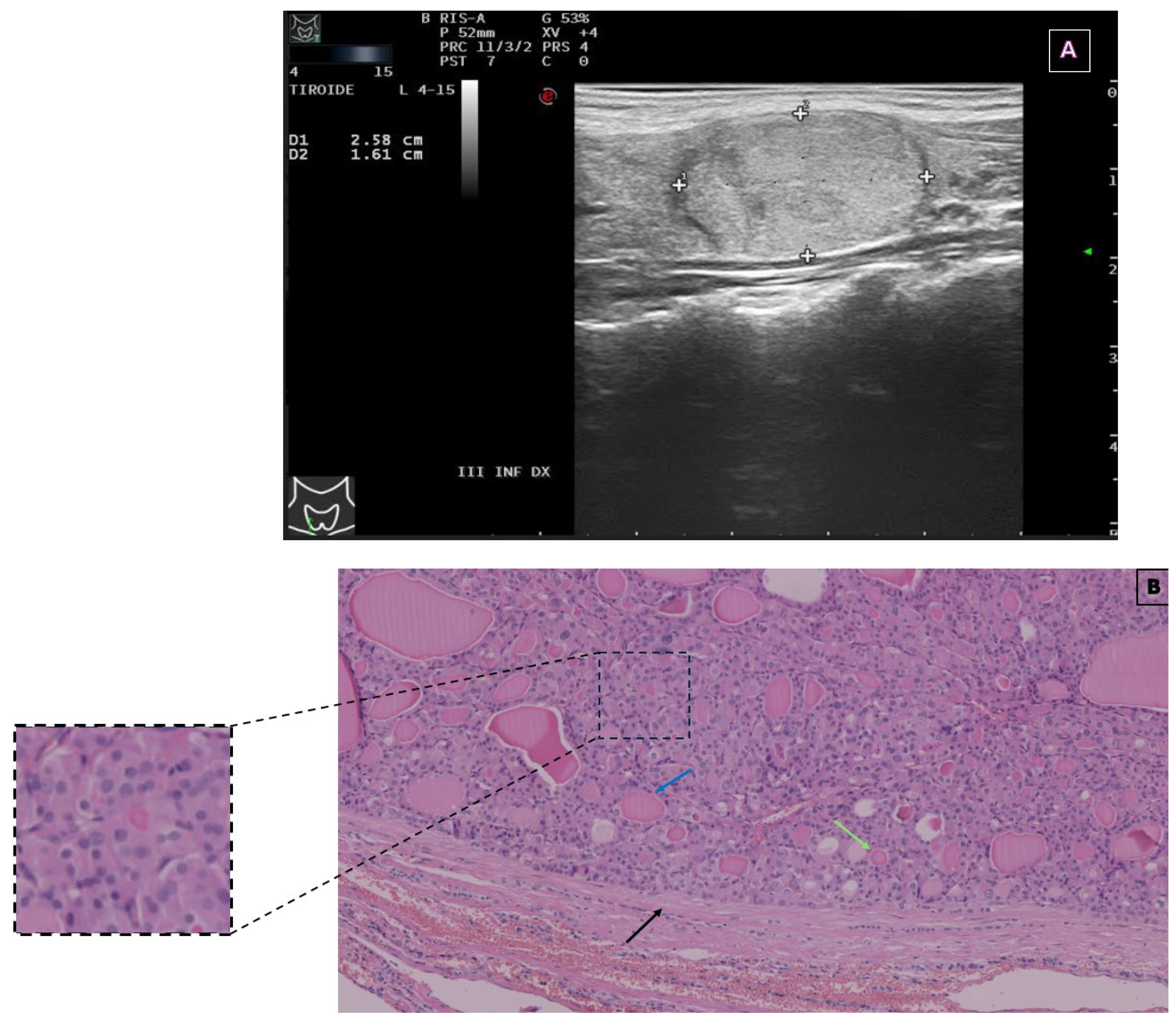
2. Case Presentation

A 13-year-old female presented to our Emergency Department with a palpable anterior neck mass, which had progressively increased in size over the past few months. She reported no symptoms of dysphagia, hoarseness, or systemic thyroid dysfunction such as weight changes, fatigue, or heat/cold intolerance. Her medical history was unremarkable except for dyslexia, and there was no history of prior radiation exposure. However, her family history was significant for thyroid disorders, including acute thyroiditis in her father and maternal grandmother, thyroid nodules in her paternal uncle, and hyperthyroidism in her paternal grandmother.
On clinical examination, the patient exhibited an isolated right thyroid nodule, approximately 2.5 cm in its largest dimension, with a firm consistency but no cervical lymphadenopathy. Thyroid function tests, including thyroid-stimulating hormone (TSH), free tri-iodothyronine (FT3), and free thyroxine (FT4), were within normal limits. Autoantibody tests for thyroid peroxidase (TPOAb) and thyroglobulin (TgAb) were negative, and serum calcitonin levels were undetectable, ruling out medullary thyroid carcinoma (Table 1). Additionally, negative complete blood count and C-reactive protein results indicated no inflammation.
Neck ultrasound revealed an enlarged right thyroid lobe (antero-posterior diameter: 19 mm; left lobe antero-posterior diameter: 7 mm) containing a well-defined, solid, isoechoic nodule with a peripheral hypoechoic halo (Figure 1A). Doppler imaging revealed peripheral and intralesional vascularization, without evidence of cervical lymph node involvement. The nodule was classified as EU-TIRADS 3, indicating low malignancy risk, but due to its size (>20 mm), FNA was recommended. Cytology demonstrated a follicular neoplasm with oncocytic features (Bethesda IV). The prominent proliferation of oncocytic cells characterized by irregular nuclei, along with occasional macrophages, and the absence of colloid, meant that it was classified as TIR3B according to the Italian Society of Anatomic Pathology and Diagnostic Cytopathology (SIAPEC) [16].
SIAPEC guidelines recommend surgical removal for TIR3B lesions. Both total thyroidectomy and lobectomy are viable options. With no additional risk factors, a thyroid lobectomy was performed under general anesthesia, and the intraoperative course was uneventful. The excised thyroid lobe was sent for histopathological evaluation, which confirmed the presence of a well-encapsulated OA (Figure 1B) without evidence of capsular or vascular invasion. No additional complications arose during the immediate postoperative period, and the patient recovered without signs of hypocalcemia or recurrent laryngeal nerve dysfunction.
Postoperative thyroid function tests remained within normal ranges, and no replacement therapy was required. At her first follow-up visit, one month after surgery, the patient reported no symptoms, and a clinical examination confirmed a well-healed surgical scar with no palpable abnormalities. Serial ultrasound examinations conducted postoperatively showed no evidence of recurrence, and the contralateral thyroid lobe remained structurally normal. At her latest follow-up, twelve months after surgery, the patient continued to be asymptomatic, maintaining normal thyroid function without the need for hormonal supplementation.

3. Discussion

The exact incidence of OAs in pediatric patients remains undefined due to their extreme rarity. Most of the available data consist of single case reports or small series, highlighting how uncommon these neoplasms are in the pediatric population [12]. In a retrospective cohort of 63 children and adolescents with thyroid nodules who underwent surgery, OAs were histologically identified in only two cases (3.2%), confirming their rarity in the pediatric population [17]. Our review of the literature identified only a few well-documented pediatric cases of OA with histopathological confirmation, which are summarized in Table 2. Literature on thyroid nodules in pediatric populations emphasizes that malignancy rates are higher in pediatric thyroid nodules than in adult cases [18], although benign nodules, like adenomas, still represent a significant portion of these cases.
Within follicular thyroid lesions, a distinct subset comprises nodules displaying oncocytic features, commonly referred to as oncocytic tumors. They constitute a unique category of thyroid neoplasms, characterized by distinct genetic alterations, including mitochondrial DNA mutations and an increased number of copy number variations [10,11,24]. To qualify as an OA, more than 75% of oncocytic cells are required [25]. In 1894, Karl Hürthle named the parafollicular cells of the thyroid “Hürthle” cells. In the latest 2022 WHO classification of thyroid tumors [26], however, this term is discouraged, since it is a misnomer. In fact, Hürthle initially identified parafollicular C cells in the thyroid glands of dogs rather than oncocytic cells in the human thyroid [27]. This terminology changed to “oncocytic” cells in the 2022 WHO classification, indicating follicular cells with abundant eosinophilic granular cytoplasm caused by an excess of mitochondria. “Oncocytes” is indeed a term that generally denotes granular eosinophilic cells found in various epithelial and glandular tissues, which are particularly prevalent in the salivary gland, kidney, and thyroid [28].
Oncocytes express thyroglobulin and other enzymes characteristic of thyroid follicular cells, indicating their derivation from thyrocytes during their developmental course. Initially, oncocytic changes were perceived as indicative of cellular senescence. However, it is now understood that these cellular alterations reflect a metaplastic process induced by cellular stress, closely linked to mitochondrial adaptive homeostasis [29]. It has been postulated that defects in mitochondrial DNA lead to their dysfunction and subsequent proliferation, culminating in an accumulation within the cytosol of follicular epithelial cells. This process unfolds gradually, explaining the rarity of oncocytes in pediatric populations [30].
One challenge is discriminating between non-neoplastic and neoplastic nodules, with several cytomorphologic criteria being considered valuable. Specifically, typical features of non-neoplastic oncocytic lesions include the presence of inflammatory cells or non-Hürthle follicular cells alongside oncocytes, a honeycomb or macrofollicular arrangement of oncocytes, a lack of nucleoli, and abundant colloid. Conversely, a combination of the following four features is highly indicative of neoplastic oncocytic lesions: a non-macrofollicular architecture, absence of colloid, a lack of inflammation, and the presence of transgressing blood vessels [31]. However, cytomorphologic risk factors alone are insufficient for distinguishing between OA and OC. Histologically, according to the 2022 WHO classification, OCs are encapsulated tumors with capsular and/or vascular invasion and at least 75% oncocytic cells. They are characterized by the absence of the distinctive nuclear features of papillary thyroid carcinoma and lack high-grade features such as necrosis and ≥5 mitoses per 2 mm² [26]. In contrast, the absence of such invasive features supports the diagnosis of OA, as in our case. The differential diagnosis thus relies on postoperative histopathological examinations. While FNA can guide the surgical approach, in fact, it is unable to identify capsular and/or vascular invasion and, therefore, to reliably distinguish between OA and OC. Most oncocytic tumors are benign, but up to 40% have been reported as malignant (OCs) [32]. Surgical histopathological analysis, which provides high accuracy, remains the gold standard for the confirmation of diagnoses [12].
OAs are typically benign and often localize unilaterally. In pediatric patients, they are generally managed through hemithyroidectomy; moreover, like most patients after lobectomy for benign thyroid neoplasms, our patient was also euthyroid [21]. However, it is important to note that OA can present bilaterally in rare cases, as seen in a 2016 report of a 14-year-old female with bilateral OA who underwent sequential hemithyroidectomy after the nodules showed growth [20]. Surgical treatment is crucial for all cases due to the risk of malignancy. Although OA is generally considered benign, its clinical course can be unpredictable, and close monitoring is essential, as the lesion may enlarge or become bilateral over time.
In contrast, OC can be more aggressive and is associated with bilateral presentation in some cases. As a malignant tumor, OC is typically treated with total thyroidectomy, in contrast to OA, which can be managed with hemithyroidectomy when no invasive features are present. In the general population, OC accounts for 3–7% of all differentiated thyroid cancers [33]. In pediatric cases, it is instead a rare occurrence, with only a limited number of case reports documented in the literature and a dearth of specific reviews. A recent study examining five cases of pediatric OC states that its prevalence among thyroid cancers in childhood is 5.8% [34]. A notable characteristic of OC is its association with a more aggressive clinical course in adults, resulting in a lower 10-year survival rate when compared to other well-differentiated thyroid cancers [12]. In children, however, the prognosis is generally more favorable. In pediatric patients, OC can also be associated with other conditions, including Hashimoto thyroiditis, neurofibromatosis type 1, or osteosarcoma, highlighting the importance of considering these comorbidities in the diagnostic workup [35].
Unlike adults, where oncocytic features often prompt close surveillance and the consideration of total thyroidectomy, pediatric guidelines are less specific about the management of oncocytic neoplasms, given the lack of data. Non-neoplastic lesions may warrant follow-up FNAs [28], while neoplastic lesions typically necessitate surgical excision for diagnostic evaluation. Distinguishing benign and malignant lesions is crucial, as treatment strategies differ significantly: OC often necessitates more extensive surgical intervention, such as total thyroidectomy, and may require postoperative radioactive iodine therapy, while OA may be managed conservatively with lobectomy alone [36], as in our patient, especially when the lesion is encapsulated and shows no evidence of vascular or capsular invasion. While OAs in adults may sometimes progress to carcinoma, the current pediatric literature indicates that most thyroid nodules in children, including OAs, follow a benign course post-resection [37].

4. Conclusions

This case of OA in a 13-year-old girl highlights the distinct challenges of diagnosing and managing thyroid nodules in pediatric patients. Although thyroid nodules are relatively rare in children, they carry a higher risk of malignancy compared to adults. This case underscores the value of a multidisciplinary approach, involving clinical evaluation, imaging, FNA, and histopathological analysis, to accurately diagnose and treat these lesions. Given the rarity of OAs in pediatric populations, this report contributes valuable insights, emphasizing the need for ongoing research to better delineate the natural history, risk factors, and optimal management strategies for pediatric thyroid nodules, particularly those with oncocytic features.

Author Contributions

Conceptualization, R.P., G.B. and S.S.; writing—original draft preparation, G.B.; writing—review and editing, R.P.; resources, L.R., N.V. and G.R.; supervision, R.P., M.C. and S.S. All authors have read and agreed to the published version of the manuscript.

Funding

This research received no external funding.

Institutional Review Board Statement

Not applicable.

Informed Consent Statement

Written informed consent was obtained from the patient’s parents to publish this study.

Data Availability Statement

The original contributions presented in this study are included in the article. Further inquiries can be directed at the corresponding author.

Acknowledgments

The authors thank the patient’s family.

Conflicts of Interest

The authors declare no conflicts of interest.

Abbreviations

The following abbreviations are used in this manuscript:
EU-TIRADSEuropean Thyroid Imaging and Reporting Data System
FNAFine-needle aspiration
FT3Free tri-iodothyronine
FT4Free thyroxine
OAOncocytic adenoma
OCOncocytic carcinoma
SIAPECItalian Society of Anatomic Pathology and Diagnostic Cytopathology
TgAbAnti-thyroglobulin antibodies
TPOAbThyroid peroxidase antibodies
TSHThyroid-stimulating hormone

References

  1. Durante, C.; Hegedüs, L.; Czarniecka, A.; Paschke, R.; Russ, G.; Schmitt, F.; Soares, P.; Solymosi, T.; Papini, E. 2023 European Thyroid Association Clinical Practice Guidelines for Thyroid Nodule Management. Eur. Thyroid. J. 2023, 12, e230067. [Google Scholar] [CrossRef] [PubMed]
  2. Gharib, H.; Papini, E.; Paschke, R.; Duick, D.S.; Valcavi, R.; Hegedüs, L.; Vitti, P. AACE/AME/ETA Task Force on Thyroid Nodules; American Association of Clinical Endocrinologists, Associazione Medici Endocrinologi, and European Thyroid Association Medical Guidelines for Clinical Practice for the Diagnosis and Management of Thyroid Nodules: Executive Summary of Recommendations. Endocr. Pract. 2010, 16, 468–475. [Google Scholar] [CrossRef] [PubMed]
  3. Suzuki, S.; Suzuki, S.; Fukushima, T.; Midorikawa, S.; Shimura, H.; Matsuzuka, T.; Ishikawa, T.; Takahashi, H.; Ohtsuru, A.; Sakai, A.; et al. Comprehensive Survey Results of Childhood Thyroid Ultrasound Examinations in Fukushima in the First Four Years After the Fukushima Daiichi Nuclear Power Plant Accident. Thyroid 2016, 26, 843–851. [Google Scholar] [CrossRef]
  4. Rallison, M.L.; Dobyns, B.M.; Keating, F.R.; Rall, J.E.; Tyler, F.H. Thyroid Nodularity in Children. JAMA 1975, 233, 1069–1072. [Google Scholar] [CrossRef]
  5. Creo, A.; Alahdab, F.; Al Nofal, A.; Thomas, K.; Kolbe, A.; Pittock, S.T. Ultrasonography and the American Thyroid Association Ultrasound-Based Risk Stratification Tool: Utility in Pediatric and Adolescent Thyroid Nodules. Horm. Res. Paediatr. 2018, 90, 93–101. [Google Scholar] [CrossRef]
  6. Hanley, P.; Lord, K.; Bauer, A.J. Thyroid Disorders in Children and Adolescents: A Review. JAMA Pediatr. 2016, 170, 1008–1019. [Google Scholar] [CrossRef]
  7. Carlomagno, F.; Minnetti, M.; Angelini, F.; Pofi, R.; Sbardella, E.; Spaziani, M.; Aureli, A.; Anzuini, A.; Paparella, R.; Tarani, L.; et al. Altered Thyroid Feedback Loop in Klinefelter Syndrome: From Infancy Through the Transition to Adulthood. J. Clin. Endocrinol. Metab. 2023, 108, e1329–e1340. [Google Scholar] [CrossRef] [PubMed]
  8. Russ, G.; Bonnema, S.J.; Erdogan, M.F.; Durante, C.; Ngu, R.; Leenhardt, L. European Thyroid Association Guidelines for Ultrasound Malignancy Risk Stratification of Thyroid Nodules in Adults: The EU-TIRADS. Eur. Thyroid J. 2017, 6, 225–237. [Google Scholar] [CrossRef]
  9. Francis, G.L.; Waguespack, S.G.; Bauer, A.J.; Angelos, P.; Benvenga, S.; Cerutti, J.M.; Dinauer, C.A.; Hamilton, J.; Hay, I.D.; Luster, M.; et al. Management Guidelines for Children with Thyroid Nodules and Differentiated Thyroid Cancer: The American Thyroid Association Guidelines Task Force on Pediatric Thyroid Cancer. Thyroid 2015, 25, 716–759. [Google Scholar] [CrossRef]
  10. McFadden, D.G.; Sadow, P.M. Genetics, Diagnosis, and Management of Hürthle Cell Thyroid Neoplasms. Front. Endocrinol. 2021, 12, 696386. [Google Scholar] [CrossRef]
  11. Gopal, R.K.; Kübler, K.; Calvo, S.E.; Polak, P.; Livitz, D.; Rosebrock, D.; Sadow, P.M.; Campbell, B.; Donovan, S.E.; Amin, S.; et al. Widespread Chromosomal Losses and Mitochondrial DNA Alterations as Genetic Drivers in Hürthle Cell Carcinoma. Cancer Cell 2018, 34, 242–255.e5. [Google Scholar] [CrossRef]
  12. Wong, K.S.; Angell, T.E.; Barletta, J.A.; Krane, J.F. Hürthle Cell Lesions of the Thyroid: Progress Made and Challenges Remaining. Cancer Cytopathol. 2021, 129, 347–362. [Google Scholar] [CrossRef]
  13. Cibas, E.S.; Ali, S.Z. The 2017 Bethesda System for Reporting Thyroid Cytopathology. Thyroid 2017, 27, 1341–1346. [Google Scholar] [CrossRef] [PubMed]
  14. Ali, S.Z.; Baloch, Z.W.; Cochand-Priollet, B.; Schmitt, F.C.; Vielh, P.; VanderLaan, P.A. The 2023 Bethesda System for Reporting Thyroid Cytopathology. Thyroid 2023, 33, 1039–1044. [Google Scholar] [CrossRef] [PubMed]
  15. Grant, C.S. Operative and Postoperative Management of the Patient with Follicular and Hürthle Cell Carcinoma. Do They Differ? Surg. Clin. N. Am. 1995, 75, 395–403. [Google Scholar] [CrossRef] [PubMed]
  16. Nardi, F.; Basolo, F.; Crescenzi, A.; Fadda, G.; Frasoldati, A.; Orlandi, F.; Palombini, L.; Papini, E.; Zini, M.; Pontecorvi, A.; et al. Italian Consensus for the Classification and Reporting of Thyroid Cytology. J. Endocrinol. Investig. 2014, 37, 593–599. [Google Scholar] [CrossRef]
  17. Corrias, A.; Mussa, A.; Baronio, F.; Arrigo, T.; Salerno, M.; Segni, M.; Vigone, M.C.; Gastaldi, R.; Zirilli, G.; Tuli, G.; et al. Diagnostic Features of Thyroid Nodules in Pediatrics. Arch. Pediatr. Adolesc. Med. 2010, 164, 714–719. [Google Scholar] [CrossRef]
  18. Gupta, A.; Ly, S.; Castroneves, L.A.; Frates, M.C.; Benson, C.B.; Feldman, H.A.; Wassner, A.J.; Smith, J.R.; Marqusee, E.; Alexander, E.K.; et al. A Standardized Assessment of Thyroid Nodules in Children Confirms Higher Cancer Prevalence Than in Adults. J. Clin. Endocrinol. Metab. 2013, 98, 3238–3245. [Google Scholar] [CrossRef]
  19. Green, O.; Keisling, M.; Kambalapalli, M.; McDaniel, J.; Boulanger, S.; Fornwalt, B.E.; Jeyakumar, A. Unusual Thyroid Mass in an Adolescent Patient. Ear Nose Throat J. 2022, 101, 654–656. [Google Scholar] [CrossRef]
  20. Kochummen, E.; Tong, S.; Umpaichitra, V.; Chin, V.L. A Unique Case of Bilateral Hürthle Cell Adenoma in an Adolescent. Horm. Res. Paediatr. 2017, 87, 136–142. [Google Scholar] [CrossRef]
  21. Bremer, A.A.; Feldman, B.J.; Iezza, G.; Clark, O.H.; Rosenthal, S.M. Report of a Hürthle Cell Neoplasm in a Peripubertal Girl. Thyroid 2007, 17, 175–178. [Google Scholar] [CrossRef] [PubMed]
  22. Roggli, V.L.; Estrada, R.; Fechner, R.E. Thyroid Neoplasia Following Irradiation for Medulloblastoma: Report of Two Cases. Cancer 1979, 43, 2232–2238. [Google Scholar] [CrossRef] [PubMed]
  23. Nagamachi, Y.; Nakamura, T.; Yamada, T. Hürthle Cell Adenoma of the Thyroid in Identical Twins with 13 Year Follow-Up. Jpn. J. Surg. 1973, 3, 212–217. [Google Scholar] [CrossRef] [PubMed]
  24. Doerfler, W.R.; Nikitski, A.V.; Morariu, E.M.; Ohori, N.P.; Chiosea, S.I.; Landau, M.S.; Nikiforova, M.N.; Nikiforov, Y.E.; Yip, L.; Manroa, P. Molecular Alterations in Hürthle Cell Nodules and Preoperative Cancer Risk. Endocr. Relat. Cancer 2021, 28, 301–309. [Google Scholar] [CrossRef]
  25. Goswami, P.; Patel, T.; Dave, R.; Singh, G.; Singh, A.; Kalonia, T. WHO 2022 Updates on Follicular Cell and C-Cell Derived Thyroid Neoplasm. J. Med. Life 2024, 17, 15–23. [Google Scholar] [CrossRef]
  26. Baloch, Z.W.; Asa, S.L.; Barletta, J.A.; Ghossein, R.A.; Juhlin, C.C.; Jung, C.K.; LiVolsi, V.A.; Papotti, M.G.; Sobrinho-Simões, M.; Tallini, G.; et al. Overview of the 2022 WHO Classification of Thyroid Neoplasms. Endocr. Pathol. 2022, 33, 27–63. [Google Scholar] [CrossRef]
  27. Caturegli, P.; Ruggere, C. Karl Hürthle! Now, Who Was He? Thyroid 2005, 15, 121–123. [Google Scholar] [CrossRef]
  28. Auger, M. Hürthle Cells in Fine-needle Aspirates of the Thyroid: A Review of Their Diagnostic Criteria and Significance. Cancer Cytopathol. 2014, 122, 241–249. [Google Scholar] [CrossRef]
  29. Asa, S.L. My Approach to Oncocytic Tumours of the Thyroid. J. Clin. Pathol. 2004, 57, 225–232. [Google Scholar] [CrossRef]
  30. Montone, K.T.; Baloch, Z.W.; LiVolsi, V.A. The Thyroid Hürthle (Oncocytic) Cell and Its Associated Pathologic Conditions: A Surgical Pathology and Cytopathology Review. Arch. Pathol. Lab. Med. 2008, 132, 1241–1250. [Google Scholar] [CrossRef]
  31. Elliott, D.D.; Pitman, M.B.; Bloom, L.; Faquin, W.C. Fine-Needle Aspiration Biopsy of Hurthle Cell Lesions of the Thyroid Gland: A Cytomorphologic Study of 139 Cases with Statistical Analysis. Cancer 2006, 108, 102–109. [Google Scholar] [CrossRef] [PubMed]
  32. Okere, P.; Olusina, D.; Enyinnah, M. Hurthle Cell Tumor of the Thyroid Gland: Report of a Rare Case and Review of Literature. Niger. J. Clin. Pract. 2014, 17, 375–377. [Google Scholar] [CrossRef]
  33. Barnabei, A.; Ferretti, E.; Baldelli, R.; Procaccini, A.; Spriano, G.; Appetecchia, M. Hurthle Cell Tumours of the Thyroid. Personal Experience and Review of the Literature. ACTA Otorhinolaryngol. Ital. 2009, 29, 305–311. [Google Scholar] [PubMed]
  34. Zirilli, G.; Santucci, S.; Cuzzupè, C.; Corica, D.; Pitrolo, E.; Salzano, G. Peculiarities of Autoimmune Polyglandular Syndromes in Children and Adolescents. Acta Bio-Medica Atenei Parm. 2017, 88, 271–275. [Google Scholar]
  35. Zirilli, G.; Valenzise, M.; Dionigi, G.; Tuccari, G.; Romeo, C.; Campennì, A.; Corrias, A.; Tuli, G.; Ieni, A.; Pajno, G.B.; et al. Hurthle Cell Carcinoma in Childhood: A Retrospective Analysis of Five Cases and Review of Pediatric Literature. Pediatr. Blood Cancer 2020, 67, e28300. [Google Scholar] [CrossRef] [PubMed]
  36. Haugen, B.R.; Alexander, E.K.; Bible, K.C.; Doherty, G.M.; Mandel, S.J.; Nikiforov, Y.E.; Pacini, F.; Randolph, G.W.; Sawka, A.M.; Schlumberger, M.; et al. 2015 American Thyroid Association Management Guidelines for Adult Patients with Thyroid Nodules and Differentiated Thyroid Cancer: The American Thyroid Association Guidelines Task Force on Thyroid Nodules and Differentiated Thyroid Cancer. Thyroid 2016, 26, 1–133. [Google Scholar] [CrossRef]
  37. Micangeli, G.; Menghi, M.; Profeta, G.; Paparella, R.; Tarani, F.; Petrella, C.; Barbato, C.; Minni, A.; Greco, A.; Ferraguti, G.; et al. Malignant and Benign Head and Neck Tumors of the Pediatric Age: A Narrative Review. Curr. Pediatr. Rev. 2024, 212, 118–132. [Google Scholar] [CrossRef]
Figure 1. Ultrasound and histological features of the patient’s thyroid nodule. (A) Longitudinal scan of the right thyroid lobe. A large (2.58 × 1.61 cm), solid, isoechoic nodule with a distinct peripheral hypoechoic halo is clearly delineated by the white cross markers in the image. (B) Histological section of the thyroid nodule (H&E stain, 10× magnification). The tumor consists of oncocytic cells arranged in a mixed microfollicular (green arrow) and normofollicular (blue arrow) growth pattern, surrounded by a thick fibrous capsule (black arrow) in the absence of capsular or vascular invasion. The inset shows a higher magnification of the oncocytic cells, characterized by large polygonal morphology with uniform round nuclei, often prominent nucleoli, and granular eosinophilic cytoplasm.
Endocrines 06 00022 g001
Table 1. Blood test results for thyroid function, autoantibodies, and calcitonin levels.
Thyroid Function TestResultReference Range
FT33.4 pg/mL2.3–4.2
FT41.09 ng/dL0.8–1.6
TSH1.81 mIU/mL0.4–3.5
Calcitonin<0.5 pg/mL<6.4
TPOAb30 U/mL<60
TgAb<10 U/mL<4.5
Table 2. Summary of reported pediatric cases of oncocytic (Hürthle cell) adenomas in the literature.
StudyAge (Years)/GenderNodule
Size
PresentationFNA
Cytology
TreatmentOutcome
Current case13/Female26 × 16 mmEnlarging
neck mass
Bethesda IV
(follicular neoplasm with oncocytic features)
HemithyroidectomyNo
recurrence
Green et al., 2020 [19]18/Female5.9 × 4.4 × 3.7 cmIncidentally identified neck mass, mild hyperthyroid symptomsRare clusters of cells with cytological atypia, negative for nuclear features of
papillary carcinoma
Left
hemithyroidectomy
No
recurrence
Kochummen et al., 2017 [20]14/FemaleBilateral nodules (sizes not
specified)
Enlarging
thyroid nodules
Not specifiedSequential
hemithyroidectomy
No
recurrence
Bremer et al., 2007 [21]12/Female20 mmPalpable thyroid noduleSuspicious
for follicular
neoplasm
HemithyroidectomyNo
recurrence
Roggli et al., 1979 [22] 122/FemaleMultiple nodules (largest: 4 cm)Neck mass 18 years after
medulloblastoma treatment
Not specifiedSubtotal
thyroidectomy
(95%)
No
recurrence (8.5-month
follow-up)
Nagamachi et al., 1973 [23]12/Female (Twin 1)Left lobe: single cystic tumor (~egg-sized); right lobe: three
nodules
(~10 mm each)
Enlarging
cervical mass
Not specifiedLocal removal
(First surgery),
hemithyroidectomy (Second surgery)
No
recurrence (13-year follow-up)
Nagamachi et al., 1973 [23]12/Female (Twin 2)Left lobe: 1 cm nodule;
right lobe: 3 cm cystic nodule
Enlarging
cervical mass
Not specifiedLocal removal
(First surgery),
hemithyroidectomy (Second surgery)
No
recurrence (13-year follow-up)
1 This case is not strictly pediatric, as the patient developed Hürthle cell adenoma at the age of 22, 18 years after receiving radiation therapy for medulloblastoma. However, it has been included for completeness, given the potential risk after childhood radiation exposure.
Disclaimer/Publisher’s Note: The statements, opinions and data contained in all publications are solely those of the individual author(s) and contributor(s) and not of MDPI and/or the editor(s). MDPI and/or the editor(s) disclaim responsibility for any injury to people or property resulting from any ideas, methods, instructions or products referred to in the content.

