Case Report

Calycovesicostomy, Ureterocalycostomy, and Ileocalycostomy: Rare Reconstructive Options for Transplant Ureteral Strictures

1
MedStar Georgetown Transplant Institute, MedStar Georgetown University Hospital, Washington, DC 20007, USA
2
Urology Unit, Department of Surgery, Al-Amiri General Hospital, Kuwait City 13041, Al Asimah Governate, Kuwait
*
Author to whom correspondence should be addressed.
Transplantology 2025, 6(3), 27; https://doi.org/10.3390/transplantology6030027
Submission received: 15 July 2025 / Revised: 21 August 2025 / Accepted: 2 September 2025 / Published: 4 September 2025
(This article belongs to the Section Solid Organ Transplantation)

Abstract

Transplant ureteral stricture (TUS) reconstruction remains a significant challenge in renal transplantation, particularly when conventional access to the transplant ureter and renal pelvis is not feasible. This report presents two rare and complex surgical reconstructions: a combined calico-vesicostomy and uretero-calycostomy in one patient, and an ileo-calycostomy in another. Both cases involved patients with prior failed minimally invasive interventions and complex anatomy that precluded standard repairs. A hybrid surgical approach incorporating open reconstruction with fluoroscopic and endoscopic guidance was used to access the renal calyces directly, enabling successful re-establishment of urinary tract continuity. The first patient underwent dual reconstruction with native ureteral reimplantation to the middle calyx and a simultaneous calycovesicostomy, bladder to the It lower pole calyx. The second patient, with prior ileal conduit urinary diversion, underwent ileocalycostomy—anastomosing the ileal conduit to the middle calyx. Both reconstructions were performed using modified partial nephrectomies to preserve nephron mass. Long-term follow-up (34 and 40 months) demonstrated excellent graft function and minimal complications. These cases represent the first reported long-term outcomes of calycovesicostomy and ileocalycostomy in transplant ureteral reconstruction and reinforce the utility of calyceal access in complex TUSs. These cases highlight rare but viable surgical options in complex scenarios of transplant ureteral strictures. Hence, a multidisciplinary approach and meticulous preoperative planning are essential to optimize outcomes in these challenging scenarios.

1. Introduction

Renal transplantation is the gold standard treatment for end-stage renal disease. Despite surgical advances in the field and refinements in the techniques of ureteral anastomoses, transplant ureteric strictures (TUSs) continue to poise the outcome with many risk factors identified [1,2,3,4]. Although minimally invasive options are attractive, patients who are fit for surgery and reluctant to undergo a major reconstructive option should be counselled regarding the high recurrence rate and need for multiple procedures [5]. Surgical repair remains the optimum approach with a higher success rate, and translates into better long-term graft survival [6,7]. Endourological and radiologic approaches should ideally be reserved for nonsurgical candidates or patients with distal, short (<1–2 cm), nonischemic strictures [5]. Since its first description, ureterocalycostomy has been reserved for rare and unique scenarios [8,9,10,11,12,13]. In patients with non-conventional anatomy, a hybrid technique combining surgical and fluoroscopic/endoscopic-guided access to the renal collecting system and calyces is a unique approach to these complex scenarios when availability/access to the transplant ureter or renal pelvis is not an option. Herein, we report two cases of rare reconstructive options of re-establishment of urinary tract continuity using a combined calycovesicostomy and ureterocalycostomy in one patient, and Ileo-calycostomy in the other. Full informed consent was obtained from the patients for intraoperative pictures and postoperative imaging. Ethical review was not required for this study as per national Ethical Guidelines for Biomedical research; Kuwait institution of Medical Specialties KIMS. Ethical Guidelines for Biomedical Research. Kuwait; 2009.

2. Case Presentation

2.1. Case 1

A 67-year-old female with history of chronic kidney disease (CKD) secondary to hypertension underwent a living-related renal transplant in August 2016 complicated with a long ureteral stricture 4 months post-transplant. She was managed initially by repeated dilatations and long-term ureteral stenting with exchanges every 3–6 months at another center. She was suffering on average from two to three urinary tract infections a year, of which two were documented admissions for urosepsis. She was seen in clinic and counselled on surgical reconstruction. Preoperative imaging demonstrated a markedly dilated calyceal system with thin renal parenchyma, and a renal pelvis rotated inferiorly facing the iliac vessels (Figure 1). She had a normal pre-operative creatinine of 66.3 mmol/L. As part of operative planning, a percutaneous nephrostomy tube (PCN) was placed, and the ureteral stent was removed for ureteral rest. On 11 January 2022, she underwent an open and fluoroscopic hybrid approach with access to the calyces obtained via fluoroscopic guidance. Initially the right native ureter to middle calyx, ureterocalycostomy, was performed after resecting the parenchyma overlying the calyx in a diamond configuration and completing the anastomosis in an end-to-end fashion using interrupted 5-0 polydioxanone (PDS II) sutures over an 18 cm 6F JJ stent. The caliber of the native ureter was not very reassuring, and there was minimal bleeding from the spatulated end. Although the intra-operative nephrostogram demonstrated a patent anastomosis, there was minimal peristalsis, and the ureter did not opacify adequately. To ensure adequate drainage and to optimize the outcome, a lower-pole partial nephrectomy was simultaneously performed to expose the dilated lower calyx and an anastomosis of the lower calyx to the bladder, a calycovesicostomy (Figure 2) was performed in a continuous fashion using 3-0 polydioxanone (PDS) suture over an 8F 16 cm JJ stent. A second layer of interrupted 4-0 PDS sutures was placed between the parenchyma/capsule and the detrusor muscle to ensure a watertight and tension free anastomosis. A final nephrostogram was performed at the end (Figure 3), and a 19F pelvic drain was placed. The estimated blood loss during the case was 500 cc.
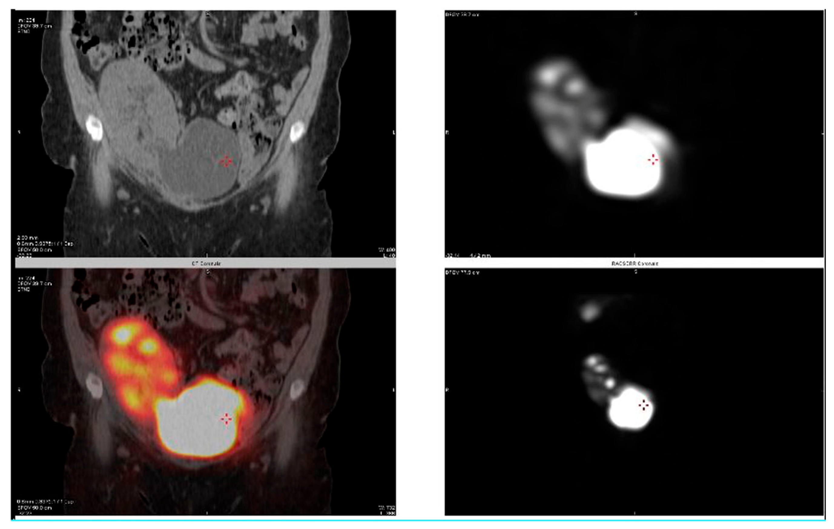
The post-operative course was smooth, and after ensuring adequate drainage at 2 weeks post-operation, the PCN was clamped followed by stent removal at 6 weeks. A week after the stent removal, a nephrostogram was performed demonstrating brisk flow and drainage (at 7 weeks post-repair). At 6 months, a MAG3 renal scan and CT SPECT were performed demonstrating excellent drainage of the kidney (Figure 4). During the first 30 days post-operation, there were no complications encountered. She developed a UTI at approximately 15 months post-repair that required outpatient management (Clavien–Dindo II). The patient continues to do very well, infection- and procedure-free, with a creatinine of 57 mmol/L at 40 months post-repair.

2.2. Case 2

A 22-year-old male with a history of Neurofibromatosis Type 1 and CKD secondary to posterior urethral valve and obstructive uropathy. He had undergone bilateral native nephrectomy and ileal conduit urinary diversion at age 8, and has had two renal transplants, one in 2008 that was complicated with graft thrombosis, and a second graft in 2009 that failed in 2020 rendering him back on dialysis. On 7 January 2022, he received a third-time deceased donor transplant onto the left iliac system onto his left sided ileal conduit. During the transplant, the kidney was intentionally placed inverted so that the transplant ureter could reach the left-sided ileal conduit. He initially did very well with immediate graft function and a creatinine nadir of 133 mmol/L. Five months after transplant, he was admitted with acute kidney injury (AKI) and hydronephrosis and was found to have a stricture/twist in the proximal third of his ureter after insertion of the nephrostomy tube, along with BK viraemia. After control of his BK viremia, and a creatinine baseline of 177 mmol/L, the patient was counselled on options for surgical reconstruction. After informed consent, he underwent surgery on 17 July 2022. An intra-operative nephrostogram was taken, and after midline incision the ileal conduit was mobilized completely, and the ureter was dissected and transected off the conduit. The ureteral tissue was friable and deemed unsuitable for reconstruction. Intra-operative ureteroscopy of the transplant ureter was performed, and given the intrarenal nature of the renal pelvis, and the location of the obstruction, the options were narrowed down to using his calyces. The operative field was densely scarred, and the kidney was implanted in an inverted manner to reach the conduit originally, the upper pole was inferior, and the lower pole was superior. Inherently, the calyces were all lateral and hence the middle calyx was the most medial calyx for us to access. The size of the ileal conduit, and the ability to tunnel a segment of it through the sigmoid mesentery to complete the anastomosis without tension, also made it more technically feasible to the middle calyx. After delineating the middle calyx with ureteroscopy and fluoroscopy, a direct needle puncture under vision was performed and a guidewire used to direct the scope through the middle calyx. The middle calyx was exposed after a modified partial nephrectomy in a diamond configuration of the parenchyma, and the proximal end of the ileal conduit was tunneled through the mesentery of the sigmoid colon, followed by anastomosing the proximal end of his ileal conduit to the middle calyx in an end-to-end fashion using 3-0 polydioxanone (PDS), achieving an ileo-calycostomy (Figure 5) over a 6F 22 cm single J stent and 16F Malecot drain into the ileal conduit. A 19F pelvic drain was placed. The estimated blood loss was 400 cc.
His post-operative course was complicated by a urinary tract infection that was treated with IV antibiotics (Clavien–Dindo II). His PCN was kept open to drainage, and the Malecot drain was used to gently flush the conduit daily with 20 cc of normal saline. At 4 weeks, an antegrade CT nephrostogram was performed demonstrating brisk drainage into the conduit. The PCN was clamped, and two weeks later the stent and drain were removed followed by removal of the PCN at the 7-week mark. Figure 6 demonstrates his 6 month follow-up MAG3 renal scan showing excellent emptying and drainage of the allograft. At 32 months follow up, he had developed a small incisional hernia and incurred another urinary tract infection that required hospitalization (Clavien–Dindo II). He remains asymptomatic, free of BK/urinary tract infections, with a creatinine of 190 mmol/L during his last follow up at 34 months post reconstruction.

3. Discussion

In scenarios where access to the transplant ureter or renal pelvis is limited, the hybrid approach of surgical, fluoroscopic- and endoscopic-guided access to the calyces proves to be an effective and viable complex reconstructive option. We hereby demonstrate excellent long-term outcomes of a case of combined calycovesicostomy and ureterocalycostomy, and a rare case of ileo-calycostomy in the management of complicated transplant ureteral strictures. The use of advanced open urologic reconstructive techniques in managing TUSs after failed endourological attempts is technically challenging; however, it is feasible, safe, and associated with good long-term patient and graft outcomes even when bowel segments are interposed to re-establish continuity [7]. Hence, we advocate further for utilizing this hybrid approach in attempting to definitively repair and restore continuity of the urinary tract when the transplant ureter or renal pelvis is inaccessible.
Transplant ureteral strictures have a negative impact on long-term graft survival [6], and recipients who are initially treated with a minimally invasive approach, over a definitive surgical approach, had shorter kidney graft survival compared with recipients with no ureteral strictures. When addressing patients with TUSs, a standardized algorithm previously published by the primary author facilitates the approach to these complicated cases [7]. Undoubtedly, a minimally invasive option is always attractive; however, solid evidence exists to question the longevity and durability of those approaches. Distal TUS are most common, and in a systematic review, the open reconstructive approach had a higher success rate than endourological treatments [5]. In all cases combined, the open approach for primary treatment had an 85% success rate versus a 64% success rate with the endourological technique. The most common primary open and endourological treatments were ureteric reimplantation and balloon dilatation, with 82% and 58% success rates, respectively [5].
Management of proximal and pan-ureteral transplant strictures is very challenging, especially when standard common approaches are not feasible. Ureterocalycostomy is a valuable surgical option and is indicated in cases of complicated native ureteropelvic junction obstructions or upper ureteral injuries with inaccessible renal pelvis [13,14,15]. Multiple series using the open, laparoscopic, and robotic approach have demonstrated excellent outcomes in native ureteral reconstruction in adult and pediatric patients [14,16,17,18,19,20,21], with patency rates reported between 76% and 92%. However, the application of ureterocalycostomy in the transplant setting remains sparse. To date, only three case reports of ureterocalycostomy for managing TUSs have been described with good short-term (<1 year) results [22,23,24]. Similarly, the reporting of calycovesicostomy has been limited in the literature. The first successful description of calycovesicostomy for managing an obstructed transplant kidney presented a case of a 13-year old patient who had previously undergone bilateral native nephroureterectomy, and at time of exploration the transplant ureter was unsuitable for reconstruction [25]. Hence, similar to our scenarios where no other option was available, the authors demonstrated feasibility and patency of calycovesicostomy for 6 months before allograft loss due to rejection [25]. In describing their approach, a psoas hitch was utilized to facilitate proximity of the bladder to the calyx, and a lower pole partial nephrectomy to expose the calyx was necessary. To our knowledge, we present the first report of ileo-calycostomy in managing a transplant ureteral stricture, and the description of a hybrid approach. Interposing bowel segments in reconstruction of transplanted kidneys has excellent long term patency outcomes; however, it comes at the risk of urinary tract infections and bowel-related complications [7].
We would like to highlight multiple points in congruence with principles of approaching these challenging cases.
First, preoperative optimization played a critical role in these cases. Placement of a percutaneous nephrostomy tube and ureteral rest (removing the ureteral stent) prior to tackling these cases is paramount to aid in system decompression, intra-operative decision making (via antegrade contrast and methylene blue injection), and post-operative resting and healing of the anastomosis. Moreover, calyceal exposure through a partial nephrectomy proved to be a key step. In prior reports, the authors describe the importance of lower-pole partial nephrectomy to facilitate exposure of the lower calyx and to prevent recurrent ureteral obstruction secondary to fibrosis of the cortex overlying the intrarenal segment [22,23,24]. Traditionally, the lower-pole renal cortex is thin and easily amputated to identify the lower pole calyx. In cases where the cortex is substantial and in a solitary transplanted kidney, the importance of nephron sparing is paramount. To our knowledge, no successful description of utilizing the middle calyx with a modified partial nephrectomy for calyceal exposure has been described as we have in our ileocalycostomy case, hence the novel approach of utilizing fluoroscopic and endoscopic techniques to facilitate that exposure. Furthermore, a modified diamond configured partial nephrectomy whereby nephron mass is spared, and the calyx is exposed, has proven patency of greater than 3 years with no recurrence due to fibrosis or scarring around the anastomosis in both lower and middle calyx approach. Lastly, we highlight the approach of fluoroscopic and endoscopic guidance in enabling utility of accessing calyces even when the lower most dependent calyx is not the most favorable. Inherently, our limitation is a small sample size and hence strong inferences on the ease and replicability of the approach is challenging; however, we solidify the prior reports by demonstrating excellent outcomes with far longer follow up. The approach in renal allografts does not mirror that of native kidneys, and hence experience in both urology and transplantation is pivotal.

4. Conclusions

The objective of restoring the continuity of the urinary tract and preserving renal function with the lowest morbidity possible is the trifecta of reconstruction for transplant ureteral strictures. In scenarios where conventional options are not feasible, the advanced hybrid approach of open and endoscopic/fluoroscopic proves to be an excellent option. We demonstrated long-term survival of patients and preservation of their renal allografts with complex TUSs using calycovesicostomy, ureterocalycostomy, and ileocalycostomy. When dealing with these difficult scenarios, a multi-faceted urologic team with transplant, reconstructive and advanced endourologic training and experience is key for surgical success.

Author Contributions

Conceptualization, T.A.-Q., A.A.-T.; Supervision, T.A.-Q.; Writing-original draft, T.A.-Q.; Writing-review and editing, B.B., A.A.-T., R.A.-Y.; Resources, R.A.-Y.; Data collection, B.B. All authors have read and agreed to the published version of the manuscript.

Funding

This research received no external funding.

Institutional Review Board Statement

Ethical review was not required for this study as per national Ethical Guidelines for Biomedical research; Kuwait institution of Medical Specialities KIMS. Ethical Guidelines for Biomedical Research. Kuwait; 2009.

Informed Consent Statement

Informed consent was obtained from all subjects for intra-operative pictures and follow up imaging.

Data Availability Statement

The original contributions presented in this study are included in the article. Further inquiries can be directed to the corresponding author.

Conflicts of Interest

The authors declare no conflicts of interest.

Abbreviations

The following abbreviations are used in this manuscript:
TUSTransplant Ureteral Stricture
CV Calycovesicostomy
UCUreterocalycostomy
ICIleocalycostomy
AKIAcute Kidney Injury
CKDChronic Kidney Disease
PCNPercutaneous Nephrostomy Tube
MAG3Mercaptoacetyltriglycine

References

  1. Dalgic, A.; Boyvat, F.; Karakayali, H.; Moray, G.; Emiroglu, R.; Haberal, M. Urologic complications in 1523 renal transplantations: The Baskent University experience. Transplant. Proc. 2006, 38, 543–547. [Google Scholar] [CrossRef]
  2. Koch, M.; Kantas, A.; Ramcke, K.; Drabik, A.I.; Nashan, B. Surgical complications after kidney transplantation: Different impacts of immunosuppression, graft function, patient variables, and surgical performance. Clin. Transplant. 2015, 29, 252–260. [Google Scholar] [CrossRef]
  3. Haberal, M.; Karakayali, H.; Sevmis, S.; Moray, G.; Arslan, G. Urologic complication rates in kidney transplantation after a novel ureteral reimplantation technique. Exp. Clin. Transplant. 2006, 4, 503–505. [Google Scholar]
  4. Haberal, M.; Emiroglu, R.; Karakayali, H.; Torgay, A.; Moray, G.; Arslan, G.; Sozen, H.; Dalgic, A. A corner-saving ureteral reimplantation technique without stenting. Transplant. Proc. 2006, 38, 548–551. [Google Scholar] [CrossRef]
  5. Kwong, J.; Schiefer, D.; Aboalsamh, G.; Archambault, J.; Luke, P.P.; Sener, A. Optimal management of distal ureteric strictures following renal transplantation: A systematic review. Transpl. Int. 2016, 29, 579–588. [Google Scholar] [CrossRef]
  6. Arpali, E.; Al-Qaoud, T.; Martinez, E.; Redfield, R.R., III; Leverson, G.E.; Kaufman, D.B.; Odorico, J.S.; Sollinger, H.W. Impact of ureteral stricture and treatment choice on long-term graft survival in kidney transplantation. Am. J. Transplant. 2018, 18, 1977–1985. [Google Scholar] [CrossRef]
  7. Al-Qaoud, T.M.; Al-Adra, D.P.; Mezrich, J.D.; Fernandez, L.A.; Kaufman, D.B.; Odorico, J.S.; Sollinger, H.W. Complex Ureteral Reconstruction in Kidney Transplantation. Exp. Clin. Transplant. 2021, 19, 425–433. [Google Scholar] [CrossRef]
  8. Neuwirt, K. Implantation of the ureter into the lower calyx of the renal pelvis. Urol. Cutaneous Rev. 1948, 52, 351. [Google Scholar]
  9. Michalowski, E.; Modelski, W.; Kmak, A. End to end anastomosis between the lower renal calix and ureter (ureterocalicostomy). Z. Urol. Nephrol. 1970, 63, 1–7. [Google Scholar]
  10. Wesolowski, S. Ureterocalicostomy. Pol. Przegl Chir. 1975, 47, 807–820. [Google Scholar]
  11. Selli, C.; Carini, M.; Turini, D.; Masini, G.; Costantini, A. Experience with ureterocalyceal anastomosis. Urology 1982, 20, 7–12. [Google Scholar] [CrossRef]
  12. Ross, J.H.; Streem, S.B.; Novick, A.C.; Kay, R.; Montie, J. Ureterocalicostomy for reconstruction of complicated pelviureteric junction obstruction. Br. J. Urol. 1990, 65, 322–325. [Google Scholar] [CrossRef]
  13. Haouas, N.; Youssef, A.; Sahraoui, W.; Thabet, I.; Ben Sorba, N.; Jaidane, M.; Mosbah, A.T. Ureterocalicostomy: Indications and results based on a series of 16 patients. Prog. Urol. 2005, 15, 641–645. [Google Scholar]
  14. Ramanitharan, M.; Lalgudi Narayanan, D.; Sreenivasan, S.R.; Sidhartha, K.; Mehra, K.; Rajiv, K.; Khelge, V. Outcomes of Robot-Assisted Ureterocalicostomy in Secondary Ureteropelvic Junction in Adults: Initial Experience Using Da Vinci Xi System with Near-Infrared Fluorescence Imaging. J. Laparoendosc. Adv. Surg. Tech. A 2020, 30, 48–52. [Google Scholar] [CrossRef]
  15. So, W.Z.; Tiong, H.Y. Robotic-Assisted Laparoscopic Ureterocalicostomy for Persistent Uretero-Pelvic Junction (UPJ) Obstruction after Failed Renal Pyeloplasty. Eur. Urol. Open Sci. 2022, 37, 1–2. Available online: https://www.sciencedirect.com/science/article/pii/S2666168321033917?via%3Dihub (accessed on 1 September 2025).
  16. Adamic, B.L.; Lombardo, A.; Andolfi, C.; Hatcher, D.; Gundeti, M.S. Pediatric robotic-assisted laparoscopic ureterocalycostomy: Salient tips and technical modifications for optimal repair. BJUI Compass 2021, 2, 53–57. [Google Scholar] [CrossRef]
  17. Ansari, M.S.; Danish, N.; Yadav, P.; Kaushik, V.N.; Kakoti, S.; Kumar, A.; Banthia, R.; Srivastava, A. Role of ureterocalicostomy in management of giant hydronephrosis in children in contemporary practice: Indications, outcomes and challenges. J. Pediatr. Urol. 2021, 17, 657.e1–657.e7. [Google Scholar] [CrossRef]
  18. Mittal, S.; Aghababian, A.; Eftekharzadeh, S.; Saxena, S.; Janssen, K.; Lombardo, A.; Adamic, B.; Dinardo, L.; Weaver, J.; Fischer, K.; et al. Robot-Assisted Laparoscopic Ureterocalicostomy in the Setting of Ureteropelvic Junction Obstruction: A Multi-Institutional Cohort. J. Urol. 2022, 208, 180–185. [Google Scholar] [CrossRef]
  19. Korets, R.; Hyams, E.S.; Shah, O.D.; Stifelman, M.D. Robotic-assisted laparoscopic ureterocalicostomy. Urology 2007, 70, 366–369. [Google Scholar] [CrossRef]
  20. Gill, I.S.; Cherullo, E.E.; Steinberg, A.; Desai, M.M.; Abreu, S.C.; Ng, C.; Kaouk, J.H. Laparoscopic ureterocalicostomy: Initial experience. J. Urol. 2004, 171, 1227–1230. [Google Scholar] [CrossRef]
  21. Schimpf, M.O.; Wagner, J.R. Case report: Robotic-assisted laparoscopic ureterocalicostomy with long-term follow-up. J. Endourol. 2009, 23, 293–295. [Google Scholar] [CrossRef]
  22. Astolfi, R.H.; Patavino, G.A.D.; Felipe Rosso, C.; Tedesco Silva Junior, H.; Medina Pestana, J.O.; Meller, A.E.; Aguiar, W. Ureterocalicostomy with lower pole nephrectomy in a renal transplant: A case report. Am. J. Transplant. 2018, 18, 2347–2351. [Google Scholar] [CrossRef]
  23. Salehipour, M.; Hosseini, N.; Adib, A. Ureterocalicostomy Using Native Ureter in an Allograft Kidney: A Case Report. Exp. Clin. Transplant. 2023, 21, 361–364. [Google Scholar] [CrossRef]
  24. Jarowenko, M.V.; Flechner, S.M. Recipient ureterocalycostomy in a renal allograft: Case report of a transplant salvage. J. Urol. 1985, 133, 844–845. [Google Scholar] [CrossRef]
  25. Ehrlich, R.M.; Whitmore, K.; Fine, R.N. Calycovesicostomy for total ureteral obstruction after renal transplantation. J. Urol. 1983, 129, 818–819. [Google Scholar] [CrossRef]
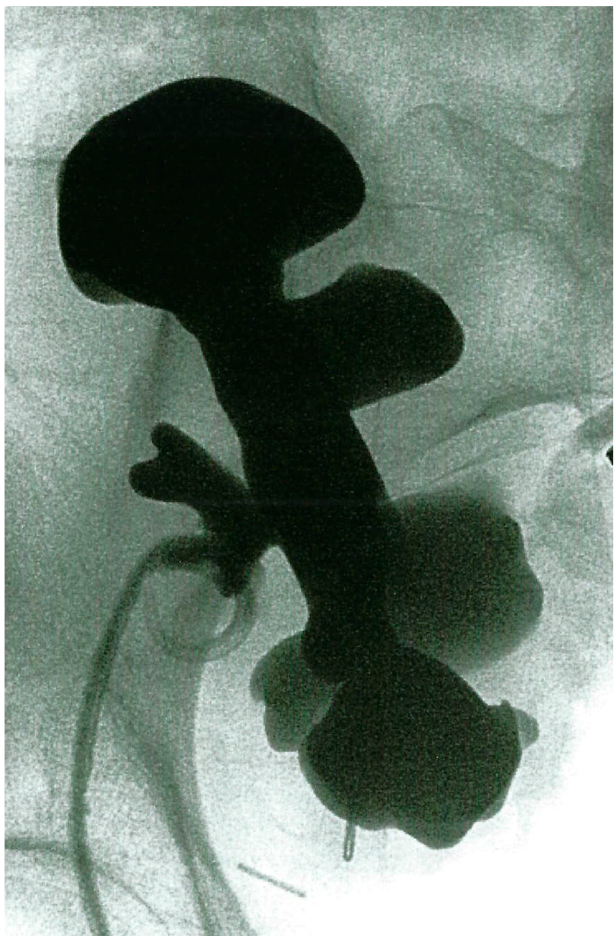
Figure 1. Pre-operative nephrostogram of Case 1 demonstrating the dilated calyces and absence of opacification of the transplant ureter.
Transplantology 06 00027 g001
Figure 2. Calycovesicostomy, CV (lower pole transplant calyx to bladder) and ureterocalycostomy, UC (native ureter to transplant middle calyx) after completion.
Transplantology 06 00027 g002
Figure 3. Intra-operative nephrostogram at completion of calycovesicostomy (CV) and ureterocalycostomy (UC).
Transplantology 06 00027 g003
Figure 4. CT SPECT of the kidney demonstrating excellent drainage into the bladder.
Transplantology 06 00027 g004
Figure 5. Completion of ileocalycostomy (IC) over JJ stent and 16F Malecot catheter.
Transplantology 06 00027 g005
Figure 6. MAG3 renal scan 6 months post operatively demonstrating brisk emptying of urine via the conduit into the urinary bag.
Transplantology 06 00027 g006
Disclaimer/Publisher’s Note: The statements, opinions and data contained in all publications are solely those of the individual author(s) and contributor(s) and not of MDPI and/or the editor(s). MDPI and/or the editor(s) disclaim responsibility for any injury to people or property resulting from any ideas, methods, instructions or products referred to in the content.