Article

Absence of Exogenous Glucose in the Perfusate During Kidney Hypothermic Machine Perfusion Does Not Affect Mitochondrial Function

by
Dafni Efraimoglou
1,*,
L. Annick van Furth
1,
Albert Gerding
2,
Barbara M. Bakker
2,
Jan-Luuk Hillebrands
3,
Henri G. D. Leuvenink
1 and
Leonie H. Venema
1,*
1
Department of Surgery, University Medical Center Groningen, University of Groningen, 9713 GZ Groningen, The Netherlands
2
Laboratory of Pediatrics, Systems Medicine of Metabolism and Signaling, University Medical Center Groningen, University of Groningen, 9713 GZ Groningen, The Netherlands
3
Department of Pathology and Medical Biology, University Medical Center Groningen, University of Groningen, 9713 GZ Groningen, The Netherlands
*
Authors to whom correspondence should be addressed.
Transplantology 2025, 6(2), 8; https://doi.org/10.3390/transplantology6020008
Submission received: 1 November 2024 / Revised: 24 February 2025 / Accepted: 18 March 2025 / Published: 22 March 2025
(This article belongs to the Section Organ and Tissue Donation and Preservation)

Abstract

:
Background: Optimizing organ preservation techniques is imperative in the face of donor kidney shortage and high waiting list mortality. Hypothermic machine perfusion (HMP) has emerged as an effective method to improve graft function post-transplantation, particularly for deceased donor kidneys, prone to ischemia reperfusion injury (IRI). The perfusion solution includes glucose to support kidney metabolism; however, its effect on mitochondrial function remains unclear. The present study investigated the effect of glucose supplementation during 24 h of oxygenated HMP on mitochondrial function in porcine kidneys. Methods: After 30 min of warm ischemia, porcine slaughterhouse kidneys were preserved for 24 h using HMP with one of the following three solutions: the standard HMP preservation solution, University of Wisconsin machine perfusion (UW-MP) solution, which contains glucose; the solution used for static cold storage, University of Wisconsin cold storage (UW-CS) solution, which lacks glucose; or the UW-CS supplemented with 10 mmol/L glucose. Tissue and perfusate samples were collected before, during, and after perfusion for further analysis. Results: ATP production, mitochondrial respiration, and oxidative stress markers were not significantly different between groups. Glucose was released into the perfusion solution even from kidneys without exogenous glucose supplementation in the perfusate. Conclusions: These results suggest that kidney mitochondrial respiration does not depend on the presence of glucose in the HMP perfusion solution at the start of perfusion, underscoring the need for further exploration of nutrient supplementation and mitochondrial function in organ preservation strategies.

1. Introduction

Kidney transplantation is used as a treatment for end-stage renal disease (ESRD) [1]. The persistent disparity between the availability of donor organs and the growing number of patients on the transplantation waiting list exacerbates mortality rates [1,2]. To address this shortage, kidneys from donation after circulatory death (DCD) are increasingly being utilized [1,2,3,4,5]. The duration of oxygen absence makes DCD kidneys prone to ischemia reperfusion injury (IRI) when oxygen is reintroduced after transplantation [4,5,6]. IRI initiates oxidative damage which causes mitochondrial dysfunction, and this injury is associated with a higher incidence of primary nonfunction (PNF) or delayed graft function (DGF), necessitating post-transplant dialysis [4,7,8,9]
To mitigate these challenges, preservation techniques of organ storage between donation and transplantation have evolved. Static cold storage (SCS), using University of Wisconsin cold storage (UW-CS) solution, has been the traditional method [3,5,7]. Hypothermic machine perfusion (HMP), using University of Wisconsin machine perfusion (UW-MP) solution, has emerged as a superior alternative, proven to reduce the duration and incidence of DGF compared with SCS [5,8,10,11,12,13,14].
Both SCS and HMP utilize hypothermic temperatures (approximately 4 °C) to reduce cellular metabolism and oxygen demand [11,12]. However, even at these low temperatures, approximately 5–10% of the physiological metabolic rate remains active [11,12,15,16]. HMP supports this residual metabolism by circulating a glucose-containing solution (UW-MP) through the renal vasculature, unlike SCS where the UW-CS solution lacks glucose and is not circulated [14,17,18,19].
Under physiological normothermic conditions, glucose metabolism in the kidney fuels the tricarboxylic acid (TCA) cycle and the electron transport chain (ETC) in the mitochondria to produce ATP (Figure 1) [19]. Importantly, isotope studies have demonstrated active glucose metabolism even under hypothermic conditions and during HMP [20]. Thus, it is not unlikely that glucose consumption during HMP might correlate with the mitochondrial ATP production capacity.
Despite these insights, direct evidence linking glucose supplementation to improved mitochondrial function during HMP is lacking. Therefore, this study aimed to investigate whether adding glucose during HMP enhances mitochondrial function over a 24 h perfusion period. We hypothesized that kidneys perfused with exogenous glucose would demonstrate superior mitochondrial function compared to those perfused without glucose. Proving this correlation between glucose and mitochondrial function may aid in improving perfusion protocols.

2. Materials and Methods

2.1. Animal Model

A porcine DCD model was used in the present study. Kidneys were obtained from a local abattoir after a standardized slaughtering process. The pigs were anesthetized using electric shock followed by exsanguination.

2.2. Experimental Design

Each kidney underwent 30 min of warm ischemia to induce injury. Three experimental groups were established: kidneys in the CS group (n = 4) were perfused in HMP with UW-CS solution (Belzer CS, Bridge to Life Ltd., London, UK); kidneys in the MP group (n = 5) were perfused in HMP with UW-MP (Belzer MP, Bridge to Life Ltd., London, UK) solution; and kidneys in the CS + glucose group (n = 5) were perfused in HMP with UW-CS solution supplemented with 10 mmol/L glucose (Sigma Aldrich, Steinheim, Germany). The complete perfusate composition of each group is presented in Table S1.

2.3. Hypothermic Machine Perfusion

After 30 min of warm ischemia, a 5 mm cortical punch biopsy (Stiefel, Dublin, Ireland) and a needle biopsy (BioPince™, Argon Medical Devices, Athens, GA, USA) were collected. The punch biopsy was stored in sonification solution (SONOP; containing 0.372 g EDTA in 130 mL H2O and NaOH (pH 10.9) + 370 mL 96% ethanol) and snap-frozen for ATP analysis. The needle biopsy was stored in UW-CS on melting ice for direct measurement of mitochondrial respiration (0 h). Kidneys were flushed with 500 mL 4 °C lactated Ringer’s solution (B Braun Melsungen AG, Melsungen, Germany). Punch biopsies were also performed after the flush for ATP analysis. Next, kidneys were connected to a Kidney Assist Transporter (XVIVO, Gothenburg, Sweden) with oxygen supplementation (100% O2, 100 mL/min). All kidneys were perfused for 24 h in HMP with the perfusion solution according to their experimental group, at a set mean pressure of 25 mmHg at 3–5 °C. Perfusion parameters such as pressure, temperature, and flow rates were continuously monitored. Perfusion solution samples were collected at 0, 1, 4, 8, and 24 h of HMP and needle biopsies for mitochondrial respiration measurements were collected after the end of warm ischemia (0 h) and after 1, 4, 8, and 24 h of perfusion. Except from the timepoint after warm ischemia (SH0), punch biopsies were also collected after flush-out, immediately before the start of perfusion (HMP0) for ATP analysis, and after 24 h HMP for ATP and thiobarbituric acid reactive substances (TBARS) analysis.

2.4. Mitochondrial Function Measurements

Needle biopsies were stored in UW-CS for transport to the laboratory. Biopsies were weighed before being placed into the two-channel high resolution Oroboros Oxygraph-2K (Oroboros Instruments GmbH, Innsbruck, Austria) chamber. The Oxygraph-2K uses a Clark electrode (Rank Brothers Ltd., Cambridge, UK) to measure the oxygen concentration in the buffer. Prior to measurements, the two channels were washed and calibrated by adding 1 mL of the Mitochondrial Respirometry Solution (MiR05: 0.5 mM EGTA, 3 mM MgCl2·6H2O, 20 mM taurine, 10 mM KH2PO4, 20 mM HEPES, 1 g/l BSA, 60 mM potassium-lactobionate, 110 mM sucrose, pH 7.1 at 30 °C). The chambers of the Oxygraph-2K were connected to a water bath to maintain 37 °C, and constant flow was achieved with a magnetic stirrer. To begin the measurements, Digitonin (10 mg/mL) was added to permeabilize the cell membranes, allowing the measurement of basal respiration. Subsequently, a combination of 2.5 μL pyruvate (800 mM), 2.5 μL malate (800 mM), and 5 μL glutamate (2 M) was introduced to initiate stimulated (state 2) respiration. To achieve maximal respiration (state 3), 4 μL ADP (0.5 M) was added. Proton leakage (state 4) was then assessed by adding 1 μL of oligomycin (4 mg/mL). Uncoupled respiration was induced by two separate additions of 1.5 μL FCCP (1 mM). The procedure was concluded with the addition of 10 μL dithionite (10% solution) to halt respiratory activity (Figure 2) [21]. The O2 flux after dithionite addition was used as a negative control for the zero calibration of obtained measurements. Data were collected and analyzed in DatLab (Version 5.1, Oroboros, Innsbruck, Austria) and are expressed as O2 flux normalized to the biopsy wet weight.
The Respiratory Control Ratio (RCR) was defined as the ratio ADP stimulated basal (state 3) respirationand proton leak (state 4) (O2 flux), and the Spare Respiratory Capacity (SRC) was calculated as the difference between maximal respiration (uncoupled state) and ADP stimulated basal (state 3) respiration (Figure 2) [21,23].
ATP levels were measured in tissue punch biopsies using the ATP Bioluminescence Assay Kit CLS II (Roche Diagnostics, Mannheim, Germany) according to a standardized protocol and expressed relative to tissue protein concentration (Pierce™ BCA Protein Assay Kit, Rockford, IL, USA) [24].

2.5. Oxidative Stress

Thiobarbituric acid-reactive substances (TBARS) were measured as indicators of oxidative stress in the kidney after perfusion. The lipid peroxidation product malondialdehyde (MDA) was measured in tissue homogenates using a standardized protocol, as previously described [24]. Data are expressed relative to the tissue protein concentration (Pierce™ BCA Protein Assay Kit, Rockford, IL, USA).

2.6. Perfusate Samples

Glucose and lactate concentrations in perfusion samples were measured using routine procedures at the clinical chemistry lab of the University Medical Centre Groningen (UMCG).

2.7. Flow and HMP Resistance

The HMP flow was continuously measured and documented at 1, 4, 8, and 24 h of perfusion. Intrarenal vascular resistance was calculated by dividing the mean arterial pressure by the flow at the specific time point (expressed in (mmHg mL−1 min)).

2.8. Histological Analysis

Surgical biopsies were obtained from the renal cortex at the end of each perfusion. Thereafter, biopsies were fixed in 4% buffered formaldehyde, embedded in paraffin, cut into 4 um sections and stained with Periodic Acid–Schiff (PAS) to evaluate tissue morphology. The biopsies were blindly scored by an experimental pathologist, for interstitial fibrosis, tubular atrophy, interstitial inflammation, arterial intimal fibrosis, arteriolar hyalinosis, acute tubular injury and necrosis, tubular dilation, and loss of brush border and interstitial edema, utilizing a semi-quantitative scale. Grades 0–3 range from no signs (0) to mild (1), moderate (2), and severe (3). Acute tubular necrosis was assessed on a scale ranging between 0 and 4 (0 = none, 1 = sporadic, 2 = cluster and 3 = confluent areas, 4 = massive) with an additional classification of the morphology as severe and moderate (scaled as 0.67 and 0.33, respectively) [25]. Sections were scanned using a Hamamatsu NanoZoomer 2.0-HT digital slide scanner (Hamamatsu Photonics, Shizuoka, Japan).

2.9. Statistical Analysis

Statistical analyses were performed using GraphPad Prism version 8.0.1. Data are presented as means with 95% confidence intervals (CI). The outliers were identified using GraphPad Prism 8.0.1. and removed from the data (Q = 1%). Depending on the distribution of the data, Kruskal–Wallis tests, multiple comparisons analysis of variance (ANOVA), and mixed-effects analyses were performed. The area under the curve (AUC) for glucose was computed using GraphPad Prism 8.0.1 which uses the trapezoidal method. Delta glucose was calculated as the perfusate glucose concentration after 24 h of perfusion minus the concentration at the beginning of perfusion; Δ C = C final C initial .

3. Results

3.1. Mitochondrial Function During Hypothermic Machine Perfusion

During HMP, the ADP stimulated basal mitochondrial respiration levels did not differ significantly between the experimental groups (Figure 3A). SRC and RCR were similar between groups over the total perfusion period (Figure 3B,C).

3.2. ATP Concentration and Oxidative Stress

No significant differences were observed between ATP levels in the different experimental groups (Figure 4A). In the MP and CS + glucose groups, ATP levels significantly increased from baseline (SH0) to the end of perfusion (HMP24) (MP, p = 0.007; CS + glucose, p = 0.011) and before perfusion (HMP0) to HMP24 (MP, p = 0.041; CS + glucose, p = 0.017). TBARS, a measure of oxidative stress, did not vary among groups at HMP24 (Figure 4B).

3.3. Glucose and Lactate Released into the Perfusion Solution

As intended by the study design, the perfusate of the CS group contained no glucose at the start of perfusion, whereas the MP and CS + glucose groups both contained 10 mmol/L glucose. Hence, the glucose concentration in the CS group was significantly lower than that in the MP (p < 0.0001) and CS + glucose (p < 0.0001) groups at the start of HMP (Figure 5A). The MP and CS + glucose groups continued to have significantly higher glucose levels than the CS group throughout the perfusion (MP vs. CS 1 h p = 0.0005, 4 h p = 0.027, 8 h p = 0.003, 24 h p = 0.007; CS + glucose vs. CS 1 h p < 0.0001, 4 h p = 0.0002, 8 h p = 0.0002, 24 h p = 0.0007) (Figure 5A).
The glucose concentration in the CS group increased after a one-hour HMP, achieving statistical significance at 4, 8, and 24 h of perfusion (4 h p = 0.008, 8 h p = 0.002, 24 h p = 0.002) (Figure 5A).
The area under the curve (AUC) of the CS + glucose group was significantly higher than that of the CS group (p = 0.014) (Figure S1A). Additionally, the delta glucose of the MP and CS + glucose groups was lower than that of the CS group, but only that of the MP group was significantly lower (p = 0.048) (Figure S1B).
No statistically significant difference was found between the lactate concentrations in the experimental groups. The lactate concentration in all three groups showed a significant increase after one hour of perfusion (p < 0.0001) (Figure 5B).

3.4. Flow and Intrarenal Vascular Resistance

No statistical differences in the flow rate and intrarenal vascular resistance during HMP were observed between experimental groups (Figure 6A,B).

3.5. Histological Assessment

No statistically significant differences were observed among the histological scores of the three experimental groups (Figure 7A,B). Furthermore, no evidence of interstitial fibrosis, arterial intimal fibrosis, or arteriolar hyalinosis was detected in any of the biopsies. Small, isolated infiltrates were present in a limited number of biopsies; however, all were assigned a score of zero for interstitial inflammation.

4. Discussion and Conclusions

This study aimed to determine the effect of glucose supplementation on mitochondrial function during 24 h of HMP. Contrary to our initial hypothesis, the addition of glucose to the perfusion solution did not significantly enhance mitochondrial function, as evidenced by mitochondrial respiration, ATP production, and oxidative stress levels. The results indicated that the kidneys released glucose and lactate particularly in the absence of exogenous glucose supply. These findings are intriguing, suggesting that kidneys can mobilize endogenous glucose or alternative substrates for metabolism during HMP.
More specifically, ADP stimulated basal mitochondrial respiration, RCR, and SRC were comparable among the experimental groups during perfusion. RCR is used to estimate the degree of coupling of ATP production and oxygen consumption [21,23]. SRC demonstrates the ability of substrate supply and electron transport to respond to an increase in energy demand [21]. The absence of significant differences among the experimental groups shows that the higher concentration of exogenous glucose in the perfusion solution of HMP had no effect on the mitochondria’s ability to respond to energy demands and produce ATP. This observation is supported by previous studies demonstrating that mitochondria adapt to varying glucose levels by utilizing alternative substrates for ATP production and that these homeostatic mechanisms remain active during hypothermia [18,26].
Perfusate glucose and lactate levels were measured to provide further insight into mitochondrial function. The accumulation of these metabolic substrates in the perfusate during perfusion suggests their release from renal tissue, either as a result of new synthesis or pre-existing stores within the kidney [16,19,20]. Conversely, a reduction in substrate concentration during perfusion indicates their utilization by renal cells to sustain ongoing cell processes [16,19,20]. Notably, kidneys in the group without exogenous glucose in the beginning of perfusion (CS group) were able to later release glucose while achieving similar levels of RCR, SRC, and ATP as those of kidneys supplied with exogenous glucose (MP and CS + glucose groups).
The observed glucose released from the kidneys without exogenous glucose supply raises questions about its source. Possible explanations include the mobilization of intracellular glucose reserves. This hypothesis could account for the sharp glucose increase in the perfusate in the first hour of perfusion, at which point the glucose reserves begin to circulate (Figure 5A). However, previous studies have reported a gradual decline in perfusate glucose levels over time, indicating that intracellular reserves alone would not be sufficient to maintain stable glucose levels throughout perfusion (Figure 5A) [27,28,29,30].
An alternative explanation is that the mitochondria utilized lactate to produce glucose in a process called gluconeogenesis. We hypothesize that lactate consumption was not evident in our measurements because the levels of excess lactate excreted were significantly higher than lactate utilization. While previous studies have reported an increase in lactate levels over time, lactate concentrations in our study remained stable across all groups [16,18,27,28,29,30]. This discrepancy may be attributed to the use of lactated Ringer’s solution (which contains 28 mmol/L lactate), for flushing before perfusion. The introduction of exogenous lactate likely provided an abundant substrate for gluconeogenesis and for maintaining stable levels in the perfusate despite ongoing metabolic activity.
Under physiological conditions, the kidney functions as both a lactate producer and consumer, with the distal nephron segment relying on glycolysis to generate ATP, resulting in lactate generation [31]. The lactate produced is used for cortical gluconeogenesis, leading to a corticomedullary glucose–lactate recycling loop, which might also be active during HMP [31]. At the onset of HMP, a portion of circulating lactate was released into the perfusate, and the cortex may have utilized the remainder for glucose production. Some of this glucose could subsequently be detected in the perfusate, with the kidneys lacking exogenous glucose supply at the beginning of perfusion, later demonstrating higher absolute glucose production (delta glucose) than the kidneys of the other groups.
Another explanation for this surprising glucose release is that the mitochondria of the CS group used lactobionate for glucose production. Lactobionate is provided in the UW-CS (CS and CS + glucose groups) but not in the UW-MP (MP group) (Table S1) and is an oxidized form of lactose with a net negative charge [32,33]. Kidneys can convert lactose-derived galactose into glucose, although this has not been observed in HMP kidneys [19,34]. Nevertheless, the CS + glucose group also included lactobionate but did not release significantly more glucose than the MP group, which did not include lactobionate. This pathway might be active only when insufficient amounts of glucose are present, although to the best of our knowledge, no study supports this hypothesis.
As previously mentioned, the elevated lactate levels observed after one hour of perfusion in all groups may be attributed to flushing with Ringer’s lactate and active glycolysis before perfusion. Ringer’s lactate was utilized to facilitate the rapid removal of blood and as its efficacy as a flush-out and short-term preservation solution has been previously demonstrated [35,36]. Lactate release during HMP has also been noted in previous studies, although the kidneys were flushed with different solutions [16,17,18,27,28,29,37]. However, these studies have shown a progressive increase in lactate over time, in contrast to the stabilization of lactate levels after 4 h of HMP in our results, suggesting the potential metabolic utilization of lactate [16,20,27,28,29,30].
Furthermore, all experimental groups exhibited comparable histological scores, further corroborating the hypothesis that kidneys adapt to nutrient availability, as the absence of glucose in the perfusate did not significantly affect tissue morphology.
The present study is the first to use a glucose-free solution during oxygenated HMP of porcine kidneys and investigate its effects on the mitochondria. The small number of kidneys in each group might lack the power to detect significant differences. Nevertheless, a significant increase in ATP levels over time was shown, comparable to the literature [11,17,38,39]. Additionally, the absence of differences between groups might be attributed to the unexpected glucose presence in kidneys without glucose supplementation. Another possible point of improvement is that the glucose and lactate perfusate analyses determined their concentration only in the extracellular environment of the kidneys. While intracellular activity can be inferred from this, concentrations within the perfusate might point to the release of pre-existing intracellular substrates, as well as the de novo metabolism of substrates derived from the perfusion fluid [37]. Analysis of the kidney tissue would have been useful to provide a more comprehensive description of intracellular differences among the groups.
In conclusion, kidneys perfused with HMP in the absence of glucose may adapt by mobilizing intracellular glucose reserves, lactate, or alternative substrates for mitochondrial respiration. The high glucose concentration during the perfusion of the group without glucose supplementation was an unexpected result that emphasizes that the complexity of kidney metabolism during HMP is still not fully understood. Further research would be of value in the development of perfusion solutions that consider the metabolic needs of the kidneys during HMP.

Supplementary Materials

The following supporting information can be downloaded at: https://www.mdpi.com/article/10.3390/transplantology6020008/s1, Figure S1: Area under the curve of glucose concentration in de perfusate of kidneys in HMP; Table S1: Contents of the perfusion fluids of the three experimental groups.

Author Contributions

Conceptualization, D.E., L.A.v.F., H.G.D.L. and L.H.V.; methodology, D.E., L.A.v.F., A.G., B.M.B., J.-L.H., H.G.D.L. and L.H.V.; data analysis, D.E., L.A.v.F. and J.-L.H., writing-original draft preparation, D.E.; writing-review and editing, D.E., L.A.v.F., A.G., B.M.B., J.-L.H., H.G.D.L. and L.H.V.; visualization, D.E.; investigation, D.E. and L.A.v.F.; supervision, B.M.B., A.G., H.G.D.L. and L.H.V.; resources, A.G., J.-L.H. and H.G.D.L. All authors have read and agreed to the published version of the manuscript.

Funding

This research received no external funding.

Institutional Review Board Statement

Ethical review and approval were waived for this study due to the use of slaughterhouse waste materials.

Informed Consent Statement

Not applicable.

Data Availability Statement

All data available upon request.

Acknowledgments

The authors are grateful to Kroon Vlees for their support in providing slaughterhouse kidneys for this research. Furthermore, many thanks to Petra J. Ottens and Janneke Wiersma-Buist for performing the analysis described in this paper. Figure 1 and Figure 2 were created using Biorender.

Conflicts of Interest

The authors declare no conflicts of interest.

References

  1. Brat, A.; Pol, R.A.; Leuvenink, H.G.D. Novel Preservation Methods to Increase the Quality of Older Kidneys. Curr. Opin. Organ Transplant. 2015, 20, 438–443. [Google Scholar] [CrossRef] [PubMed]
  2. Giwa, S.; Lewis, J.K.; Alvarez, L.; Langer, R.; Roth, A.E.; Church, G.M.; Markmann, J.F.; Sachs, D.H.; Chandraker, A.; Wertheim, J.A.; et al. The Promise of Organ and Tissue Preservation to Transform Medicine. Nat. Biotechnol. 2017, 35, 530–542. [Google Scholar] [CrossRef] [PubMed]
  3. Moers, C.; Leuvenink, H.G.D.; Ploeg, R.J. Non-Heart Beating Organ Donation: Overview and Future Perspectives. Transpl. Int. 2007, 20, 567–575. [Google Scholar] [CrossRef] [PubMed]
  4. Heylen, L.; Jochmans, I.; Samuel, U.; Tieken, I.; Naesens, M.; Pirenne, J.; Sprangers, B. The Duration of Asystolic Ischemia Determines the Risk of Graft Failure after Circulatory-Dead Donor Kidney Transplantation: A Eurotransplant Cohort Study. Am. J. Transplant. 2018, 18, 881–889. [Google Scholar] [CrossRef]
  5. Jochmans, I.; Moers, C.; Smits, J.M.; Leuvenink, H.G.D.; Treckmann, J.; Paul, A.; Rahmel, A.; Squifflet, J.-P.; van Heurn, E.; Monbaliu, D.; et al. Machine Perfusion Versus Cold Storage for the Preservation of Kidneys Donated After Cardiac Death. Ann. Surg. 2010, 252, 756–764. [Google Scholar] [CrossRef]
  6. Salvadori, M.; Rosso, G.; Bertoni, E. Update on Ischemia-Reperfusion Injury in Kidney Transplantation: Pathogenesis and Treatment. World J. Transplant. 2015, 5, 52. [Google Scholar] [CrossRef]
  7. Deng, R.; Gu, G.; Wang, D.; Tai, Q.; Wu, L.; Ju, W.; Zhu, X.; Guo, Z.; He, X. Machine Perfusion versus Cold Storage of Kidneys Derived from Donation after Cardiac Death: A Meta-Analysis. PLoS ONE 2013, 8, e56368. [Google Scholar] [CrossRef]
  8. Jiao, B.; Liu, S.; Liu, H.; Cheng, D.; Cheng, Y.; Liu, Y. Hypothermic Machine Perfusion Reduces Delayed Graft Function and Improves One-Year Graft Survival of Kidneys from Expanded Criteria Donors: A Meta-Analysis. PLoS ONE 2013, 8, e81826. [Google Scholar] [CrossRef]
  9. Foguenne, M.; MacMillan, S.; Kron, P.; Nath, J.; Devresse, A.; De Meyer, M.; Michel, M.; Hosgood, S.; Darius, T. Current Evidence and Future Perspectives to Implement Continuous and End-Ischemic Use of Normothermic and Oxygenated Hypothermic Machine Perfusion in Clinical Practice. J. Clin. Med. 2023, 12, 3207. [Google Scholar] [CrossRef]
  10. Watson, C.J.E.; Wells, A.C.; Roberts, R.J.; Akoh, J.A.; Friend, P.J.; Akyol, M.; Calder, F.R.; Allen, J.E.; Jones, M.N.; Collett, D.; et al. Cold Machine Perfusion Versus Static Cold Storage of Kidneys Donated After Cardiac Death: A UK Multicenter Randomized Controlled Trial. Am. J. Transplant. 2010, 10, 1991–1999. [Google Scholar] [CrossRef]
  11. Venema, L.H.; Brat, A.; Moers, C.; ‘t Hart, N.A.; Ploeg, R.J.; Hannaert, P.; Minor, T.; Leuvenink, H.G.D. Effects of Oxygen During Long-Term Hypothermic Machine Perfusion in a Porcine Model of Kidney Donation After Circulatory Death. Transplantation 2019, 103, 2057–2064. [Google Scholar] [CrossRef] [PubMed]
  12. Jochmans, I.; Brat, A.; Davies, L.; Hofker, H.S.; van de Leemkolk, F.E.M.; Leuvenink, H.G.D.; Knight, S.R.; Pirenne, J.; Ploeg, R.J.; Abramowicz, D.; et al. Oxygenated versus Standard Cold Perfusion Preservation in Kidney Transplantation (Compare): A Randomised, Double-Blind, Paired, Phase 3 Trial. Lancet 2020, 396, 1653–1662. [Google Scholar] [CrossRef] [PubMed]
  13. Moers, C.; Smits, J.M.; Maathuis, M.-H.J.; Treckmann, J.; van Gelder, F.; Napieralski, B.P.; van Kasterop-Kutz, M.; van der Heide, J.J.H.; Squifflet, J.-P.; van Heurn, E.; et al. Machine Perfusion or Cold Storage in Deceased-Donor Kidney Transplantation. N. Engl. J. Med. 2009, 360, 7–19. [Google Scholar] [CrossRef]
  14. Husen, P.; Boffa, C.; Jochmans, I.; Krikke, C.; Davies, L.; Mazilescu, L.; Brat, A.; Knight, S.; Wettstein, D.; Cseprekal, O.; et al. Oxygenated End-Hypothermic Machine Perfusion in Expanded Criteria Donor Kidney Transplant. JAMA Surg. 2021, 156, 517. [Google Scholar] [CrossRef]
  15. Hendriks, K.D.W.; Brüggenwirth, I.M.A.; Maassen, H.; Gerding, A.; Bakker, B.; Porte, R.J.; Henning, R.H.; Leuvenink, H.G.D. Renal Temperature Reduction Progressively Favors Mitochondrial ROS Production over Respiration in Hypothermic Kidney Preservation. J. Transl. Med. 2019, 17, 265. [Google Scholar] [CrossRef]
  16. Nath, J.; Smith, T.; Hollis, A.; Ebbs, S.; Canbilen, S.W.; Tennant, D.A.; Ready, A.R.; Ludwig, C. 13C Glucose Labelling Studies Using 2D NMR Are a Useful Tool for Determining Ex Vivo Whole Organ Metabolism during Hypothermic Machine Perfusion of Kidneys. Transplant. Res. 2016, 5, 7. [Google Scholar] [CrossRef]
  17. Patel, K.; Smith, T.B.; Neil, D.A.H.; Thakker, A.; Tsuchiya, Y.; Higgs, E.B.; Hodges, N.J.; Ready, A.R.; Nath, J.; Ludwig, C. The Effects of Oxygenation on Ex Vivo Kidneys Undergoing Hypothermic Machine Perfusion. Transplantation 2019, 103, 314–322. [Google Scholar] [CrossRef]
  18. Nath, J.; Smith, T.B.; Patel, K.; Ebbs, S.R.; Hollis, A.; Tennant, D.A.; Ludwig, C.; Ready, A.R. Metabolic Differences between Cold Stored and Machine Perfused Porcine Kidneys: A 1 H NMR Based Study. Cryobiology 2017, 74, 115–120. [Google Scholar] [CrossRef]
  19. Liu, R.X.; Koyawala, N.; Thiessen-Philbrook, H.R.; Doshi, M.D.; Reese, P.P.; Hall, I.E.; Mohan, S.; Parikh, C.R. Untargeted Metabolomics of Perfusate and Their Association with Hypothermic Machine Perfusion and Allograft Failure. Kidney Int. 2023, 103, 762–771. [Google Scholar] [CrossRef]
  20. Guy, A.J.; Nath, J.; Cobbold, M.; Ludwig, C.; Tennant, D.A.; Inston, N.G.; Ready, A.R. Metabolomic Analysis of Perfusate During Hypothermic Machine Perfusion of Human Cadaveric Kidneys. Transplantation 2015, 99, 754–759. [Google Scholar] [CrossRef]
  21. Brand, M.D.; Nicholls, D.G. Assessing Mitochondrial Dysfunction in Cells. Biochem. J. 2011, 435, 297–312. [Google Scholar] [CrossRef] [PubMed]
  22. van Furth, L.A.; Efraimoglou, D.; Gerding, A.; Bakker, B.M.; Olinga, P.; Leuvenink, H.G.D.; Venema, L.H. Macronutrient Supplementation during Prolonged Normothermic Incubation Increases Mitochondrial Function of Precision-Cut Kidney Slices after Ischemia. Transplantology 2024. submitted. [Google Scholar]
  23. Divakaruni, A.S.; Jastroch, M. A Practical Guide for the Analysis, Standardization and Interpretation of Oxygen Consumption Measurements. Nat. Metab. 2022, 4, 978–994. [Google Scholar] [CrossRef] [PubMed]
  24. Mahboub, P.; Ottens, P.; Seelen, M.; ’t Hart, N.; Van Goor, H.; Ploeg, R.; Martins, P.N.; Leuvenink, H. Gradual Rewarming with Gradual Increase in Pressure during Machine Perfusion after Cold Static Preservation Reduces Kidney Ischemia Reperfusion Injury. PLoS ONE 2016, 11, e0152006. [Google Scholar] [CrossRef]
  25. Pool, M.B.F.; Hamelink, T.L.; Van Goor, H.; Van Den Heuvel, M.C.; Leuvenink, H.G.D.; Moers, C. Prolonged Ex-Vivo Normothermic Kidney Perfusion: The Impact of Perfusate Composition. PLoS ONE 2021, 16, e0251595. [Google Scholar] [CrossRef]
  26. Casanova, A.; Wevers, A.; Navarro-Ledesma, S.; Pruimboom, L. Mitochondria: It Is All about Energy. Front. Physiol. 2023, 14, 1114231. [Google Scholar] [CrossRef]
  27. Bon, D.; Claire, B.; Thuillier, R.; Hebrard, W.; Boildieu, N.; Celhay, O.; Irani, J.; Seguin, F.; Hauet, T. Analysis of Perfusates During Hypothermic Machine Perfusion by NMR Spectroscopy. Transplantation 2014, 97, 810–816. [Google Scholar] [CrossRef]
  28. Darius, T.; Vergauwen, M.; Smith, T.; Gerin, I.; Joris, V.; Mueller, M.; Aydin, S.; Muller, X.; Schlegel, A.; Nath, J.; et al. Brief O2 Uploading during Continuous Hypothermic Machine Perfusion Is Simple yet Effective Oxygenation Method to Improve Initial Kidney Function in a Porcine Autotransplant Model. Am. J. Transplant. 2020, 20, 2030–2043. [Google Scholar] [CrossRef]
  29. Patel, K.; Nath, J.; Smith, T.; Darius, T.; Thakker, A.; Dimeloe, S.; Inston, N.; Ready, A.; Ludwig, C. Metabolic Characterization of Deceased Donor Kidneys Undergoing Hypothermic Machine Perfusion Before Transplantation Using 13C-Enriched Glucose. Transplant. Direct 2024, 11, e1736. [Google Scholar] [CrossRef]
  30. Verstraeten, L.; Den Abt, R.; Ghesquière, B.; Jochmans, I. Current Insights into the Metabolome during Hypothermic Kidney Perfusion—A Scoping Review. J. Clin. Med. 2023, 12, 3613. [Google Scholar] [CrossRef]
  31. Legouis, D.; Faivre, A.; Cippà, P.E.; de Seigneux, S. Renal Gluconeogenesis: An Underestimated Role of the Kidney in Systemic Glucose Metabolism. Nephrol. Dial. Transplant. 2022, 37, 1417–1425. [Google Scholar] [CrossRef] [PubMed]
  32. King, M.W. Carbohydrates: Galactose Metabolism. In Integrative Medical Biochemistry Examination and Board Review; McGraw-Hill Education: New York, NY, USA, 2014. [Google Scholar]
  33. t Hart, N.A.; der van Plaats, A.; Leuvenink, H.G.D.; van Goor, H.; Wiersema-Buist, J.; Verkerke, G.J.; Rakhorst, G.; Ploeg, R.J. Determination of an Adequate Perfusion Pressure for Continuous Dual Vessel Hypothermic Machine Perfusion of the Rat Liver. Transpl. Int. 2007, 20, 343–352. [Google Scholar] [CrossRef] [PubMed]
  34. Gao, A.W.; Cantó, C.; Houtkooper, R.H. Mitochondrial Response to Nutrient Availability and Its Role in Metabolic Disease. EMBO Mol. Med. 2014, 6, 580–589. [Google Scholar] [CrossRef] [PubMed]
  35. Akdogan, M.; Demirbakan, K.; Baydilek, Y.; Yuksel, Y. Lactated Ringer as Preservation Solution in Living Donor Renal Transplantation. Transplant. Proc. 2023, 55, 1134–1139. [Google Scholar] [CrossRef]
  36. Tan, H.P.; Vyas, D.; Basu, A.; Randhawa, P.; Shah, N.; Donaldson, J.; Marcos, A.; Simmons, R.L.; Starzl, T.E.; Shapiro, R. Cold Heparinized Lactated Ringers with Procaine (HeLP) Preservation Fluid in 266 Living Donor Kidney Transplantations. Transplantation 2007, 83, 1134–1136. [Google Scholar] [CrossRef]
  37. Nath, J.; Guy, A.; Smith, T.B.; Cobbold, M.; Inston, N.G.; Hodson, J.; Tennant, D.A.; Ludwig, C.; Ready, A.R. Metabolomic Perfusate Analysis during Kidney Machine Perfusion: The Pig Provides an Appropriate Model for Human Studies. PLoS ONE 2014, 9, e114818. [Google Scholar] [CrossRef]
  38. Darius, T.; Nath, J.; Mourad, M. Simply Adding Oxygen during Hypothermic Machine Perfusion to Combat the Negative Effects of Ischemia-Reperfusion Injury: Fundamentals and Current Evidence for Kidneys. Biomedicines 2021, 9, 993. [Google Scholar] [CrossRef]
  39. Ravaioli, M.; Baldassare, M.; Vasuri, F.; Pasquinelli, G.; Laggetta, M.; Valente, S.; De Pace, V.; Neri, F.; Siniscalchi, A.; Zanfi, C.; et al. Strategies to Restore Adenosine Triphosphate (ATP) Level After More than 20 Hours of Cold Ischemia Time in Human Marginal Kidney Grafts. Ann. Transplant. 2018, 23, 34–44. [Google Scholar] [CrossRef]
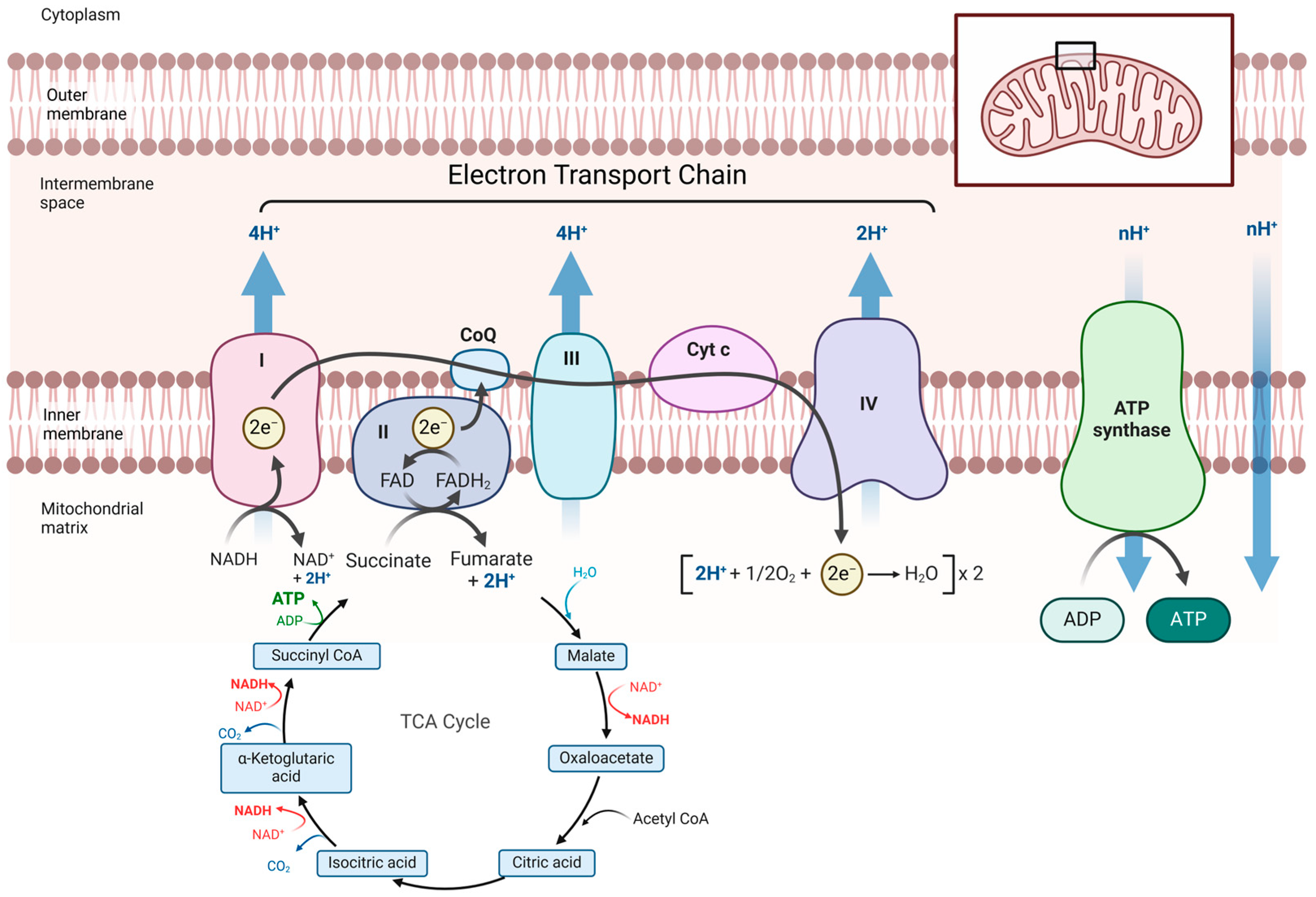
Figure 1. Schematic representation of the electron transport chain. The electron transport chain in the mitochondria where oxygen is consumed to produce ATP. ATP, adenosine triphosphate; ADP, adenosine diphosphate; e, electron; CoQ, coenzyme Q; NAD, nicotinamide adenine dinucleotide; NADH, Dihydronicotinamide adenine dinucleotide; TCA cycle, tricarboxylic acid cycle; Cyt c, cytochrome c. Created using biorender.com.
Transplantology 06 00008 g001
Figure 2. Protocol followed during the mitochondrial respiration measurements with the Oroboros Oxygraph-2K [22]. ADP, adenosine diphosphate; FCCP, Carbonyl cyanide-p-trifluoromethoxyphenylhydrazone.
Transplantology 06 00008 g002
Figure 3. Mitochondrial respiration during different points of perfusion. (A) ADP stimulated basal respiration (state 3) levels. (B) Spare Respiratory Capacity (SRC). (C) Respiratory Control Ratio (RCR). CS, kidneys perfused on HMP with University of Wisconsin cold storage solution; MP, kidneys perfused on HMP with University of Wisconsin machine perfusion solution; CS + glucose, kidneys perfused on HMP with University of Wisconsin cold storage solution supplemented with glucose. Data are presented as the mean with its 95% confidence interval.
Transplantology 06 00008 g003
Figure 4. Kidney energy production and oxidative stress (A) ATP content in kidney cortex tissue after 30 min warm ischemia (SH0), before preservation (HMP0) and at the end of 24 h of HMP (HMP24). (B) Total TBARS production after 24 h of HMP. ATP; adenosine triphosphate; HMP; hypothermic machine perfusion with 100% oxygen; CS, kidneys perfused on HMP with University of Wisconsin cold storage solution; MP, kidneys perfused on HMP with University of Wisconsin machine perfusion solution; CS + glucose, kidneys perfused on HMP with University of Wisconsin cold storage solution supplemented with glucose. The data are shown as the mean with its 95% confidence interval. * p < 0.05; ** p < 0.01.
Transplantology 06 00008 g004
Figure 5. Metabolites in the perfusion solution. (A) Glucose concentrations at different time points of perfusion, p value represents differences between 0 h and 24 h HMP for the CS group. (B) Lactate concentrations at different time points of perfusion. p value represents differences between 0 h and 1 h of HMP for all experimental groups. CS, kidneys perfused on HMP with University of Wisconsin cold storage solution; MP, kidneys perfused on HMP with University of Wisconsin machine perfusion solution; CS + glucose, kidneys perfused on HMP with University of Wisconsin cold storage solution supplemented with glucose. Data are shown as the mean with its 95% confidence interval. ** p < 0.01 ****; p < 0.0001.
Transplantology 06 00008 g005
Figure 6. Flow rates and intrarenal vascular resistance. (A) Flow rates at different time points of perfusion. (B) Intrarenal vascular resistance, calculated as the ratio of the mean arterial pressure and the flow at the specific time point HMP, hypothermic machine perfusion with 100% oxygen; CS, kidneys perfused on HMP with University of Wisconsin cold storage solution; MP, kidneys perfused on HMP with University of Wisconsin machine perfusion solution; CS + glucose, kidneys perfused on HMP with University of Wisconsin cold storage solution supplemented with glucose. Data are shown as the mean with its 95% confidence interval.
Transplantology 06 00008 g006
Figure 7. Histological assessment of cortical kidney biopsies collected after 24 h of HMP. (A) Biopsy scores for tubular atrophy, tubular dilation, and loss of brush border and interstitial edema. (B) Tubular injury and necrosis score for cortical biopsies per experimental group. (C) High-power magnification of a cortical biopsy from a kidney perfused with UW-MP. The biopsy was classified as exhibiting mild tubular atrophy, mild tubular dilation, mild interstitial edema, and sporadic ATN. (D) High-power magnification of a biopsy from a kidney perfused with UW-CS. The biopsy was classified as exhibiting moderate tubular atrophy, mild tubular dilation, moderate interstitial edema (red arrow, in area with atrophic tubules), and cluster ATN. (E) High-power magnification of a biopsy from a kidney perfused with UW-CS + glucose. The biopsy demonstrated no tubular atrophy, mild tubular dilation, and severe ATN. (F) Higher magnification of the area in the rectangle depicted in panel (E), illustrating tubular necrosis and vacuolization (yellow arrow). ATN, acute tubular necrosis; CS, kidneys perfused on HMP with University of Wisconsin cold storage solution; MP, kidneys perfused on HMP with University of Wisconsin machine perfusion solution; CS + glucose, kidneys perfused on HMP with University of Wisconsin cold storage solution supplemented with glucose. The data are shown as the mean with its 95% confidence interval.
Transplantology 06 00008 g007
Disclaimer/Publisher’s Note: The statements, opinions and data contained in all publications are solely those of the individual author(s) and contributor(s) and not of MDPI and/or the editor(s). MDPI and/or the editor(s) disclaim responsibility for any injury to people or property resulting from any ideas, methods, instructions or products referred to in the content.