Case Report

Persistent Right Umbilical Vein: Clinical Case and Literature Review

Faculty of Medicine, Institute of Clinical Medicine, Clinic of Obstetrics and Gynecology, Vilnius University Hospital Santaros Klinikos Obstetrics and Gynecology Center, 08661 Vilnius, Lithuania
*
Author to whom correspondence should be addressed.
Reprod. Med. 2025, 6(4), 36; https://doi.org/10.3390/reprodmed6040036
Submission received: 26 June 2025 / Revised: 5 October 2025 / Accepted: 8 October 2025 / Published: 6 November 2025

Abstract

Background and Clinical Significance: This article presents a clinical case of persistent umbilical vein of the extrahepatic type in a fetus. The features, diagnosis, and prognosis of this rare vascular anomaly, as well as the applied pregnancy monitoring tactics, are reviewed. Case Presentation: A 34-year-old woman was referred to a tertiary-level hospital at 25th weeks’ gestation for evaluation of a suspected fetal heart defect. Persistent right umbilical vein of the extrahepatic type, cardiomegaly with predominant atrial enlargement, a primum atrial septal defect, and hydropericardium were diagnosed. At 33 + 5 weeks of gestation, signs of decompensation emerged, including progressive cardiomegaly, hydropericardium, and newly developed ascites, leading to the decision to induce labor. Imaging was performed using a Voluson E8 ultrasound system (GE Healthcare, Zipf, Austria). Despite all efforts, the severity of the condition ultimately proved fatal in this clinical case. Conclusions: Patients carrying fetuses suspected or diagnosed with PRUV should receive coordinated management by a multidisciplinary team of specialists. Delivery should be planned in a tertiary-level hospital.

1. Introduction

In early embryogenesis, the right umbilical vein has left and right branches. In the fourth week of normal development, the umbilical vein begins to atrophy gradually, and by the seventh week, it disappears completely. Persistent right umbilical vein (PRUV) is a vascular anomaly that occurs during embryonic development when the right umbilical vein does not undergo atrophy [1]. Recent advances in ultrasound technology, together with heightened attention to the evaluation of the fetal venous system, have demonstrated that PRUV is not as uncommon as once believed. The prevalence rate is up to 0.46%. Two variants of this vascular anomaly are distinguished: intrahepatic (PRUV-I) and extrahepatic (PRUV-E). Among all diagnosed cases of PRUV, approximately 5% are classified as PRUV-E. Unlike PRUV-I, PRUV-E bypasses the hepatic circulation and drains directly into the systemic venous system. This configuration can significantly affect fetal hemodynamics. It is often associated with more pronounced clinical symptoms, concomitant abnormalities, and a worse prognosis [2]. Typically, PRUV-I is associated with a more favorable prognosis. In contrast, the prognosis of PRUV-E depends on the type of associated pathology and the severity of hemodynamic disturbances.
Despite its clinical significance, PRUV-E is rarely described in the literature. Greater recognition of this pathology may improve diagnostic accuracy, allow for more comprehensive counseling of prospective parents, and ultimately support more informed clinical decisions. This article presents a clinical case of PRUV-E and reviews the scientific literature on its manifestation, diagnostic approaches, prenatal management, and obstetric outcomes.

2. Case Report

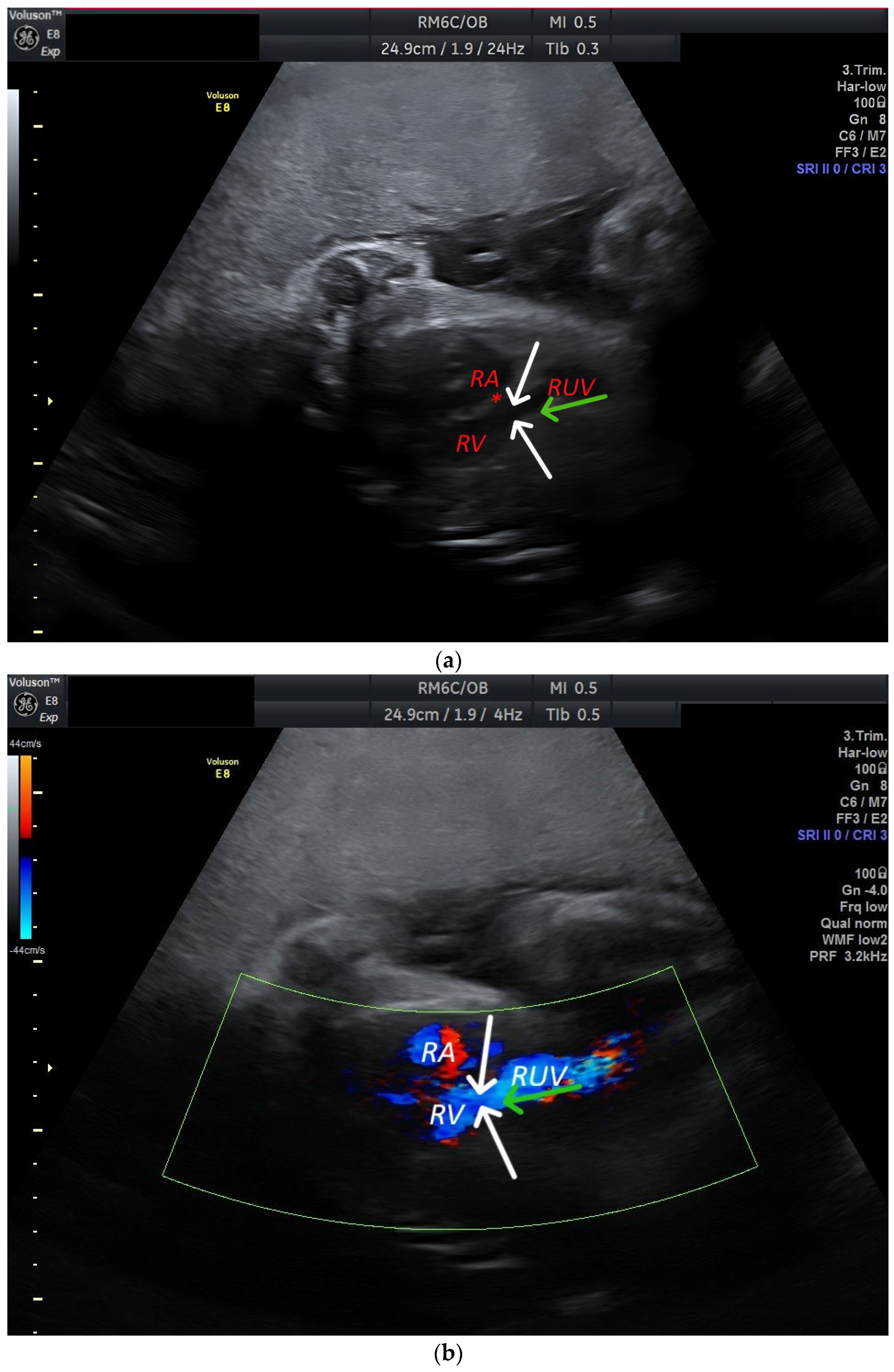
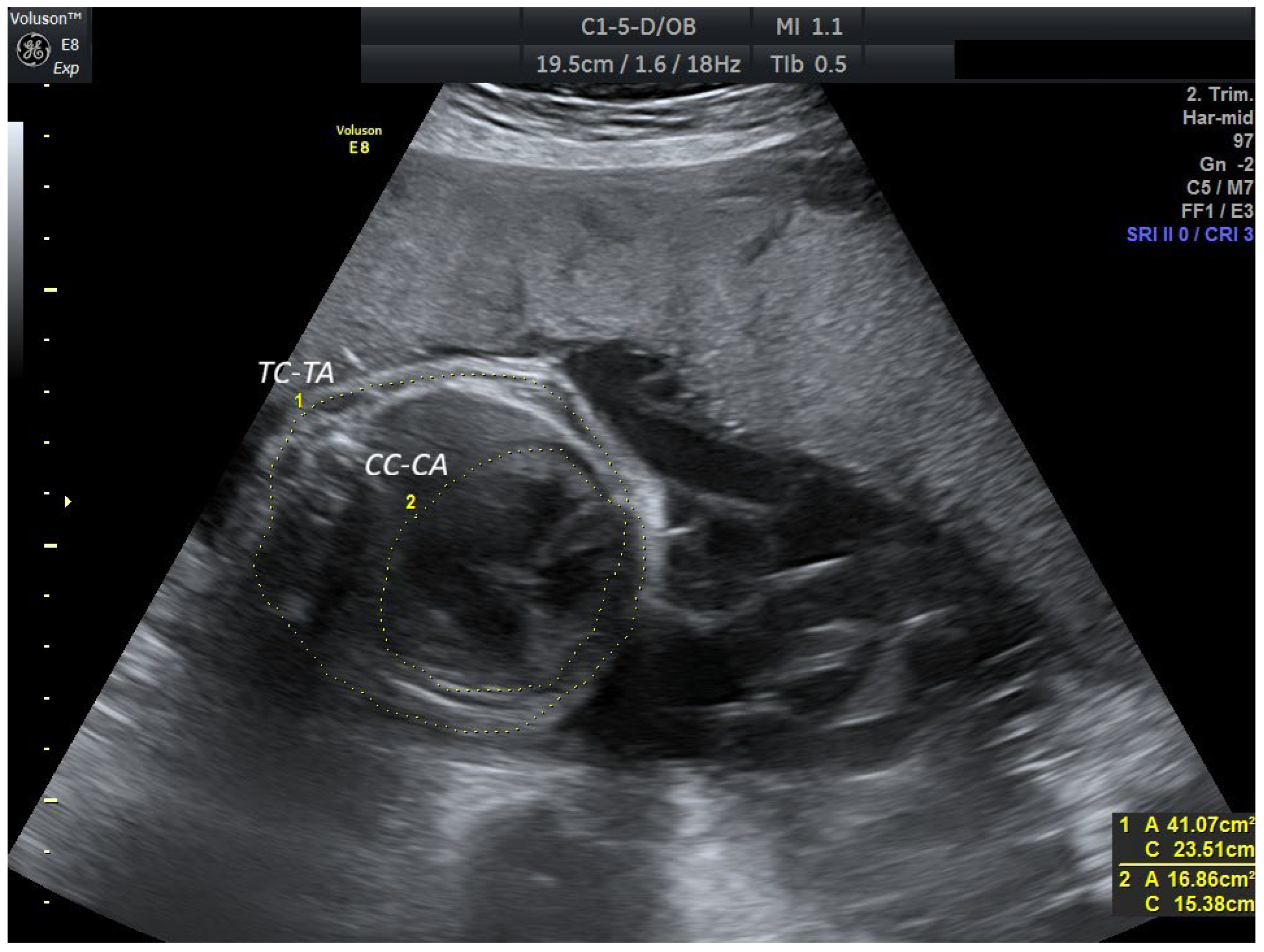
A 34-year-old woman was evaluated at Lithuanian University of Health Sciences (LUHS) Hospital in Kaunas at 25 weeks’ gestation for a suspected fetal cardiac anomaly. The patient had no known comorbidities and a history of two previous preterm deliveries. Infectious diseases did not complicate the current pregnancy, and the patient denied the use of any medications. For further fetal evaluation and diagnostic clarification, the woman was referred to the Perinatology Coordination Center (PCC) at Santaros Klinikos, Vilnius University Hospital (VUH). A week after visiting LUHS Hospital Kaunas Klinikos, the patient was consulted at VUH Santaros Klinikos by an obstetrician–gynecologist, a perinatologist, and a pediatric cardiologist. A detailed fetal ultrasound examination and fetal cardioechoscopy were performed. Fetal growth was appropriate for gestational age, and the amount of amniotic fluid was within normal limits. Findings included PRUV-E, cardiomegaly with atrial enlargement, a primum atrial septal defect, and hydropericardium. Karyotype analysis revealed no abnormalities. A follow-up evaluation was advised after one month. The pregnant woman was repeatedly consulted at the PCC at 31 weeks of gestation. Fetal biometry corresponded to 31 + 5 weeks of gestation. Amniotic fluid volume was normal. A PRUV was observed, dilated along its entire length. The intra-abdominal part of the vein measured 1.26 cm, and the extra-abdominal part measured 1.01 cm (Figure 1). The extrahepatic vein drained into the right ventricle of the heart (Figure 2; Supplementary Video S1). The ductus venosus was absent. Ultrasound examination revealed cardiomegaly. The cardiothoracic area ratio was 0.41, a value above the 97th percentile for gestational age (z-score ≈ +2.5), consistent with moderate cardiomegaly [3] (Figure 3). Both atria and the left ventricle were enlarged, with the posterior wall of the left ventricle measuring 5.3 mm. Systolic function was preserved. The amount of pericardial fluid remained stable. Moderate hydrocele (anechoic fluid surrounding the testis without compression) and placental thickening were also noted. Placental thickness measured 4.06 cm, which exceeds the reported mean values for this gestational age (3.19 ± 0.27 cm to 3.43 ± 0.77 cm) [4]. As the fetal condition remained stable, continued monitoring was recommended. The case was scheduled for discussion with the multidisciplinary team at VUH Santaros Klinikos at 32 weeks of gestation. It was decided to administer treatment to promote fetal lung maturation due to the high risk of premature birth. It was further agreed to continue the pregnancy conservatively and to aim for at least 34 weeks of gestation unless signs of fetal decompensation appeared. The patient was referred to the Obstetrics Day Care Unit at VUH Santaros Klinikos for further management. At 32 + 5 weeks of gestation, ultrasound revealed progression of cardiomegaly. No evidence of significant fetal decompensation or hydrops fetalis was identified. Ultrasound examination at 33 + 5 weeks of gestation demonstrated progressive cardiomegaly, hydropericardium, and newly developed ascites. Induction of labor was recommended.
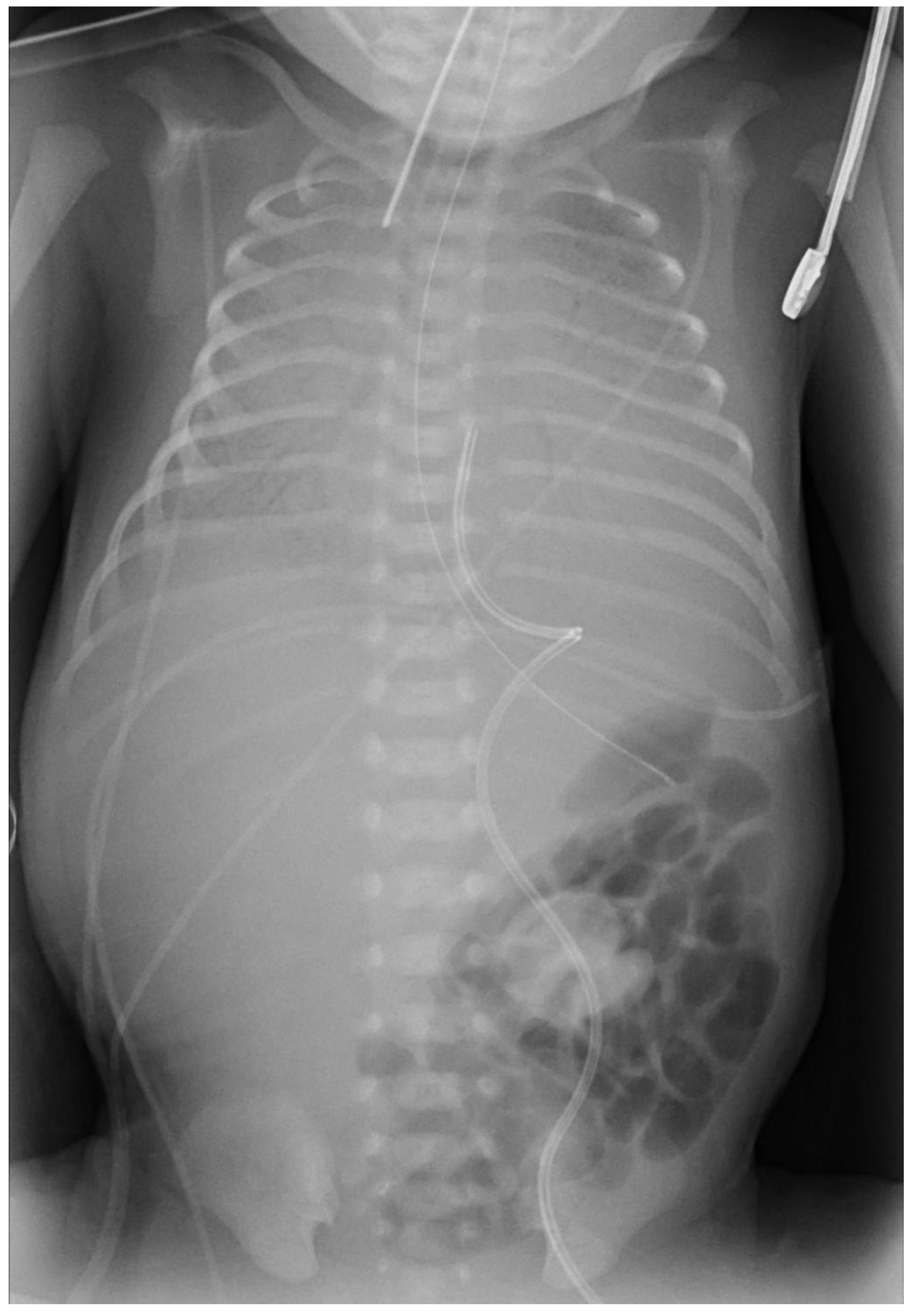
Induction of labor was initiated with a mechanical cervical dilator, followed by amniotomy and oxytocin augmentation. The woman gave birth vaginally to a male newborn weighing 2340 g. The newborn was rated 4, 3, and 5 on the Apgar scale at 1, 5, and 10 min, respectively, and the umbilical cord pH was 7.37. After birth, the newborn was resuscitated with mechanical ventilation (MV) via a mask, intubated 6 min after birth, and continued with MV. Recurrent episodes of bradycardia were treated with adrenaline infusions; however, the patient showed no clinical response. At 50 min after resuscitation, the neonate was transferred to the Neonatal Intensive Care Unit (NICU) in critical condition, where a chest X-ray showed pulmonary edema (Figure 4). Asystole developed 26 min after transfer to the NICU; neonatal resuscitation was initiated according to protocol but proved unsuccessful. One hour and 41 min after birth, the death of the newborn was declared due to exhaustion of vital functions. Autopsy revealed no structural abnormalities other than PRUV-E, cardiomegaly, and pericardial effusion. A primum atrial septal defect was not confirmed. The placenta was edematous, and the umbilical vein was dilated. The cause of death was determined to be acute heart failure secondary to cardiomegaly caused by PRUV-E.
All scans were performed using a Voluson E8 ultrasound system (GE Healthcare, Zipf, Austria). Standard B-mode imaging was supplemented with color and pulsed Doppler to optimize visualization of umbilical venous and intracardiac blood flow.

3. Discussion

3.1. Incidence and Etiology

PRUV is a vascular anomaly that forms early in embryonic development when the right umbilical vein fails to regress. During the initial stages of development, the umbilical vein consists of two branches: the left and the right. Normally, the right umbilical vein begins to regress in the fourth week of embryogenesis and disappears completely by the seventh week. The segment of the left umbilical vein near the heart also regresses. In contrast, the segment from the umbilical cord to the liver persists, dividing into the portal sinus and the ductus venosus. The ductus venosus carries oxygenated blood to the inferior vena cava. Subsequently, the left umbilical vein becomes dominant, carrying blood from the placenta to the fetus. If development is impaired and the right branch fails to regress, it remains functional, resulting in a condition referred to as PRUV [1,5]. The frequency of this anomaly varies from 0.19% to 0.46% [6]. The exact causes of this pathology are unknown. Vascular development is believed to be affected by a combination of genetic and environmental factors, including infections, medication use, exposure to toxic substances during pregnancy, folic acid deficiency in the first trimester, and early obstruction of the left umbilical vein [7].

3.2. Types of PRUV

Two types of PRUV are recognized: intrahepatic and extrahepatic [8]. This anomaly is typically identified in the second trimester of gestation. About 95% of all cases of PRUV comprise the intrahepatic variant. In this type, placental blood carried by the right umbilical vein passes through the liver before reaching the fetal heart, similar to the normal variant [8,9]. The less common extrahepatic type differs from the intrahepatic type in that the right umbilical vein bypasses the liver and connects directly to the right atrium, the iliac veins, or the intracardiac portion of the inferior vena cava. This anatomic variant is frequently associated with atresia of the ductus venosus and confers an increased risk of hemodynamic disturbance. It may lead to cardiac overload, cardiomegaly, or hydrops fetalis. These pathophysiological consequences were also evident in the clinical case we observed [2,10]. Associated structural anomalies are more common in PRUV-E than in PRUV-I. In a retrospective analysis of 39 fetuses diagnosed with PRUV across three tertiary prenatal care centers, 41% were classified as PRUV-E. Importantly, all of these cases were associated with additional comorbid anomalies, most notably a higher incidence of congenital heart disease. Other anomalies involved the digestive, urinary, skeletal, nervous, respiratory, and reproductive systems, as well as chromosomal abnormalities [1,8]. While the prognosis is typically favorable, it may be significantly compromised in instances of PRUV-E and particularly when concomitant comorbidities are present. A comparison of PRUV-I and PRUV-E is presented in Table 1.

3.3. Diagnostics

PRUV is determined by means of ultrasound examination. The umbilical vein is visualized running along the right side of the fetal abdomen, instead of its normal position on the left. The diagnosis can be confirmed with Doppler imaging. This technique allows evaluation of blood flow within the umbilical vein and helps determine the drainage site, whether intrahepatic or extrahepatic. The examination can also assess ductus venosus function, particularly when PRUV-E is suspected. In some cases, 3D ultrasound provides improved visualization, allowing more precise assessment of vascular anomalies.
It is important to differentiate this anomaly from umbilical varices, ductus venosus abnormalities, duplication of the gallbladder, intrahepatic cysts, and an abnormal course of the portal vein or its branches. Fetal umbilical vein varix typically appears as an enlargement of the umbilical vein rather than an abnormal vascular course. In ductus venosus anomalies and in cases of an abnormal course of the portal vein or its branches, blood drainage occurs through alternative venous pathways, and the umbilical vein follows its normal left-sided course into the fetal abdomen. Although gallbladder duplication and intrahepatic cysts can mimic PRUV, the absence of detectable blood flow on Doppler imaging provides a clear diagnostic distinction.
Once a venous anomaly is diagnosed, a detailed ultrasound examination of the fetus, particularly of the cardiovascular system, should be performed to identify other possible developmental anomalies. In certain complex cases, when the diagnosis remains unclear after the ultrasound examination or when other related anomalies are suspected, magnetic resonance imaging (MRI), fetal echocardiography, and genetic testing may be indicated.
Fetal echocardiography plays a complementary role in the evaluation of PRUV. It enables a detailed assessment of the fetal heart and ductus venosus. It also provides information on hemodynamics and helps identify associated cardiac anomalies. When ultrasound is limited by fetal position or maternal factors (e.g., maternal obesity, anterior placenta, oligohydramnios, and multiple pregnancy), MRI can provide detailed clarification of fetal vascular anatomy. This is particularly valuable in the assessment of PRUV–E, where ultrasound alone may not clearly demonstrate the anomalous vascular course.
The isolated PRUV-I variant is not associated with an increased frequency of genetic disorders, so fetal karyotyping is not required if this type is detected. In other cases, the diagnosis of genetic diseases should be considered [8].

3.4. Clinic and Prognosis

The presentation of symptoms can vary. In asymptomatic cases, the anomaly is usually diagnosed incidentally during second-trimester ultrasonography. Sometimes, as in our case, signs of fetal circulatory subcompensation appear during pregnancy. These may include fetal cardiomegaly, hydrops, circulatory disturbance, and polyhydramnios [6,8].
Although measurement of the PRUV lumen on ultrasound is not a major predictor of disease course, it may indirectly reflect fetal hemodynamics. In our clinical case, the PRUV measured 1.26 cm intra-abdominal and 1.01 cm extra-abdominal at 31 weeks’ gestation. In the standard variant between 20 and 39 weeks of gestation, the diameter of the umbilical vein increases from 0.38 to 0.80 cm in the extra-abdominal segment and from 0.33 to 0.70 cm in the intra-abdominal segment [11]. Enlargement of the umbilical vein is typically associated with increased blood flow volume. When the PRUV is extrahepatic, abnormal drainage into the systemic circulation may lead to fetal cardiac overload. Progressive dilation may signal a higher risk of fetal heart failure, particularly when other cardiac anomalies are present or the ductus venosus is absent.
Most often, PRUV presents as an isolated phenomenon, but approximately 20% of all PRUV cases may be associated with minor or major accompanying anomalies. PRUV-E is frequently associated with additional anomalies. This may include cardiac defects, as well as disorders of the digestive, urinary, and reproductive systems, along with neurological and chromosomal abnormalities. In contrast to the intrahepatic variant, PRUV-E is more often accompanied by atresia of the ductus venosus. As a result, significant hemodynamic disturbances may develop, worsening the prognosis [7,8,10,12,13].
The literature suggests an association between this pathology and lower fetal weight. One retrospective study conducted in two tertiary-level hospitals in Madrid reported that 22.7% of fetuses with an isolated PRUV abnormality had a predicted birth weight below the 10th percentile. It is important to note that all PRUV cases mentioned in this article were intrahepatic [14]. In our case, fetal growth restriction was not observed, and the newborn’s weight corresponded to the 50th–75th percentile range [15]. According to the study conducted in Madrid, perinatal outcomes were favorable. This included all cases of PRUV, as well as those with a concomitant anomaly, which occurred in 40.1% of cases [14]. In a prospective study conducted in Poland, all fetuses with PRUV-I in the absence of associated pathology had successful outcomes. All deliveries were uncomplicated, with 77.8% of newborns born at full term and the remainder after 35 weeks’ gestation. Postnatal outcomes were favorable [8]. Thus, the prognosis of this anomaly depends on the type of PRUV, the features of the accompanying pathology, and the extent of fetal hemodynamic disturbances.
Interestingly, incidental cases of PRUV-E with favorable outcomes have also been documented in the literature. One such case was diagnosed at Gemelli University Hospital in Rome. At 33 weeks of gestation, the woman presented with vaginal bleeding caused by placenta previa. At admission, fetal echocardiography demonstrated moderate cardiomegaly with right-sided heart overload, along with ectasia of the inferior vena cava and absence of the ductus venosus. Genetic testing yielded normal results, and no additional anomalies were identified. Due to persistent bleeding, delivery was performed by C-section at 36 weeks of gestation. No significant hemodynamic changes were observed during pregnancy. However, the authors suggest that hemodynamic alterations might have been overlooked because of the short interval between diagnosis and delivery. Notably, PRUV-E underwent spontaneous obliteration three months after birth [2]. In contrast, in our case, the vascular anomaly persisted despite the expectation of postnatal regression. S.V. Ramanan and colleagues also described an incidentally identified case of PRUV-E. The vascular anomaly was detected in an 8-year-old boy during surgical correction of a secundum atrial septal defect. Intraoperatively, an aberrant vascular channel draining into the right atrium was observed. An additional tricuspid valve anomaly (14 mm displacement of the septal leaflet of the valve) was identified, though it was of no clinical significance. As PRUV-E had no hemodynamic impact, the atrial septal defect was closed, and the PRUV-E was preserved [6].
In our case, unlike typical cases, PRUV drained directly into the right ventricle, resulting in a high-volume systemic venous return. This direct flow caused right ventricular volume overload, leading to ventricular dilatation, impaired systolic function, hemodynamic decompensation, and ultimately, a poor clinical outcome.

3.5. Limitations

The main limitation of our clinical case is the scarcity of literature on PRUV-E, which restricted meaningful comparison with similar cases. In addition, the lack of standardized management guidelines for this anomaly limited the interpretation of the clinical course and prognosis. As a single case observation, it cannot establish a causal relationship between the vascular anomaly and the neonatal decompensation. Finally, the absence of long-term follow-up, due to the neonatal death, precluded assessment of potential compensatory mechanisms or progression of associated complications.

4. Conclusions

Considering the limited literature available, it can be stated that both in Lithuania and globally, there is a lack of defined recommendations regarding the prenatal and postnatal care of newborns diagnosed with PRUV. The prognosis is usually favorable, but outcomes may be poor in cases of PRUV-E and/or when comorbidities are present. Early diagnosis facilitates timely assessment of associated anomalies and the planning of appropriate monitoring strategies. Therefore, pregnancies with a suspected or confirmed diagnosis of PRUV, due to the risk of concomitant abnormalities and hemodynamic complications, must be managed by multidisciplinary specialist teams, with delivery taking place in tertiary-level maternity hospitals.

Supplementary Materials

The following supporting information can be downloaded at: https://www.mdpi.com/article/10.3390/reprodmed6040036/s1, Video S1: Demonstration of PRUV drainage into the right ventricle.

Author Contributions

Conceptualization, J.V. and D.K.; methodology, J.V. and D.K.; formal analysis, J.V. and D.K.; investigation, D.K.; resources, D.K.; data curation, J.V. and D.K.; writing—original draft preparation, D.K.; writing—review and editing, J.V.; supervision, J.V.; All authors have read and agreed to the published version of the manuscript.

Funding

This research received no external funding.

Institutional Review Board Statement

Not applicable.

Informed Consent Statement

Written informed consent has been obtained from the patients and their representatives to publish this paper.

Data Availability Statement

The data presented in this study are available in the article.

Conflicts of Interest

The authors declare no conflicts of interest.

Abbreviations

The following abbreviations are used in this manuscript:
FIUVIntra-abdominal umbilical vein
FUVExtra-abdominal umbilical vein
LUHSLithuanian University of Health Sciences
MRIMagnetic resonance imaging
MVMechanical ventilation
NICUNeonatal Intensive Care Unit
PCCPerinatology Coordination Center
PRUVPersistent right umbilical vein
PRUV-EPersistent right umbilical vein extrahepatic type
PRUV-IPersistent right umbilical vein intrahepatic type
RARight atrium
RVRight ventricle
RUVRight umbilical vein
VUHVilnius University Hospital

References

  1. Li, J.; Yuan, Q.; Ding, H.; Yang, Z.; Wang, B.; Wang, B. Ultrasonic detection of fetal persistent right umbilical vein and incidence and significance of concomitant anomalies. BMC Pregnancy Childbirth 2020, 20, 610. [Google Scholar] [CrossRef] [PubMed]
  2. Pinna, G.; De Carolis, M.P.; Lanzone, A.; Del Sordo, G.; Botta, A.; De Carolis, S. Persistence of right umbilical vein: A singular case. Turk. J. Pediatr. 2020, 62, 338–342. [Google Scholar] [CrossRef] [PubMed]
  3. Sompagdee, N.; Anuwutnavin, S.; Burapasikarin, C.; Ruangvutilert, P.; Thongkloung, P. Nomograms of fetal cardiothoracic ratio from 17 to 37 weeks’ gestation as assessed by three different measurement techniques and their correlation with gestational age. Prenat. Diagn. 2021, 41, 1658–1667. [Google Scholar] [CrossRef] [PubMed]
  4. Mathai, B.M.; Singla, S.C.; Nittala, P.P.; Chakravarti, R.J.; Toppo, J.N. Placental Thickness: Its correlation with ultrasonographic gestational age in normal and intrauterine growth-retarded pregnancies in the late second and third trimester. J. Obstet. Gynecol. India 2013, 63, 230–233. [Google Scholar] [CrossRef] [PubMed]
  5. Spurway, J.; Logan, P.; Pak, S. The development, structure and blood flow within the umbilical cord with particular reference to the venous system. Australas. J. Ultrasound Med. 2015, 15, 97–102. [Google Scholar] [CrossRef] [PubMed]
  6. Ramanan, S.V.; Mohammed, S.; Dharan, B.S. Intracardiac relation of extrahepatic persistent right umbilical vein. Indian J. Thorac. Cardiovasc. Surg. 2020, 37, 354–356. [Google Scholar] [CrossRef] [PubMed]
  7. Wolman, I.; Gull, I.; Fait, G.; Amster, R.; Kupferminc, M.J.; Lessing, J.B.; Jaffa, A.J. Persistent right umbilical vein: Incidence and significance. Ultrasound Obstet. Gynecol. 2002, 19, 562–564. [Google Scholar] [CrossRef] [PubMed]
  8. Krzyżanowski, A.; Swatowski, D.; Gęca, T.; Kwiatek, M.; Stupak, A.; Woźniak, S.; Kwaśniewska, A. Prenatal diagnosis of persistent right umbilical vein–incidence and clinical impact. A prospective study. Aust. N. Z. J. Obstet. Gynaecol. 2019, 59, 77–81. [Google Scholar] [CrossRef] [PubMed]
  9. Yagel, S.; Kivilevitch, Z.; Cohen, S.M.; Valsky, D.V.; Messing, B.; Shen, O.; Achiron, R.; Anteby, E.Y. The fetal venous system, part II: Ultrasound evaluation of the fetus with congenital venous system malformation or developing circulatory compromise. Ultrasound Obstet. Gynecol. 2010, 36, 93–111. [Google Scholar] [CrossRef] [PubMed]
  10. Weichert, J.; Hartge, D.; Germer, U.; Axt-Fliedner, R.; Gembruch, U. Persistent right umbilical vein: A prenatal condition worth mentioning? Ultrasound Obstet. Gynecol. 2011, 37, 543–548. [Google Scholar] [CrossRef] [PubMed]
  11. Wang, L.; Zhou, Q.; Zhou, C.; Wang, J.; Shi, C.; Long, B.; Zhang, M.; Luo, G. Z-score reference ranges for umbilical vein diameter and blood flow volume in normal fetuses. J. Ultrasound Med. 2022, 41, 907–916. [Google Scholar] [CrossRef]
  12. Toscano, P.; Saccone, G.; Di Meglio, L.; Di Meglio, L.; Mastantuoni, E.; Riccardi, C.; Sirico, A.; Berghella, V. Intrahepatic persistent fetal right umbilical vein: A retrospective study. J. Matern. Fetal Neonatal Med. 2021, 34, 4025–4028. [Google Scholar] [CrossRef]
  13. Chan, E.S.; Suchet, I.; Yu, W.; Somerset, D.; Soliman, N.; Kuret, V.; Liu, H.; Keating, S. Absence of ductus venosus: A comparison of two distinctive fetal autopsy cases and embryologic perspectives. Pediatr. Dev. Pathol. 2023, 27, 139–144. [Google Scholar] [CrossRef]
  14. Martinez, R.; Gamez, F.; Bravo, C.; Sánchez, P.; Orizales, C.; Ortiz, L.; Garcia-Talavera, J.; Gorostidi, M.A. Perinatal outcome after ultrasound prenatal diagnosis of persistent right umbilical vein. Eur. J. Obstet. Gynecol. Reprod. Biol. 2013, 168, 36–39. [Google Scholar] [CrossRef] [PubMed]
  15. Morkuniene, R.; Cole, T.J.; Jakimaviciene, E.M.; Bankauskiene, A.; Isakova, J.; Drazdiene, N.; Vitrikas, T.; Vasiliauskiene, L.; Ramasauskaite, D. Regional references vs international standards for assessing weight and length by gestational age in Lithuanian neonates. Front. Pediatr. 2023, 11, 1173685. [Google Scholar] [CrossRef] [PubMed]
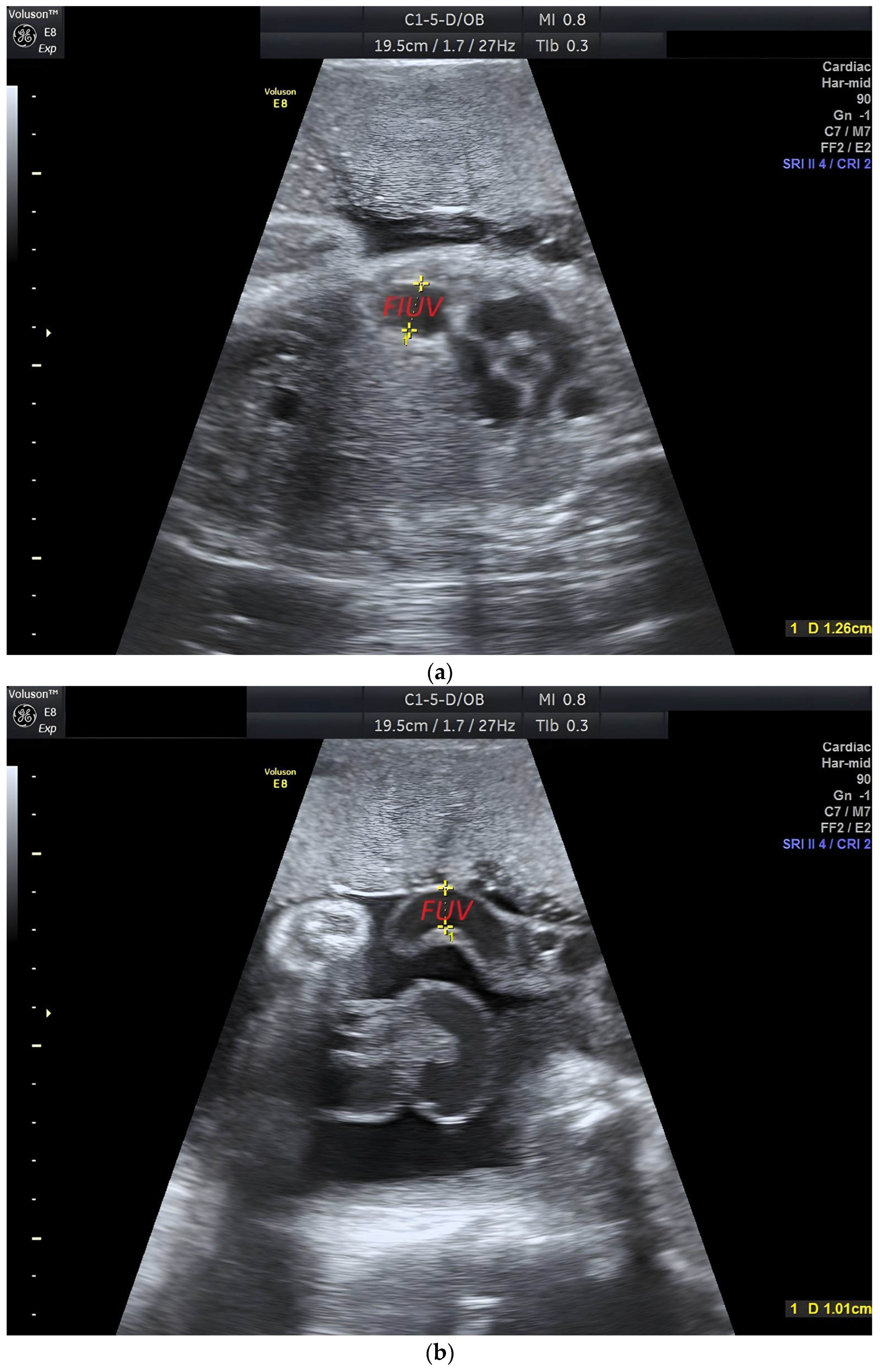
Figure 1. Ultrasound at 31 + 3 weeks’ gestation demonstrates the fetus in the transverse abdominal plane. (a) The intra-abdominal umbilical vein (FIUV) is markedly dilated, measuring 1.26 cm in diameter. (b) The extra-abdominal umbilical vein (FUV) is dilated, measuring 1.01 cm in diameter.
Reprodmed 06 00036 g001
Figure 2. Ultrasound at 31 + 3 weeks’ gestation in a transverse thoracic plane demonstrates PRUV-E draining directly into the right ventricle. (a) Grayscale image. RA = right atrium; RV = right ventricle; RUV = right umbilical vein; * = right atrioventricular valve. Green arrow: blood flow direction in the PRUV. White arrows: entry site of the PRUV into the right ventricle. (b) Color Doppler imaging shows abnormal flow from the RUV draining into the RV.
Reprodmed 06 00036 g002
Figure 3. Four-chamber view of the fetal thorax at 31 + 3 weeks’ gestation. Cardiothoracic measurements for circumference and area ratios are obtained. The thoracic area measures 41.07 cm2, and the cardiac area 16.86 cm2, yielding a cardiothoracic area ratio of 0.41. CC-CA, cardiac circumference and cardiac area; TC-TA, thoracic circumference and thoracic area.
Reprodmed 06 00036 g003
Figure 4. Chest radiograph of the newborn demonstrating pulmonary edema. Reduced pulmonary aeration is noted due to bilateral infiltrates. The hilar regions, cardiac borders, and diaphragmatic outlines are poorly defined.
Reprodmed 06 00036 g004
Table 1. Key differences between PRUV-I and PRUV–E, including frequency, anatomical course, ductus venosus involvement, hemodynamic impact, associated anomalies, prognosis, and typical timing of prenatal diagnosis.
FeaturesPRUV-IPRUV-E
Frequency95%5%
DrainageRight portal veinDirectly into systemic veins
Associated abnormalitiesRareMore frequent (cardiac, genitourinary, chromosomal, gastrointestinal, nervous, skeletal, respiratory)
Hemodynamic impactUsually minimalCan significantly alter fetal hemodynamics
Connection with the ductus venosusUsually remains patentOften absent or altered
PrognosisGenerally favorableOften worse due to associated complications. Close monitoring needed
Typical timing of diagnosisSecond trimesterSecond trimester
Disclaimer/Publisher’s Note: The statements, opinions and data contained in all publications are solely those of the individual author(s) and contributor(s) and not of MDPI and/or the editor(s). MDPI and/or the editor(s) disclaim responsibility for any injury to people or property resulting from any ideas, methods, instructions or products referred to in the content.