Article

Hemodynamic Comparison of Inferior Vena Cava Collapsibility Index in Patients with Preeclampsia vs. Controls: A Pilot Study

1
Department of Obstetrics & Gynecology, Division of Maternal-Fetal Medicine, Norton College of Medicine, SUNY Upstate Medical University, Syracuse, NY 13210, USA
2
Department of Pulmonary Critical Care and Sleep Medicine, Norton College of Medicine, SUNY Upstate Medical University, Pulmonary Critical Care, Crouse Hospital, Syracuse, NY 13210, USA
*
Author to whom correspondence should be addressed.
Reprod. Med. 2025, 6(4), 35; https://doi.org/10.3390/reprodmed6040035
Submission received: 2 August 2025 / Revised: 4 October 2025 / Accepted: 16 October 2025 / Published: 1 November 2025

Abstract

Background/Objectives: There is a paucity of research studying point-of-care ultrasound in the pregnant population, despite the rising incidence of maternal medical complications and, in particular, preeclampsia. This study sought to compare the inferior vena cava collapsibility index (IVC-CI) between patients with preeclampsia with severe features (PECS) and gestational age-matched controls and to assess whether pulse pressure (PP) correlates with IVC-CI in PECS. Methods: This was a prospective pilot study of patients recruited at an inpatient hospital and an outpatient office. The case group included admitted patients with a diagnosis of PECS considered to be stable for prolonged antepartum expectant management. One patient per gestational age week from 23 to 34 weeks with PECS was compared to a gestational age-matched patient without any form of preeclampsia and/or gestational hypertension. Patients on magnesium sulfate, those with multiple gestation, large-for-gestational age fetus, fetal growth restriction, polyhydramnios, oligohydramnios, and/or an anomalous fetus were excluded. Results: IVC-CI was significantly lower in the PECS group compared with controls (mean 20.1% vs. 48.3%, 95% CI = −0.40–−0.16, p < 0.001). There was an inverse relationship between IVC-CI and pulse pressure in patients with PECS. For each additional 1 mmHg in pulse pressure, the odds of having IVC-CI greater than 50% decreased by 13%. Conclusions: Patients with preeclampsia exhibit detectable changes in inferior vena cava diameter that can be assessed with bedside ultrasound and correlated with maternal PP. Future research should focus on validating these findings and exploring the clinical significance of these measurements.

1. Introduction

Preeclampsia is a significant hypertensive disorder of pregnancy, characterized by the release of vasoactive substances leading to new-onset hypertension, vascular fluid shifts, and the potential for multi-organ damage [1,2,3]. This condition frequently results in a state of fluid overload, despite intravascular volume depletion [4,5]. In non-obstetric populations, fluid status can be effectively evaluated using bedside ultrasound, particularly through measurement of the inferior vena cava collapsibility index (IVC-CI) [6,7,8,9]. However, there is limited research on the application of IVC-CI for assessing fluid status in pregnant patients [10,11].
Monitoring the inferior vena cava (IVC) during pregnancy has been proposed as a valuable non-invasive method for assessing maternal hemodynamic status [10,11,12]. The IVC, which returns deoxygenated blood from the lower body to the heart, is sensitive to intravascular volume changes and can be evaluated via ultrasound for its diameter and collapsibility. These parameters provide indirect insights into central venous pressure and fluid responsiveness. In pregnancy, especially in the second and third trimesters, the gravid uterus can exert pressure on the IVC, potentially leading to reduced venous return, hypotension, and symptoms of supine hypotensive syndrome [7,8,9]. This phenomenon is particularly relevant in women with multiple gestation, polyhydramnios, high body mass index (BMI), or other conditions leading to increased intra-abdominal pressure. Furthermore, assessing IVC parameters may be clinically beneficial in populations at risk for preeclampsia, hemorrhage, or cardiovascular complications, where volume status assessment is crucial for guiding fluid management and preventing adverse maternal outcomes [10,11]. Regular measurement of IVC dimensions may therefore support early identification of hemodynamic compromise and guide individualized management strategies to optimize maternal and fetal health.
While normal pregnancy is marked by physiologic vasodilation and volume expansion to support increased maternal blood volume and cardiac output, thus ensuring adequate uteroplacental perfusion, preeclampsia exhibits distinct cardiovascular maladaptation [1,13]. Emerging evidence indicates that hypertension during pregnancy can manifest through two distinct hemodynamic profiles [13,14]. The first profile, referred to as hyperdynamic, is marked by increased cardiac output, driven by elevated heart rate and stroke volume [12,15,16,17]. In contrast, the second profile is hypodynamic and characterized by normal or reduced cardiac output and increased systemic vascular resistance [18,19,20,21]. In this scenario, systemic vascular resistance rises out of proportion to maternal blood pressure, thought to be due to significant endothelial dysfunction and persistent vasoconstriction [21,22]. In prior studies, pulse pressure (PP), defined as the difference between systolic and diastolic blood pressure, has been used as proxy for assessing hemodynamic profiles in patients with preeclampsia [23,24,25]. This hemodynamic heterogeneity underscores the complexity of preeclampsia pathophysiology and the need for individualized assessment and management strategies. This study aimed to compare the IVC-CI between patients diagnosed with PECS and gestational age-matched controls, and to evaluate the correlation between pulse pressure and IVC-CI.

2. Materials and Methods

2.1. Patient Selection

This IRB-approved pilot study was conducted from 1 June 2024 through 1 September 2024. All patients underwent and signed informed consent. Patients were prospectively recruited via convenience sampling at the two participating clinical sites, an inpatient hospital and an outpatient office. The case group included admitted patients with a diagnosis of PECS considered to be stable for prolonged antepartum expectant management of the disease in accordance with the American College of Obstetricians and Gynecologists guidelines. One patient per gestational age week from 23 to 34 weeks gestation was examined. PECS was defined as the onset of hypertension (≥140/90 mmHg) and proteinuria after 20 weeks of gestation, accompanied by end-organ damage, which may include abnormalities in the liver, kidneys, nervous system, or clotting functions. Gestational age-matched patients without any form of preeclampsia and/or gestational hypertension were included for the comparison group. Patients on magnesium sulfate, those with multiple gestation, large-for-gestational age fetus, fetal growth restriction, polyhydramnios, oligohydramnios, and/or an anomalous fetus were excluded (Figure 1). Patient demographic characteristics were collected using electronic health records. Blood pressure measurements were collected at the bedside, and pulse pressure was calculated and recorded.

2.2. Measurements

Bedside substernal ultrasound was performed to obtain inferior vena cava measurements via a curvilinear ultrasound probe (Canon Aplio i700 (Tochigi, Japan), i8CX1 convex probe and GE Voluson E10 (Vienna, Austria), C1-6 convex probe). The standard curvilinear probe with frequency range used on most labor and delivery units was chosen due to its widespread accessibility in clinical obstetrics settings.
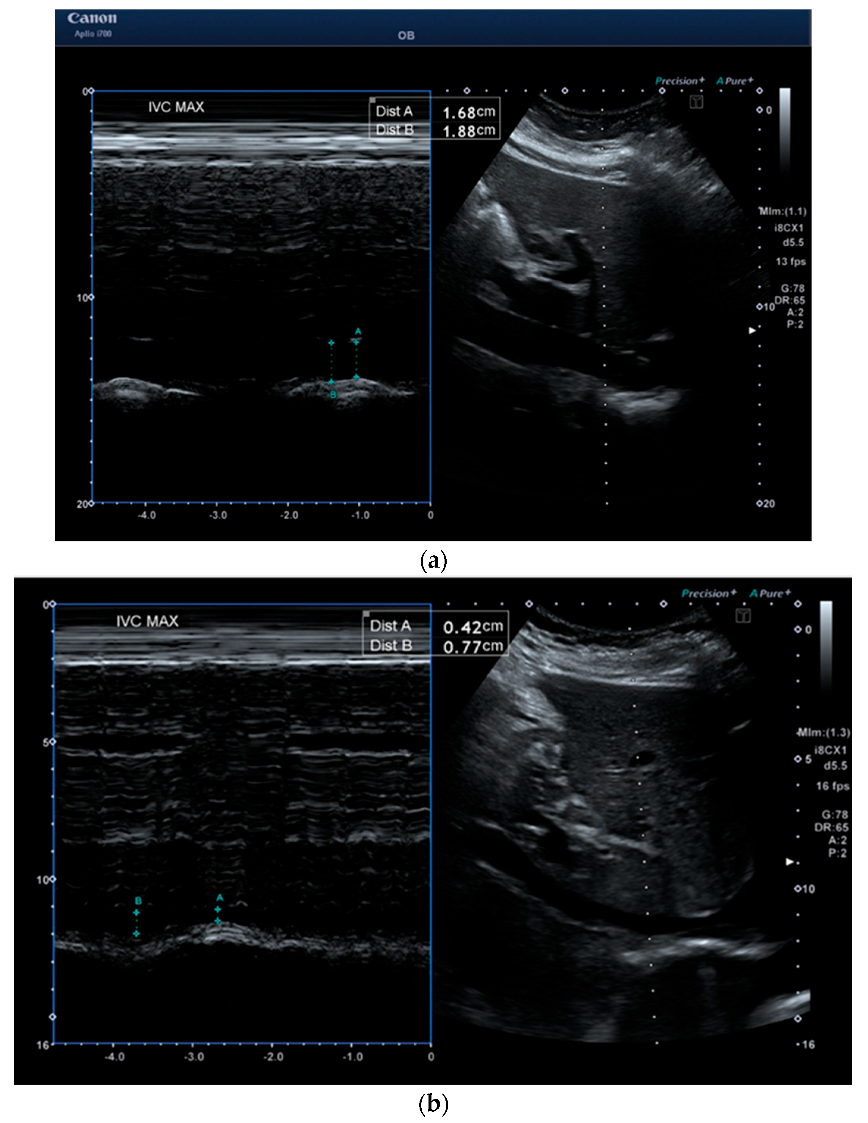
The inferior vena cava (IVC) was assessed via M-mode through the subxiphoid window 2 cm from the right atrium distal to the confluence of the hepatic veins. The diameter of the IVC was measured during inhalation (IVCmin) and after exhalation (IVCmax) (Figure 2). IVC-CI was calculated as (IVCmax minus IVCmin) divided by (IVCmax). All demographic information and obtained measurements were de-identified and recorded.

2.3. Statistical Analysis

Data were analyzed using SPSS statistical software (version 29.0, IBM/SPSS, Chicago, IL, USA). Bivariate tests were conducted with independent sample t-tests for continuous variables and chi-squared tests for dichotomous variables. We dichotomized IVC-CI to greater than or less than 50% collapsibility, as this is the threshold commonly used in clinical practice. To evaluate the relationship between IVC-CI and changes in pulse pressure, a simple logistic regression analysis was conducted. A p-value of <0.05 was established as the threshold for statistical significance across all analyses.

3. Results

A total of 24 patients (12 in the PECS group and 12 in the comparison group) were included. There were no statistically significant differences in age, parity, body mass index, race and ethnicity, pre-pregnancy diabetes, gestational diabetes, or chronic hypertension between the two groups (Table 1).
The IVC collapsibility trend across gestational age was examined for both groups (Figure 3). The IVC-CI was significantly lower in the PECS group compared to the control group (mean 20.1% vs. 48.3%, 95% CI = −0.40–−0.16, p < 0.001). The IVC diameter with inhalation was greater in the PECS group compared to the control group (1.5 cm vs. 0.6 cm, 95% CI = −1.16–−0.68, p < 0.001). The IVC diameter with exhalation was also greater in the PECS group compared to the controls (1.9 cm vs. 1.1 cm, 95% CI = −1.02–−0.50, p < 0.001).
Patients with PECS had wider pulse pressures as a group when compared to controls (Table 2). There was an inverse relationship between IVC-CI and pulse pressure; for each additional 1 mmHg in pulse pressure, the odds of having IVC collapsibility index greater than 50% decreased by 13 percent.

4. Discussion

While the concept of hemodynamic changes in the pathophysiology of preeclampsia is far from novel [4,26,27,28,29]. this pilot study supports the concept that point-of-care ultrasound measurement of inferior vena cava collapsibility index correlates inversely with pulse pressure elevation in pregnancies complicated by preeclampsia with severe features.

4.1. Advantages of Ultrasound over Traditional Hemodynamic Monitoring

Ultrasound is noninvasive, offering advantages over hemodynamic monitoring of the past which depended mainly on invasive techniques of pulmonary artery or central venous catheterization using the Fick method, dye dilution, or thermodilution [30,31,32]. More recently, Doppler noninvasive techniques, maternal echocardiogram, and transthoracic bioimpedance have been applied to assess maternal hemodynamic status in pregnancy [33,34]. However, these methods require an investment in additional equipment and a specialized skillset. Conversely, point-of-care ultrasound has been shown to be an effective tool in the obstetrician’s armamentarium by utilizing the standard curvilinear probe present on the obstetric wards and in the outpatient setting via basic competency in ultrasound, which many obstetric providers already possess [10,11].
Pregnancy, childbirth, and the postpartum period are marked by continual changes in maternal hemodynamics to meet physiological demand [35,36,37]. Understanding these changes is crucial for providing effective clinical care, not only in healthy pregnant women but also in more complicated cases like preeclampsia. Preeclampsia is characterized by complex pathophysiological alterations in blood volume distribution, including reduced intravascular volume and increased capillary permeability, likely resulting from altered vascular resistance and endothelial dysfunction [1,2,3,38,39]. Fluid management is essential for maintaining cardiac output, blood pressure, oxygen delivery, and overall tissue and organ perfusion [40]. Preeclampsia is of particular clinical focus in relation to fluid management due to the increased risk for pulmonary edema [41,42]. Point-of-care ultrasound offers a noninvasive, rapid, and reproducible method for assessing fluid responsiveness, which is crucial for effectively managing fluid therapy in pregnant patients [10,11,40]. Inferior vena cava ultrasound in spontaneously breathing patients determines diameter variability, predicts fluid responsiveness, and is feasible even late in pregnancy [43,44,45].

4.2. Insights into Tailored Treatment for Hypertensive Disorders of Pregnancy

In addition to guiding the appropriateness of intravenous fluid resuscitation, our results suggest that IVC-CI may provide insight into the gravida’s various factors influencing movement of blood through the heart, blood vessels, and organs, namely the hemodynamic status. A normal hemodynamic state indicates balanced circulation, where blood pressure, cardiac output, and tissue perfusion are within expected ranges. A disturbed hemodynamic state may suggest cardiovascular pathology. Growing evidence suggests different phenotypes of preeclampsia, including those with a high-cardiac-output, hyperdynamic profile and those complicated by a vasoconstrictive state, uteroplacental insufficiency, and a low-cardiac-output, hypodynamic profile [18,19,20,21,22,23,24,25,46,47]. Pulse pressure is proportional to the ratio of stroke volume to compliance of the arterial tree. As pulse pressure widens in the hypertensive state, it can serve as a readily available surrogate for stroke volume without requiring an advanced ultrasound or Doppler hemodynamic monitor [25,48].
In the present study, IVC-CI was reduced in PECS as compared to controls and the pulse pressure was increased. Specifically, for each additional 1 mmHg in pulse pressure, the odds of having IVC collapsibility index greater than 50% decreased by 13 percent. The IVC diameters were larger in the PECS group and there was less respiratory-induced change in diameter, signifying that fluids could be potentially harmful. We hypothesize that IVC-CI is reduced in PECS due to the complex interplay between the maternal cardiovascular system and the fetal–placental unit. Future studies can expand upon the hyperdynamic and hypodynamic subtypes by examining IVC-CI and additional point-of-care ultrasound parameters to better delineate the gravida’s stroke volume, heart rate, cardiac output, total vascular resistance, and blood pressure. Additionally, a Cochrane systematic review that involved 3573 women found no significant differences regarding either efficacy or safety between hydralazine and labetalol or between hydralazine and calcium channel blockers [49]. Any of these agents can be used to treat acute severe hypertension in pregnancy under current standard-of-care national guidelines [50]. However, mounting evidence suggests that a tailored pharmacologic intervention aimed at correcting not only the blood pressure values but also the underlying maternal hemodynamic imbalance is more effective in reducing the occurrence of severe hypertension [46,47]. This forms the basis of the proposed future research direction, whereby IVC-CI is examined in relationship to antihypertensive choice.
Our study is limited by the small sample size and the convenience sampling method utilized. IVC measurements were taken at non-standard times of the day, and we were unable to account for IV fluid balance at the varying time points. Although we deliberately excluded patients on magnesium sulfate, the incorporation of net fluid balance would provide valuable insight into fluid management. The clinical significance of IVC-CI in preeclampsia lies in its potential role as a noninvasive hemodynamic marker to guide individualized management. In particular, IVC-CI may help inform fluid administration strategies and support decisions regarding antihypertensive therapy by providing insight into intravascular volume status. Given the risks of both fluid overload and under-resuscitation in preeclampsia, such an approach could improve maternal hemodynamic stability and outcomes. However, our findings are preliminary, and larger, standardized studies are needed to validate the utility of IVC-CI before it can be recommended for routine clinical application. Furthermore, ultrasound measurements are inherently subject to operator experience and inter-observer variability. A strength of this study is that two providers (RS and PRM) independently performed and concurred on all IVCmax and IVCmin measurements across the 24 patients, which supports internal consistency. However, inter-observer variability was not formally assessed through statistical analysis, and this remains a methodological limitation.

5. Conclusions

Although IVC-CI should not be used as a single parameter to assess fluid responsiveness or hemodynamic status, the study demonstrates a correlation between maternal pulse pressure and IVC-CI in the severe form of preeclampsia. As point-of-ultrasound becomes increasingly adopted in labor and delivery settings, through formal training and continuing medical education, this tool can be effectively combined with other markers of perfusion, such as cardiac and lung ultrasound. Future studies could explore the use of point-of-care ultrasound, including IVC-CI, in assessing preeclampsia without severe features, gestational hypertension, chronic hypertension, and the presence or absence of fetal growth restriction.

Author Contributions

Conceptualization, R.S., P.R.M. and V.K.; methodology, R.S., P.R.M. and V.K.; software, R.S. and P.R.M.; validation, R.S., P.R.M. and V.K.; formal analysis, R.S. and P.R.M.; investigation, R.S. and P.R.M.; resources, R.S.; data curation, R.S. and P.R.M.; writing—original draft preparation, R.S.; writing—review and editing, R.S.; visualization, R.S.; supervision, V.K.; project administration, R.S. and V.K. All authors have read and agreed to the published version of the manuscript.

Funding

This research received no external funding.

Institutional Review Board Statement

This study was conducted in accordance with the Declaration of Helsinki and approved by the Institutional Review Boards of SUNY Upstate Medical University (2191066-2) and Crouse Hospital (IRB Study# 2024.0213). Approval date: 1 June 2024.

Informed Consent Statement

Informed consent was obtained from all subjects involved in the study.

Data Availability Statement

Dataset available on request from the authors.

Conflicts of Interest

The authors declare no conflicts of interest.

References

  1. Erez, O.; Romero, R.; Jung, E.; Chaemsaithong, P.; Bosco, M.; Suksai, M.; Gallo, D.M.; Gotsch, F. Preeclampsia and eclampsia: The conceptual evolution of a syndrome. Am. J. Obstet. Gynecol. 2022, 226, S786–S803. [Google Scholar] [CrossRef] [PubMed] [PubMed Central]
  2. Roberts, J.M. Preeclampsia epidemiology(ies) and pathophysiology(ies). Best. Pract. Res. Clin. Obstet. Gynaecol. 2024, 94, 102480. [Google Scholar] [CrossRef] [PubMed]
  3. Westerberg, A.C.; Degnes, M.L.; Andresen, I.J.; Roland, M.C.P.; Michelsen, T.M. Angiogenic and vasoactive proteins in the maternal-fetal interface in healthy pregnancies and preeclampsia. Am. J. Obstet. Gynecol. 2024, 231, 550.e1–550.e22. [Google Scholar] [CrossRef]
  4. Sibai, B.M.; Mabie, W.C. Hemodynamics of preeclampsia. Clin Perinatol. 1991, 18, 727–747. [Google Scholar] [CrossRef]
  5. Salas, S.P.; Marshall, G.; Gutiérrez, B.L.; Rosso, P. Time course of maternal plasma volume and hormonal changes in women with preeclampsia or fetal growth restriction. Hypertension 2006, 47, 203–208. [Google Scholar] [CrossRef] [PubMed]
  6. Szabó, M.; Bozó, A.; Darvas, K.; Horváth, A.; Iványi, Z.D. Role of inferior vena cava collapsibility index in the prediction of hypotension associated with general anesthesia: An observational study. BMC Anesthesiol. 2019, 19, 139. [Google Scholar] [CrossRef] [PubMed] [PubMed Central]
  7. Ni, T.T.; Zhou, Z.F.; He, B.; Zhou, Q.H. Inferior Vena Cava Collapsibility Index Can Predict Hypotension and Guide Fluid Management After Spinal Anesthesia. Front. Surg. 2022, 9, 831539. [Google Scholar] [CrossRef] [PubMed] [PubMed Central]
  8. Orso, D.; Paoli, I.; Piani, T.; Cilenti, F.L.; Cristiani, L.; Guglielmo, N. Accuracy of Ultrasonographic Measurements of Inferior Vena Cava to Determine Fluid Responsiveness: A Systematic Review and Meta-Analysis. J. Intensive Care Med. 2020, 35, 354–363. [Google Scholar] [CrossRef] [PubMed]
  9. Sethi, D.; Jadhav, V.L.; Garg, G. Role of Inferior Vena Cava Collapsibility Index in the Prediction of Hypotension Associated With Central Neuraxial Block: A Prospective Observational Study. J. Ultrasound Med. 2023, 42, 1977–1985. [Google Scholar] [CrossRef] [PubMed]
  10. Easter, S.R.; Hameed, A.B.; Shamshirsaz, A.; Fox, K.; Zelop, C.M. Point of care maternal ultrasound in obstetrics. Am. J. Obstet. Gynecol. 2023, 228, 509.e1–509.e13. [Google Scholar] [CrossRef] [PubMed]
  11. Martins, J.G.; Waller, J.; Horgan, R.; Kawakita, T.; Kanaan, C.; Abuhamad, A.; Saade, G. Point-of-Care Ultrasound in Critical Care Obstetrics: A Scoping Review of the Current Evidence. J. Ultrasound Med. 2024, 43, 951–965. [Google Scholar] [CrossRef] [PubMed]
  12. Tomsin, K.; Mesens, T.; Molenberghs, G.; Peeters, L.; Gyselaers, W. Characteristics of heart, arteries, and veins in low and high cardiac output preeclampsia. Eur. J. Obstet. Gynecol. Reprod. Biol. 2013, 169, 218–222. [Google Scholar] [CrossRef]
  13. Yagel, S.; Cohen, S.M.; Admati, I.; Skarbianskis, N.; Solt, I.; Zeisel, A.; Beharier, O.; Goldman-Wohl, D. Expert review: Preeclampsia Type I and Type II. Am. J. Obstet. Gynecol. MFM 2023, 5, 101203. [Google Scholar] [CrossRef] [PubMed]
  14. Roberts, J.M.; Rich-Edwards, J.W.; McElrath, T.F.; Garmire, L.; Myatt, L.; Collaboration, G.P. Subtypes of preeclampsia: Recognition and determining clinical usefulness. Hypertension 2021, 77, 1430–1441. [Google Scholar] [CrossRef]
  15. Stott, D.; Bolten, M.; Paraschiv, D.; Papastefanou, I.; Chambers, J.B.; Kametas, N.A. Longitudinal hemodynamics in acute phase of treatment with labetalol in hypertensive pregnant women to predict need for vasodilatory therapy. Ultrasound Obstet. Gynecol. 2017, 49, 85–94. [Google Scholar] [CrossRef] [PubMed]
  16. Stott, D.; Papastefanou, I.; Paraschiv, D.; Clark, K.; Kametas, N. Serial hemodynamic monitoring to guide treatment of maternal hypertension leads to reduction in severe hypertension. Ultrasound Obstet. Gynecol. 2017, 49, 95–103. [Google Scholar] [CrossRef] [PubMed]
  17. Valensise, H.; Vasapollo, B.; Gagliardi, G.; Novelli, G.P. Early and late preeclampsia: Two different maternal hemodynamic states in the latent phase of the disease. Hypertension 2008, 52, 873–880. [Google Scholar] [CrossRef]
  18. Foo, F.L.; Mahendru, A.A.; Masini, G.; Fraser, A.; Cacciatore, S.; MacIntyre, D.A.; McEniery, C.M.; Wilkinson, I.B.; Bennett, P.R.; Lees, C.C. Association Between Prepregnancy Cardiovascular Function and Subsequent Preeclampsia or Fetal Growth Restriction. Hypertension 2018, 72, 442–450. [Google Scholar] [CrossRef] [PubMed]
  19. Tay, J.; Foo, L.; Masini, G.; Bennett, P.R.; McEniery, C.M.; Wilkinson, I.B.; Lees, C.C. Early and late preeclampsia are characterized by high cardiac output, but in the presence of fetal growth restriction, cardiac output is low: Insights from a prospective study. Am. J. Obstet. Gynecol. 2018, 218, 517.e1–517.e12. [Google Scholar] [CrossRef]
  20. Perry, H.; Binder, J.; Gutierrez, J.; Thilaganathan, B.; Khalil, A. Maternal haemodynamic function differs in pre-eclampsia when it is associated with a small-for-gestational-age newborn: A prospective cohort study. BJOG Int. J. Obstet. Gynaecol. 2021, 128, 167–175. [Google Scholar] [CrossRef]
  21. Ornaghi, S.; Caricati, A.; Di Martino, D.D.; Mossa, M.; Di Nicola, S.; Invernizzi, F.; Zullino, S.; Clemenza, S.; Barbati, V.; Tinè, G. Non-invasive maternal hemodynamic assessment to classify high-risk pregnancies complicated by fetal growth restriction. Front. Clin. Diabetes Healthc. 2022, 3, 851971. [Google Scholar] [CrossRef]
  22. Di Martino, D.D.; Stampalija, T.; Zullino, S.; Fusè, F.; Garbin, M.; Parasiliti, M.; Sterpi, V.; Farina, A.; Ferrazzi, E. Maternal hemodynamic profile during pregnancy and in the post-partum in hypertensive disorders of pregnancy and fetal growth restriction. Am. J. Obstet. Gynecol. MFM 2023, 5, 100841. [Google Scholar] [CrossRef]
  23. Mullan, S.J.; Vricella, L.K.; Edwards, A.M.; Powel, J.E.; Ong, S.K.; Li, X.; Tomlinson, T.M. Pulse pressure as a predictor of response to treatment for severe hypertension in pregnancy. Am. J. Obstet. Gynecol. MFM 2021, 3, 100455. [Google Scholar] [CrossRef] [PubMed]
  24. Maykin, M.M.; Mercer, E.; Saiki, K.M.; Kaneshiro, B.; Miller, C.B.; Tsai, P.S. Furosemide to lower antenatal severe hypertension: A randomized placebo-controlled trial. Am. J. Obstet. Gynecol. MFM 2024, 6, 101348. [Google Scholar] [CrossRef] [PubMed]
  25. Sampson, R.; Davis, S.; Wong, R.; Baranco, N.; Silverman, R.K. Pulse Pressure as a Hemodynamic Parameter in Preeclampsia with Severe Features Accompanied by Fetal Growth Restriction. J. Clin. Med. 2024, 13, 4318. [Google Scholar] [CrossRef] [PubMed] [PubMed Central]
  26. Rafferty, T.D.; Berkowitz, R.L. Hemodynamics in patients with severe toxemia during labor and delivery. Am. J. Obstet. Gynecol. 1980, 138, 263–270. [Google Scholar] [CrossRef] [PubMed]
  27. Phelan, J.P.; Yurth, D.A. Severe preeclampsia. I. Peripartum hemodynamic observations. Am. J. Obstet. Gynecol. 1982, 144, 17–22. [Google Scholar] [CrossRef] [PubMed]
  28. Benedetti, T.J.; Kates, R.; Williams, V. Hemodynamic observations in severe preeclampsia complicated by pulmonary edema. Am. J. Obstet. Gynecol. 1985, 152, 330–334. [Google Scholar] [CrossRef] [PubMed]
  29. Sibai, B.M. Maternal and uteroplacental hemodynamics for the classification and prediction of preeclampsia. Hypertension 2008, 52, 805–806. [Google Scholar] [CrossRef] [PubMed]
  30. Lee, A.J.; Cohn, J.H.; Ranasinghe, J.S. Cardiac output assessed by invasive and minimally invasive techniques. Anesthesiol. Res. Pract. 2011, 2011, 475151. [Google Scholar] [CrossRef] [PubMed] [PubMed Central]
  31. Sorensen, M.B.; Bille-Brahe, N.E.; Engell, H.C. Cardiac output measurement by thermal dilution: Reproducibility and comparison with the dye-dilution technique. Ann. Surg. 1976, 183, 67–72. [Google Scholar] [CrossRef] [PubMed]
  32. Fegler, G. The reliability of the thermodilution method for determination of the cardiac output and the blood flow in central veins. Q. J. Exp. Physiol. Cogn. Med. Sci. 1957, 42, 254–266. [Google Scholar] [CrossRef]
  33. Vinayagam, D.; Patey, O.; Thilaganathan, B.; Khalil, A. Cardiac output assessment in pregnancy: Comparison of two automated monitors with echocardiography. Ultrasound Obstet. Gynecol. 2017, 49, 32–38. [Google Scholar] [CrossRef] [PubMed]
  34. Lavie, A.; Ram, M.; Lev, S.; Blecher, Y.; Amikam, U.; Shulman, Y.; Avnon, T.; Weiner, E.; Many, A. Maternal cardiovascular hemodynamics in normotensive versus preeclamptic pregnancies: A prospective longitudinal study using a noninvasive cardiac system (NICaS™). BMC Pregnancy Childbirth 2018, 18, 229. [Google Scholar] [CrossRef] [PubMed] [PubMed Central]
  35. Hunter, S.; Robson, S.C. Adaptation of the maternal heart in pregnancy. Br. Heart J. 1992, 68, 540–543. [Google Scholar] [CrossRef] [PubMed] [PubMed Central]
  36. Ouzounian, J.G.; Elkayam, U. Physiologic changes during normal pregnancy and delivery. Cardiol. Clin. 2012, 30, 317–329. [Google Scholar] [CrossRef] [PubMed]
  37. Kuhn, J.C.; Falk, R.S.; Langesæter, E. Haemodynamic changes during labour: Continuous minimally invasive monitoring in 20 healthy parturients. Int. J. Obstet. Anesth 2017, 31, 74–83. [Google Scholar] [CrossRef] [PubMed]
  38. Schrier, R.W. Pathogenesis of sodium and water retention in high-output and low-output cardiac failure, nephrotic syndrome, cirrhosis, and pregnancy (2). N. Engl. J. Med. 1988, 319, 1127–1134. [Google Scholar] [CrossRef]
  39. Opichka, M.A.; Rappelt, M.W.; Gutterman, D.D.; Grobe, J.L.; McIntosh, J.J. Vascular Dysfunction in Preeclampsia. Cells 2021, 10, 3055. [Google Scholar] [CrossRef] [PubMed] [PubMed Central]
  40. Gevaerd Martins, J.; Saad, A.; Saade, G.; Pacheco, L.D. The role of point-of-care ultrasound to monitor response of fluid replacement therapy in pregnancy. Am. J. Obstet. Gynecol. 2024, 231, 563–573. [Google Scholar] [CrossRef] [PubMed]
  41. Melchiorre, K.; Sharma, R.; Thilaganathan, B. Cardiovascular implications in preeclampsia: An overview. Circulation 2014, 130, 703–714. [Google Scholar] [CrossRef] [PubMed]
  42. Gandhi, S.; Sun, D.; Park, A.L.; Hladunewich, M.; Silversides, C.K.; Ray, J.G. The Pulmonary Edema Preeclampsia Evaluation (PEPE) Study. J. Obstet. Gynaecol. Can. 2014, 36, 1065–1070. [Google Scholar] [CrossRef]
  43. Qasem, F.; Hegazy, A.F.; Fuller, J.G.; Lavi, R.; Singh, S.I. Inferior vena cava assessment in term pregnant women using ultrasound: A comparison of the subcostal and right upper quadrant views. Anaesth. Intensive Care. 2021, 49, 389–394. [Google Scholar] [CrossRef] [PubMed]
  44. Gagné, M.P.; Richebé, P.; Loubert, C.; Drolet, P.; Gobert, Q.; Denault, A.; Zaphiratos, V. Ultrasound evaluation of inferior vena cava compression in tilted and supine term parturients. Can. J. Anaesth. 2021, 68, 1507–1513. (In English) [Google Scholar] [CrossRef] [PubMed]
  45. Chong, Y.; Yu, Y.; Zhao, Y.; Zhang, Y. Value of inferior vena cava diameter and inferior vena cava collapse index in the evaluation of peripartum volume: A prospective cohort study. Eur. J. Obstet. Gynecol. Reprod. Biol. 2023, 285, 69–73. [Google Scholar] [CrossRef] [PubMed]
  46. Di Pasquo, E.; Giannubilo, S.R.; Valentini, B.; Salvi, S.; Rullo, R.; Fruci, S.; Filippi, E.; Ornaghi, S.; Zullino, S.; Rossi, F.; et al. The “Preeclampsia and Hypertension Target Treatment” study: A multicenter prospective study to evaluate the effectiveness of the antihypertensive therapy based on maternal hemodynamic findings. Am. J. Obstet. Gynecol. MFM 2024, 6, 101368. [Google Scholar] [CrossRef] [PubMed]
  47. Davis, A.M.; Blanchard, C.T.; Subramaniam, A.; Sinkey, R.G.; Tita, A.T.; Battarbee, A.N. Physiologic Treatment of Severe Hypertension in Pregnancy and Postpartum. Obstet. Gynecol. 2024, 143, 277–280. [Google Scholar] [CrossRef] [PubMed] [PubMed Central]
  48. Convertino, V.A.; Cooke, W.H.; Holcomb, J.B. Arterial pulse pressure and its association with reduced stroke volume during progressive central hypovolemia. J. Trauma 2006, 61, 629–634. [Google Scholar] [CrossRef] [PubMed]
  49. Duley, L.; Meher, S.; Jones, L. Drugs for treatment of very high blood pressure during pregnancy. Cochrane Database Syst Rev. 2013, 2013, CD001449. [Google Scholar] [CrossRef] [PubMed] [PubMed Central]
  50. American College of Obstetricians and Gynecologists. Gestational Hypertension and Preeclampsia: ACOG Practice Bulletin, Number 222. Obstet. Gynecol. 2020, 135, e237–e260. [Google Scholar] [CrossRef] [PubMed]
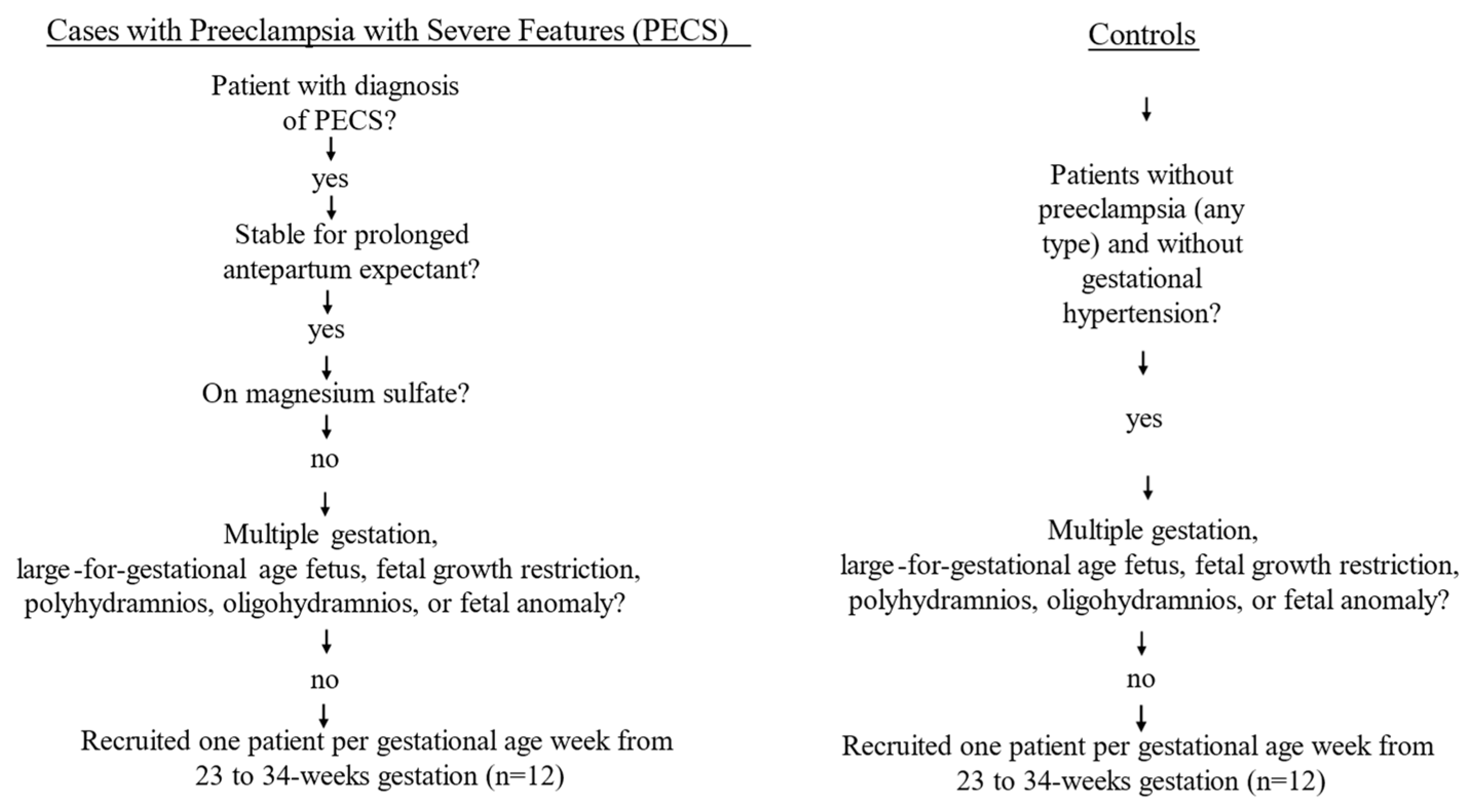
Figure 1. Flow chart for inclusion and exclusion criteria of study population.
Reprodmed 06 00035 g001
Figure 2. (a) M-mode of the maternal inferior vena cava assessed through the subxiphoid window 2 cm from the right atrium distal to the confluence of the hepatic veins in a patient with preeclampsia with severe features at 30 weeks. (b) M-mode in the control patient at 30 weeks. Inferior vena cava diameters are measured during inspiration (here noted Dist A) and expiration (here noted Dist B).
Reprodmed 06 00035 g002
Figure 3. The trend in IVC collapsibility index across gestational age for both groups.
Reprodmed 06 00035 g003
Table 1. Baseline Characteristics of Gestational Age-Matched Controls versus Preeclampsia.
Gestational Age Matched Controls (n = 12)PECS
(n = 12)
Age (years)30.8 (5.6) *28.3 (6.6) *
Parity
    Nulliparous5 (41.7)8(66.7)
    Multiparous7 (58.3)4 (33.3)
BMI (kg/m2)34.3 (7.4)37.8 (10.5)
Race/Ethnicity
    White, non-Hispanic3 (60.0)2 (40.0)
    Not white, non-Hispanic9 (47.4)10 (52.6)
Pre-pregnancy diabetes01 (8.3)
Gestational diabetes1 (8.3)0
Chronic hypertension6 (50.0)5 (41.7)
n, number; BMI, body mass index; Data are mean (standard deviation) *, or number (percentage).
Table 2. Comparison of maternal caval parameters and maternal pulse pressure.
ControlsPECS
IVC inhalation (cm)0.6 (0.2)1.5 (0.4)p < 0.001
IVC exhalation (cm)1.1 (0.3)1.9 (0.3)p < 0.001
IVC-CI (%)48.3 (13.6)20.1 (14.5)p < 0.001
Pulse pressure (mmHg)40.8 (4.1)61.4 (10.4)p < 0.001
IVC, inferior vena cava; IVC-CI, inferior vena cava collapsibility index. Data are mean (standard deviation).
Disclaimer/Publisher’s Note: The statements, opinions and data contained in all publications are solely those of the individual author(s) and contributor(s) and not of MDPI and/or the editor(s). MDPI and/or the editor(s) disclaim responsibility for any injury to people or property resulting from any ideas, methods, instructions or products referred to in the content.