Article

Can We Safely Decrease Early-Term Delivery and Cesarean Section Rate in Pregnancies Complicated by Fetal Transposition of Great Arteries?

by
Angel Chimenea
1,2,
Lutgardo García-Díaz
1,3,
Ana Méndez
4 and
Guillermo Antiñolo
1,2,3,5,*
1
Department of Materno-Fetal Medicine, Genetics and Reproduction, Institute of Biomedicine of Seville (IBIS), Hospital Universitario Virgen del Rocio/CSIC/University of Seville, 41013 Seville, Spain
2
Fetal, IVF and Reproduction Simulation Training Centre (FIRST), 41010 Seville, Spain
3
Department of Surgery, University of Seville, 41002 Seville, Spain
4
Department of Paediatric Cardiology, Hospital Universitario Virgen del Rocio, 41013 Seville, Spain
5
Centre for Biomedical Network Research on Rare Diseases (CIBERER), 41013 Seville, Spain
*
Author to whom correspondence should be addressed.
Reprod. Med. 2023, 4(3), 233-241; https://doi.org/10.3390/reprodmed4030021
Submission received: 19 July 2023 / Revised: 20 August 2023 / Accepted: 8 September 2023 / Published: 14 September 2023

Abstract

:
Background: Transposition of the great arteries (TGA) is a common critical neonatal congenital heart defect. After birth, physiological shunts close rapidly, necessitating early treatment with prostaglandin infusion and balloon-atrial septostomy. Timing of delivery is challenging, balancing the risks and advantages of early-term delivery and specialized care. The aim of this study is to assess the safety of a full-term delivery policy in fetuses diagnosed with TGA. Methods: A retrospective chart review was conducted of 17 women with a prenatal diagnosis of fetal TGA at Virgen del Rocío University Hospital between 2015 and 2021. Primary outcomes included: incidence of preterm, early-term, full-term, and late-term delivery, and rate of cesarean section. Secondary outcomes included: Saturday to Sunday admission and birth, and delivery between 0:00 a.m. and 8:00 a.m. Results: Full-term birth was achieved in 94.1%, reaching a low cesarean delivery rate (17.6%). A total of 82.4% of infants were born on weekdays, and only in three of the cases (17.6%) did delivery occur between 0 a.m. and 8 a.m. The median birth weight was 3300 g. Intravenous prostaglandins were administered in all cases, and 94.1% required balloon-atrial septostomy. Conclusions: In our study favoring full-term delivery, we reduce early-term deliveries and the cesarean section rate in prenatally diagnosed TGA.

1. Introduction

Transposition of the great arteries (TGA) is one of the most common forms of critical neonatal congenital heart defects (CHDs), with an estimated incidence of 315 cases per million live births, accounting for 5% to 7% of all congenital heart diseases [1,2,3]. It frequently goes undiagnosed during pregnancy, with prenatal detection rates typically falling below 50% [3]. It involves a unique anatomical arrangement where the aorta emerges from the right ventricle, and the pulmonary artery originates from the left ventricle, along with a concordant atrio-ventricular connection. This arrangement creates two parallel circulatory systems, a configuration that the fetus can accommodate well due to the presence of essential physiological shunts: the foramen ovale (FO) and the ductus arteriosus (DA). The prenatal diagnosis is established by 2-D and color doppler ultrasound, which reveals the presence of two vessels instead of three in the three-vessel view, the parallel alignment of the great arteries, and the identification of the origin of each artery (Figure 1). Employing 3D ultrasound can also prove beneficial for diagnosis [3,4].
Throughout gestation, these shunts play a vital role by permitting blood to bypass certain non-functional fetal structures. However, as the newborn takes its first breaths, the shunts begin to close spontaneously and gradually, causing a sudden deterioration in the heart’s hemodynamics [5]. This shift in circulation can lead to serious consequences, such as reduced oxygenation of tissues, cyanosis, and respiratory difficulties.
Newborns with TGA require early treatment after birth to maintain these shunts open. Prostaglandin infusion and, when necessary, urgent balloon-atrial septostomy (BAS) are commonly employed to ensure adequate blood flow [6,7,8]. Due to the critical nature of their condition, it is strongly recommended that newborns with TGA be delivered in medical centers equipped with expertise in pediatric cardiology and cardiac surgery [7,9,10,11]. However, centralizing care in specialized centers may pose logistical challenges, as families may have to travel long distances to reach the referral center for delivery. Centralization can limit the organization and scheduling of full-term delivery, because these patients may have to travel long distances to reach the referral center. Thus, most of these births occur at early term to favor easier access and care coordination at tertiary-care centers [12,13,14].
Extensive research has shown that early-term delivery, especially in cases where CHDs are prenatally diagnosed, can have adverse effects [15,16,17,18,19]. Elective delivery before full term does not provide any advantages and, in fact, poses additional technical challenges for surgeons when attempting to perform complete surgical repair [20,21,22]. This is particularly evident in newborns with TGA, as they experience a higher mortality rate during surgery if their birth weight is less than 2.5 kg [23,24].
However, there is no consensus on the optimal time of delivery for a fetus with known TGA. The obstetrician is faced with the challenging task of carefully balancing various factors to make the best decision for both the mother and the baby. On the one hand, early-term delivery may carry disadvantages and potential risks for the infant’s development and health [19]. On the other hand, delaying delivery until full term could increase the risk of hemodynamic failure shortly after birth.
In this paper, we present our experience following the adoption of a full-term delivery policy when prenatal diagnosis of TGA had been established. We hypothesize that it would be possible to safely reduce the number of early-term deliveries, allowing immediate neonatal cardiological care in a high-level center, while reducing the rate of cesarean sections in these patients.

2. Materials and Methods

2.1. Study Design, Setting, and Population

This was a retrospective observational study in an unselected population, conducted at Virgen del Rocío University Hospital of Seville, a tertiary referral center for fetal and pediatric CHD, with an average of 6000 births per year.
All cases of suspected fetal TGA between January 2015 and November 2021 were identified from the prenatal ultrasound database and were included in the study. All pregnancy terminations (n = 1), stillbirths (n = 1), and patients with a postnatal diagnosis of TGA (n = 3) were excluded.
Gestational age was determined by fetal biometry at the first-trimester scan (11.0–13.6 weeks of gestation). A detailed malformation scan was performed at 18.0–21.6 weeks of gestation. After the suspected diagnosis of TGA, the patients were referred to our department, where the diagnosis was confirmed by a multidisciplinary team, comprising specialists in fetal medicine and pediatric cardiology. All the fetal echocardiographic images were reviewed for the purposes of this study.
In this study, written informed consent was obtained from all patients who were included in the research. The data for the study were extracted from the institution’s electronic health record system. As part of the research process, the local research ethics committee thoroughly evaluated the study and determined it to be an audit of practice, not necessitating a full review. The management of patients in this study has been conducted in accordance with current clinical practice guidelines. Throughout the entire process of medical care, the recommendations and directives established by the current clinical guidelines for managing patients with TGA were strictly adhered to.

2.2. Data Collection

Data concerning maternal characteristics, obstetric history, prenatal diagnosis, and pregnancy follow-up were retrieved from the electronic health record, including: maternal age, body mass index (BMI) at the first prenatal visit, smoking during pregnancy, gravidity, parity, previous cesarean section, twin pregnancy, CHD in a previous pregnancy, gestational age (GA) at first consultation in our department, prenatal diagnosis (isolated TGA, TGA + ventricular septal defect, or complex TGA), invasive procedure of prenatal diagnosis, GA at last visit, and estimated fetal weight (EFW) at last visit.
Other data, such as postnatal diagnosis, gender, mode of delivery, GA at birth, birthweight, hour at delivery, Saturday to Sunday admission and birth, Apgar score, umbilical cord arterial pH, and newborn mortality, were recorded.
We also collected information related to the treatment and support received at birth, such as infusion of prostaglandins, BAS (Rashkind), cardiac surgery (Switch) as well as intubation or mechanical ventilation, along with other complications, such as metabolic acidosis and post-surgery complications, and the length of hospital stay.
Data were complete for all variables, except the umbilical cord arterial pH (15/17, 88%).

2.3. Perinatal Management

In our center, vaginal delivery is preferred following a prenatal diagnosis of CHD, as cesarean sections are reserved for obstetric indications. Since 2015, in TGA patients, induction of labor is considered from 39.0 weeks of gestation on. From that gestational age on, the GA for induction of labor is individualized, depending on parity and patient distance from the hospital.
Induction is undertaken using mechanical methods (Cook® Cervical Ripening Balloon, Bloomington, IN, USA) or the vaginal prostaglandin E2 delivery system (Propess®, Saint-Prex, Switzerland). Later, amniotomy is performed, and oxytocin infusion is started, depending on uterine dynamics. Antepartum fetal monitoring was conducted in accordance with the guidelines set by the American College of Obstetricians and Gynecologists (ACOG) [25]. The mode of delivery was determined on a case-by-case basis, taking obstetrical criteria into consideration. In all instances, the fetal cardiac condition did not influence the choice of the delivery mode.
All patients with a prenatal diagnosis of TGA were born in our center regardless of the maternal residence and were managed by a multidisciplinary team, including an obstetrician, fetal medicine specialist, neonatologist, pediatric cardiologist, and nurse specialist. After birth, the newborns were admitted to the neonatal intensive care unit for prostaglandin infusion, including mechanical ventilation and inotropic agents when necessary. BAS was performed in newborns with severe hypoxemia.
After 24 to 48 h of clinical stability, the patient was evaluated in a surgical committee where the surgical indication was established. Following individualization of the clinical condition, the patient undergoes an arterial switch intervention, usually in the first two weeks of life.

2.4. Outcomes

The co-primary outcomes of the study included the incidence of preterm deliveries (birth under 37.0 weeks of gestation), early-term deliveries (birth between 37.0 and 38.6 weeks of gestation), full-term deliveries (birth between 39.0 and 40.6 weeks of gestation) and late-term deliveries (birth between 41.0 and 41.6 weeks of gestation), as well as the rate of cesarean sections in fetal TGA pregnancies. Secondary outcomes included Saturday to Sunday admission and birth, and delivery between 0:00 a.m. and 8:00 a.m. We further assessed the neonatal management and the time in days until hospital discharge.

2.5. Statistical Analysis

Statistical analysis was performed using the SPSS 25.0 software package (SPSS Inc., Chicago, IL, USA), and statistical significance was assumed at p < 0.05. All hypothesis tests were two-sided. Discrete variables were presented as numbers and percentages (%). Continuous variables were described with medians and ranges, or with means and standard deviations (SD), since data were normally distributed.

3. Results

3.1. Study Population and Participant Characteristics

Over a period of 6 years, 39 582 children were born at Virgen del Rocío University Hospital (Seville). A total of 17 cases of TGA were prenatally diagnosed.
The baseline maternal and obstetric characteristics are summarized in Table 1. All the patients included in the study had singleton pregnancies. The median maternal age was 35 years, presenting a low rate of obesity (5.9%, defined as BMI < 30 kg/m2). More than half of the patients (52.9%) had a previous pregnancy, and 11.8% had a previous cesarean section. Only one of the patients had a diagnosis of CHD in a previous pregnancy (5.9%, pulmonary stenosis).

3.2. Diagnostic Features and Follow-Up

Data related to the diagnosis of TGA and follow-up of the pregnancy are displayed in Table 2. Most patients have an isolated TGA (64.7%). A ventricular septal defect was present in five cases (29.4%), and one fetus had a complex form of TGA associated with ventricular septum defect, and pulmonary stenosis.
In 70.6% of the cases, the patients accepted an invasive procedure, and we found genetic abnormalities in only one of them (an interstitial gain affecting the region of the 16p11.2 was identified in array-CGH).
The last ultrasound control was performed close to delivery (median 37.6 weeks of gestation), and no gestational complications, including intrauterine growth restriction, were identified.

3.3. Obstetric Outcomes

All fetuses were born at term. In 88.2% of pregnancies (15/17), we reached the target gestational age for delivery (≥39.0 weeks of gestation). The median gestational age at birth was 39.2 weeks (range 37.2–41.0). Only one was a non-elective early-term delivery (spontaneous onset of labor at 38.3 weeks, with a favorable post-natal outcome). In the remaining case, an elective cesarean section had to be performed at 37.2 weeks due to the presence of placenta previa. Therefore, excluding elective early-term delivery for obstetric indications, 94.1% of the pregnancies reached full term.
All the obstetric outcomes are summarized in Table 3.
Applying this delivery policy, we achieved a low cesarean delivery rate (3/17, 17.6%), even well below the cesarean section rate in our center (24.5%). Regarding emergent cesarean sections (2/3), only one was performed that was related to pathological CTG (Apgar score of 6, 7, and 10 at 1, 5, and 10 min, and umbilical cord arterial pH of 7.32).
We found satisfactory results in the Apgar score (median 5-min Apgar score: 9; 5-min Apgar score < 7: n = 2) as well as in the umbilical cord arterial pH (median 7.28, range 7.18–7.44). One of the newborns died after 24 h of life due to a very restrictive FO, refractory to performing BAS. None of the fetuses exhibited acidemia at birth.
Fourteen infants (82.4%) were born on weekdays. The median hour at delivery was 5 p.m. (2 a.m.–10 p.m.), and only in three of the cases (17.6%) did delivery occur between 0 a.m. and 8 a.m.
The median birth weight was 3300 g. Only one of the cases had a low birth weight (<2500 g), and it corresponded to the elective cesarean section for placenta previa mentioned above. In our series favoring full-term delivery, a higher birth weight was achieved, which facilitates postnatal medical and surgical management.

3.4. Initial Management and Surgical Treatment

Data referring to postnatal management are presented in Table 4. After delivery, echocardiography was performed, modifying the diagnosis in three cases. In all cases, intravenous prostaglandins were administered after birth, and 94.1% required BAS (16/17) (median 5 h after delivery), similar to data previously reported [26].
During the study period, neonatal corrective surgery was performed in 14 patients (82.4%). The median hospital stay was 29 days (range 11–71 days).

4. Discussion

In our series of cases, the implementation of a policy promoting full-term delivery in fetuses diagnosed with TGA resulted in a high success rate, with 94.1% of pregnancies achieving full-term births. This shift towards full-term deliveries had an additional positive impact, as it led to a significantly low cesarean delivery rate of only 17.6%. This reduction in cesarean deliveries is noteworthy, as it may contribute to improved perinatal outcomes and reduce potential complications associated with surgical deliveries.
Furthermore, our experience suggests that full-term delivery may be associated with higher birth weights. This can be a critical factor when planning surgical correction for TGA, as infants with higher birth weights may have improved physiological reserves and a better tolerance for the surgical procedure. This association could lead to safer surgical interventions and potentially enhance overall outcomes for infants born with TGA.
Since the early 2010s, multiple studies have paid careful attention to GA at delivery in fetuses diagnosed with CHD [15,16,17,18,27]. The studies published by the group of Costello et al. represented a change in clinical practice in many centers worldwide. The authors concluded that in neonates who underwent cardiac surgery, early-term birth was associated with a higher in-hospital mortality, complication rates, and postoperative length of stay [19,28]. Since 2015, we have implemented a delivery policy aimed at increasing the rate of full-term births in pregnancies where the newborn would foreseeably undergo postnatal surgery, such as TGA. However, in recent published studies on fetuses with TGA, the mean GA at delivery is less than 39.0 weeks, within a range of 38.0 to 38.3 weeks [29,30], and some authors continue to advocate for elective termination after 38.0 weeks [31].
Recently, there has been growing interest in standardizing the mode of delivery for fetuses with CHD to address concerns about the increasing cesarean section rate and the percentage of early-term deliveries. A multi-institutional study, which focused on 496 fetal TGA pregnancies, aimed to evaluate and optimize the delivery approach [32]. The study’s findings revealed a mean GA at birth that was close to full-term (38.9 weeks), with a significant proportion of vaginal deliveries, accounting for 61% of cases.
Our results align with those of previous publications and further contribute to the evidence supporting the benefits of promoting full-term deliveries in fetuses diagnosed with TGA. Achieving a high rate of full-term deliveries has proven to be feasible and safe while simultaneously reducing the prevalence of cesarean sections. Moreover, in our series, more than 80% of deliveries occurred on weekdays, between 8 a.m. and 11 p.m. (median hour at delivery: 5 p.m.).
Survival in newborns with TGA depends on the time of prostaglandin infusion initiation after birth [33]. Due to this critical factor, many obstetricians have concerns today about attempting a full-term delivery in fetuses with TGA, especially when the reference hospital is located far away from the delivery location. However, the advantages of a full-term delivery together with the centralization of pediatric heart surgery far outweigh the risks associated with an out-of-hospital birth [11]. In this sense, full-term delivery did not lead to an increase in maternal and fetal morbidity or mortality, but it did lead to a decrease in neonatal respiratory morbidity [17,19,27,31,34]. Our findings are consistent with those reported previously. In one case within our series, labor commenced spontaneously before 39.0 weeks. Fortunately, the patient reached the hospital in time for optimal care conditions, demonstrating that even in instances of spontaneous labor before the recommended term, prompt access to specialized medical facilities can lead to favorable outcomes.
Despite the described association between TGA and SGA and FGR [35,36], none of the cases in our series presented either of these diagnoses during pregnancy, and the birth weight was appropriate for the gestational age. In these cases, management should be tailored according to the guidelines set for growth restriction.
It is important to recognize the inherent limitations in our retrospective study. Depending on existing data introduces potential inaccuracies, and the study’s retrospective nature hinders the ability to establish causal relationships due to uncontrolled confounding factors. Nonetheless, the study provides valuable historical insights, serving as a basis for future prospective research. It should be noted that a longer series of cases is necessary to reaffirm this preliminary evidence. Additional variables impacting the decision-making process for delivery timing in TGA pregnancies should be explored in future investigations. Despite this limitation, the study emphasizes the significance of considering the timing of delivery in TGA pregnancies to optimize outcomes for both the mother and the baby, highlighting the potential benefits of waiting for full-term delivery in these cases.

5. Conclusions

In our study, by favoring full-term delivery in prenatally diagnosed TGA, we can safely decrease the incidence of early-term deliveries, reduce the rate of cesarean sections, and lower the percentage of infants with a low birth weight. These improvements in delivery timing contribute to enhanced postnatal care and facilitate more favorable conditions for surgical correction procedures. Overall, this approach significantly improves the management of TGA cases and offers better outcomes for infants and their families.

Author Contributions

Conceptualization, A.C., L.G.-D., A.M. and G.A.; Data curation, A.C.; Formal analysis, A.C. and G.A.; Investigation, A.C., L.G.-D., A.M. and G.A.; Methodology, A.C. and G.A.; Project administration, A.C.; Resources, A.C.; Software, A.C.; Supervision, L.G.-D., A.M. and G.A.; Visualization, A.C., L.G.-D., A.M. and G.A.; Writing—original draft, A.C.; Writing—review & editing, A.C., L.G.-D., A.M. and G.A. All authors have read and agreed to the published version of the manuscript.

Funding

This research received no external funding.

Institutional Review Board Statement

The study was conducted in accordance with the Declaration of Helsinki. The Andalusian research ethics committee (Virgen del Rocío—Virgen Macarena) assessed the present study as being an audit of practice and not requiring a full review.

Informed Consent Statement

Informed consent was obtained from all subjects involved in the study.

Data Availability Statement

The datasets used during the current study are available from the corresponding author on reasonable request.

Conflicts of Interest

The authors declare no conflict of interest.

References

  1. Marek, J.; Tomek, V.; Škovránek, J.; Povýšilová, V.; Šamánek, M. Prenatal ultrasound screening of congenital heart disease in an unselected national population: A 21-year experience. Heart 2011, 97, 124–130. [Google Scholar] [CrossRef] [PubMed]
  2. Hoffman, J.I.; Kaplan, S. The incidence of congenital heart disease. J. Am. Coll. Cardiol. 2002, 39, 1890–1900. [Google Scholar] [CrossRef] [PubMed]
  3. Bravo-Valenzuela, N.J.; Peixoto, A.B.; Júnior, E.A. Prenatal diagnosis of transposition of the great arteries: An updated review. Ultrasonography 2020, 39, 331–339. [Google Scholar] [CrossRef]
  4. Quaresima, P.; Fesslova, V.; Farina, A.; Kagan, K.O.; Candiani, M.; Morelli, M.; Crispi, F.; Cavoretto, P.I. How to do a fetal cardiac scan. Arch Gynecol Obstet. 2023, 307, 1269–1276. [Google Scholar] [CrossRef]
  5. Jouannic, J.M.; Gavard, L.; Fermont, L.; Le Bidois, J.; Parat, S.; Vouhé, P.R.; Dumez, Y.; Sidi, D.; Bonnet, D. Sensitivity and specificity of prenatal features of physiological shunts to predict neonatal clinical status in transposition of the great arteries. Circulation 2004, 110, 1743–1746. [Google Scholar] [CrossRef] [PubMed]
  6. Bonnet, D.; Coltri, A.; Butera, G.; Fermont, L.; Le Bidois, J.; Kachaner, J.; Sidi, D. Detection of transposition of the great arteries in fetuses reduces neonatal morbidity and mortality. Circulation 1999, 99, 916–918. [Google Scholar] [CrossRef]
  7. Blyth, M.; Howe, D.; Gnanapragasam, J.; Wellesley, D. The hidden mortality of transposition of the great arteries and survival advantage provided by prenatal diagnosis. BJOG 2008, 115, 1096–1100. [Google Scholar] [CrossRef]
  8. van Velzen, C.L.; Haak, M.C.; Reijnders, G.; Rijlaarsdam, M.E.B.; Bax, C.J.; Pajkrt, E.; Hruda, J.; Galindo-Garre, F.; Bilardo, C.M.; de Groot, C.J.M.; et al. Prenatal detection of transposition of the great arteries reduces mortality and morbidity. Ultrasound Obstet. Gynecol. 2015, 45, 320–325. [Google Scholar] [CrossRef]
  9. Hellström-Westas, L.; Hanséus, K.; Jögi, P.; Lundström, N.-R.; Svenningsen, N. Long-distance transports of newborn infants with congenital heart disease. Pediatr. Cardiol. 2001, 22, 380–384. [Google Scholar] [CrossRef]
  10. Sarris, G.E.; Balmer, C.; Bonou, P.; Comas, J.V.; da Cruz, E.; Di Chiara, L.; Di Donato, R.M.; Fragata, J.; Jokinen, T.E.; Kirvassilis, G.; et al. Clinical guidelines for the management of patients with transposition of the great arteries with intact ventricular septum. Eur. J. Cardio-Thoracic Surg. 2017, 51, e1–e32. [Google Scholar] [CrossRef]
  11. Lundström, N.; Berggren, H.; Björkhem, G.; Jögi, P.; Sunnegårdh, J. Centralization of pediatric heart surgery in Sweden. Pediatr. Cardiol. 2000, 21, 353–357. [Google Scholar] [CrossRef] [PubMed]
  12. Tworetzky, W.; McElhinney, D.B.; Reddy, V.M.; Brook, M.M.; Hanley, F.L.; Silverman, N.H. Improved surgical outcome after fetal diagnosis of hypoplastic left heart syndrome. Circulation 2001, 103, 1269–1273. [Google Scholar] [CrossRef]
  13. Bartlett, J.M.; Wypij, D.; Bellinger, D.C.; Rappaport, L.A.; Heffner, L.J.; Jonas, R.A.; Newburger, J.W. Effect of prenatal diagnosis on outcomes in D-transposition of the great arteries. Pediatrics 2004, 113, e335–e340. [Google Scholar] [CrossRef]
  14. Levey, A.; Glickstein, J.S.; Kleinman, C.S.; Levasseur, S.M.; Chen, J.; Gersony, W.M.; Williams, I.A. The impact of prenatal diagnosis of complex congenital heart disease on neonatal outcomes. Pediatr. Cardiol. 2010, 31, 587–597. [Google Scholar] [CrossRef]
  15. ACOG. Committee Opinion No. 764: Medically Indicated Late-Preterm and Early-Term Deliveries. Obstet. Gynecol. 2019, 133, e151–e155. [Google Scholar] [CrossRef]
  16. Clark, S.L.; Miller, D.D.; Belfort, M.A.; Dildy, G.A.; Frye, D.K.; Meyers, J.A. Neonatal and maternal outcomes associated with elective term delivery. Am. J. Obstet. Gynecol. 2009, 200, 156.e1–156.e4. [Google Scholar] [CrossRef]
  17. Laas, E.; on behalf of the EPICARD study group; Lelong, N.; Ancel, P.-Y.; Bonnet, D.; Houyel, L.; Magny, J.-F.; Andrieu, T.; Goffinet, F.; Khoshnood, B. Impact of preterm birth on infant mortality for newborns with congenital heart defects: The EPICARD population-based cohort study. BMC Pediatr. 2017, 17, 1–8. [Google Scholar] [CrossRef]
  18. McLaren, R., Jr.; London, V.; Stein, J.L.; Minkoff, H. Adverse outcomes in early term versus full-term deliveries among higher-order cesarean births. J. Matern. Fetal. Neonatal. Med. 2021, 35, 5464–5469. [Google Scholar] [CrossRef]
  19. Costello, J.M.; Pasquali, S.K.; Jacobs, J.P.; He, X.; Hill, K.D.; Cooper, D.S.; Backer, C.L.; Jacobs, M.L. Gestational age at birth and outcomes after neonatal cardiac surgery: An analysis of the Society of Thoracic Surgeons Congenital Heart Surgery Database. Circulation 2014, 17, 2511–2517. [Google Scholar] [CrossRef]
  20. Hickey, E.J.; Nosikova, Y.; Zhang, H.; Caldarone, C.A.; Benson, L.; Redington, A.; Van Arsdell, G.S. Very low-birth-weight infants with congenital cardiac lesions: Is there merit in delaying intervention to permit growth and maturation? J. Thorac. Cardiovasc. Surg. 2012, 143, 126–136.e1. [Google Scholar] [CrossRef]
  21. Donofrio, M.T.; Levy, R.J.; Schuette, J.J.; Skurow-Todd, K.; Sten, M.-B.; Stallings, C.; Pike, J.I.; Krishnan, A.; Ratnayaka, K.; Sinha, P.; et al. Specialized delivery room planning for fetuses with critical congenital heart disease. Am. J. Cardiol. 2013, 111, 737–747. [Google Scholar] [CrossRef] [PubMed]
  22. Craigo, S.D. Indicated preterm birth for fetal anomalies. Semin. Perinatol. 2011, 35, 270–276. [Google Scholar] [CrossRef] [PubMed]
  23. Curzon, C.L.; Milford-Beland, S.; Li, J.S.; O’Brien, S.M.; Jacobs, J.P.; Jacobs, M.L.; Welke, K.F.; Lodge, A.J.; Peterson, E.D.; Jaggers, J. Cardiac Surgery in Infants with Low Birth Weight is Associated with Increased Mortality: Analysis of the Society of Thoracic Surgeons Congenital Heart Database. J. Thorac. Cardiovasc. Surg. 2008, 135, 546–551. [Google Scholar] [CrossRef] [PubMed]
  24. Kansy, A.; Tobota, Z.; Maruszewski, P.; Maruszewski, B. Analysis of 14,843 Neonatal Congenital Heart Surgical Procedures in the European Association for Cardiothoracic Surgery Congenital Database. Ann. Thorac. Surg. 2010, 89, 1255–1259. [Google Scholar] [CrossRef]
  25. American College of Obstetricians and Gynecologists ACOG. Practice Bulletin No. 106: Intrapartum Fetal Heart Rate Monitoring: Nomenclature, Interpretation, and General Management Principles. Obstet. Gynecol. 2009, 114, 192–202. [Google Scholar] [CrossRef]
  26. Hraska, V.; Podnar, T.; Kunovsky, P.; Kovacikova, L.; Kaldararova, M.; Horvathova, E.; Masura, J.; Mayer, J. Is a learning curve for arterial switch operation in small countries still acceptable? Model for cooperation in Europe. Eur. J. Cardio-Thoracic Surg. 2003, 24, 352–357. [Google Scholar] [CrossRef]
  27. Cnota, J.F.; Gupta, R.; Michelfelder, E.C.; Ittenbach, R.F. Congenital heart disease infant death rates decrease as gestational age advances from 34 to 40 weeks. J. Pediatr. 2011, 159, 761–765. [Google Scholar] [CrossRef]
  28. Costello, J.M.; Polito, A.; Brown, D.W.; McElrath, T.F.; Graham, D.A.; Thiagarajan, R.R.; Bacha, E.A.; Allan, C.K.; Cohen, J.N.; Laussen, P.C. Birth before 39 weeks’ gestation is associated with worse outcomes in neonates with heart disease. Pediatrics 2010, 126, 277–284. [Google Scholar] [CrossRef]
  29. Słodki, M.; Axt-Fliedner, R.; Zych-Krekora, K.; Wolter, A.; Kawecki, A.; Enzensberge, C.; Gulczyńska, E.; Respondek-Liberska, M. The international prenatal cardiology collaboration group new method to predict need for Rashkind procedure in fetuses with dextro-transposition of the great arteries. Ultrasound Obstet. Gynecol. 2018, 51, 531–536. [Google Scholar] [CrossRef]
  30. Escobar-Diaz, M.C.; Freud, L.R.; Bueno, A.; Brown, D.W.; Friedman, K.G.; Schidlow, D.; Emani, S.; Del Nido, P.J.; Tworetzky, W. Prenatal diagnosis of transposition of the great arteries over a 20-year period: Improved but imperfect. Ultrasound Obstet. Gynecol. 2015, 45, 678–682. [Google Scholar] [CrossRef]
  31. Sanapo, L.; Moon-Grady, A.J.; Donofrio, M.T. Perinatal and Delivery Management of Infants with Congenital Heart Disease. Clin. Perinatol. 2016, 43, 55–71. [Google Scholar] [CrossRef] [PubMed]
  32. Afshar, Y.; Hogan, W.J.; Conturie, C.; Sunderji, S.; Duffy, J.Y.; Peyvandi, S.; Boe, N.M.; Melber, D.; Fajardo, V.M.; Tandel, M.D.; et al. Multi-Institutional Practice-Patterns in Fetal Congenital Heart Disease Following Implementation of a Standardized Clinical Assessment and Management Plan. J. Am. Heart Assoc. 2021, 10, e021598. [Google Scholar] [CrossRef] [PubMed]
  33. Quaegebeur, J.M.; Rohmer, J.; Ottenkamp, J.; Buis, T.; Kirklin, J.W.; Blackstone, E.H.; Brom, A.G. The arterial switch operation. An eight-year experience. J. Thorac. Cardiovasc. Surg 1986, 92, 361–384. [Google Scholar] [CrossRef] [PubMed]
  34. Kadour-Peero, E.; Bleicher, I.; Vitner, D.; Sloma, R.; Bahous, R.; Levy, E.; Sagi, S.; Gonen, R. When should repeat cesarean delivery be scheduled, after two or more previous cesarean deliveries? J. Matern. Fetal. Neonatal Med. 2018, 31, 474–480. [Google Scholar] [CrossRef] [PubMed]
  35. Giorgione, V.; Fesslova, V.; Boveri, S.; Candiani, M.; Khalil, A.; Cavoretto, P. Adverse perinatal outcome and placental abnormalities in pregnancies with major fetal congenital heart defects: A retrospective case-control study. Prenat. Diagn. 2020, 40, 1390–1397. [Google Scholar] [CrossRef]
  36. Inversetti, A.; Fesslova, V.; Deprest, J.; Candiani, M.; Giorgione, V.; Cavoretto, P. Prenatal Growth in Fetuses with Isolated Cyanotic and Non-Cyanotic Congenital Heart Defects. Fetal. Diagn. Ther. 2020, 47, 411–419. [Google Scholar] [CrossRef]
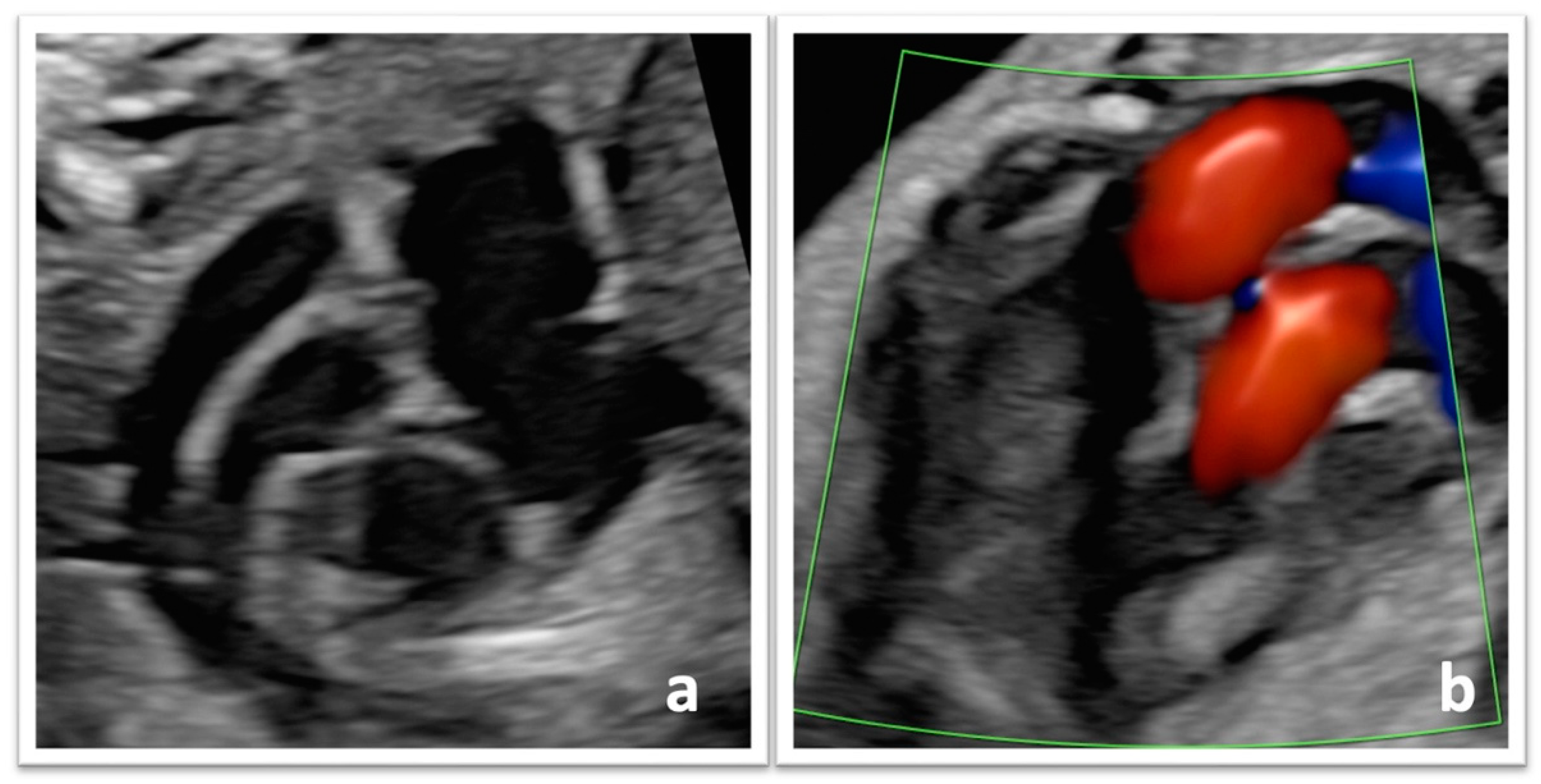
Figure 1. Prenatal diagnosis of TGA using (a) 2-D ultrasound and (b) color Doppler ultrasound. Green lines: color doppler region of interest.
Reprodmed 04 00021 g001
Table 1. Maternal and obstetric characteristics.
All TGA (n = 17)
Maternal age (years), median (range)35 (19–39)
BMI, kg/m2, median (range)23.2 (16.4–31.7)
BMI ≥ 30 kg/m2, n (%)1 (5.9%)
Smoking, n (%)2 (11.8%)
Gravidity, median (range)2 (1–3)
Previous pregnancy, n (%)9 (52.9%)
Previous cesarean section, n (%)2 (11.8%)
Twin pregnancy, n (%) 0
CHD in previous pregnancy, n (%)1 (5.9%)
         Type of CHDPulmonary stenosis
Table 2. Diagnostic features and follow-up.
All TGA (n = 17)
GA at first consultation in our department (weeks), median (range)20.5 (14.1–32.6)
Prenatal diagnosis
         Isolated TGA, n (%)11 (64.7%)
         TGA + VSD, n (%)5 (29.4%)
         Complex TGA, n (%)1 (5.9%)
Invasive procedure of prenatal diagnosis, n (%)12 (70.6%)
         Abnormal, n (%)1 (5.9%)
GA at last visit (weeks), median (range)37.6 (34.6–41.0)
EFW at last visit (grams), median (range)3251 (2239–3502)
         EFW centile, median (range)56 (24–85)
         RCIU, n (%)0
Table 3. Obstetric outcomes.
All TGA (n = 17)
Gender (female), n (%)7 (41.2%)
Elective cesarean section, n (%)1 (5.9%)
Vaginal birth, n (%)14 (82.4%)
Operative birth, n (%)4 (23.5%)
         Vacuum extraction, n (%)4 (100%)
Urgent cesarean section, n (%)2 (11.8%)
Gestational age at delivery (weeks), median (range)39.2 (37.2–41.0)
         Gestational age < 39 weeks, n (%)2 (11.8%)
         Gestational age < 40 weeks, n (%)12 (70.6%)
         Gestational age < 41 weeks, n (%)16 (94.1%)
Hour at delivery, median (range)5 p.m. (2 a.m.–10 p.m.)
Saturday to Sunday admission and birth, n (%)3 (17.6%)
Birthweight (grams), median (range)3300 (2399–3891)
5-min APGAR score, median (range)9 (4–10)
         5-min APGAR score < 7, n (%)2 (11.8%)
Umbilical cord arterial pH, median (range)7.28 (7.18–7.44)
         Umbilical cord arterial pH < 7.1, n (%)0
Mortality before surgery, n (%)1 (5.8%)
Table 4. Initial management and surgical treatment.
All TGA (n = 17)
Postnatal diagnosis
         Isolated TGA, n (%)10 (58.8%)
         TGA + VSD, n (%)3 (17.7%)
         Complex TGA, n (%)4 (23.5%)
Use of prostaglandin E2, n (%)17 (100%)
BAS (Rashkind), n (%)16 (94.1%)
         Age at BAS (Rashkind) (hours), median (range)5 (2–408)
Surgery (switch), n (%)14 (82.4%)
         Age at surgery (days), median (range)8 (3–30)
Intubation or mechanical ventilation, n (%)4 (23.5%)
Metabolic acidosis, n (%)1 (5.9%)
Length of hospital stays (days), median (range)29 (11–71)
Post-surgery complications, n (%)6 (46.2%)
Disclaimer/Publisher’s Note: The statements, opinions and data contained in all publications are solely those of the individual author(s) and contributor(s) and not of MDPI and/or the editor(s). MDPI and/or the editor(s) disclaim responsibility for any injury to people or property resulting from any ideas, methods, instructions or products referred to in the content.