Article

Implementation of Minimally Invasive Mitral Valve Surgery in a Novice Center

by
Andre Korshin
1,*,
Peter Hasse Møller-Sørensen
1,
Jacob Eifer Møller
2 and
Christian Lildal Carranza
3
1
Department of Cardiothoracic Anesthesiology, The Rigshospital, 2300 Copenhagen, Denmark
2
Department of Cardiology, The Rigshospital, 2300 Copenhagen, Denmark
3
Department of Cardiothoracic Surgery, The Rigshospital, 2300 Copenhagen, Denmark
*
Author to whom correspondence should be addressed.
Hearts 2025, 6(2), 11; https://doi.org/10.3390/hearts6020011
Submission received: 3 March 2025 / Revised: 31 March 2025 / Accepted: 1 April 2025 / Published: 17 April 2025

Abstract

:
Background/Objectives: The complexity of Minimally Invasive Mitral Valve Surgery (MIMVS) could cause a slow learning curve and potentially patient harm. We thus investigated if a novice mitral valve center encountered difficulties implementing MIMVS. Methods: We investigated seven hundred and forty-eight mitral valve surgery patients, two years before and after MIMVS introduction. Results: We propensity score matched two hundred and sixty elective mitral valve patients for comparison, with one hundred and thirty patients in each group. Surgical- (5.5 vs. 4.3 h), Cardiopulmonary bypass- (180 vs. 102 min) and aortic cross-clamp times (98 vs. 81 min) became longer after MIMVS introduction. One-year mortality and in-hospital outcomes remained unaffected. Hospital length of stay shortened significantly after MIMVS (5 d vs. 7 d; p < 0.001). Conclusions: Adopting MIMVS in a mitral valve center without prior experience in the procedure showed feasibility, equally good outcome and shorter hospital stay when compared to conventional sternotomy.

1. Introduction

Minimizing the extent of surgical trauma through endoscopic techniques remains a cornerstone in modern surgery. Minimally Invasive Mitral Valve Surgery (MIMVS) combines right-sided thoracic endoscopy (thoracoscopy) with a minimal right-sided anterior thoracotomy and enables access to and surgery on the mitral valve. MIMVS has gradually been recognized as an alternative to mitral valve surgery by conventional sternotomy (CS) for treatment of degenerative mitral valve regurgitation [1]. MIMVS and CS show equally good survival and surgical durability [2]. In addition, MIMVS appears to shorten hospital length of stay, lower financial costs [3,4], decrease bleeding [5,6], improve patient satisfaction [7] and a newly published propensity scored matched metanalysis suggests a mortality benefit [8]. However, the technique in MIMVS differs widely from CS and demands unique surgical skills and dexterity [9]. MIMVS also differs from other aspects of heart surgery. The minimal access necessitates peripheral cannulation for cardiopulmonary bypass (CPB), one-lung ventilation, vacuum-assisted CPB, single-dose crystalloid cardioplegia, comprehensive echocardiography as well as specific heart exposure-, de-airing- and CPB weaning procedures [1]. These altered practices in MIMVS introduce more complex interactions between perfusion, surgery and anesthesia, making the procedure hard for the entire team to master [10]. A gradual learning curve has correspondingly been associated with MIMVS [11,12]. Thus, when implementing MIMVS, concerns arise of an increased risk of serious adverse effects, such as stroke and aortic dissection [7,13,14], with some studies showing poorer outcomes after MIMVS [15,16]. To study the safety of adopting MIMVS, we comprehensively investigated mitral valve surgery, before and after implementing MIMVS in a center without prior experience in the procedure.

2. Materials and Methods

2.1. Implementation

The introduction of MIMVS at the institution followed “the seven pillars of governance”. This framework provides guidance for commencing new treatments and/or practices in health care [17]. The National Health Service in the United Kingdom created these guidelines to ensure efficient, safe and patient-centered new treatment and has previously guided MIMVS implementation [17].
Two senior surgeons experienced in mitral valve- and minimally invasive surgery, but not in MIMVS, performed all MIMVS procedures. Two senior cardiac anesthesiologist/intensivists certified and proficient in perioperative transesophageal echocardiography provided all the anesthesia. The anesthesiologist/intensivists also attended the patients in the Intensive Care Unit (ICU). Other team members initially consisted of the same group of operating room (OR) nurses (three) and perfusionists (three). This latter group gradually added members. The first four cases were proctor supervised (>5 years of MIMVS experience). Age (<70 years), size (body surface area [BSA] < 2.3 m2), co-morbidities (Euroscore 2 < 2%) and mitral valve pathology (primarily P2 prolapse) served as selection criteria in the first twenty patients. After the first twenty patients, selection for MIMVS was based on the same criteria as CS, but with exclusion criteria for MIMVS continuing to be: dilated ascending aorta (<40 mm); aortic valve regurgitation of more than mild to moderate; other concomitant valve surgery; peripheral vascular calcification of more than mild degree on preoperative computer tomography of the aorta (CTA), which all MIMVS candidates received; previous right-sided thoracic interventions or an expectation of right pleural cavity adhesions due to other reasons. Unavailability of the MIMVS team precluded MIMVS, leaving a group of patients receiving CS fulfilling MIMVS criteria after implementation for use in propensity score matching analysis.

2.2. Procedure

2.2.1. Anesthesia

Anesthesia for MIMVS consisted of propofol instead of sevoflurane to avoid environmental contamination during lung isolation. The double lumen tube employed for lung isolation in MIMVS was exchanged for a single lumen tube at the end of surgery. MIMVS monitoring differed by having near infrared spectroscopy (NIRS) for monitoring cerebral oximetry and by placing external defibrillation to enable external cardioversion/defibrillation. In MIMVS, a central line was placed in the left instead of the right jugular vein to monitor superior vena cava pressure (CVP) during the procedure instead of right atrial pressure. In patients with a BSA of 2.3 m2 or more, a 7 Fr venous sheath was placed in the right internal jugular vein in the draped surgical field to enable placement of an additional venous return cannula for upper venous return if warranted by inadequate venous drainage. These monitoring measures could also be employed in CS but were not standard. In MIMVS, a triangular wedged cushioning pad was placed under the patient, elevating the right hemithorax in the supine position. Straps and cushioning protected the right arm just below the level of the operating table, facilitating right-sided surgical access. Standard supine positioning with arms tucked next to the patient enabled access in CS. Otherwise, anesthesia monitoring and maintenance in MIMVS duplicated CS and consisted of left radial artery blood pressure measurement, five lead electrocardiogram, TEE (Philips Healthcare, Inc., Andover, MA, USA), foley catheter with temperature monitoring and anesthesia machine monitoring (Philips Healthcare, Inc., Andover, MA, USA). The attending anesthesiologist decided perioperative care such as intra/postoperative blood transfusion, timing of tracheal extubation and ICU management and discharge.

2.2.2. Surgery

Surgery by MIMVS only necessitated a 4–6 cm skin incision and retraction with a Soft Tissue Retractor (Edwards Lifesciences, Irvine, California, USA) gently aided by a small standard retractor in the right 4th intercostal space at the anterior axillary line. The camera port, the atrial retractor, and the aortic clamp required additional small (2–10 mm) puncture incisions in the 2nd/3rd parasternal, 2nd and 6th lateral intercostal spaces respectively on the right side. An Endo Close™ trocar Site Closure Device (Medtronic Plc, Minneapolis, MN, USA) facilitated placing a pericardial stay suture through the aortic clamp incision. A transverse right-sided groin incision (3–5 cm) exposed the femoral vessels for cardiopulmonary bypass cannulation. Left-sided vessels replaced right-sided vessels in the event of an inaccessible right groin. Careful TEE examination ensured correct tip positioning in the superior vena cava of a 25 Fr multistage Biomedicus Quickdraw venous cannula (Medtronic Plc, Minneapolis, MN, USA/Edwards Lifesciences Inc., Irvine, CA, USA) and guidewire in the aorta from a 17–19 Fr Biomedicus arterial cannula (Medtronic Plc, Minneapolis, MN, USA). Seldinger technique facilitated the placement of both arterial and venous cannulas through the femoral vessels. Vacuum-assisted (−40 to −70 mmHg) roller-pump cardiopulmonary bypass (CPB) targeted a cardiac index of at least 2.4 L/min and an activated clotting time (ACT) above 480 s using heparin anticoagulation. A standard heater/cooler system cooled the patients to 34 °C. Norepinephrine administration during CPB ensured a mean arterial pressure (MAP) minus superior CVP of 45–60 mmhg. A LigaSure (Medtronic Plc, Minneapolis, MN, USA) sealer ensured hemostatic pericardial fat resection and division of the pericardium anterior to the phrenic nerve. The division opened the pericardium from the superior vena cava to approximately two centimeters superior to the diaphragm, allowing access to the heart. A needle vent cannula placed in the ascending aorta served as cardioplegia administration site as well as a vent for de-airing. A transthoracic Chitwood (Fehling Instruments GMBH & CO. KG, Karlstein, Germany) clamp cross-clamped the aorta and 1500 mL of crystalloid cardioplegia (Custodiol-CE; Dr. Franz Köhler Chemie, Bensheim, Germany) arrested the heart. In case of cardiac activity any time during the procedure, an additional dose of cardioplegia (1000 mL) and aortic cross-clamp repositioning mitigated the risk of inadequate myocardial protection. Carbon dioxide flooded the surgical field through the camera port. Paraseptal atriotomy (Sondergaards grove access) exposed the mitral valve by an Obadia 3D Atrial Retractor with Flex Blade (Delacroix-Chevalier, Paris, France) fixed through the small parasternal intercostal space incision. Adjusting the retractor in case of CVP increase or NIRS decrease reduced the risk of inadequate cerebral venous drainage. After closure of the left atrium, but before aortic clamp release, pacing-wires were placed exiting the chest via the atrial retractor incision. Weaning from CPB was done twice. The first wean served to carefully de-air the heart via the aorta needle vent catheter and by gradually levelling the patient from the deep Trendelenburg position (initiated just before clamp release), while on full lung ventilation for maximum venting. The patient returned to CPB after careful TEE evaluation of the surgical result and to evaluate for possible complications as well as remaining air. Vent needle removal from the aorta could thus be done safely while on CPB. Electrocautery and occasional suturing completed hemostatic maneuvers of surgical sites in the mediastinum and chest wall, which received an intercostal nerve injection for postoperative pain relief. During peripheral CPB, increased risk of watershed phenomenon exists whenever the aorta is not cross-clamped if the heart ejects poorly oxygenated blood. One-lung ventilation thus persisted whenever possible to avoid poorly oxygenated blood getting ejected from the heart and preferentially going to the head and coronary arteries. Second and final wean from CPB, cannula removal and protamine administration followed hemostatic maneuvers, but preceded skin and soft tissue closure in the right hemithorax.

2.2.3. Post-Surgical-/ICU Care

At the end of surgery, the double lumen tube was exchanged for a single lumen tube and the patients were transferred intubated to the ICU for standardized postoperative care.

2.2.4. CS Patients

CS patients received standard median sternotomy, pericardial opening, intermittent cold blood cardioplegia, aortic and bicaval venous cannulation. MIMVS employed Cor-Knot fastener (LSI solutions, Victor, NY, USA) instead of manual surgical knots to secure ring or valve position and a self-made leaflet separator facilitating neo-chord placement. The method of mitral valve repair and replacement was performed according to the experience of the senior mitral valve surgeons. Direct visual inspection aided by comprehensive intraoperative TEE determined surgical decision-making regarding mitral valve operability and focused especially on lesion location, repairability, calcifications and systolic anterior motion (SAM) risk.

2.3. Statistics

Propensity score adjustment included age at surgery, gender, BSA, EuroScore 2, creatinine, LVEF, stenotic component of mitral valve disease, history of smoking, cerebrovascular disease, diabetes, hypertension, hyperlipidemia or atrial fibrillation. The nearest-neighbour method matched the patient with a maximum caliper width of 0.05. Analyses were performed using IBM SPSS Statistics version 28.0 for macOS (IBM Corp, Armonk, NY, USA) and GraphPad Prism version 9.5.0 for macOS, (GraphPad Software, San Diego, CA, USA). Continuous variables, when normally distributed, are expressed as mean including standard deviation (when appropriate) and as median with range when non-normally distributed. Categorical data are presented as frequencies and percentages. Differences between groups were assessed using T-test, Fisher’s exact test for categorical variables and the Mann–Whitney U-test or Kolmogorov–Smirnov test as appropriate. A p-value of 0.05 was considered statistically significant.

2.4. Data Acquisition
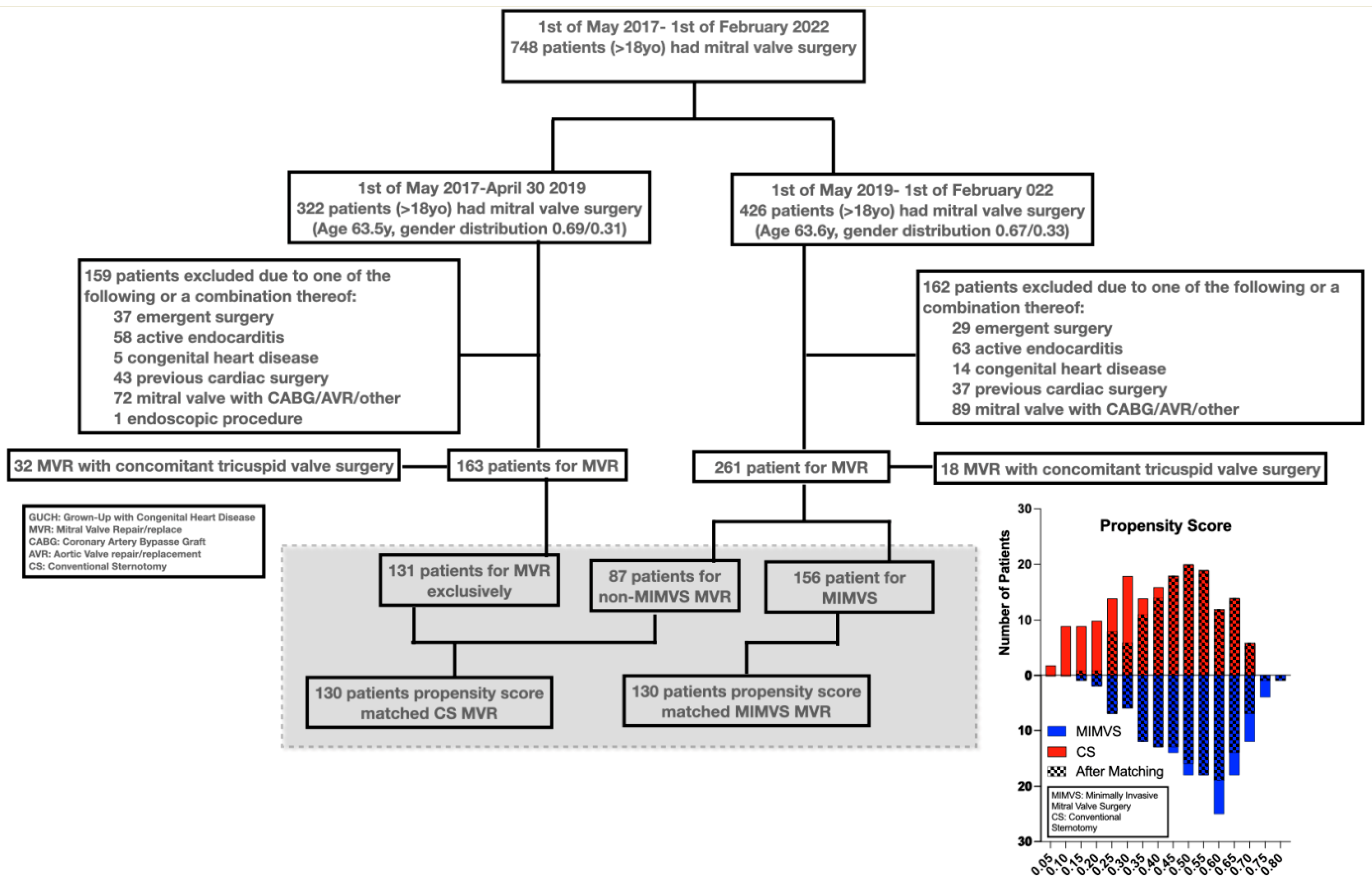
The primary investigator (AK) surveyed all completed cardiac surgeries in the timespan (1 May 2017–28 February 2022) via the operating room schedule using the Snap Board featured in the center’s electronic medical record (EMR) system (EPIC System, Madison, WI, United States). The EMR preoperative standardized heart team conference, admission history and physical chart note provided data on patient demographics and co-morbidities. The standardized surgical post procedure note provided surgical data such as ring/valve type and size, left atrial closure and cryo-maze procedure. A search in the automated barcode-scanned implant history system of the EMR verified the surgical data. A Patient Advocate Tracking System (PATS) online data spreadsheet also provided perfusion-related information in the center’s data management. PATS is registered in real time by the perfusionists during surgery at the institution. Both PATS and EPIC thus provided perfusion-related data (CPB time, cross-clamp time, BSA and EuroScore 2). If minor inconsistencies existed between data in PATS and EPIC, such as in the EuroScore 2, the least favorable data point would be used (e.g., highest EuroScore 2). The Phillips Intellispace Carciovascular (Philips Healthcare, Inc., Andover, MA, USA) and Xeroviewer (AGFA healthcare, Mortsel, Belgium) systems provided echocardiographic data. The cardiologist report of the echocardiography conducted before (closest date) and after the surgery (latest date up to one year) served as data entry points. In case of non-description of relevant echocardiographic findings in the report, the next closest echocardiography would be used as entry point. The EMR’s “Patient Station” provided real time admission, transfer, discharge and readmission data. Patient outcome data was also collected from the EMR. Either from automated integrated electronic transfer to the EMR, such as from ICU ventilators and medicine infusion pumps or from hourly electronic observation charts in the EMR, such as for bleeding or urine output, in both the ICU and operating room. The blood bank’s EMR integrated data system provided blood product administration information and consisted of blood products handed out and not returned to the blood bank. The EMR’s integrated laboratory value reporting system provided laboratory values. Neuroimaging data was recorded from XeroViewer (AGFA healthcare, Mortsel, Belgium) as scans performed and reported. Neurologic incidents were recorded as any neuro consult that described neuro deficits in any patient note up to one year following surgery. The outcomes of atrial fibrillation, pacemaker, pleural or pericardial effusion were likewise collected based on any patient note describing these occurrences. The EMR’s free-standing word search system served to cross-check for these outcomes by searching the following: “atrial…”, “neuro…”, ”pace…”, “pleu…” and “peri…”. The Danish central citizen registry served to cross-check the outcome of death and the centralized Danish EMR system (“Sundhedsjournalen”) served to cross-check on other outcomes. The “Sundhedsjournal” provides centralized data on all health data across all vendors, independent of geographical location or healthcare settings in all healthcare facilities. Availability of datapoint is expressed in parenthesis in tables.

3. Results

Seven hundred and forty-eight patients underwent screening in the study—424 underwent assessment. Figure 1 shows patient selection and the histographic alignment in the propensity score. Tables S1–S4 provides an overview of data from the propensity matched patients.

3.1. Matching

  • Mean and median propensity score margins were both 0.006 (range −0.015–0.05, interquartile range −0.002–0.014).
  • Matching mitigated pre-existing significant differences (Table 1) in gender (34% vs. 32%, p = 0.7), size (BSA of 1.93 m2 vs. 1.94 m2, p = 0.7), age (64 vs. 64 years, p = 0.6), rate of atrial fibrillation (30% vs. 34%, p = 0.6) and Euroscore 2 (1.38% vs. 1.43%, p = 0.7) in patients receiving CS compared to MIMVS. Significant differences persisted in CAD frequency after matching (19% vs. 36%, p = 0.003).

3.2. Surgical Technique

Table 2 displays surgical interventions in the study. After patient matching, the repair versus replace rate was similar (79% vs. 85%, p = 0.2). Repair technique changed significantly after MIMVS implementation.
  • More chord placement (57% vs. 83%, p < 0.001) and smaller ring sizes (34 mm vs. 36 mm, p < 0.001)
  • Less left atrial appendage closure (28% vs. 43%, p = 0.009)
Rate of tissue valve (11% vs. 17%, p = 0.15) and mechanical valve replacement (3.1% vs. 4.6%, p = 0.5) did not differ significantly.

3.3. Patient Outcome

Table 3 shows outcomes. MIMVS took longer.
  • Operating, CPB (180 vs. 102 min, p < 0.001) and aortic cross-clamp times (98 vs. 81 min, p < 0.001) (5.5 vs. 4.3 h, p < 0.001) increased.
  • Longer procedure time appeared to affect extubation in MIMVS slightly (10 vs. 9 h, p = 0.009).
  • However, MIMVS in-hospital time decrased significantly.
  • ICU re-admissions occurred less (0 vs. 3.1%, p = 0.045) and hospital discharge shortened (p < 0.001, median 5 vs. 7) after MIMVS.
  • Patient-centered outcomes such as neurologic-, effusion- and reintervention endpoints showed non-significant small differences.
  • Postoperative atrial fibrillation and endocarditis/mediastinitis occurred significantly less in MIMVS (42% vs. 70%, p < 0.001/0 vs. 3.1%, p = 0.044)

4. Discussion

This retrospective report shows that MIMVS introduction in a mitral valve surgical center without prior experience appears to be safe and feasible.
Longer surgical-, CPB- and aortic cross-clamp times seemed not to affect one-year survival, hospital length of stays or subordinate in-hospital outcomes such as ICU length of stay, bleeding or transfusion requirement.
Interestingly, the study showed that surgical repair technique changed with MIMVS introduction.
The patient population presenting for mitral valve surgery in this study resembles previously published large databases [18]. The findings thus seem generalizable for the surgical mitral valve patient population. The more frequent occurrence of mild CAD, also after propensity score matching, could likely be explained by a transition from evaluating selected risk patients by catheter-based angiogram to broadly screening all cardiac surgery patients with CT-based angiogram during the timespan of the study.
In this study, MIMVS adoption correlated to a changed surgical repair technique. Thus, smaller ring size and more chord placement occurred after MIMVS introduction, likely reflecting a move away from correcting lesions by resection and stitching to a strategy of displacing scallops using artificial chords. Editorials discuss these two diverging strategies [19]. A recent large meta-analysis of more than 6000 patients favored the displacement technique [20] by showing hard endpoints such as operative mortality differing, supporting the change in strategy observed in the current study. Minimally invasive approaches combine more readily with the displacement technique [19,21], because of the constrained instrument movement, when repairing the valve, contributing to the transition to the displacement strategy.
The current study showed that mitral valve repair takes longer by MIMVS. Several previous studies support this finding [11,21]. Counterintuitively, the longer duration of MIMVS contrasted to a finding of shorter hospital stays of up to two days. Meta-analyses and a randomized clinical trial have found similar benefits of approximately two days shorter hospital stay after MIMVS [21]. Hospital stays in MIMVS could likely shorten due to factors such as less pain and early mobilization. Shorter hospitalization with the potential of lowering cost to both patients and hospitals highly favors adopting MIMVS, along with a possible decrease in rare events such as ICU re-admission or serious chest infections. However, Chikwe et al. [22] showed that low-volume mitral valve centers (<50 mitral valve operations per surgeon per year) might encounter challenges in outcomes due to lack of exposure and hence slow advancement on the learning curve. Thus, Müller [13] recently suggested the need for at least two MIMVS per week to ensure excellence, which equals more than 100 surgeries needed per year. However, in the current study we found no indication that a volume of 50–60 MIMVS per year at start-up correlated with poorer outcomes and hence impeded the learning curve when adopting MIMVS. A recent well-conducted multicenter randomized trial with approximately the same number of patients as in the current trial reported almost identical outcomes in the safety parameters of death, stroke and ICU length of stay [21]. The current study therefore suggests that high-quality treatment and advancement on the learning curve can be achieved by firm adherence to a strict implementation strategy.
Several limitations exist in this investigation. The single-center retrospective study design creates a risk of detection and selection bias. Matching by propensity score balances known diverging variables among patients, but unmeasured variables still might skew results. Furthermore, treatment success depends on a longer period of follow-up than the one year reported in the current study and preferably should also include data on patient-centered outcomes such as quality of life post-surgery, which could strengthen the patient-oriented relevance of the findings.

5. Conclusions

This retrospective implementation study of adopting MIMVS in a center without prior experience in the procedure showed feasibility and equally good outcomes when compared to CS.

Supplementary Materials

The following supporting information can be downloaded at: https://www.mdpi.com/article/10.3390/hearts6020011/s1.

Author Contributions

Conceptualization, A.K., P.H.M.-S., J.E.M. and C.L.C.; methodology, A.K. and C.L.C.; formal analysis, A.K.; investigation, A.K.; data curation, A.K.; writing—original draft preparation, A.K.; writing—review and editing, A.K., P.H.M.-S., J.E.M. and C.L.C. All authors have read and agreed to the published version of the manuscript.

Funding

This research received no external funding.

Institutional Review Board Statement

The study was conducted in accordance with the Declaration of Helsinki, and approved Danish Capital Region Regional Ethics Committee waived the requirement for additional informed study consent (protocol# F-22051524, 21 October 2021).

Informed Consent Statement

Danish law (Danish Health Care Law §42d), which mandates physicians to follow patients’ outcomes after implementing new treatments. Individual patients consented to surgery and data collection, but due to the retrospective nature of the investigation, the Danish Capital Region Regional Ethics Committee waived the requirement for additional informed study consent (protocol# F-22051524).

Data Availability Statement

All data will be shared upon reasonable request to the corresponding author.

Conflicts of Interest

The authors declare no conflicts of interest.

Abbreviations

The following abbreviations are used in this manuscript.
ACTActivated clotting time
BSABody surface area
CADCoronary artery disease
CPBCardiopulmonary bypass
CSConventional sternotomy
CTCA Computer tomography coronary angiogram
CVP Central venous pressure
EMRElectronic medical record
ICUIntensive care unit
LVEFLeft ventricular ejection fraction
NIRSNear infra-read spectrometry
MAPMean arterial pressure
MIMVSMinimally invasive mitral valve surgery
MRMitral regurgitation
MVMitral valve
MVRMitral valve repair/replacement
SAMSystolic anterior motion

References

  1. Modi, P.; Hassan, A.; Chitwood, W.R. Minimally invasive mitral valve surgery: A systematic review and meta-analysis. Eur. J. Cardio-Thoracic Surg. 2008, 34, 943–952. [Google Scholar] [CrossRef]
  2. Goldstone, A.B.; Atluri, P.; Szeto, W.Y.; Trubelja, A.; Howard, J.L.; MacArthur, J.W.; Newcomb, C.; Donnelly, J.P.; Kobrin, D.M.; Sheridan, M.A.; et al. Minimally invasive approach provides at least equivalent results for surgical correction of mitral regurgitation: A propensity-matched comparison. J. Thorac. Cardiovasc. Surg. 2013, 145, 748–756. [Google Scholar] [CrossRef]
  3. Iribarne, A.; Easterwood, R.; Russo, M.J.; Wang, Y.C.; Yang, J.; Hong, K.N.; Smith, C.R.; Argenziano, M. A minimally invasive approach is more cost-effective than a traditional sternotomy approach for mitral valve surgery. J. Thorac. Cardiovasc. Surg. 2011, 142, 1507–1514. [Google Scholar] [CrossRef] [PubMed]
  4. Perin, G.; Shaw, M.; Toolan, C.; Palmer, K.; Al-Rawi, O.; Modi, P. Cost Analysis of Minimally Invasive Mitral Valve Surgery in the UK National Health Service. Ann. Thorac. Surg. 2021, 112, 124–131. [Google Scholar] [CrossRef]
  5. Svensson, L.G.; Atik, F.A.; Cosgrove, D.M.; Blackstone, E.H.; Rajeswaran, J.; Krishnaswamy, G.; Jin, U.; Gillinov, A.M.; Griffin, B.; Navia, J.L.; et al. Minimally invasive versus conventional mitral valve surgery: A propensity-matched comparison. J. Thorac. Cardiovasc. Surg. 2010, 139, 926–932.e2. [Google Scholar] [CrossRef] [PubMed]
  6. Moscarelli, M.; Fattouch, K.; Casula, R.; Speziale, G.; Lancellotti, P.; Athanasiou, T. What Is the Role of Minimally Invasive Mitral Valve Surgery in High-Risk Patients? A Meta-Analysis of Observational Studies. Ann. Thorac. Surg. 2016, 101, 981–989. [Google Scholar] [CrossRef] [PubMed]
  7. Cheng, D.C.; Martin, J.; Lal, A.; Diegeler, A.; Folliguet, T.A.; Nifong, L.W.; Perier, P.; Raanani, E.; Smith, J.M.; Seeburger, J.; et al. Minimally Invasive Versus Conventional Open Mitral Valve Surgery. Innov. Technol. Tech. Cardiothorac. Vasc. Surg. 2011, 6, 84–103. [Google Scholar] [CrossRef]
  8. Hussain, S.; Swystun, A.G.; Caputo, M.; Angelini, G.D.; Vohra, H.A. A review and meta-analysis of conventional sternotomy versus minimally invasive mitral valve surgery for degenerative mitral valve disease focused on the last decade of evidence. Perfusion 2023, 39, 988–997. [Google Scholar] [CrossRef]
  9. Gammie, J.S.; Bartlett, S.T.; Griffith, B.P. Small-Incision Mitral Valve Repair: Safe, Durable, and Approaching Perfection. Ann. Surg. 2009, 250, 409–415. [Google Scholar] [CrossRef]
  10. Han, J.J.; Smood, B.; Atluri, P. Commentary: Transitioning to Minimally Invasive Mitral Valve Repair—Navigating the Gauntlet. Semin. Thorac. Cardiovasc. Surg. 2021, 32, 838–839. [Google Scholar] [CrossRef]
  11. Abu-Omar, Y.; Fazmin, I.T.; Ali, J.M.; Pelletier, M.P. Minimally invasive mitral valve surgery. J. Thorac. Dis. 2020, 13, 1960–1970. [Google Scholar] [CrossRef]
  12. Holzhey, D.M.; Seeburger, J.; Misfeld, M.; Borger, M.A.; Mohr, F.W. Learning minimally invasive mitral valve surgery: A cumulative sum sequential probability analysis of 3895 operations from a single high-volume center. Circulation 2013, 128, 483–491. [Google Scholar] [CrossRef] [PubMed]
  13. Müller, L.C. Is minimally invasive mitral valve surgery equal to or worse than a full sternotomy: A perspective from the Netherlands National Heart Registration. Eur. J. cardio-Thorac. Surg. Off. J. Eur. Assoc. Cardio-Thorac. Surg. 2021, 61, 1107–1108. [Google Scholar] [CrossRef] [PubMed]
  14. Barbero, C.; Marchetto, G.; Ricci, D.; Rinaldi, M. Temporary Neurological Dysfunction After Minimal Invasive Mitral Valve Surgery: Influence of Type of Perfusion and Aortic Clamping Technique. Ann. Thorac. Surg. 2017, 103, 691–692. [Google Scholar] [CrossRef]
  15. Olsthoorn, J.R.; Heuts, S.; Houterman, S.; Maessen, J.G.; Sardari Nia, P. Effect of minimally invasive mitral valve surgery compared to sternotomy on short- and long-term outcomes: A retrospective multicentre interventional cohort study based on Netherlands Heart Registration. Eur. J. Cardio-Thorac. Surg. Off. J. Eur. Assoc. Cardio-Thorac. Surg. 2021, 61, 1099–1106. [Google Scholar] [CrossRef] [PubMed]
  16. Renner, J.; Lorenzen, U.; Borzikowsky, C.; Schoeneich, F.; Cremer, J.; Haneya, A.; Hensler, J.; Panholzer, B.; Huenges, K.; Broch, O. Unilateral pulmonary oedema after minimally invasive mitral valve surgery: A single-centre experience. Eur. J. Cardio-Thoracic Surg. 2017, 53, 764–770. [Google Scholar] [CrossRef]
  17. Hunter, S. How to start a minimal access mitral valve program. Ann. Cardiothorac. Surg. 2013, 2, 774–778. [Google Scholar] [CrossRef]
  18. Gammie, J.S.; Chikwe, J.; Badhwar, V.; Thibault, D.P.; Vemulapalli, S.; Thourani, V.H.; Gillinov, M.; Adams, D.H.; Rankin, J.S.; Ghoreishi, M.; et al. Isolated Mitral Valve Surgery: The Society of Thoracic Surgeons Adult Cardiac Surgery Database Analysis. Ann. Thorac. Surg. 2018, 106, 716–727. [Google Scholar] [CrossRef]
  19. Dreyfus, G.D.; Dulguerov, F.; Marcacci, C.; Haley, S.R.; Gkouma, A.; Dommerc, C.; Albert, A. “Respect when you can, resect when you should”: A realistic approach to posterior leaflet mitral valve repair. J. Thorac. Cardiovasc. Surg. 2018, 156, 1856–1866.e3. [Google Scholar] [CrossRef]
  20. Sá, M.P.; Cavalcanti, L.R.P.; Eynde, J.V.D.; Amabile, A.; Neto, A.C.E.; Perazzo, A.M.; Weymann, A.; Ruhparwar, A.; Sicouri, S.; Bisleri, G.; et al. Respect versus resect approaches for mitral valve repair: A study-level meta-analysis. Trends Cardiovasc. Med. 2023, 33, 225–239. [Google Scholar] [CrossRef]
  21. Maier, R.H.; Kasim, A.S.; Zacharias, J.; Vale, L.; Graham, R.; Walker, A.; Laskawski, G.; Deshpande, R.; Goodwin, A.; Kendall, S.; et al. Minimally invasive versus conventional sternotomy for Mitral valve repair: Protocol for a multicentre randomised controlled trial (UK Mini Mitral). BMJ Open 2021, 11, e047676. [Google Scholar] [CrossRef] [PubMed]
  22. Chikwe, J.; Toyoda, N.; Anyanwu, A.C.; Itagaki, S.; Egorova, N.N.; Boateng, P.; El-Eshmawi, A.; Adams, D.H. Relation of Mitral Valve Surgery Volume to Repair Rate, Durability, and Survival. J. Am. Coll. Cardiol. 2017, 69, 2397–2406. [Google Scholar] [CrossRef] [PubMed]
Figure 1. Patient selection flowchart.
Hearts 06 00011 g001
Table 1. Demography and co-morbidity.
MIMVS (130)CS (130)p
Age64.0 y (130)64.0 y (130)0.556
Female32.3% (130)34.6% (130)0.695
Body surface area 1.94 m2 (130)1.93 m2 (130)0.720
Diabetes4.6% (130)3.8% (130)0.760
Hypertension42.3% (130)40.1% (130)0.802
Cerebrovascular disease6.9% (130)5.4% (130)0.607
Mild CAD on angiogram/CTCA 36.2% (130)19.4% (129)0.003 *
History of smoking39.2% (130)42.3% (130)0.615
Hyperlipidemia31.5% (130)31.5% (130)0.999
Atrial fibrillation30.1% (130)33.9% (130)0.600
EuroScore 21.38% (130)1.43% (130)0.676
CAD: Coronary artery disease. CCTA: Coronary computer tomographic angiogram. * Denotes significant difference.
Table 2. Surgical interventions.
MIMVS
(130)
CS (130)p
Repair84.6% (110 ptt)78.5% (102 ptt)0.202
Tissue valve10.8% (14 ptt)16.9% (22 ptt)0.152
Mechanical valve4.6% (6 ptt)3.1% (4 ptt)0.521
Chords placed83.1% (108 ptt)56.7% (74 ptt)<0.001 *
Mitral ring size (M, range)34 (28–40) mm36 (28–40) mm<0.001 *
Left atrial closure27.7% (36 ptt)43.1% (56 ptt)0.009 *
Cryomaze21.5% (28 ptt)17.7% (23 ptt)0.437
* Denotes significant difference.
Table 3. Outcomes.
Table 3. Outcomes.
MIMVS (130)CS (130)p
Cardiopulmonary bypass time (m)180 (96–391) min (130)102 (43–214) min (129)<0.001 *
Aortic cross-clamp time (m)98 (58–204) min (125)81 (28–160) min (129)<0.001 *
Time in operating room (anesthesia time)5.5 (3.75–9.0) h (130)4.3 (2.8–7.0) h (129)<0.001 *
Time on ventilator10 (7–120) h (130)9 (4–46) h (130)0.009 *
ICU discharge (m/range)1 d (1–16 d) (130)1 d (1–6 d) (130)0.974
ICU re-admission0% (130)3.1% (4 ptt) (130)0.045 *
Hospital discharge postoperatively (m/range)5 (3–29) d (130)7 (4–72) d (130)<0.001 *
Bleeding total (m/range)450 (105–4675) mL (130)465 (40–5357) mL (130)0.5136
Urine output 24 h (m/range)3398 (1665–7610) mL (130)2893 (270–5700) mL (130)<0.001 *
Red blood cell transfusion per- or postoperatively24.6 (32 ptt) (130)23.8 (31 ptt) (130)0.886
Neuro-scan postoperatively12.3% (16 ptt) (130)7.7% (10 ptt) (130)0.2163
Neuro-scan abnormal4.6% (6 ptt) (130)4.6% (6 ptt) (130)0.998
Neuro sequela3.9% (5 ptt) (130)3.9% (5 ptt) (130)0.998
Major neurologic injury0 (130)1.5% (2 ptt) (130)0.158
Permanent cardiac implanted electronic device (CIED)4.6% (6 ptt) (130)5.4% (7 ptt) (130)0.778
Pericardial effusion requiring intervention8.5% (11 ptt) (130)13.4% (18 ptt) (130)0.169
Pleural effusion requiring intervention6.9% (9 ptt) (130)10.8% (14 ptt) (130)0.277
Postoperative atrial fibrillation42.3% (55 ptt) (130)66.9% (87 ptt) (130)<0.001 *
Re-exploration for bleeding2.3% (3 ptt) (130)4.6% (6 ptt) (130)0.331
Re-op medias-/endocarditis0 (130)3.1 (4 ptt) (130)0.044 *
One year mortality1.5% (2 ptt) (130)1.5% (2 ptt) (130)0.999
One year mortality cardiac0.8 (1 ptt) (130)1.5% (2 ptt) (130)0.974
ICU: Intensive Care Unit. * Denotes significant difference.
Disclaimer/Publisher’s Note: The statements, opinions and data contained in all publications are solely those of the individual author(s) and contributor(s) and not of MDPI and/or the editor(s). MDPI and/or the editor(s) disclaim responsibility for any injury to people or property resulting from any ideas, methods, instructions or products referred to in the content.

