Current ECG Aspects of Interatrial Block
Abstract
1. Introduction
2. ECG Diagnosis of Interatrial Blocks (IAB)
2.1. How to Measure P-Wave Duration?
2.2. First Degree (Partial) Interatrial Block
2.3. Third-Degree (Advanced) Interatrial Block
2.3.1. Typical ECG Pattern
2.3.2. Atypical Patterns
2.3.3. Atypical A-IAB Due to Changes in P-Wave Morphology
- (i)
- (ii)
- (iii)
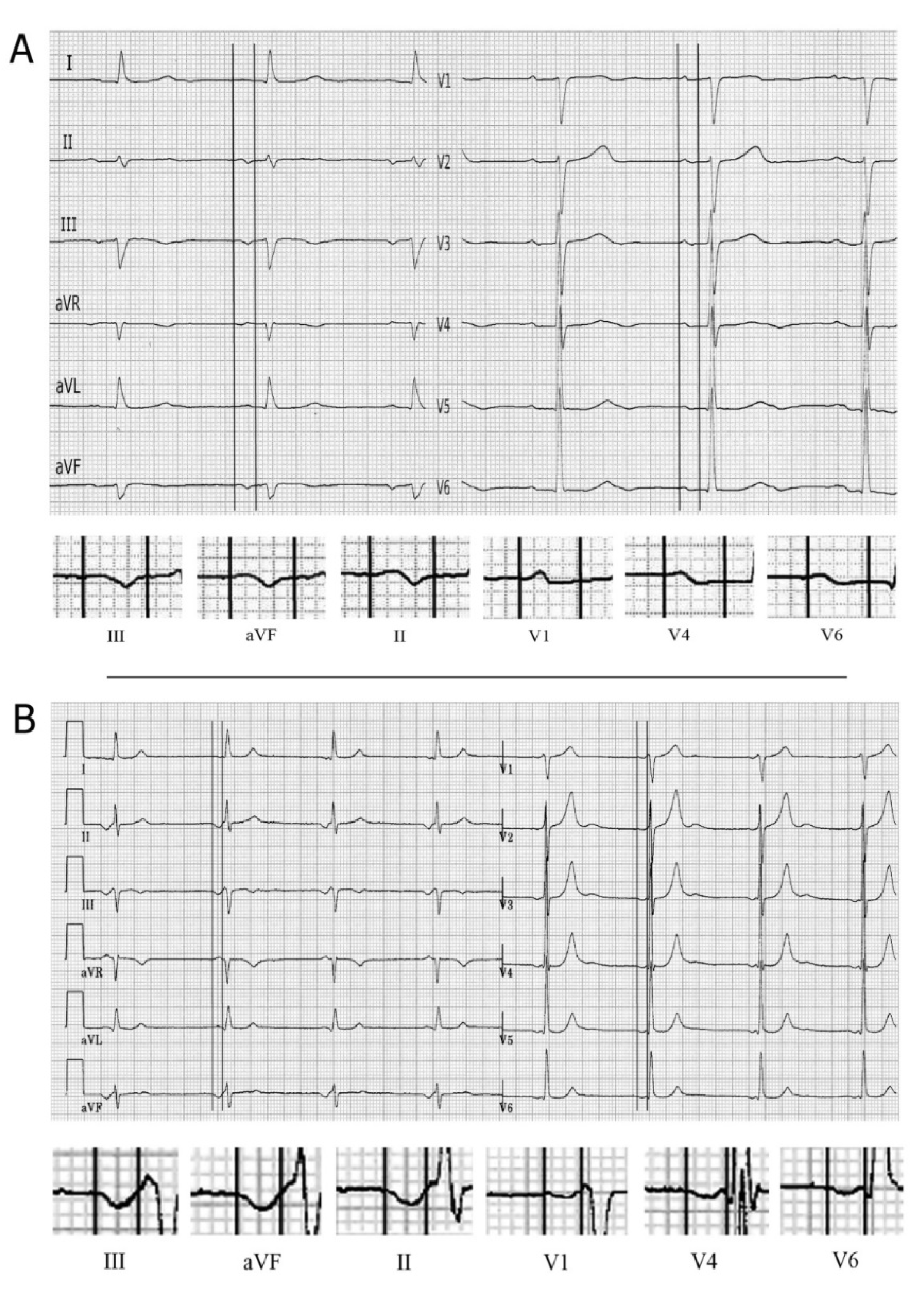
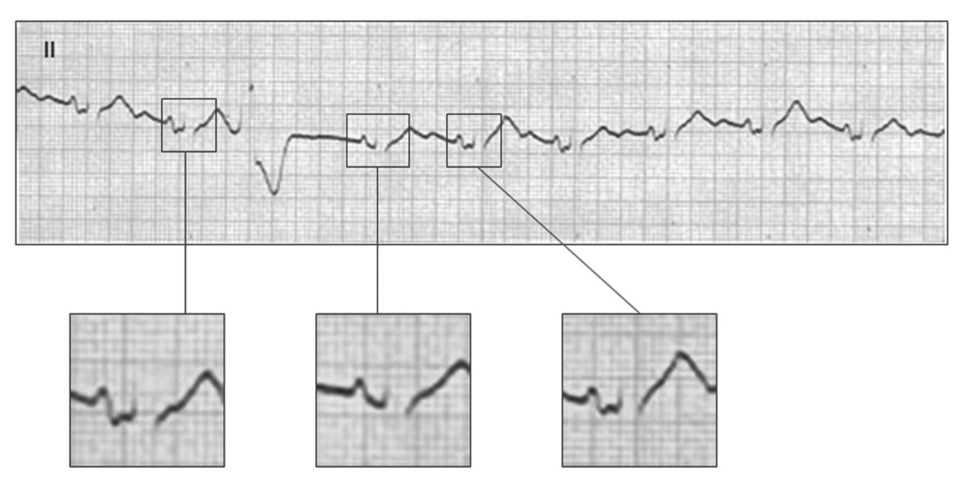
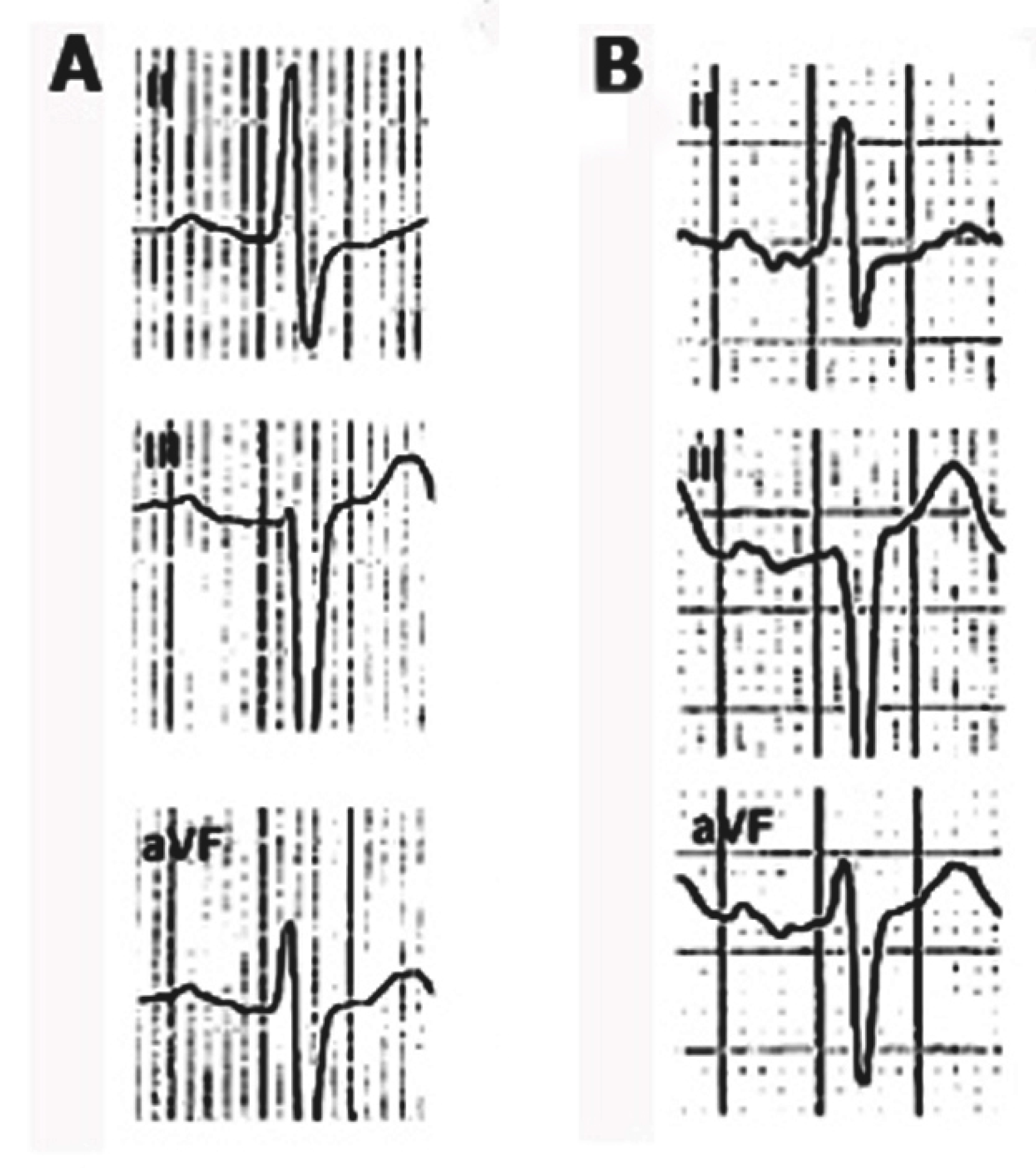
- Type III (Figure 8E and Figure 9F) The P-wave morphology in leads III and aVF is completely negative, but started being isodiphasic, and the P-wave in lead II is biphasic (±). This pattern requires a differential diagnosis with junctional rhythm (Figure 10) [37]. If the polarity of the P-waves in leads V5-V6 is positive, then atypical A-IAB is diagnosed, while if it is negative, then junctional rhythm is the final diagnosis.
2.3.4. Atypical A-IAB Due to Changes in P-Wave Duration
2.4. Second-Degree Atrial Block
3. Clinical Implications
Author Contributions
Funding
Institutional Review Board Statement
Informed Consent Statement
Data Availability Statement
Conflicts of Interest
References
- Eugene, B. Foreword. In Clinical Electrocardiography. A Textbook, 2nd ed.; Futura Publishing Company, Inc.: Armonk, NY, USA, 1998. [Google Scholar]
- Fuster, V.; Narula, J.; Harrington, R.A.; Eapen, Z.J. (Eds.) Hurst’s The Heart, 14th ed.; McGraw-Hill Education: New York, NY, USA, 2017. [Google Scholar]
- Camm, J.; Lüscher, T.F.; Maurer, G.; Serruys, P.W. (Eds.) The ESC Textbook of Cardiovascular Medicine, 3rd ed.; Oxford University Press: Oxford, UK, 2019. [Google Scholar]
- Bayés de Luna, A. Clinical Electrocardiography; Wiley-Blackwell: Hoboken, NJ, USA, 2012. [Google Scholar]
- Bayés de Luna, A.; de Ribot, R.F.; Trilla, E.; Julia, J.; Garcia, J.; Sadurni, J.; Riba, J.; Sagues, F. Electrocardiographic and vectorcardiographic study of interatrial conduction disturbances with left atrial retrograde activation. J. Electrocardiol. 1985, 18, 1–13. [Google Scholar] [CrossRef]
- Strauss, D.G.; Schocken, D.D. Marriott’s Practical Electrocardiography, 13th ed.; Wolters Kluwer: Philadelphia, PA, USA, 2021. [Google Scholar]
- Bachmann, G. The significance of splitting of the P-wave in the ECG. Ann. Intern. Med. 1941, 14, 1702–1709. [Google Scholar] [CrossRef]
- Puech, P. L’activité Électrique Auriculaire. Normale et Pathologique; Masson & Editeurs: Paris, France, 1956. [Google Scholar]
- Castillo Fenoy, A.; Vernant, P. Les troubles de la conduction interauriculaire pour bloc du faisceau du Bachmann. Arch. Mal. Coeur. Vaiss. 1971, 64, 1490–1503. [Google Scholar]
- Di Biase, M.; Rizzon, P. Blocco interatriale con attivazione caudocraneale dell’atrio sinistro. G. Ital. Cardiol. 1975, 5, 323. [Google Scholar]
- García Civera, R.; Llácer Escorihuela, A.; Benages Martínez, A.; López Merino, V. Estudio de la activación auricular y de la conducción AV en el bloqueo del haz de Bachmann en el corazón humano. [Study of auricular activation and A-V conduction in Bachmann bundle block in the human heart]. Rev. Esp. Cardiol. 1972, 25, 341. [Google Scholar] [PubMed]
- Bayés de Luna, A.; Bonnin, O.; Ferriz, J.; Fort De Ribot, R.; Julia, J.; Oter, R.; Trilla, E.; Roman, M.; Vernis, J.; Vilaplana, J.; et al. Trastorno de conducción intraauricular con conducción retrógrada auricular izquierda. Estudio electrocardiológico y clínico a propósito de 24 casos. Rev. Esp. Cardiol. 1978, 31, 173–178. [Google Scholar] [PubMed]
- Bayés de Luna, A.; Cladellas, M.; Oter, R.; Torner, P.; Guindo, J.; Martí, V.; Rivera, I.; Iturralde, P. Interatrial conduction block and retrograde activation of the left atrium and paroxysmal supraventricular tachycarrhythmias. Eur. Heart J. 1988, 9, 1112. [Google Scholar] [CrossRef]
- Bayés de Luna, A.; Oter, M.C.; Guindo, J. Interatrial conduction block with retrograde activation of the left atrium and paroxysmal supraventricular tachyarrhythmic treatment. Int. J. Cardiol. 1989, 22, 147–150. [Google Scholar] [CrossRef]
- Bayés de Luna, A.; Guindo, J.; Viñolas, X.; Martinez-Rubio, A.; Oter, R.; Bayés-Genís, A. Third-degree interatrial block and supraventricular tachyarrhythmias. EP Eur. 1999, 1, 43–46. [Google Scholar] [CrossRef]
- Ariyarajah, V.; Puri, P.; Apiyasawat, S.; Spodick, D.H. Interatrial block: A novel risk factor for embolic stroke? Ann. Noninvasive Electrocardiol. 2007, 12, 15–20. [Google Scholar] [CrossRef]
- Garcia Cosio, F.; Martín-Peñato, A.; Pastor, A.; Núñez, A.; Montero, M.A.; Cantale, C.P.; Schames, S. Atrial activation mapping in sinus rhythm in the clinical electrophysiology laboratory. Observations in Bachmann’s bundle block. J. Cardiovasc. Electrophysiol. 2004, 15, 524–531. [Google Scholar] [CrossRef]
- Platonov, P.G.; Mitrofanova, L.B.; Chirreikin, L.V.; Olsson, S.B. Morphology of inter-atrial conduction routes in patients with atrial fibrillation. Europace 2002, 4, 183–192. [Google Scholar] [CrossRef]
- Bayés de Luna, A.; Platonov, P.; Cosio, F.G.; Cygankiewicz, I.; Pastore, C.; Baranowski, R.; Bayés-Genis, A.; Guindo, J.; Viñolas, X.; Garcia-Niebla, J.; et al. Interatrial blocks. A separate entity from left atrial enlargement: A consensus report. J. Electrocardiol. 2012, 45, 445–451. [Google Scholar] [CrossRef] [PubMed]
- Bayés de Luna, A.; Baranchuk, A.; Niño Pulido, C.; Martínez-Sellés, M.; Bayés-Genís, A.; Elosua, R.; Elizari, M.V. Second-Degree Interatrial block: Brief review and concept. Ann. Noninvasive Electrocardiol. 2018, 23, e12583. [Google Scholar] [CrossRef] [PubMed]
- Waldo, A.; Bush, H.L., Jr.; Gelband, H.; Zorn, G.L., Jr.; Vitikainen, K.J.; Hoffman, B.F. Effects on the canine P waves of discrete lesions in the specialized atrial tracts. Circ. Res. 1971, 29, 452–467. [Google Scholar] [CrossRef] [PubMed]
- Guerra, J.; Vilahur, G.; Bayés de Luna, A.; Cabrera, J.A.; Martínez-Sellés, M.; Mendieta, G.; Baranchuk, A.; Sánchez-Quintana, D. Interatrial block can occur in the absence of left atrial enlargement: New experimental model. Pacing Clin. Electrophysiol. 2020, 43, 427. [Google Scholar] [CrossRef] [PubMed]
- Bayés de Luna, A.; Escobar-Robledo, L.A.; Aristizabal, D.; Weir Restrepo, D.; Mendieta Badimon, G.; Massó-van Roessel, A.; Elosua, R.; Bayés-Genís, A.; Martínez-Sellés, M.; Baranchuk, A. Atypical advanced interatrial block: Definition electrocardiographic recognition. J. Electrocardiol. 2018, 51, 1091–1093. [Google Scholar] [CrossRef] [PubMed]
- Kottkamp, H. Human atrial fibrillation substrate: Towards a specific fibrotic atrial cardiomyopathy. Eur. Heart J. 2013, 34, 2731–2738. [Google Scholar] [CrossRef]
- Bayés de Luna, A.; Martínez-Sellés, M.; Elosua, R.; Bayés-Genís, A.; Mendieta, G.; Baranchuk, A.; Breithardt, G. Relation of Advanced Interatrial block to risk of atrial fibrillation and stroke. Am. J. Cardiol. 2020, 125, 1745–1748. [Google Scholar] [CrossRef] [PubMed]
- Bayés de Luna, A.; Martínez-Sellés, M.; Bayés-Genís, A.; Elosua, R.; Baranchuk, A. Lo que todo clínico debe conocer. What every clinician should know about Bayés Syndrome. Rev. Esp. Cardiol. (Engl. Ed.) 2020, 73, 758–762. [Google Scholar] [CrossRef]
- Baranchuk, A. Interatrial Block and Supraventricular Arrhythmias. Clinical Implications of Bayés’ Syndrome; Cardiotext Publishing: Minneapolis, MN, USA, 2017. [Google Scholar]
- Morris, J.J., Jr.; Estes, E.H., Jr.; Whalen, R.E.; Thompson, H.K., Jr.; Mcintosh, H.D. P wave analysis in valvular heart disease. Circulation 1964, 29, 242. [Google Scholar] [CrossRef] [PubMed]
- Rasmussen, M.U.; Fabricius-Bjerre, A.; Kumarathurai, P.; Larsen, B.S.; Dominguez, H.; Kanters, J.K.; Sajadieh, A. Common source of miscalculation and misclassification of P-wave negativity and P-wave terminal force in lead V1. J. Electrocardiol. 2019, 53, 85–88. [Google Scholar] [CrossRef] [PubMed]
- Sajeev, J.K.; Koshy, A.N.; Dewey, H.; Kalman, J.M.; Bhatia, M.; Roberts, L.; Cooke, J.C.; Frost, T.; Denver, R.; Teh, A.W. Poor reliability of P-wave terminal force V1 in ischemic stroke. J. Electrocardiol. 2019, 52, 47–52. [Google Scholar] [CrossRef]
- Bacharova, L.; Wagner, G.S. The time for naming the interatrial block syndrome: Bayes syndrome. J. Electrocardiol. 2015, 48, 133–134. [Google Scholar] [CrossRef] [PubMed]
- Escobar-Robledo, L.A.; Bayés de Luna, A.; Lupón, J.; Baranchuk, A.; Moliner, P.; Martínez-Sellés, M.; Zamora, E.; de Antonio, M.; Domingo, M.; Cediel, G.; et al. Advanced Interatrial Block Predicts New-onset Atrial Fibrillation and Ischemic Stroke in Patients with Heart Failure: The “Bayes Syndrome-HF” Study. Int. J. Cardiol. 2018, 271, 174–180. [Google Scholar] [CrossRef] [PubMed]
- Martínez-Sellés, M.; Elosua, R.; Ibarrola, M.; de Andrés, M.; Díez-Villanueva, P.; Bayés-Genis, A.; Baranchuk, A.; Bayés-de-Luna, A.; BAYES Registry Investigators. Advanced interatrial block and P-wave duration are associated with atrial fibrillation and stroke in older adults with heart disease: The BAYES registry. Europace 2020, 22, 1001–1008. [Google Scholar] [CrossRef] [PubMed]
- Martínez-Sellés, M.; Martínez-Larrú, E.; Ibarrola, M.; Santos, A.; Díez-Villanueva, P.; Bayés-Genis, A.; Baranchuk, A.; Bayés-de-Luna, A.; Elosua, R. Interatrial block and cognitive impairment in the BAYES prospective registry. Int. J. Cardiol. 2020, 321, 95–98. [Google Scholar] [CrossRef] [PubMed]
- Massó-van Roessel, A.; Escobar-Robledo, L.A.; Dégano, I.R.; Grau, M.; Sala, J.; Ramos, R.; Marrugat, J.; Bayés de Luna, A.; Elosua, R. Analysis of the association between electrocardiographic P-wave characteristics and atrial fibrillation in the REGICOR Study. Rev. Esp. Cardiol. (Engl. Ed.) 2017, 70, 841–847. [Google Scholar] [CrossRef]
- Martínez-Sellés, M.; Massó-van Roessel, A.; Álvarez-Garcia, J.; Garcia de la Villa, B.; Cruz-Jentoft, A.; Vidán, M.T.; López, J.; Felix-Redondo, F.J.; Durán, J.M.; Bayés-Genís, A.; et al. (The Investigators of the Cardiac and Clinical Characterization of Centenarians (4C) registry). Interatrial block and atrial arrhythmias in centenarians: Prevalence, associations, and clinical implications. Heart Rhythm. 2016, 13, 645–651. [Google Scholar] [CrossRef]
- de Luna, A.B.; Platonov, P.G.; García-Niebla, J.; Baranchuk, A. Atypical Advanced Interatrial block or junctional rhythm? J. Electrocardiol. 2019, 5, 85–86. [Google Scholar] [CrossRef]
- Gentille-Lorente, D.I.; Scott, L.; Escobar-Robledo, L.A.; Mesa-Maya, M.A.; Carreras-Costa, F.; Baranchuk, A.; Martínez-Sellés, M.; Elosua, R.; Bayés-Genís, A.; Bayés-de-Luna, A. Atypical advanced interatrial block due to giant atrial lipoma. Pacing Clin. Electrophysiol. 2021, 44, 737–739. [Google Scholar] [CrossRef]
- Chung, E.K. Aberrant atrial conduction: Unrecognized electrocardiographic entity. Br. Heart J. 1972, 34, 341–346. [Google Scholar] [CrossRef] [PubMed][Green Version]
- Julià, J.; Bayés de Luna, A.; Candell, J.; Fiol, M.; Pons, G.; Obrador, D.; Oca, F.; Trilla, E.; Vilaplana, J.; Wilke, M. Aberrancia auricular: A propósito de 21 casos. Rev. Esp. Cardiol. 1978, 31, 207. [Google Scholar] [PubMed]
- Benito, E.M.; Bayés de Luna, A.; Baranchuk, A.; Mont, L. Extensive atrial fibrosis assessed by late gadolinium enhancement cardiovascular magnetic resonance associated with advanced interatrial block electrocardiogram pattern. Europace 2017, 19, 377. [Google Scholar] [CrossRef] [PubMed]
- Agarwal, Y.K.; Aronow, W.S.; Levy, J.A.; Spodick, D.H. Association of interatrial block with development of atrial fibrillation. Am. J. Cardiol. 2003, 91, 882. [Google Scholar] [CrossRef]
- Holmqvist, F.; Platonov, P.; Carlson, J.; Zareba, W.; Moss, A.J.; MADIT II Investigators. Abnormal P wave morphology is a predictor of atrial fibrillation in MADIT II patients. Ann. Noninvasive Electrocardiol. 2010, 15, 63–72. [Google Scholar] [CrossRef]
- Enriquez, A.; Conde, D.; Hopman, W.; Mondragon, I.; Chiale, P.A.; Bayés de Luna, A.; Baranchuk, A. Advanced interatrial block is associated with recurrence of atrial fibrillation post pharmacological cardioversion. Cardiovasc. Ther. 2014, 32, 52–56. [Google Scholar] [CrossRef]
- Enriquez, A.; Sarrias, A.; Villuendas, R.; Ali, F.S.; Conde, D.; Hopman, W.M.; Redfearn, D.P.; Michael, K.; Simpson, C.; Bayés de Luna, A.; et al. New-onset atrial fibrillation after cavotricuspid isthmus ablation: Identification of advanced interatrial block is key. Europace 2015, 17, 1289–1293. [Google Scholar] [CrossRef]
- Sadiq Ali, F.; Enriquez, A.; Conde, D.; Redfearn, D.; Michael, K.; Simpson, C.; Abdollah, H.; Bayes de Luna, A.; Hopman, W.; Baranchuk, A. Advanced Interatrial Block Predicts New Onset Atrial Fibrillation in Patients with Severe Heart Failure and Cardiac Resynchronization Therapy. Ann. Noninvasive Electrocardiol. 2015, 20, 586–591. [Google Scholar] [CrossRef]
- Alexander, B.; MacHaalany, J.; Lam, B.; van Rooy, H.; Haseeb, S.; Kuchtaruk, A.; Glover, B.; Bayés de Luna, A.; Baranchuk, A. Comparison of the extent of coronary artery disease in patients with versus without interatrial block and implications for new-onset atrial fibrillation. Am. J. Cardiol. 2017, 119, 1162–1165. [Google Scholar] [CrossRef]
- Wu, J.T.; Wang, S.L.; Chu, Y.J.; Long, D.Y.; Dong, J.Z.; Fan, X.W.; Yang, H.T.; Duan, H.Y.; Yan, L.J.; Qian, P. CHADS2 and CHA2DS2-VASc scores predict the risk of ischemic stroke outcome in patients with interatrial block without atrial fibrillation. J. Atheroscler. Thromb. 2017, 24, 176–184. [Google Scholar] [CrossRef][Green Version]
- O’Neal, W.T.; Kamel, H.; Zhang, Z.M.; Chen, L.Y.; Alonso, A.; Soliman, E.Z. Advanced interatrial block and ischemic stroke. The atherosclerosis risk in communities study. Neurology 2016, 87, 352–356. [Google Scholar] [CrossRef] [PubMed]
- Skov, M.W.; Ghouse, J.; Kühl, J.T.; Platonov, P.G.; Graff, C.; Fuchs, A.; Rasmussen, P.V.; Pietersen, Ä.; Nordestgaard, B.G.; Torp-Pedersen, G.; et al. Risk Prediction of Atrial Fibrillation Based on Electrocardiographic Interatrial Block. J. Am. Heart Assoc. 2018, 7, e008247. [Google Scholar] [CrossRef]
- O’Neal, W.T.; Zhang, Z.M.; Loehr, L.R.; Chen, L.Y.; Alonso, A.; Soliman, E.Z. Electrocardiographic advanced interatrial block and atrial fibrillation risk in the general population. Am. J. Cardiol. 2016, 117, 1755–1759. [Google Scholar] [CrossRef] [PubMed]
- Magnani, J.W.; Gorodeski, E.Z.; Johnson, V.M.; Sullivan, L.M.; Hamburg, N.M.; Benjamin, E.J.; Ellinor, P.T. P wave duration is associated with cardiovascular and all-cause mortality outcomes: The National Health and Nutrition Examination Survey. Heart Rhythm. 2011, 8, 93–100. [Google Scholar] [CrossRef]
- Maheshwari, A.; Norby, F.L.; Soliman, E.Z.; Alraies, M.C.; Adabag, S.; O’Neal, W.T.; Alonso, A.; Chen, L.Y. Relation of prolonged P-wave duration to risk of sudden cardiac death in the general population (from the Atherosclerosis risk in Communities Study). Am. J. Cardiol. 2017, 119, 1302–1306. [Google Scholar] [CrossRef] [PubMed]
- Gutierrez, A.; Norby, F.L.; Maheshwari, A.; Rooney, M.R.; Gottesman, R.F.; Mosley, T.H.; Lutsey, P.L.; Oldenburg, N.; Soliman, E.Z.; Alonso, A.; et al. Association of abnormal P-wave indices with dementia and cognitive decline over 25 years: ARIC-NCS (The Atherosclerosis Risk in Communities Neurocognitive Study). J. Am. Heart Assoc. 2019, 8, e014553. [Google Scholar] [CrossRef] [PubMed]

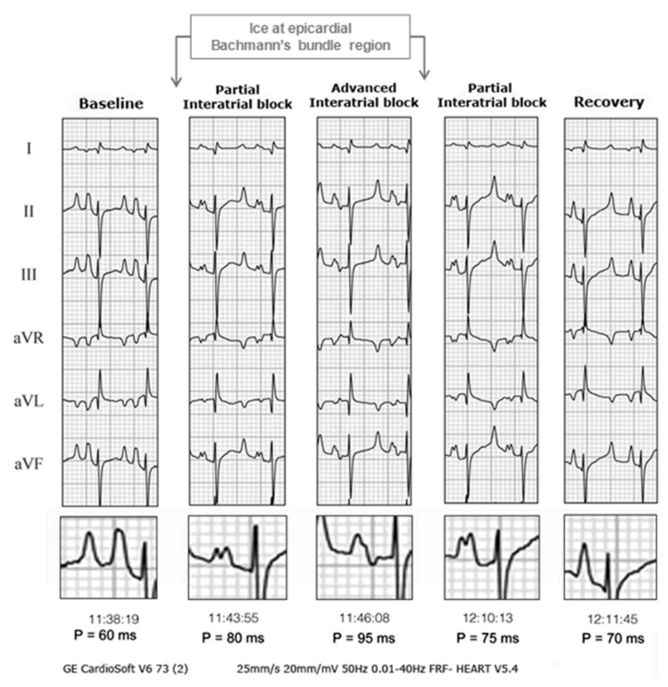
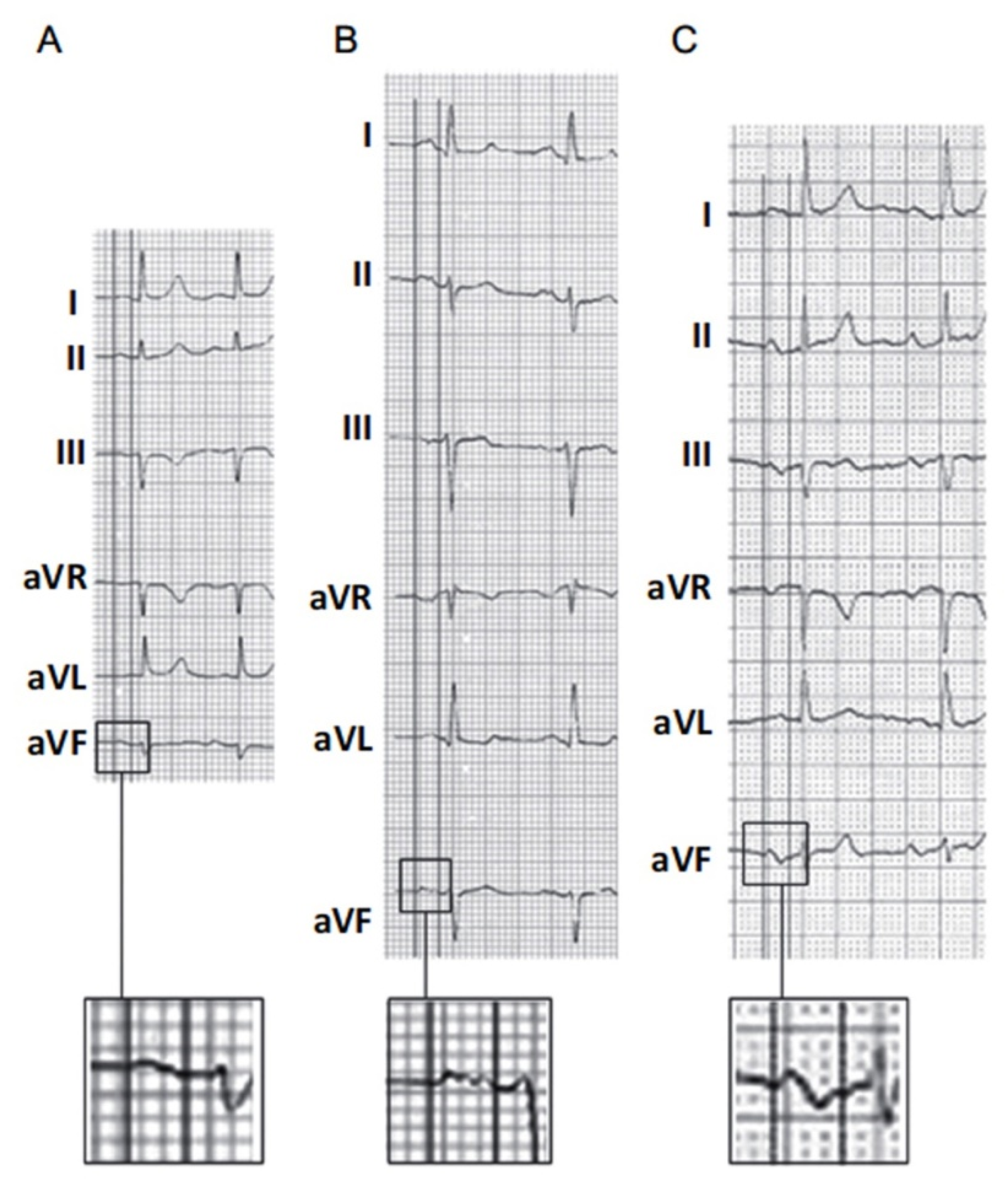
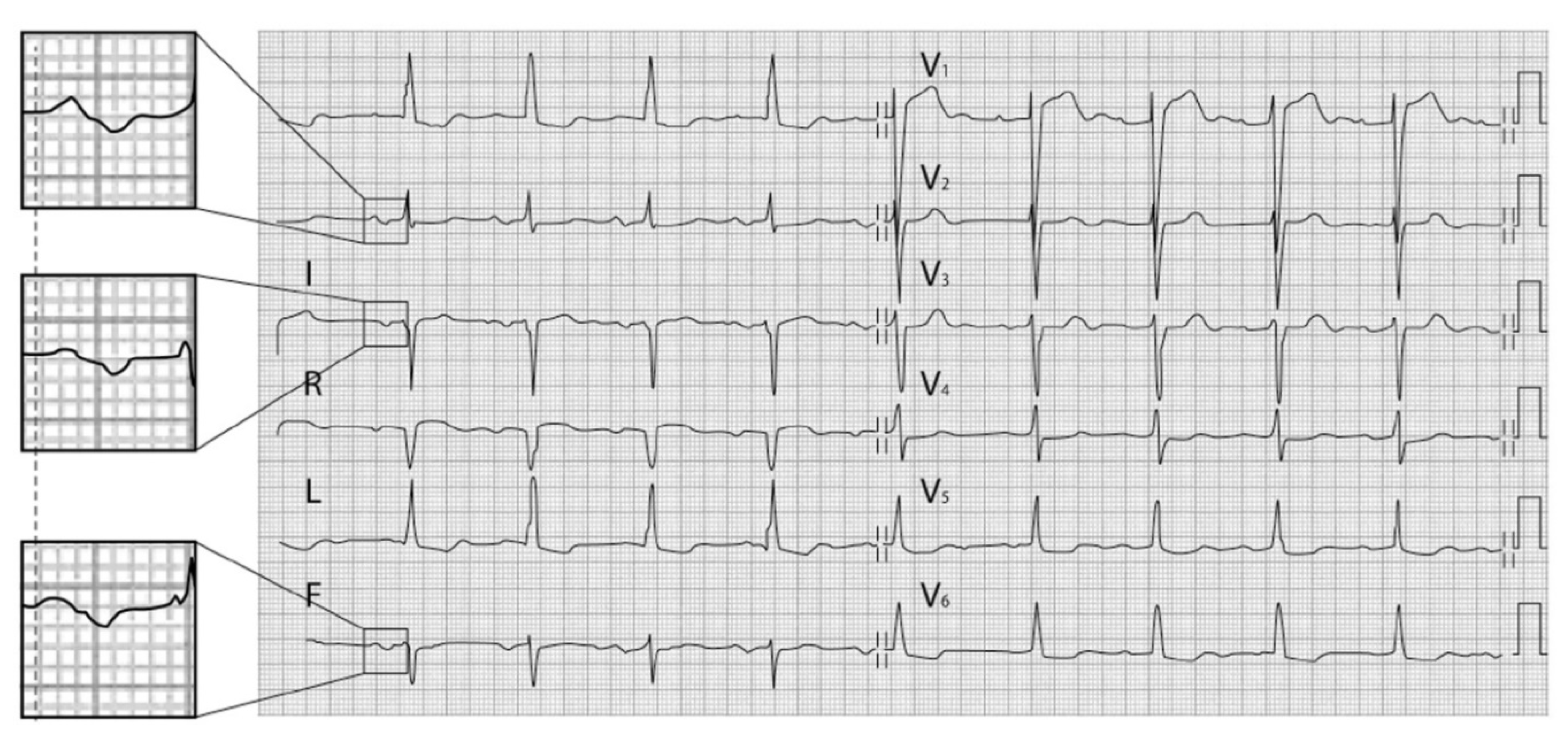
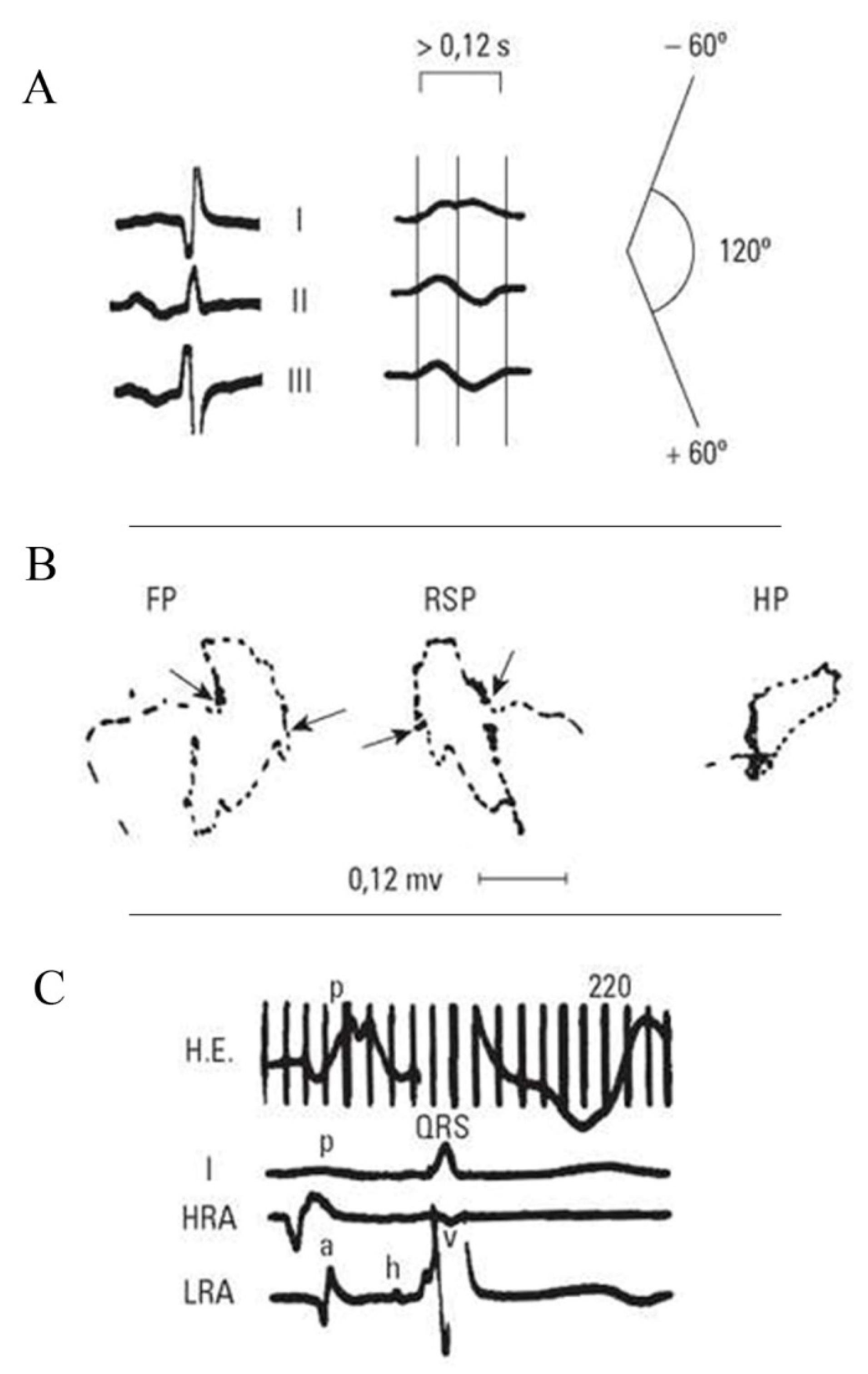
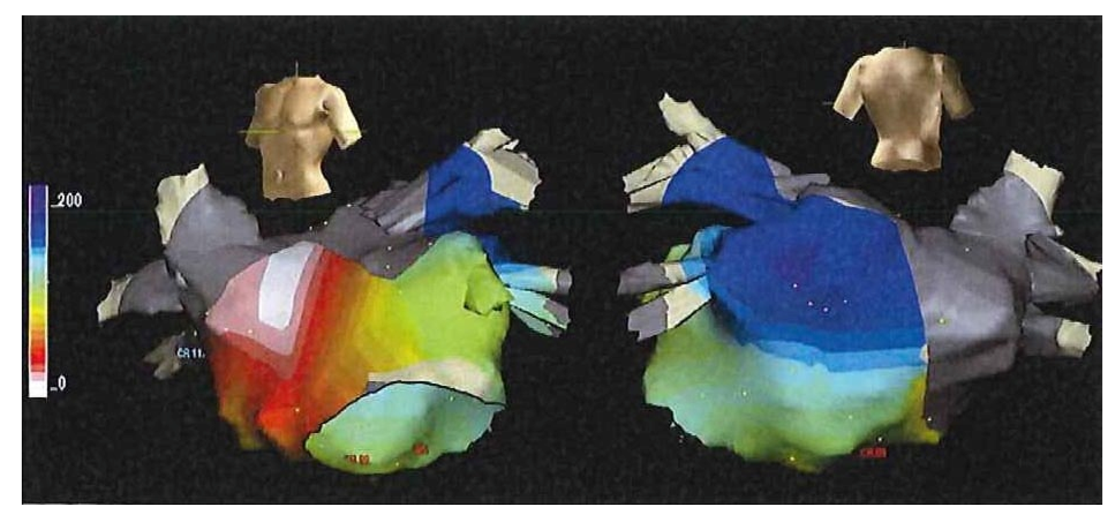
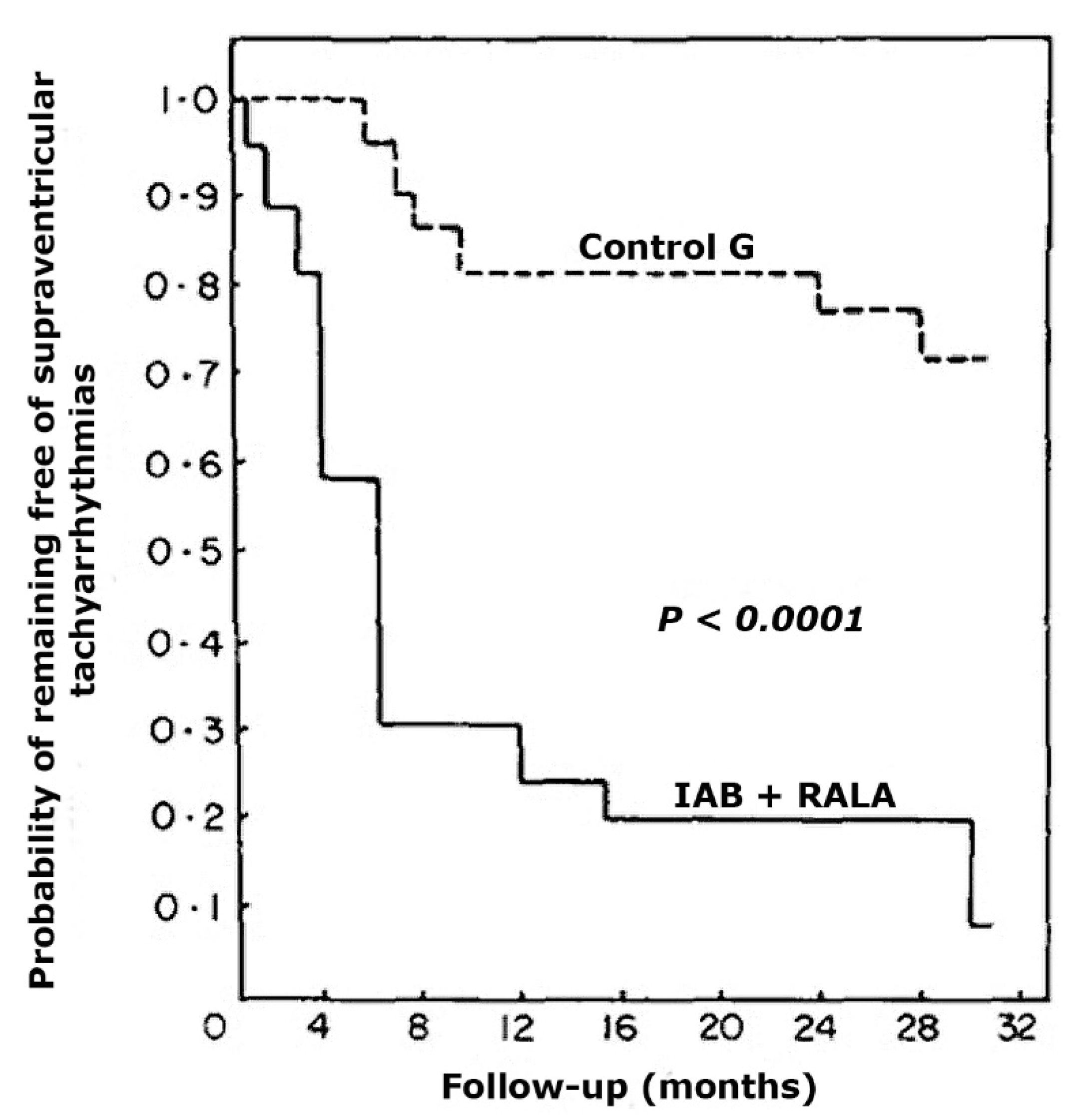
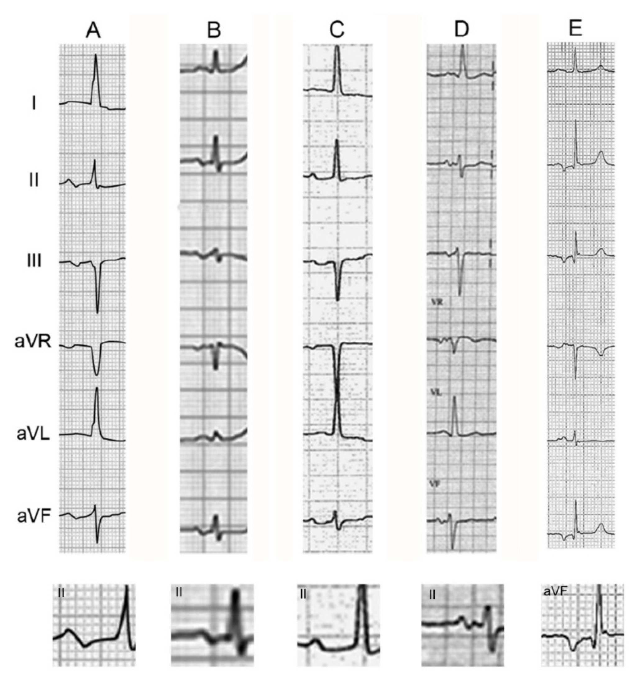
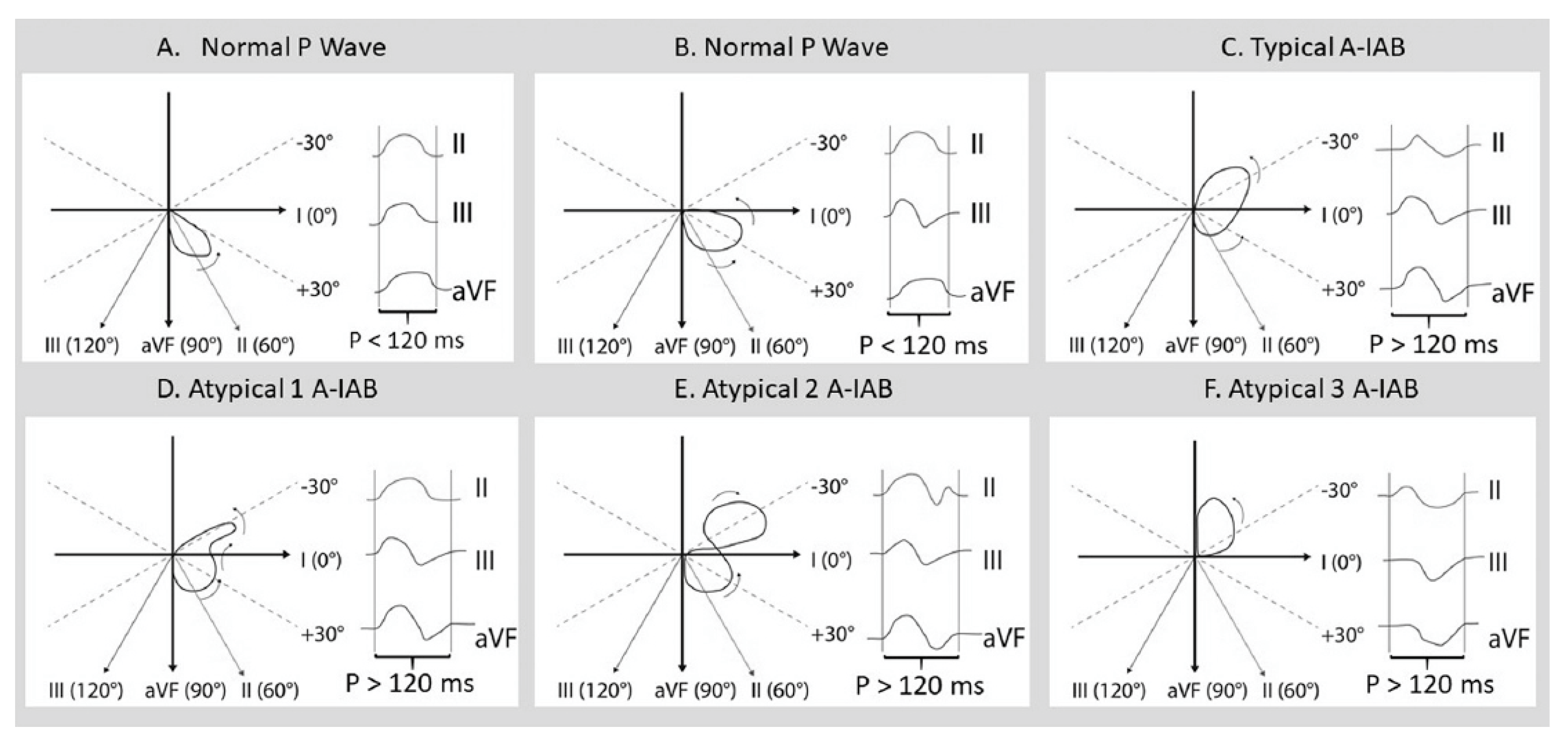
| 1. Partial Interatrial Block (First Degree) (P-IAB) P-wave ≥ 120 ms without negative terminal component in the inferior leads |
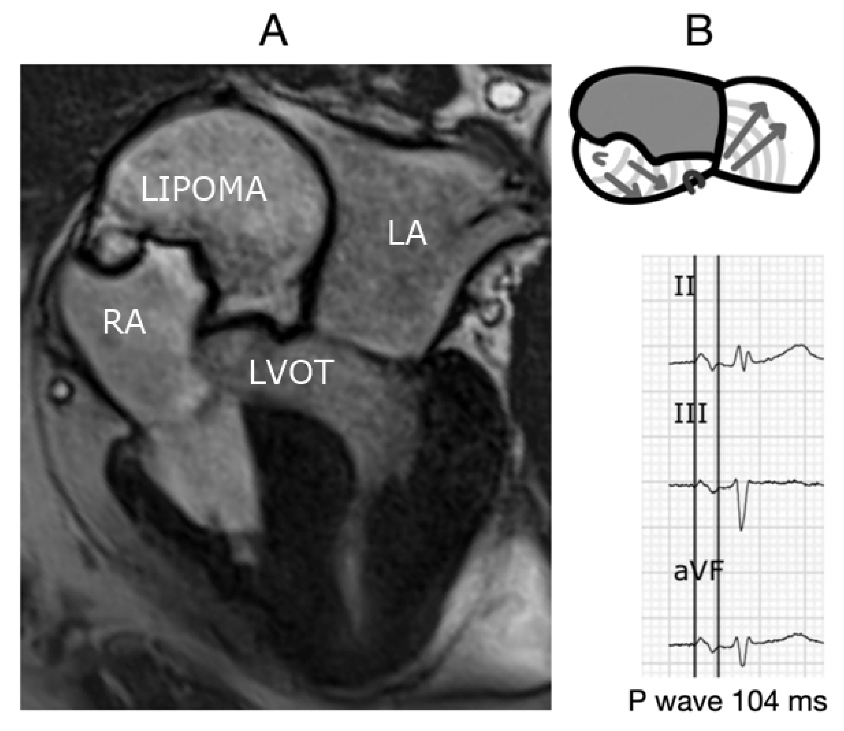
| 2. Advanced interatrial block (third degree) (A-IAB) Typical pattern P-wave ≥ 120 ms with biphasic morphology in leads II, III aVF (±) Atypical A-IAB may be atypical by morphology or by duration: (i): Morphological criteria Type 1: P-wave ≥ 120 ms with biphasic morphology in leads III and aVF and the final component of the P-wave in lead II is isodiphasic. Type 2: P-wave ≥ 120 ms. The second part of The P-wave is biphasic (∓). This means that the global P-wave is triphasic (+ − +). Type 3: P-wave ≥ 120 ms. The first part of P-wave in leads III and aVF is isoelectric, but the last part is negative. Therefore, it is necessary to perform differential diagnoses with junctional rhythm. (ii) Duration criteria P-wave < 120 ms with typical morphology (Biphasic (±) P-wave in leads II, III, and aVF) |
| 3. Second degree: The presence of ECG pattern of IAB is intermittent |
Publisher’s Note: MDPI stays neutral with regard to jurisdictional claims in published maps and institutional affiliations. |
© 2021 by the authors. Licensee MDPI, Basel, Switzerland. This article is an open access article distributed under the terms and conditions of the Creative Commons Attribution (CC BY) license (https://creativecommons.org/licenses/by/4.0/).
Share and Cite
Bayés-de-Luna, A.; Fiol-Sala, M.; Martínez-Sellés, M.; Baranchuk, A. Current ECG Aspects of Interatrial Block. Hearts 2021, 2, 419-432. https://doi.org/10.3390/hearts2030033
Bayés-de-Luna A, Fiol-Sala M, Martínez-Sellés M, Baranchuk A. Current ECG Aspects of Interatrial Block. Hearts. 2021; 2(3):419-432. https://doi.org/10.3390/hearts2030033
Chicago/Turabian StyleBayés-de-Luna, Antoni, Miquel Fiol-Sala, Manuel Martínez-Sellés, and Adrian Baranchuk. 2021. "Current ECG Aspects of Interatrial Block" Hearts 2, no. 3: 419-432. https://doi.org/10.3390/hearts2030033
APA StyleBayés-de-Luna, A., Fiol-Sala, M., Martínez-Sellés, M., & Baranchuk, A. (2021). Current ECG Aspects of Interatrial Block. Hearts, 2(3), 419-432. https://doi.org/10.3390/hearts2030033


