Review

The Use of PEEK Barriers in Bone Regeneration Procedures: A Scoping Review

1
Department of Prosthodontics, Faculty of Dentistry, University of Chile, Santiago 8380544, Chile
2
Department of Stomatology, Faculty of Dentistry, Universidad de Sevilla, 41004 Sevilla, Spain
3
Perioplastic Institute, Santiago 8380544, Chile
4
Private Practice, Puerto Varas 5550170, Chile
5
Postgraduate Program in Dentistry, University of Fortaleza, Fortaleza 60811-905, Brazil
6
Private Practice, Fortaleza 60811-905, Brazil
7
Department of Implantology, University of Santo Amaro, São Paulo 01311-000, Brazil
8
Postgraduate Implant Dentistry Department, School of Dentistry, Universidad Andrés Bello, Santiago 8380544, Chile
9
Department of Conservative Dentistry, Faculty of Dentistry, University of Chile, Santiago 8380544, Chile
10
Department of Oral and Maxillofacial Surgery, Plastic Operations, University Medical Center Mainz, 55116 Mainz, Germany
11
Oral Surgery and Implantology, Faculty of Medicine and Health Sciences, University of Barcelona, 08002 Barcelona, Spain
*
Authors to whom correspondence should be addressed.
Prosthesis 2025, 7(4), 101; https://doi.org/10.3390/prosthesis7040101
Submission received: 18 June 2025 / Revised: 9 July 2025 / Accepted: 23 July 2025 / Published: 19 August 2025

Abstract

Introduction: Guided bone regeneration (GBR) is a key approach for managing alveolar ridge defects. Although titanium meshes are widely used for non-resorbable space maintenance, their limitations have prompted interest in alternative materials. Polyetheretherketone (PEEK), a high-performance thermoplastic, has emerged as a potential barrier due to its mechanical strength, radiolucency, and compatibility with digital workflows. Objective: To map the current evidence on the use of PEEK barriers in GBR, focusing on biological performance, mechanical properties, and clinical outcomes in animal and human studies. Methods: A scoping review was conducted following PRISMA-ScR guidelines. Eligible studies included in vivo animal models or clinical trials involving PEEK barriers for alveolar bone regeneration. Data on study design, defect type, barrier characteristics, surgical protocol, outcomes, and complications were extracted. Results: Five studies met the inclusion criteria: two animal models and three clinical trials. All reported successful space maintenance and bone gain with PEEK barriers, with outcomes comparable to titanium meshes. Customization through CAD/CAM or 3D printing was common. Complications such as soft tissue dehiscence and exposure occurred but generally did not affect regeneration. Evidence was limited by small sample sizes, short follow-up, and single-center designs. Conclusions: PEEK barriers show promise as customizable alternatives to traditional GBR membranes. However, current evidence is limited and geographically concentrated. Future multicenter studies with long-term follow-up and standardized outcome measures are needed to validate the clinical potential of PEEK in bone regeneration.

1. Introduction

Guided bone regeneration (GBR) is a surgical technique that has revolutionized the field of oral and maxillofacial surgery. The principle of GBR is that bone defect regeneration can be predictably achieved through the use of occlusive membranes, which mechanically exclude non-osteogenic cell populations from the surrounding soft tissues, thus allowing osteogenic cell populations from the adjacent bone to migrate to the bone defect [1]. In this context, using regenerative barriers is essential for graft protection and guidance of bone growth. The selection of the barrier material should be adapted to the specific needs of the patient and the clinical context, and some aspects should be considered, including biocompatibility, mechanical stability, biological activity, exposure tolerance, biodegradability, antimicrobial properties, cost-effectiveness, and radiographic visibility [2]. Traditionally, membranes made of materials such as collagen, titanium mesh, and expanded polytetrafluoroethylene (e-PTFE) or dense polytetrafluoroethylene (d-PTFE) have been used. A systematic review has recently been published on the advantages of the use of computer-aided design and manufacturing (CAD/CAM) fabricated zirconia barriers in bone augmentation procedures [3], and in recent years, there has been growing interest in the use of PEEK-manufactured barriers [4].
PEEK is a semicrystalline thermoplastic polymer composed of repeating units of aromatic ethers and ketones (–[C6H4–O–C6H4–O–C6H4–CO]n–). This molecular configuration provides high thermal stability, chemical resistance, and notable mechanical rigidity. PEEK has an elastic modulus of 3.6 to 4 GPa, closely approximating that of cortical bone, and demonstrates excellent fatigue resistance. These properties make it a valuable material in various biomedical applications requiring mechanical strength, biocompatibility, and long-term structural integrity [5,6]. This polymer has been widely used in orthopedics as an excellent alternative to titanium [6]. The use of PEEK barriers in GBR has emerged as an innovative solution due to its exceptional mechanical properties and low biological reactivity. PEEK has demonstrated adequate mechanical behavior under tensile and flexural forces, which allows it to be designed, fabricated, and used with thicknesses between 0.5 and 1 mm [7]. Furthermore, PEEK is biocompatible, which means that it does not induce adverse reactions in the surrounding tissues and is generally well tolerated, although it shows limited osseointegration capacity [5,6,8]. Among the advantages of PEEK as a barrier in GBR is the ability to maintain its structural integrity in challenging bone defects, where a prolonged period of protection and stability of the graft material is required, which makes it an ideal barrier material for advanced or complex regenerative procedures. In addition, PEEK barriers can be custom-designed and manufactured using CAD/CAM technology to adapt to the specific needs of each patient, optimizing the space for the formation of new bone tissue, reducing surgical time, and minimizing the risk of complications due to poor membrane adaptation [9].
In recent years, significant advances have been made in manufacturing PEEK, leading to substantial improvements in its antimicrobial properties [10,11]. Infections can seriously compromise the success of GBR procedures, and its antimicrobial properties may help reduce this risk and improve the prognosis of regenerative procedures. Additionally, PEEK is radiolucent, allowing a better follow-up and analysis of the operated area in postoperative controls [5,6]. Although the rigidity of PEEK is an advantage in terms of mechanical resistance, it can also be a disadvantage. In some cases, the lack of flexibility may hinder its adaptation to particular complex bone morphologies, limiting its use in some bone defects, especially in thicknesses of less than 0.5 mm. However, when comparing PEEK with other CAD/CAM materials used as barriers in GBR, PEEK is more ductile than ceramics and less rigid than titanium, offering a balance between flexibility and strength [7]. In fact, conventional GBR materials such as titanium meshes and PTFE membranes have demonstrated predictable outcomes in bone regeneration but are not without drawbacks. Titanium meshes, while mechanically stable, are frequently associated with mucosal dehiscence and membrane exposure, with exposure rates ranging from 20–30%, potentially compromising regenerative outcomes [12]. PTFE membranes offer good barrier function but are prone to bacterial colonization and often require early removal upon exposure [13]. These limitations have prompted growing interest in alternative barrier materials that offer greater biocompatibility, design flexibility, and lower complication profiles. In this context, PEEK has emerged as a candidate material with promising mechanical and biological features that warrant systematic exploration.
The potential of PEEK in medical and dental applications continues to be explored, and its efficacy and accessibility could improve, making it an increasingly viable material for regenerative applications. Despite its promising properties, evidence regarding the clinical efficacy and safety of PEEK barriers in oral regenerative procedures remains limited. Further studies are needed to evaluate clinical outcomes and long-term performance. Therefore, the aim of this scoping review was to systematically map the available scientific literature on the use of PEEK barriers in GBR and bone augmentation procedures, both in preclinical and clinical studies, with particular emphasis on design features, clinical outcomes, and potential complications.

2. Materials and Methods

2.1. Protocol and Population–Context–Concept Framework

This scoping review was conducted in accordance with the Preferred Reporting Items for Systematic Reviews and Meta-Analyses extension for Scoping Reviews (PRISMA-ScR) guidelines [14] using the following population–context–concept (PCC) framework:
  • Population: Human adults (aged ≥ 18) with vertical and/or horizontal bone resorption in the maxillofacial area or animals with bone defects, with the indication of bone augmentation techniques.
  • Context: Laboratories, universities, dental clinics, and hospitals.
  • Concept: Vertical and/or horizontal bone formation using PEEK scaffolds as a barrier in bone augmentation procedures.

2.2. Eligibility Criteria

A comprehensive search for relevant studies was conducted without language or date restrictions; the last search was performed on 1 April 2025. Articles that met the following criteria were included in this review:
  • Human studies: (1) all primary studies in adult humans, including clinical (i.e., randomized clinical trials (RCTs)), prospective and retrospective cohort studies, case–control studies, case series and case reports reporting the use of PEEK scaffolds as a barrier in bone augmentation procedures in the maxillofacial area; (2) studies reporting the area of use and time of removal of the PEEK barriers; (3) studies reporting the surgical protocol of placement, including the use of grafts and their composition; and (4) studies reporting possible complications.
  • Animal studies: (1) studies reporting the use of PEEK scaffolds as barriers in augmentation procedures or bone regeneration of defects and (2) studies reporting the number of specimens, defect area, characteristics of PEEK barriers, filler material, and postoperative endpoint.
Therefore, the exclusion criteria were as follows: (1) reviews, cadaveric and in vitro studies, editorials, technical notes, communications, and studies where full-texts were not obtained were all excluded; (2) studies involving the use of PEEK scaffolds in bone regeneration procedures but not as a barrier; (3) studies that did not report results or sufficient information; and (4) studies reporting the use of zirconia, titanium or hydroxyapatite meshes or scaffolds were also excluded.

2.3. Information Sources and Search Strategy

Two independent reviewers (L.D. and X.U.) conducted electronic and manual literature searches using PubMed/Medline, Web of Science, Scopus, EBSCO, and Cochrane Library. A search string was created combining Boolean operators “AND” and “OR” and the following search terms/MeSH/keywords: (“Alveolar Ridge Augmentation” [Mesh] OR “Bone Regeneration” [Mesh] OR “Guided Tissue Regeneration, Periodontal” [Mesh] OR “Alveolar Bone Augmentation” OR “Guided Bone Augmentation” OR “Guided Bone Regeneration” OR “Bone Augmentation” OR “Bone Regeneration” OR “Bone Reconstruction” OR “Guided Tissue Regeneration”) AND (“Membranes, Artificial” [Mesh] OR “Membrane” OR “Barrier” OR “Matrix” OR “Mesh” OR “Scaffold” OR “Sheet” OR “Tissue Scaffolds” [Mesh]) AND (“polyetheretherketone” OR “Polyether ether ketone” OR “PEEK” OR “Ketones” [Mesh]). Additionally, a manual search was performed in the reference lists of selected articles, Google Scholar and all issues of the following journals: Annals of Maxillofacial Surgery, British Journal of Oral and Maxillofacial Surgery, Clinical Advances of Periodontics, Clinical Implant Dentistry and Related Research, Clinical Oral Implants Research, Clinical Oral Investigations, Clinical Trials in Dentistry, Dental Traumatology, Implant Dentistry, International Journal of Implant Dentistry, International Journal of Oral and Maxillofacial Implants, International Journal of Oral and Maxillofacial Surgery, International Journal of Oral Implantology, International Journal of Oral Science, International Journal of Periodontics and Restorative Dentistry, Journal of Clinical Periodontology, Journal of Cranio-Maxillo-Facial Surgery, Journal of Dental Research, Journal of Dentistry, Journal of Evidence-Based Dental Practice, Journal of Maxillofacial and Oral Surgery, Journal of Oral and Maxillofacial Surgery, Journal of Oral Implantology, Journal of Oral Pathology and Medicine, Journal of Orofacial Orthopedics, Journal of Periodontal and Implant Science, Journal of Periodontal Research, Journal of Periodontology, Journal of Stomatology, Oral and Maxillofacial Surgery, Journal of the Korean Association of Oral and Maxillofacial Surgeons, Maxillofacial Plastic and Reconstructive Surgery, Oral and Maxillofacial Surgery, Oral and Maxillofacial Surgery Clinics of North America, Oral Oncology, Oral Surgery, Oral Surgery Oral Medicine Oral Pathology Oral Radiology and Endodontology, Orthodontics & Craniofacial Research, and Periodontology 2000. These journals were selected based on their recognized relevance and frequency of publication of original research in oral and maxillofacial surgery, implant dentistry, bone regeneration, and biomaterials, including innovative applications such as PEEK. Table S1 (Supplementary File S1) shows the completed search queries.

2.4. Study Selection/Screening

After removing duplicates, two reviewers (LD and XU) independently screened the titles and abstracts of all studies retrieved from the above-mentioned search strategies and voted for inclusion or exclusion. Subsequently, full-text screening was independently performed. A Cohen’s Kappa test was computed in Microsoft Excel 2022 (Microsoft Corporation, Redmond, OR, USA) to assess inter-rater agreement between the two reviewers. Inter-rater agreement was interpreted according to the categories proposed by Landis and Koch [15]. Disagreements were discussed among the authors until an agreement was reached or by the decision of the arbiter reviewer (A.L.).

2.5. Data Extraction

Data extraction was performed by one reviewer (L.D.) and then checked by another (X.U.). Authors were contacted by e-mail to request article full-texts in case they were unavailable or to obtain key information not reported in the included studies. The variables collected included the following: author(s), year of publication, country, type of study, number of patients/samples, mean age and gender, bone defect, area, barrier characteristics, fixation methods, presence of bone perforations, filling material, cover materials, postoperative endpoint or removal time, amount of bone gained (vertical and/or horizontal), and complications. Quantitative data were collected on a Microsoft Excel 2018 spreadsheet.

2.6. Quality Assessment

To assess the methodological quality and risk of bias of the included studies, three standardized tools were employed depending on the study design: the SYRCLE Risk of Bias tool [16], the Risk of Bias 2 (RoB 2) tool [17], and the Risk Of Bias In Non-randomized Studies—of Interventions (ROBINS-I) tool [18]. The SYRCLE (Systematic Review Centre for Laboratory Animal Experimentation) tool was applied to animal studies and is based on the Cochrane Risk of Bias tool, adapted for preclinical research. It evaluates ten domains, including selection bias (sequence generation and baseline characteristics), performance bias (allocation concealment and random housing), detection bias (random outcome assessment and blinding), attrition bias, reporting bias, and other biases. Each domain is classified as “low risk,” “high risk,” or “unclear risk” based on the reported methodology. The RoB 2 tool, developed by the Cochrane Collaboration, was utilized for randomized clinical trials. This instrument comprises five bias domains: (1) bias arising from the randomization process, (2) bias due to deviations from intended interventions, (3) bias due to missing outcome data, (4) bias in the measurement of the outcome, and (5) bias in the selection of the reported result. Each domain is rated as “low risk,” “some concerns,” or “high risk” of bias, with an overall judgment derived from the collective assessment of these domains. Non-randomized clinical studies were evaluated using the ROBINS-I tool. This tool systematically assesses the risk of bias across seven domains: (1) bias due to confounding, (2) bias in the selection of participants, (3) bias in the classification of interventions, (4) bias due to deviations from intended interventions, (5) bias due to missing data, (6) bias in the measurement of outcomes, and (7) bias in the selection of the reported results. Each domain is judged as “low,” “moderate,” “serious,” “critical,” or “no information,” with an overall risk of bias score corresponding to the most severe domain rating. Two reviewers (L.D. and A.L.) conducted all assessments independently, and any discrepancies were resolved by consensus or consultation with a third reviewer (X.U.) to ensure methodological rigor and reproducibility.

3. Results

3.1. Study Selection

A total of 364 studies were retrieved from the comprehensive electronic database search and 4 through a manual search in Google Scholar and the list of journals detailed before. After removing duplicates, 215 articles were screened based on title and abstract, leaving 59 reports eligible (substantial agreement, κ = 0.86). After full-text reading and a subsequent search for relevant citations, three studies in humans and two in animals were included in this scoping review. The reviewers (L.D. and X.U.) agreed 100% on the final selection of studies. Of the 60 articles reviewed, 55 were excluded from the final analysis because they reported insufficient information (n = 2), the full-text version of the article was not obtained (n = 2), PEEK scaffolds were not used as a barrier, but rather as plates, implants, discs, or pins (n = 48), and/or by type of study, specifically in vitro studies (n = 2) and a review (n = 1). The PRISMA flow diagram of the screening process is shown in Figure 1.

3.2. Characteristics of the Included Studies, Samples, and Bone Defects

The articles included two animal studies [19,20] and three human clinical studies corresponding to two RCTs [21,22] and one case series [23]. Animal studies were conducted in Japan [19] and China [20]. Human studies were conducted by the same team at Cairo University, Egypt [21,22,23]. The animal studies presented biomechanical, gene expression, histological, and radiographic analyses of PEEK barriers and newly formed bone. In contrast, the human studies presented radiographic and histomorphometric analyses of the percentage of newly formed bone.
Concerning animal studies, a sample of 30 Wistar rats (300–350 G, 10 weeks old) and 3 Beagle dogs (12–16 kg, 2 years old) was obtained. The bone defects in the rats, totaling 30, were created in the left femurs, in 5 mm long sections of the diaphysis, and removed with a micro-cutting saw [19]. On the other hand, in Beagles, the defects were created from mandibular premolar and molar extractions. After 8 weeks of healing, bilateral bone defects of 7 × 7 mm, 5 mm apart, were made using an auxiliary template. This study treated six bone defects with PEEK barriers [20].
Regarding the human studies, the samples consisted of 30 patients with 30 bone defects treated with PEEK barriers, nine males and 13 females, with an average age of 39.0 years in one study [21] and 29.0 years in the other [23]. One study did not report the age or sex of the eight patients treated [22]. The included patients had no systemic alterations that could interfere with bone healing and no previous bone augmentation surgery history. They also had partial or total maxillary edentulism with 3D bone defects, evaluated clinically and radiographically through Cone-Beam Computed Tomography (CBCT). The sizes and locations of the defects were not detailed for each case. However, two studies specified that the 16 bone defects analyzed due to tooth loss were less than 6 mm in height, measured from the alveolar ridge to the basal bone, and less than 2 mm in width horizontally [21,22], and the other included study reported the analysis of 14 sites of massive maxillary resorption, with an average initial height in the vertical direction of 9.56 ± 1.54 mm and 2.59 ± 0.92 mm horizontally [23]. The complete characteristics of the included studies are presented in Table 1.

3.3. Design, Manufacture, and Cleaning of PEEK Barriers

The first animal study was the only one that reported PEEK barriers with a tubular shape [19]. These barriers had an external diameter of 5 mm, an internal diameter of 3 mm, and a height of 5 mm. Additionally, they featured four elliptical holes measuring 3 × 1.5 mm on the lateral surface. These tubular barriers were manufactured from TECAPEEK CLASSIX polymers (Ensinger, Nufringen, Germany), revealing no characteristics of the barriers’ design, manufacturing, and cleaning process. In the study reported in Beagle dogs [20], after 8 weeks of healing after premolar and molar extractions, CBCT was performed, exporting the DICOM to 3D design software (Mimics Research and 3-Matic, Materialise, Leuven, Belgium) for the design of the defects, the template for their creation and the PEEK barriers to contain the grafts, with a thickness of 0.6 mm and perforations of 2 mm along the entire length of the barrier. These were subsequently manufactured using a 3D printer (FDM 3D printer, Funmat Ht, Intamisys, Shanghai, China) and cleaned in an ultrasonic bath for 60 min with 75% ethanol, followed by distilled water, and then sterilized at high temperature (121 °C) and high pressure (0.12 MPa). The two RCTs performed in humans presented the same workflow and characteristics of the manufactured PEEK barriers [21,22]. A CBCT of the patients was required to evaluate the volume of the alveolar ridge and quantify the bony defects. The DICOMs were exported to design software (Mimics 19, Materialize NV, Belgium) to virtually fill the defect and design the PEEK and titanium barriers, with thicknesses of 2 mm and multiple round perforations along the entire length of the barriers. The PEEK barriers were intended to cover the alveolar bone’s crestal, buccal, and palatal surfaces and then manufactured from medical-grade PEEK blocks using a 5-axis milling machine. They were cold sterilized before surgery by placing them in 2.4% glutaraldehyde for 20 min, while titanium meshes were steam sterilized using an autoclave. The third study [23] reported the treatment of maxillary bone defects with the same previously described protocol of bone volume acquisition and software design of the barriers; however, the barriers were designed by contouring only the buccal and palatal surfaces and then creating an interconnection between them, and these barriers were not designed with perforations or thickness specifications, only with holes designed for micro-titanium fixation screws into areas of sufficient bone, irrelevant to the proposed future implant positions. The sterilization protocol for these barriers was established by immersing them in 2.4% glutaraldehyde for 12 h. Finally, among the included studies evaluating PEEK membranes, few addressed the influence of perforation size on regenerative outcomes, and no comparative analysis was available to determine the effect of varying pore diameters on osteogenesis or soft tissue ingrowth.

3.4. Characteristics of Bone Grafts, Fixation Methods, and Presence of Bone Perforations

Concerning animal studies, femur bone defects in rats were treated with tubular barriers with an endpoint of 28 days; these were divided into three groups according to their content: peptide hydrogel, autogenous bone, and no content, respectively. The barriers were fixed with an external tutor to the femurs, in addition to 4 pins of 1.4 mm diameter, without bone perforations [19]. The other animal study [20] investigated bone formation in 18 mandibular defects by comparing the use of PEEK barriers, titanium CAD/CAM meshes, and collagen pericardium membranes, all fixed using titanium pins, containing a mixture of calcined bovine bone and autogenous bone chips. Neither study reported the presence of bone perforations or corticotomies. Human studies used the same graft combination to treat bone defects from PEEK barriers. A 50:50 mixture of xenogeneic and autologous bone was obtained from the iliac crest [21,22] or intraoral sites such as the chin or retromolar area [23]. Regarding the fixation of the barriers, all reported using 3–4 mini-screws for this purpose and small round surgical drills to generate bleeding points or bone perforations to expose the underlying marrow for easier graft consolidation.

3.5. Radiographic Analysis of Bone Formation

The included studies reported Bone formation from radiographic analysis using soft X-ray radiography, micro-CT, and CBCT. Nakahara et al., 2010 [19] utilized soft X-ray radiography to calculate the newly formed bone length ratio to the defect length. In rats, the average bridging ratio of femoral bone defects was 96.5 ± 4.7% for the PEEK/autogenous bone group and 29.5 ± 9.7% for the PEEK/empty group (p < 0.01). Li et al., 2020 [20], from micro-CT analysis, observed that the regenerated bone was 50.18 ± 7.26% for the PEEK barriers group and 52.62 ± 3.61% for the titanium meshes group, both significantly higher than the collagen membrane control group, with 41.90 ± 5.20% (p < 0.05). About alveolar bone height, this was significantly reduced in the control group, with 0.99 ± 0.21 mm (p < 0.001), compared to the PEEK barriers and titanium meshes groups, with 0.29 ± 0.31 mm and 0.28 ± 0.32 mm, respectively.
Regenerated bone in human maxillary defects was assessed by CBCT from a cross-sectional view of each proposed future implant before surgery, at 1 week, and at 6 months. Mounir et al., 2019 [21] reported the percentage of bone gained and not by linear measurements because all defects did not have the same dimensions, with values of 20.9 ± 13.3% for the titanium meshes group and 31.8 ± 22.7% for the PEEK barriers group, finding no statistically significant differences between them (p = 0.2). El Morsy et al., 2020 [23] used the nasal floor and the maxillary sinus as a fixed anatomical reference for the measurements. In addition, a radiographic stent was used for each patient with radiopaque material (barium sulfate mixed with acrylic powder), filling the area of interest to ensure that the calculations were taken in the same location. The average bone gained vertically was 3.47 ± 1.46 mm and horizontally 3.42 ± 1.10 mm, with a statistically significant difference in both dimensions compared to the preoperative defect measurements (p = 0.0001). Gouda et al., 2023 [22] did not report radiographic analysis of bone gain.

3.6. Histological Analysis

The animal studies performed histological analyses of the tissue samples obtained after removing the barriers. The first [19], after 28 days of scaffold placement, samples were removed, fixed in 4% paraformaldehyde, and demineralized for another 7 days. The 6 um thick tissue sections were stained with hematoxylin–eosin and Masson’s trichome, finding that in the unfilled/empty PEEK scaffold group, cartilage formation and a small amount of bone formation were observed around the edges. In the PEEK/autogenous bone group, nearly all of the transplanted bone was left behind, resulting in a certain degree of contact between the bone sections. The other study [20] that performed histological analysis removed the samples 3 months after the installation of the barriers, and the operated areas were excised in dog jaws and immediately fixed in 4% paraformaldehyde solution. After micro-CT analysis, they were dehydrated in ethanol and embedded in methyl methacrylate to be sectioned into 50 μm thicknesses. Stevenel’s blue and van Gieson’s picrofuchsin staining was used to observe bone regeneration. The percentage of new bone for the PEEK and titanium barrier groups was 28.18 ± 9.46% and 27.30 ± 14.76%, respectively. Both were significantly higher than the collagen membrane control group, with 8.61 ± 5.42% (p < 0.05). The percentage of new bone mixed with bone substitutes was the lowest in the PEEK barrier group (42.11 ± 2.94%) when compared to the other two groups but the highest in relation to the percentage of soft tissue mixed with bone substitutes (29.01 ± 12.03%), with a non-statistically significant difference (p > 0.05). In the vertical new bone height measurement, the PEEK and titanium barrier groups showed a greater increase, with 3.17 ± 1.67 mm and 3.15 ± 1.25 mm, respectively, when compared to the control group, which presented 1.56 ± 0.82 mm, with a non-statistically significant difference (p > 0.05).
In humans, only one study reported histological analysis of regenerated bone after 6 months from transcortical bone biopsies obtained using 3 mm trephine burs in areas where implants were installed [22]. After being processed and sectioned, they were stained using Masson’s trichrome stain for histomorphometric analysis. A notable difference in bone quality was reported between the titanium mesh group and the PEEK barrier group. Mature and organized lamellar bone was detected in the titanium group, while, on the other hand, the PEEK group showed a less mature woven bone with interfering xenograft particles. In addition, histomorphometric analysis showed that the percentage of newly formed bone was higher in the titanium group (26.3 ± 4.35%) compared to the PEEK group (19.5 ± 2.38%), with a statistically significant difference (p = 0.000).

3.7. Complications

Only clinical studies involving humans reported complications. Among the 38 patients examined, 30 bone defects were treated with PEEK barriers. In three of these cases (10%), mucosal dehiscence occurred, resulting in barrier exposure during the first [23], second [21], and third [22] postoperative weeks. These situations were addressed with daily saline irrigation until healing by secondary intention was successfully achieved. After obtaining the CBCT scans at 6 months postoperatively, it was verified that bone regeneration of the defects was satisfactorily accomplished in those cases, and the planned implants could be placed. On the other hand, one case (3.3%) reported very poor bone quality and massive fibrointegration upon the second stage of surgery, which directed the authors to modify the barrier design to include an interconnecting part covering the graft’s crestal portion. This case was treated with GBR using a resorbable collagen membrane, postponing the implant placement [23]. The number of bone defects treated with CAD/CAM titanium meshes amounted to 16 in total, with two (12.5%) presenting mesh exposure at two weeks postoperatively and resolved with the same protocol previously described [21,22].

3.8. Biomechanical and Gene Expression Analysis

Mechanical tests were reported only in animal studies; the first one, from uniaxial compressive strength tests to PEEK scaffolds, reported an average maximum stress of 71.8 ± 0.18 MPa, with an average maximum displacement of 0.677 ± 0.017 mm [19]. The second study evaluated the barriers to the maximum bending strength, obtaining values of 56.03 ± 2.17 MPa in the three-point bending test, exhibiting a long plastic deformation before the maximum bending stress. In the finite element analysis, the maximum von Mises stress was 14.06 MPa, and the maximum deformation was 0.04 mm [20].
Only one study performed gene expression analysis [19]. After 7 days of PEEK barrier placement on the left femur of rats, total RNA was extracted from the tissue inside the scaffolds, and the expression of Runx2, VEGF-A, osteocalcin, and a housekeeping gene (GAPDH) was analyzed with RT-PCR. Gene expression when comparing VEGF-A with GAPDH was 6.87-fold for group 1 (PEEK/Peptide Hydrogel), 1.81-fold for group 2 (PEEK/Autogenous bone), and 0.95-fold for group 3 (PEEK/empty). When Runx2 was compared with GAPDH, the ratio was 0.93-fold, 0.84-fold, and 0.31-fold for groups 1, 2, and 3; finally, when osteocalcin was compared with GAPDH, the ratio was 0.80-fold, 3.8-fold, and 0.16-fold for groups 1, 2, and 3, respectively.

3.9. Risk of Bias Assessment

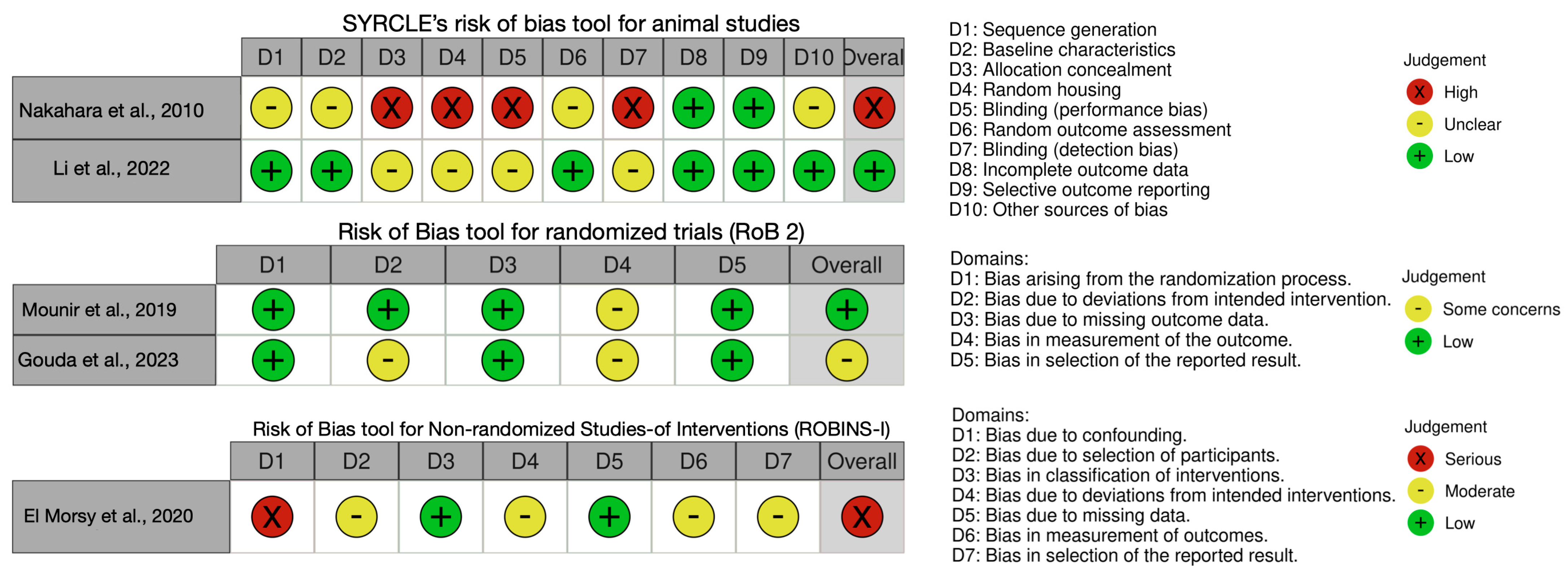
The included studies demonstrated variable risk of bias, with certain methodological limitations that warrant cautious interpretation of their findings. Overall, the animal studies generally exhibited a moderate risk of bias due to unclear reporting of allocation concealment and blinding of outcome assessment, which is crucial for minimizing performance and detection biases. Moreover, the limited sample sizes and absence of preregistered protocols further reduce the internal validity of these preclinical experiments. The human clinical trials showed better methodological rigor, with most employing randomized designs and clearly defined inclusion and exclusion criteria; however, concerns remain regarding blinding of outcome assessors, potential selection bias due to convenience sampling, and insufficient detail on allocation concealment procedures. Notably, while two of the clinical studies utilized split-mouth designs to reduce intersubject variability, the lack of long-term follow-up and relatively small sample sizes may limit the generalizability of their results. These limitations underscore the need for future research employing standardized reporting guidelines, larger sample sizes, and robust bias mitigation strategies to strengthen the evidence base regarding the comparative effectiveness of PEEK versus titanium in GBR. The risk of bias in the included studies is shown in Figure 2.

4. Discussion

This scoping review aimed to synthesize and critically appraise the evidence on using PEEK barriers for GBR in animal and human models. Five studies—two animal models and three human clinical studies—highlight the emerging yet limited body of evidence supporting the application of PEEK as a space-maintaining biomaterial in alveolar bone reconstruction. The findings of this review suggest that PEEK barriers may represent a suitable and customizable alternative for alveolar ridge augmentation, especially in cases of severe maxillary resorption. However, given the comparable performance to titanium meshes and the lack of long-term outcome data, PEEK cannot yet be considered superior or universally preferred.
The animal studies [19,20] demonstrated the feasibility of PEEK barriers in maintaining space for bone regeneration, with substantial new bone formation confirmed histologically and radiographically. In the study by Nakahara et al. [19], PEEK scaffolds with a tubular design yielded a bridging rate of up to 96.5% when filled with autologous bone. Conversely, Li et al. [20] demonstrated comparable outcomes between PEEK barriers and titanium meshes in mandibular defects in dogs, although alveolar bone height gain was slightly lower in the PEEK group. However, qualitative assessment of bone quality and integration appeared similar, suggesting comparable biological behavior despite slight dimensional differences. These results indicate that PEEK barriers may be equivalent to titanium in specific parameters of bone regeneration, at least in the preclinical setting.
In human studies [21,22,23], PEEK barriers were primarily applied in the context of severe anterior maxillary defects, with vertical and horizontal dimensions less than 6 mm and 2 mm, respectively. Bone gain values varied, with 3D volume increases and histomorphometric outcomes suggesting clinical effectiveness. Notably, Mounir et al. [21] and Gouda et al. [22] both found that PEEK and titanium barriers yielded similar bone formation outcomes, albeit with slight variation in complication rates (e.g., exposure). The case series by El Morsy et al. [23] also demonstrated significant bone augmentation with PEEK barriers, though two complications were reported, including fibrous integration instead of osteointegration in one case. In summary, comparative analysis of histological and radiographic outcomes reveals mixed findings regarding the regenerative capacity of PEEK barriers. While some studies report bone gain comparable to titanium meshes, others show lower histomorphometric bone percentages or higher rates of fibrous tissue encapsulation. In animal models, the observed variability may reflect differences in scaffold porosity, defect location, or fixation technique. Moreover, radiographic measurements in clinical trials were often limited to early follow-up periods (<12 months), limiting the ability to assess long-term volumetric stability. The lack of standardization in outcome measures further complicates interpretation. These inconsistencies underscore the need for more rigorous, standardized protocols when evaluating the biological performance of PEEK barriers in future research.
Customized 3D titanium meshes (Ti-mesh) and non-resorbable membranes such as e-PTFE and d-PTFE are widely regarded as the gold standard for GBR [24,25,26,27]. Traditional non-resorbable membranes, such as Ti-meshes, have long been valued for their outstanding mechanical stability and space-maintaining capacity in GBR [28]. However, one major limitation of manually shaped Ti-meshes is their poor adaptability to complex anatomical contours, often necessitating intraoperative bending and trimming. This prolongs surgical time and may increase the risk of mucosal irritation and postoperative exposure [29,30]. Exposure rates of conventional Ti-meshes can be as high as 23.1% in some reports, compared to 7.7% in customized Ti-meshes [30], suggesting that pre-contoured, patient-specific devices may reduce complication rates due to smoother edges and anatomical fit. Recent advancements in CAD/CAM and 3D printing technologies have enabled the development of customized barrier membranes made from Ti, zirconia, unsintered hydroxyapatite/poly-l-lactide (uHA/PLLA), and PEEK, designed to conform precisely to the bone defect area [4,31,32,33,34,35]. However, PEEK has emerged as an up-and-coming alternative from a biological standpoint due to its favorable mechanical and biological properties. Unlike Ti, PEEK exhibits an elastic modulus closer to cortical bone, potentially enhancing mechanical compatibility under masticatory loading while minimizing stress shielding [36]. It is also biocompatible, radiolucent, and less irritating to soft tissues, which may lower the incidence of dehiscence and exposure [21]. Nevertheless, PEEK lacks intrinsic osteoconductivity and requires secondary surgical removal, similar to Ti [37]
Regarding design and fabrication considerations, one of the key advantages of PEEK lies in its customizable fabrication potential [20,21,22,23]. All human studies utilized patient-specific, CAD/CAM-designed barriers based on CBCT-derived DICOM files [21,22,23]. Milling and 3D-printing techniques produced barriers with anatomically adapted geometries and perforation patterns. This individualized approach potentially improves the stability and fit of the barriers, favoring predictable bone regeneration. However, the studies varied in design features such as barrier thickness (0.6 mm to 2 mm), presence or absence of perforations, and surface coverage (crestal, buccal, and/or palatal) [19,20,21,22,23]. These design inconsistencies reflect a lack of consensus, highlighting the need to standardize both manufacturing protocols and mechanical design parameters.
Several of the included studies reported the use of glutaraldehyde immersion (typically 2–2.4%) as the sterilization method for PEEK barriers prior to their in vivo application. This cold chemical sterilization technique is known for its broad-spectrum antimicrobial activity and material compatibility at low temperatures. However, it also poses risks of cytotoxicity and tissue irritation if not properly neutralized and rinsed. Studies have shown that glutaraldehyde can leave residual compounds that compromise biocompatibility, especially in sensitive surgical applications [38]. Although it may have been selected to avoid thermal deformation of the PEEK structure, other low-temperature sterilization methods—such as hydrogen peroxide plasma or ethylene oxide—have demonstrated greater safety profiles and material compatibility for implantable polymer-based devices [39]. Future research should report sterilization protocols more transparently and prioritize biocompatible methods validated for implantable materials.
Perforation size also plays a pivotal role in the biological performance of barrier membranes. While larger pores (e.g., 2 mm in PEEK lattices) may support angiogenesis and nutrient diffusion [20], they also risk soft tissue invasion and formation of pseudo-periosteum—a fibrous connective layer often observed under Ti meshes [40]. This tissue may limit the direct contact between the graft and bone surface, potentially compromising proper osseous regeneration. The ideal pore configuration remains controversial, as large apertures may promote early bone healing but offer limited benefits in final bone volume [41,42].
The use of PEEK as a barrier material in bone regeneration presents several advantages, including its high dimensional stability, radiolucency, resistance to degradation, and adaptability to custom digital design and manufacturing workflows. These properties make it attractive as an alternative to traditional titanium meshes. However, drawbacks remain. PEEK is bioinert and does not naturally promote cell adhesion or bone integration, which may result in fibrous encapsulation and limited regenerative response in some cases [4,6]. Using PEEK combined with grafting materials (autogenous and xenogeneic) was a common strategy across all human-included studies [21,22,23], which complicates the isolated assessment of PEEK’s osteopromotive potential. Therefore, future studies should explore the osteopromotive potential of PEEK in isolation, using standardized grafting or graft-free conditions, to determine its contribution to new bone formation. Nevertheless, the consistent outcomes in terms of bone fill and volume gain, even when compared to titanium mesh, suggest that PEEK may serve as a viable alternative with certain advantages—particularly in aesthetics, as PEEK lacks the metallic translucency of titanium and may be better tolerated in anterior maxillary zones. However, clinical complications, including barrier exposure, were observed in all human studies, regardless of the barrier material [21,22,23]. This highlights the inherent risk of non-resorbable GBR materials and the need for meticulous flap management, tension-free closure, and postoperative follow-up. Interestingly, only one case across the studies showed an aberrant healing response (fibrointegration), which might be related to patient-specific factors or mechanical instability of the barrier.
Despite promising outcomes, the current body of literature presents several limitations. First, the small number of studies and limited sample sizes, particularly in animal studies and case series, restrict the generalizability of findings and reflects the limited availability of published research specifically addressing the use of PEEK as a barrier material in bone regeneration. The inclusion of both animal and human data was essential to capture the breadth of available evidence, but the low volume underscores the need for further research in this emerging field. Second, heterogeneity in study design, including defect locations, dimensions, barrier configurations, grafting protocols, and evaluation methods, impedes direct comparison across studies. Third, none of the included studies offered long-term follow-up data or assessed implant survival or peri-implant tissue health post-regeneration, which are critical for evaluating the clinical relevance of the GBR procedure. Furthermore, all three human studies originated from the same research group at Cairo University [21,22,23], potentially introducing geographic or institutional bias. This concentration of evidence from a single institution may reflect specific surgical techniques, patient selection criteria, or workflow efficiencies not generalizable to broader settings. Independent replication by other research centers is necessary to validate these preliminary results and establish broader applicability. Therefore, caution must be exercised when extrapolating these findings to broader clinical contexts.
Future research should focus on conducting high-quality, randomized controlled clinical trials with long-term follow-up to validate the efficacy and safety of PEEK barriers in GBR. Standardization of defect models, barrier design parameters, and grafting protocols is essential for meaningful study comparisons. Moreover, investigations into the biological response of periosteal and mucosal tissues to PEEK surfaces—especially in different surface-treated or coated variants—may offer insights into optimizing soft tissue integration and minimizing exposure risks. Comparative studies assessing cost-effectiveness, patient-reported outcomes, and digital workflow efficiency versus traditional titanium meshes will also be valuable. Finally, integrating biomechanical modeling and 3D imaging analytics could support the development of patient-specific, functionally optimized PEEK barriers tailored to individual anatomical and mechanical needs.

5. Conclusions

This scoping review highlights the emerging role of PEEK barriers in bone regeneration, particularly in digitally driven, customized approaches. While preclinical and early clinical studies report favorable mechanical handling and regenerative outcomes, the overall evidence remains limited, predominantly derived from single-center reports, and lacks long-term follow-up. To advance the clinical translation of PEEK barriers, future research should prioritize multicenter, prospective trials using standardized outcome measures such as radiographic volumetric bone gain, incidence of membrane exposure, and the need for secondary intervention. These efforts are essential to clarify indications, long-term safety, and comparative effectiveness relative to conventional barrier materials.

Supplementary Materials

The following supporting information can be downloaded at https://www.mdpi.com/article/10.3390/prosthesis7040101/s1. Table S1: Search Queries. Table S2: Excludes studies by reasons.

Author Contributions

L.D.: conceptualization, methodology, validation, formal analysis, investigation, data curation, writing—original draft, writing—review and editing, visualization, supervision, and project administration. X.U.: conceptualization, methodology, investigation, data curation, writing—original draft, writing—review and editing, and visualization. A.L.: writing—original draft and writing—review and editing. H.F.M.: writing—original draft and writing—review and editing. P.U.: writing—original draft and writing—review and editing. A.T.: methodology, validation, formal analysis, writing—original draft, writing—review and editing, and visualization. S.F.: conceptualization, methodology, writing—review and editing, visualization, and supervision. All authors have read and agreed to the published version of the manuscript.

Funding

This research received no external funding.

Institutional Review Board Statement

Not applicable.

Informed Consent Statement

Not applicable.

Data Availability Statement

The data sets used and/or analyzed during the current study are available from the corresponding author upon reasonable request.

Conflicts of Interest

The authors declare no conflicts of interest.

References

  1. Retzepi, M.; Donos, N. Guided Bone Regeneration: Biological Principle and Therapeutic Applications. Clin. Oral Implant. Res. 2010, 21, 567–576. [Google Scholar] [CrossRef]
  2. Sanz, M.; Dahlin, C.; Apatzidou, D.; Artzi, Z.; Bozic, D.; Calciolari, E.; De Bruyn, H.; Dommisch, H.; Donos, N.; Eickholz, P.; et al. Biomaterials and Regenerative Technologies Used in Bone Regeneration in the Craniomaxillofacial Region: Consensus Report of Group 2 of the 15th European Workshop on Periodontology on Bone Regeneration. J. Clin. Periodontol. 2019, 46 (Suppl. S21), 82–91. [Google Scholar] [CrossRef]
  3. Uriarte, X.; Landázuri, A.; Marão, H.F.; Lucena, N.; Schiegnitz, E.; Díaz, L. Zirconia Barriers in Bone Regeneration Procedures: A Scoping Review. Clin. Oral Implants Res. 2025, 36, 411–422. [Google Scholar] [CrossRef]
  4. Shi, Y.; Liu, J.; Du, M.; Zhang, S.; Liu, Y.; Yang, H.; Shi, R.; Guo, Y.; Song, F.; Zhao, Y.; et al. Customized Barrier Membrane (Titanium Alloy, Poly Ether-Ether Ketone and Unsintered Hydroxyapatite/Poly-l-Lactide) for Guided Bone Regeneration. Front. Bioeng. Biotechnol. 2022, 10, 916967. [Google Scholar] [CrossRef]
  5. Panayotov, I.V.; Orti, V.; Cuisinier, F.; Yachouh, J. Polyetheretherketone (PEEK) for Medical Applications. J. Mater. Sci. Mater. Med. 2016, 27, 118. [Google Scholar] [CrossRef]
  6. Kurtz, S.M.; Devine, J.N. PEEK Biomaterials in Trauma, Orthopedic, and Spinal Implants. Biomaterials 2007, 28, 4845–4869. [Google Scholar] [CrossRef] [PubMed]
  7. Papia, E.; Brodde, S.A.C.; Becktor, J.P. Deformation of Polyetheretherketone, PEEK, with Different Thicknesses. J. Mech. Behav. Biomed. Mater. 2022, 125, 104928. [Google Scholar] [CrossRef] [PubMed]
  8. Najeeb, S.; Khurshid, Z.; Matinlinna, J.P.; Siddiqui, F.; Nassani, M.Z.; Baroudi, K. Nanomodified Peek Dental Implants: Bioactive Composites and Surface Modification-A Review. Int. J. Dent. 2015, 2015, 381759. [Google Scholar] [CrossRef]
  9. Mounir, M.; Atef, M.; Abou-Elfetouh, A.; Hakam, M.M. Titanium and Polyether Ether Ketone (PEEK) Patient-Specific Sub-Periosteal Implants: Two Novel Approaches for Rehabilitation of the Severely Atrophic Anterior Maxillary Ridge. Int. J. Oral Maxillofac. Surg. 2018, 47, 658–664. [Google Scholar] [CrossRef]
  10. Uysal, I.; Tezcaner, A.; Evis, Z. Methods to Improve Antibacterial Properties of PEEK: A Review. Biomed. Mater. 2024, 19. [Google Scholar] [CrossRef] [PubMed]
  11. Zheng, Z.; Liu, P.; Zhang, X.; Xin, J.; Wang, Y.; Zou, X.; Mei, X.; Zhang, S.; Zhang, S. Strategies to Improve Bioactive and Antibacterial Properties of Polyetheretherketone (PEEK) for Use as Orthopedic Implants. Mater. Today Bio 2022, 16, 100402. [Google Scholar] [CrossRef] [PubMed]
  12. Felice, P.; Pistilli, R.; Pellegrino, G.; Bonifazi, L.; Tayeb, S.; Simion, M.; Barausse, C. A Randomised Controlled Trial Comparing the Effectiveness of Guided Bone Regeneration with Polytetrafluoroethylene Titanium-Reinforced Membranes, CAD/CAM Semi-Occlusive Titanium Meshes and CAD/CAM Occlusive Titanium Foils in Partially Atrophic Arches. Int. J. Oral Implantol. 2024, 17, 285–296. [Google Scholar]
  13. Rocchietta, I.; Moreno, F.; Nisand, D. Management of Complications in Anterior Maxilla During Guided Bone Regeneration. In Bone Augmentation by Anatomical Region; Wiley: Hoboken, NJ, USA, 2020; pp. 235–254. [Google Scholar]
  14. Tricco, A.C.; Lillie, E.; Zarin, W.; O’Brien, K.K.; Colquhoun, H.; Levac, D.; Moher, D.; Peters, M.D.J.; Horsley, T.; Weeks, L.; et al. PRISMA Extension for Scoping Reviews (PRISMA-ScR): Checklist and Explanation. Ann. Intern. Med. 2018, 169, 467–473. [Google Scholar] [CrossRef]
  15. Landis, J.R.; Koch, G.G. The Measurement of Observer Agreement for Categorical Data. Biometrics 1977, 33, 159–174. [Google Scholar] [CrossRef]
  16. Hooijmans, C.R.; Rovers, M.M.; de Vries, R.B.M.; Leenaars, M.; Ritskes-Hoitinga, M.; Langendam, M.W. SYRCLE’s Risk of Bias Tool for Animal Studies. BMC Med. Res. Methodol. 2014, 14, 43. [Google Scholar] [CrossRef]
  17. Sterne, J.A.C.; Savović, J.; Page, M.J.; Elbers, R.G.; Blencowe, N.S.; Boutron, I.; Cates, C.J.; Cheng, H.-Y.; Corbett, M.S.; Eldridge, S.M.; et al. RoB 2: A Revised Tool for Assessing Risk of Bias in Randomised Trials. BMJ 2019, 366, l4898. [Google Scholar] [CrossRef]
  18. Sterne, J.A.; Hernán, M.A.; Reeves, B.C.; Savović, J.; Berkman, N.D.; Viswanathan, M.; Henry, D.; Altman, D.G.; Ansari, M.T.; Boutron, I.; et al. ROBINS-I: A Tool for Assessing Risk of Bias in Non-Randomised Studies of Interventions. BMJ 2016, 355, i4919. [Google Scholar] [CrossRef]
  19. Nakahara, H.; Misawa, H.; Yoshida, A.; Hayashi, T.; Tanaka, M.; Furumatsu, T.; Tanaka, N.; Kobayashi, N.; Ozaki, T. Bone Repair Using a Hybrid Scaffold of Self-Assembling Peptide PuraMatrix and Polyetheretherketone Cage in Rats. Cell Transpl. 2010, 19, 791–797. [Google Scholar] [CrossRef]
  20. Li, L.; Gao, H.; Wang, C.; Ji, P.; Huang, Y.; Wang, C. Assessment of Customized Alveolar Bone Augmentation Using Titanium Scaffolds vs Polyetheretherketone (PEEK) Scaffolds: A Comparative Study Based on 3D Printing Technology. ACS Biomater. Sci. Eng. 2022, 8, 2028–2039. [Google Scholar] [CrossRef] [PubMed]
  21. Mounir, M.; Shalash, M.; Mounir, S.; Nassar, Y.; El Khatib, O. Assessment of Three Dimensional Bone Augmentation of Severely Atrophied Maxillary Alveolar Ridges Using Prebent Titanium Mesh vs Customized Poly-Ether-Ether-Ketone (PEEK) Mesh: A Randomized Clinical Trial. Clin. Implant Dent. Relat. Res. 2019, 21, 960–967. [Google Scholar] [CrossRef] [PubMed]
  22. Gouda, A.; Mounir, M.; Noureldin, N.; El Khatib, O.; Mounir, S. Guided Bone Regeneration Using Patient Specific Titanium vs. Polyether-Ether Keton (PEEK) Meshes for Horizontal Maxillary Ridge Augmentation: A Histomorphometric Study. Egypt. Dent. J. 2023, 69, 75–84. [Google Scholar] [CrossRef]
  23. El Morsy, O.A.; Barakat, A.; Mekhemer, S.; Mounir, M. Assessment of 3-Dimensional Bone Augmentation of Severely Atrophied Maxillary Alveolar Ridges Using Patient-Specific Poly Ether-Ether Ketone (PEEK) Sheets. Clin. Implant Dent. Relat. Res. 2020, 22, 148–155. [Google Scholar] [CrossRef] [PubMed]
  24. Ito, K.; Nanba, K.; Murai, S. Effects of Bioabsorbable and Non-Resorbable Barrier Membranes on Bone Augmentation in Rabbit Calvaria. J. Periodontol. 1998, 69, 1229–1237. [Google Scholar] [CrossRef] [PubMed]
  25. Naenni, N.; Schneider, D.; Jung, R.E.; Hüsler, J.; Hämmerle, C.H.F.; Thoma, D.S. Randomized Clinical Study Assessing Two Membranes for Guided Bone Regeneration of Peri-Implant Bone Defects: Clinical and Histological Outcomes at 6 Months. Clin. Oral Implant. Res. 2017, 28, 1309–1317. [Google Scholar] [CrossRef]
  26. Mandelli, F.; Traini, T.; Ghensi, P. Customized-3D Zirconia Barriers for Guided Bone Regeneration (GBR): Clinical and Histological Findings from a Proof-of-Concept Case Series. J. Dent. 2021, 114, 103780. [Google Scholar] [CrossRef] [PubMed]
  27. Schliephake, H.; Dard, M.; Planck, H.; Hierlemann, H.; Jakob, A. Guided Bone Regeneration around Endosseous Implants Using a Resorbable Membrane vs a PTFE Membrane. Clin. Oral Implants Res. 2000, 11, 230–241. [Google Scholar] [CrossRef]
  28. Buser, D.; Urban, I.; Monje, A.; Kunrath, M.F.; Dahlin, C. Guided Bone Regeneration in Implant Dentistry: Basic Principle, Progress over 35 Years, and Recent Research Activities. Periodontol. 2000 2023, 93, 9–25. [Google Scholar] [CrossRef]
  29. Jung, G.-U.; Jeon, J.-Y.; Hwang, K.-G.; Park, C.-J. Preliminary Evaluation of a Three-Dimensional, Customized, and Preformed Titanium Mesh in Peri-Implant Alveolar Bone Regeneration. J. Korean Assoc. Oral Maxillofac. Surg. 2014, 40, 181–187. [Google Scholar] [CrossRef]
  30. Sumida, T.; Otawa, N.; Kamata, Y.U.; Kamakura, S.; Mtsushita, T.; Kitagaki, H.; Mori, S.; Sasaki, K.; Fujibayashi, S.; Takemoto, M.; et al. Custom-Made Titanium Devices as Membranes for Bone Augmentation in Implant Treatment: Clinical Application and the Comparison with Conventional Titanium Mesh. J. Craniomaxillofac. Surg. 2015, 43, 2183–2188. [Google Scholar] [CrossRef]
  31. Oberoi, G.; Nitsch, S.; Edelmayer, M.; Janjić, K.; Müller, A.S.; Agis, H. 3D Printing-Encompassing the Facets of Dentistry. Front. Bioeng. Biotechnol. 2018, 6, 172. [Google Scholar] [CrossRef]
  32. Vaquette, C.; Mitchell, J.; Ivanovski, S. Recent Advances in Vertical Alveolar Bone Augmentation Using Additive Manufacturing Technologies. Front. Bioeng. Biotechnol. 2021, 9, 798393. [Google Scholar] [CrossRef]
  33. Ikawa, T.; Shigeta, Y.; Hirabayashi, R.; Hirai, S.; Hirai, K.; Harada, N.; Kawamura, N.; Ogawa, T. Computer Assisted Mandibular Reconstruction Using a Custom-Made Titan Mesh Tray and Removable Denture Based on the Top-down Treatment Technique. J. Prosthodont. Res. 2016, 60, 321–331. [Google Scholar] [CrossRef]
  34. Malmström, J.; Anderud, J.; Abrahamsson, P.; Wälivaara, D.-Å.; Isaksson, S.G.; Adolfsson, E. Guided Bone Regeneration Using Individualized Ceramic Sheets. Int. J. Oral Maxillofac. Surg. 2016, 45, 1246–1252. [Google Scholar] [CrossRef]
  35. Souidan, A.; Ayad, S.; Sweedan, A. Augmentation of the Anterior Maxillary Bony Defects Using Custom-Made Zirconia Membranes (A Case Series). Alex. Dent. J. 2022, 48, 35–43. [Google Scholar] [CrossRef]
  36. Schwitalla, A.; Müller, W.-D. PEEK Dental Implants: A Review of the Literature. J. Oral Implant. 2013, 39, 743–749. [Google Scholar] [CrossRef] [PubMed]
  37. Zhao, H.; Wang, X.; Zhang, W.; Wang, L.; Zhu, C.; Huang, Y.; Chen, R.; Chen, X.; Wang, M.; Pan, G.; et al. Bioclickable Mussel-Derived Peptides With Immunoregulation for Osseointegration of PEEK. Front. Bioeng. Biotechnol. 2021, 9, 780609. [Google Scholar] [CrossRef] [PubMed]
  38. Rutala, W.A.; Boyce, J.M.; Weber, D.J. Disinfection, Sterilization and Antisepsis: An Overview. Am. J. Infect. Control 2023, 51, A3–A12. [Google Scholar] [CrossRef]
  39. Savaris, M.; Carvalho, G.A.; Falavigna, A.; dos Santos, V.; Brandalise, R.N. Chemical and Thermal Evaluation of Commercial and Medical Grade PEEK Sterilization by Ethylene Oxide. Mater. Res. 2016, 19, 807–811. [Google Scholar] [CrossRef]
  40. Gutta, R.; Baker, R.A.; Bartolucci, A.A.; Louis, P.J. Barrier Membranes Used for Ridge Augmentation: Is There an Optimal Pore Size? J. Oral Maxillofac. Surg. 2009, 67, 1218–1225. [Google Scholar] [CrossRef]
  41. Zellin, G.; Linde, A. Effects of Different Osteopromotive Membrane Porosities on Experimental Bone Neogenesis in Rats. Biomaterials 1996, 17, 695–702. [Google Scholar] [CrossRef]
  42. Zhang, H.Y.; Jiang, H.B.; Ryu, J.-H.; Kang, H.; Kim, K.-M.; Kwon, J.-S. Comparing Properties of Variable Pore-Sized 3D-Printed PLA Membrane with Conventional PLA Membrane for Guided Bone/Tissue Regeneration. Materials 2019, 12, 1718. [Google Scholar] [CrossRef] [PubMed]
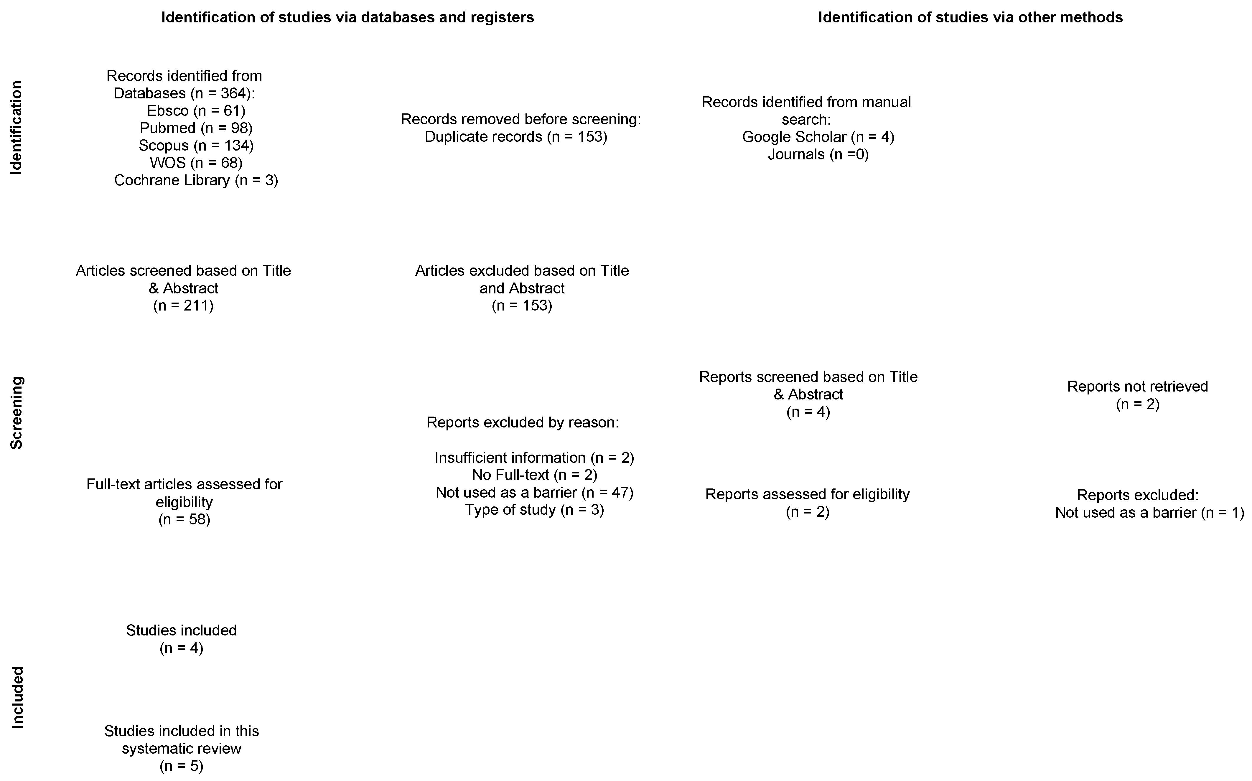
Figure 1. PRISMA flow diagram.
Prosthesis 07 00101 g001
Figure 2. Risk of bias of the included studies [19,20,21,22,23].
Prosthesis 07 00101 g002
Table 1. Characteristics of the included studies.
Article and CountryStudy DesignSamples and GenderMean Age (Range)Bone Defect AreaPEEK Barrier
Characteristics
Bone PerforationsFilling MaterialCover MaterialPostoperative Endpoint/
Removal Time
Bone GainedComplications
Nakahara et al., 2010 [19]
(Japan)
Animal model
(in vivo study)
30 female Wistar rats10 weeksLeft femur (n = 30)PEEK scaffolds with a tubular structure
Outer diameter = 5 mm
Inner diameter = 3 mm
Height = 5 mm
Perforations = 4 (3 × 1.5 mm)
Thickness = NR
Fixation method = external fixator and four pins of 1.4 mm in diameter
NoG1: Peptide hydrogel (n = 10)
G2: Autologous bone (n = 10)
G3: Empty (n = 10)
No28 daysAverage bridging ratios of bone defect:
- G1: 78.9 ± 11.8%
- G2: 96.5 ± 4.7%
- G3: 29.5 ± 9.7%
NR
Li et al., 2022 [20]
(China)
Animal model
(in vivo study)
3 Beagle dogs2 yearsMandible (bilateral premolars and first molars 7 × 7 mm defects)
(n = 18)
PEEK barriers with a bone defect shape
Perforations = 2.0 mm
Thickness = 0.6 mm
Fixation method = external fixator and 4 pins of 1.4 mm in diameter
NoG1: PEEK barrier with calcined bovine bone + autogenous bone (n = 6)
G2: Titanium mesh with autologous bone (n = 6)
G3: Collagen pericardium membrane and calcined bovine bone + autogenous bone (n = 6)
No3 monthsRegenerated bone:
- G1: 50.18 ± 7.26%
- G2: 52.62 ± 3.61%
- G3: 41.90 ± 5.20%
Alveolar bone height:
- G1: 0.29 ± 0.31 mm
- G2: 0.28 ± 0.32 mm
- G3: 0.99 ± 0.21 mm
NR
Mounir et al., 2019 [21]
(Egypt)
Human study
(RCT)
G1: 8 patients (6M/2F)
G2: 8 patients
(4M/4F)
G1: 38.0 years
G2: 39.0 years
Severely atrophied anterior maxillary alveolar ridges
<6 mm in height
<2 mm in width
PEEK barriers with a bone defect shape
Perforations = Yes (P/B)
Thickness = 2 mm
Fixation method = 3–4 fixation screws
YesG1: Titanium mesh with a 50:50 mixture of autogenous (IC) and xenogenic bone (n = 8).
G2: PEEK barrier with 50:50 autogenous bone (IC) and xenogenic bone (n = 8).
Yes
(collagen membrane)
6 monthsThree-dimensional bone gain:
G1: 20.9 ± 13.3%
G1: 31.8 ± 22.7%
G1: 1 case
(mesh exposure)
G2: 1 case
(barrier exposure)
Gouda et al., 2023 [22]
(Egypt)
Human study
(RCT)
8 patients
(NR)
NRUnilateral severe bone defect in the maxilla
<6 mm in height.
<2 mm in width
PEEK barriers with a bone defect shape
Perforations: Yes (P/B)
Thickness = 2 mm
Fixation method: 3–4 micro titanium screws.
YesG1: Titanium mesh with 50:50 mixture of autogenous bone (IC) + xenogenic bone (n = 8).
G2: PEEK barrier with 50:50 mixture of autogenous bone (IC) + xenogenic bone (n = 8).
Yes
(collagen membrane)
6 monthsPercentage of newly formed bone:
G1: 26.25 ± 4.35%
G2: 19.5 ± 2.38%
.
G1: 1 case
(mesh exposure)
G2: 1 case
(barrier exposure)
El Morsy et al., 2020 [23]
(Egypt)
Human study
(Case series)
14 patients
(5M/9F)
29.0Unilateral severe bone defect in the maxilla
<6 mm in height
<2 mm in width
PEEK barriers with a bone defect shape
Perforations: No
Thickness = NR
Fixation method: micro titanium screws
YesPEEK barrier with 50:50 autogenous bone (chin or retromolar region) + xenogenic bone (n = 14).No6 monthsAverage horizontal bone gain: 3.42 ± 1.10 mm.
Average vertical bone gain: 3.47 ± 1.46 mm.
1 case
(barrier exposure).
1 case (very poor quality of bone and massive fibrointegration)
Note: B: Buccal; F: Female; G1: Group 1; G2: Group 2; G3: Group 3; IC: Iliac Crest; M: Male; NR: Not Reported; P: Palatal.
Disclaimer/Publisher’s Note: The statements, opinions and data contained in all publications are solely those of the individual author(s) and contributor(s) and not of MDPI and/or the editor(s). MDPI and/or the editor(s) disclaim responsibility for any injury to people or property resulting from any ideas, methods, instructions or products referred to in the content.