Article

Influence of Different Fiber-Reinforced Biobases on the Marginal Adaptation of Lithium Disilicate Overlay Restorations (A Comparative In Vitro Study)

by
Maareb Abdulraheem Nabat
* and
Alaa Jawad Kadhim
Department of Restorative and Aesthetic Dentistry, College of Dentistry, University of Baghdad, Baghdad 1417, Iraq
*
Author to whom correspondence should be addressed.
Prosthesis 2025, 7(3), 55; https://doi.org/10.3390/prosthesis7030055
Submission received: 11 April 2025 / Revised: 15 May 2025 / Accepted: 18 May 2025 / Published: 22 May 2025
(This article belongs to the Section Prosthodontics)

Abstract

Background/purpose: Fiber-reinforced materials are commonly used as biobases beneath indirect restorations, potentially affecting the seating and marginal accuracy of the restorations. This study intended to assess the impact of various biobase techniques on the marginal adaptation of lithium disilicate overlay restorations. Methods: Fifty sound maxillary first premolar teeth of comparable dimensions were prepared using a full-bevel overlay design (3 mm occlusal reduction) and allocated randomly to five groups as follows (n = 10): Group A, delayed dentin sealing; Group B, immediate dentin sealing using Optibond FL; Group C, immediate dentin sealing with a 1 mm flowable composite layer (Clearfil AP-X Flow); Group D, immediate dentin sealing followed by a 1 mm short-fiber-reinforced composite layer (everX Flow); and Group E, immediate dentin sealing coated with a 1 mm flowable composite layer reinforced with polyethylene Ribbond fibers. Digital impressions were obtained using a Medit i700 intraoral scanner, and the overlays were digitally designed via the Sirona inLab CAD software and milled via a four-axis milling machine. The overlays were luted with a preheated composite (Clearfil AP-X). Marginal gap assessments were conducted pre- and post-cementation via a digital microscope at 230× magnification. The data were statistically analyzed using a one-way analysis of variance and paired t-tests. Results: The one-way ANOVA disclosed no significant differences among the groups before or after cementation (p > 0.05). Conclusions: The presence or absence of fiber-reinforced biobases did not impact the marginal adaptation of the restorations; these biobases can be incorporated to optimize the mechanical behavior of indirect restorations without adversely affecting their seating accuracy. These findings suggest that fiber-reinforced and non-reinforced biobase techniques can be safely integrated into clinical adhesive protocols to enhance the mechanical performance of restorations without comprising their marginal adaptation.

1. Introduction

The restoration of posterior teeth with substantial coronal defects remains a common issue in dentistry and can be achieved with several treatment options [1]. Indirect restorations represent a beneficial solution, offering advantages such as an enhanced tooth contour and anatomic form, improved occlusal contacts, and superior mechanical characteristics [2,3]. Partial indirect restorations are regarded as conservative alternatives to full crown restorations [4,5].
Glass ceramic materials, especially lithium disilicate, are commonly preferred for indirect restorations, due to their unique mechanical properties and aesthetic appeal [6,7]. They bond adhesively to dentin; immediate dentin sealing (IDS) was intended to improve their adhesion. IDS can be performed using the following two approaches:
  • The dentin-bonding agent is applied immediately following cavity preparation [8,9];
  • A thin film of low-viscosity flowable composite resin is placed atop the bonded dentin [10].
A biobase is a highly bonded base that reduces stress and serves as the bonding interface for indirect restorations. According to the biomimetic dentistry school protocol, a biobase consists of deep margin elevation, IDS, a resin coat (RC), and a dentin-replacing composite [11,12].
Despite lithium disilicate’s high strength, it displays an inherent brittleness and an inadequate toughness [13]. This becomes a major concern for more extensive restorations in which brittle materials replace large amounts of dentin, due to its low fracture toughness compared to natural dentin [14]. To address this challenge, materials with a higher fracture toughness have been suggested as ideal replacements for the lost dentin and the dentin–enamel junction (DEJ) [15].
Fiber reinforcement has emerged as a promising solution. The biomimetic protocol advocates using leno wave ultrahigh-molecular-weight polyethylene (UHMWPE) Ribbond fibers to reinforce the compromised tooth structure and reduce and dissipate occlusal stresses [16].
Additionally, short-fiber-reinforced composite (SFRC) material, characterized by the incorporation of multidirectional fibers, has demonstrated a fracture toughness comparable to that of natural dentine, making it a viable substitute for dentin in structurally compromised teeth [15,17].
While numerous investigations have highlighted the beneficial role of fiber reinforcements in enhancing the fracture strength and improving the failure patterns of indirect restorations [18,19,20,21], their influence on the marginal accuracy of such restorations remains underexplored.
As far as we are aware, no existing research has systematically analyzed the impact of different biobase techniques on the marginal adaptation of indirect restorations. Thus, this research aimed to assess the impact of various biobase techniques on the marginal adaptation, seating, and potential marginal inaccuracies of indirect lithium disilicate overlay restorations. By addressing this knowledge gap, the findings may guide clinicians in adopting reinforcement strategies that can improve mechanical performance without compromising marginal adaptation, particularly in structurally compromised teeth. This study sought to inform clinical decision-making by clarifying whether fiber-reinforced biobases affect the precision of the restoration fit in adhesive protocols.

2. Materials and Methods

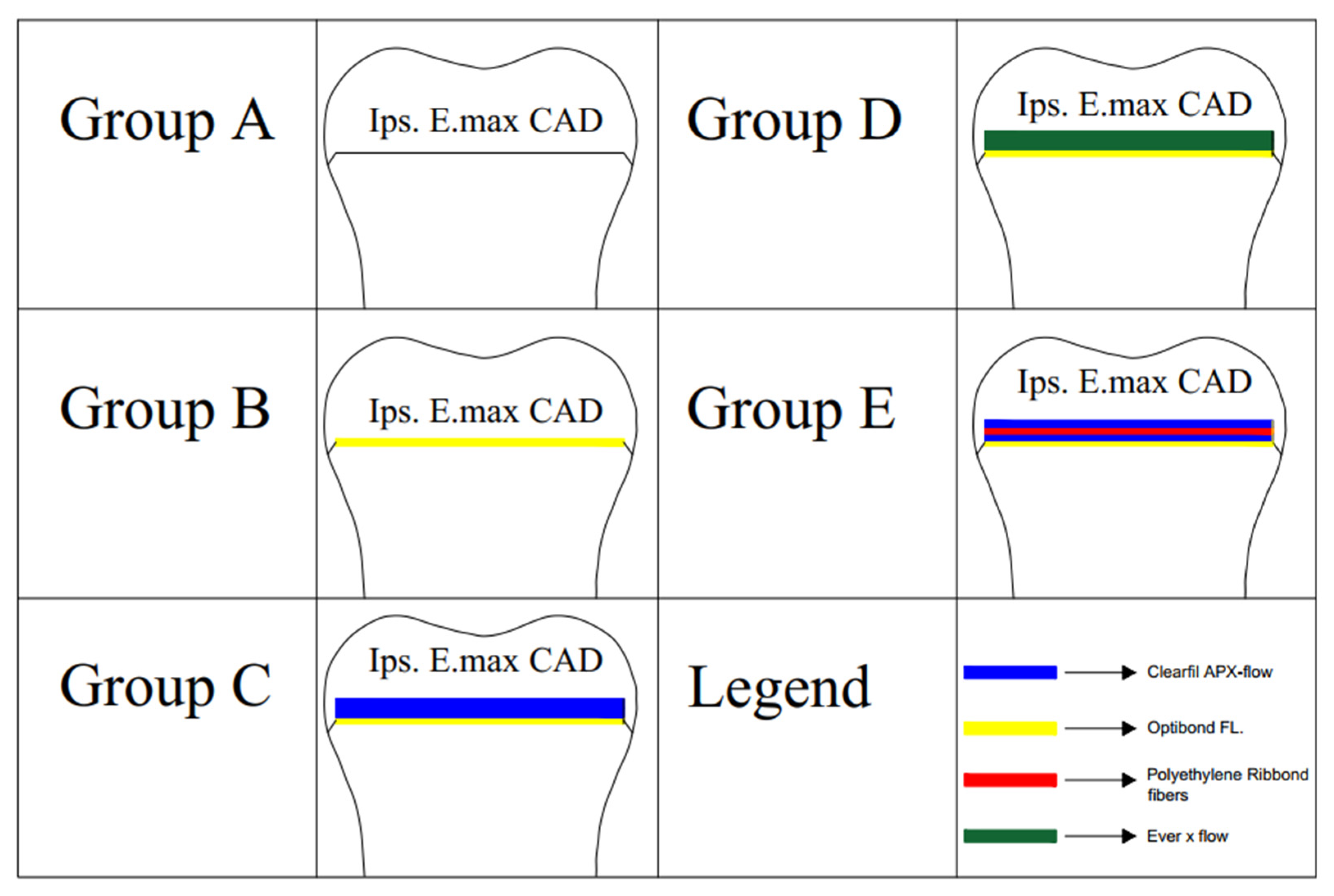
2.1. Tooth Selection and Grouping

The Committee of Research Ethics of Baghdad University, College of Dentistry, Iraq, approved this study in January 2024 (approval No. 895524). Fifty maxillary first premolar teeth with comparable dimensions were obtained from individuals between 18 and 22 years of age. The tooth samples were allocated randomly to five experimental groups (n = 10) on the basis of the biobase technique utilized as follows: Group A, delayed dentin sealing (DDS); Group B, IDS; Group C, IDS plus RC; Group D, IDS with an SFRC biobase; and Group E, IDS with a Ribbond-fiber-reinforced biobase.

2.2. Specimen Preparation and Biobase Application

A standardized full-bevel preparation design was utilized for all the samples to accommodate indirect overlay restorations, with an occlusal reduction of 3 mm, as shown in Figure 1. Following cavity preparation, dentin sealing was performed using Optibond FL (Kerr, Italy) immediately for all the groups except Group A (DDS), where dentin sealing was intentionally delayed. Immediate dentin sealing was followed for Group C by applying a 1 mm microfilled flowable composite layer (Clearfil AP-X Flow, Kurrary, Tokyo, Japan) over the sealed dentin, for Group D by incorporating a 1 mm SFRC layer (everX Flow/Dentin Shade, GC Europe, Leuven, Belgium) over the sealed dentin, and for Group E by initially applying an approximately 0.5 mm flowable composite layer (Clearfil AP-X Flow) atop the sealed dentin and leaving it uncured. The mesiodistal width of all the teeth was previously standardized during the sample selection phase, allowing for the use of a 4 mm Ribbond fiber to match the occlusal dimensions across all specimens. For each tooth, the suitable buccolingual length of the Ribbond strip was individually measured using a periodontal probe. The fiber was cut accordingly and presoaked in Ribbond Wetting Resin (Ribbond Ultra, Ribbond Inc., Seattle, WA, USA), with the excess resin gently removed. The fiber was then carefully adapted over the uncured resin in a buccolingual direction, gently pressed into place using a plastic instrument to ensure an even orientation, and light-cured for 20 s to secure its position. A second flowable composite layer was then added above the fiber-reinforced composite layer and light-cured, ensuring the complete encapsulation of the exposed fibers and a final biobase thickness of 1 mm. This layering protocol ensured that the fiber orientation remained planar, minimized folding, and contributed to a consistent fiber distribution and thickness across all specimens. The thickness was verified in each case using a periodontal probe. The experimental groups are illustrated in Figure 2.

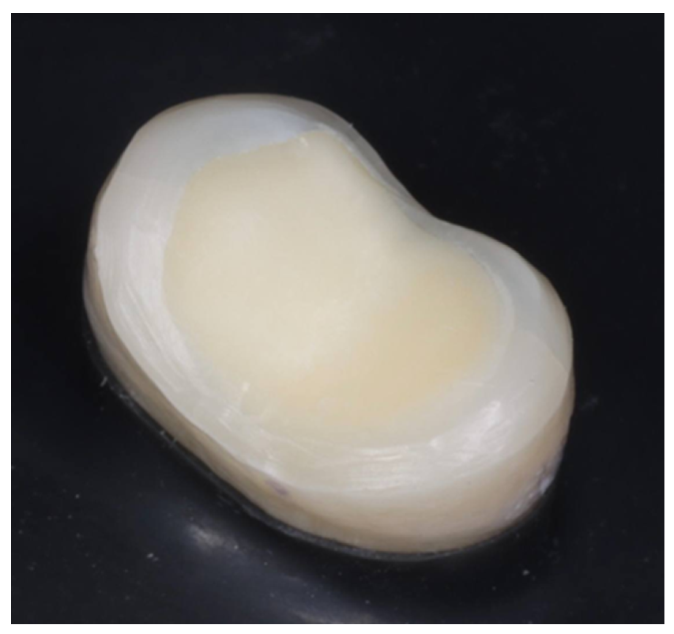
2.3. Digital Workflow and Overlay Fabrication

A Medit i700 intraoral scanner (Korea) was utilized for the prepared teeth scanning. Then, the temporary restorative material (Revotek LC, GC, Tokyo, Japan) was adapted. The overlay restorations were digitally designed via the Sirona inLab CAD software (version 20.0) and subsequently milled from (IPS e.max CAD, Ivoclar Vivadent, Schaan, Liechtenstein) lithium disilicate blocks via an inLab MC XL milling unit. The milled restorations underwent crystallization and glaze-firing in a P500 furnace at 840 °C.

2.4. Marginal Gap Assessment and Cementation

Each overlay was positioned onto its respective prepared tooth via a specially constructed specimen-holding device, ensuring stable placement under a standard fixed loading force of 5 kg (50 N) [22]. The marginal gap assessment was conducted by utilizing a direct viewing technique with a digital microscope (Dino-Lite Pro, AnMo Electronics Corp., New Taipei, Taiwan). High-resolution images that captured the marginal fit were obtained at 230× magnification.
To ensure a comprehensive evaluation, four reference points were marked on each tooth surface, covering the mesial, distal, buccal, and palatal aspects. Two of these reference points were precisely positioned at the midpoint of each surface, whereas the remaining two were spaced 1 mm mesially and distally from the midpoint [23].
Subsequently, image processing was performed using the Dino-Capture software (version 2.0), developed by AnMo Electronics Corp. The marginal gaps at each reference point were then measured using ImageJ 1.50i, a software tool developed by the National Institutes of Health in Bethesda, MD, USA.
At each of the sixteen total points (four per surface), three marginal gap measurements were performed, and the highest value was recorded to minimize operator variability. The mean of these sixteen maximum values was calculated and used as the representative of the pre-cementation marginal gap value for each specimen, as illustrated in Figure 3.
The restorations’ internal surfaces underwent 20 s of etching using hydrofluoric acid (etching gel > 5%, Ivoclar Vivadent), followed by a 15 s water rinse. Then, they were immersed in a 90% alcohol ultrasonic bath for 5 min to remove etching remnants. Afterwards, silane (Bisco Inc., Schaumburg, IL, USA) was placed for 20 s, and the mixture was subjected to drying at 100 °C (212 °F) for 2 min using a hair dryer.
After removing the provisional restoration, the prepared tooth surfaces were cleaned and reactivated using an AquaCare airborne particle abrasion device (UK). The surfaces were treated with 50 µm aluminum oxide airborne particles for 5 s under 15 and 2 mm bar pressures.
Next, 37% phosphoric acid (Kerr, Italy) was applied to all the samples for 30 s, after which the samples were rinsed thoroughly and dried for 20 and 3 s. Only the samples in Group A (DDS) received an additional primer application for 15 s; they were then dried for 5 s. An adhesive resin was employed and gently brushed for 15 s without polymerization. The restorations were then cemented using a preheated microhybrid composite (Clearfil AP-X, Kurrary, Tokyo, Japan) that had been heated in an Ena heat composite heater (Micerium S.p.A, Avegno, Italy) at 68 °C for 15 min. Following the same pre-cementation measurement points, the post-cementation marginal gap was then calculated.

2.5. Statistical Analysis
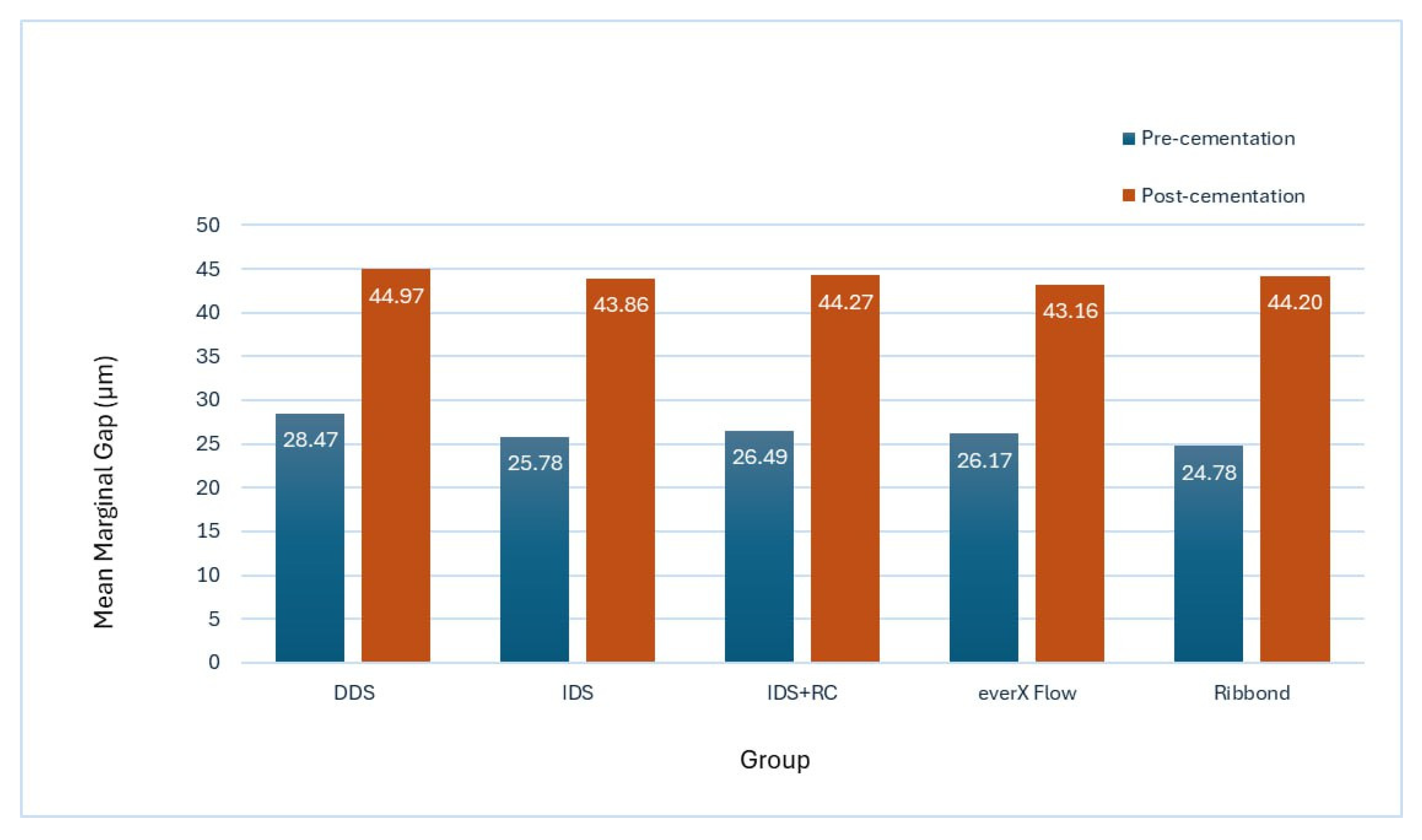
The SPSS statistical software, version 25.0 (IBM Crop., Armonk, NY, USA), was used for the statistical analyses. A one-way ANOVA and paired t-tests at a 0.05 significance level were employed for the data analyses.

3. Results

The one-way ANOVA tests presented a p-value of 0.479 and 0.981, demonstrating no significant differences among the groups before and after cementation (p > 0.05). The paired t-tests displayed a statistically significant change between the pre- and post-cementation marginal gap for each group (p < 0.05), as clarified in Table 1 and Figure 4.

4. Discussion

The marginal discrepancy refers to the vertical distance between the preparation’s finishing line and the restoration’s cervical margin [24]. Ensuring an optimal marginal adaptation is critical, as it prevents bacterial and fluid infiltration, thereby reducing the risk of periodontal complications and secondary caries [25,26,27]. Various fiber reinforcement materials have been incorporated as biobases to enhance fracture strength, and the failure mode of indirect restorations may impact the restoration’s seating accuracy, leading to marginal inaccuracies [28].
The present study assessed the marginal adaptation of lithium disilicate overlay restorations with various biobase techniques (DDS, IDS, IDS + RC, IDS + everX Flow, and IDS + Ribbond). The findings indicated that the biobase techniques employed had no statistically significant effect on the marginal adaptation at any stage, before or after cementation, and the inclusion of fiber-reinforced biobases did not compromise the external adaptation of the restorations compared to the non-reinforced groups.
Measurement of the vertical marginal gap is the predominant technique for evaluating the precision of a restoration’s fitness [22,29]. In this study, the mean marginal gaps of each sample were calculated to establish the overall mean marginal value, following previous research methodologies [23,30,31]. This approach was used to analyze the design as a single bulk instead of dividing the results. A digital microscope employing a direct viewing technique was utilized for marginal gap measurements. This method was chosen due to its cost-effectiveness, non-destructive nature, time efficiency, and ability to minimize the errors associated with multi-step procedures [32,33,34].
There is no universal agreement on the lowest marginal gap value that is considered clinically acceptable [35,36]. Some researchers suggest a value of <100 μm, while others propose <120 μm [37,38]. Another perspective places an acceptable value between 80 and 85 μm [39]. In this study, all the pre- and post-cementation vertical marginal gap measurements remained within the clinically acceptable limits for all groups.
Several factors influence the marginal discrepancy, including the preparation design, ceramic type, restorative material, fabrication method, spacer thickness, scanning method and exactness, software, processing machine characteristics, measurement techniques, and the number of measurement points [40]. The use of a full-bevel preparation design in this study probably contributed to achieving a favorable marginal adaptation. Previous research by Ferraris et al. demonstrated that a full-bevel design yielded a superior marginal fit compared to other designs, such as a butt joint [38]. The gradual transition from an interproximal to an axial preparation reduced stress concentration and enhanced restoration seating, thereby minimizing the marginal discrepancy.
Furthermore, all the restorations in this study were fabricated via a computer-aided design/computer-aided manufacturing (CAD/CAM) workflow, ensuring consistency and precision. The spacer was adjusted to a thickness of 100 μm, with the marginal adhesive interface at 0 μm, a parameter that has been shown to enhance the marginal adaptation, as indicated in previous research [41]. The use of fine milling burs in the manufacturing process likely enhanced the restoration’s accuracy, further reducing potential marginal discrepancies [42]. The use of lithium disilicate, a material known for its excellent physical properties and high marginal accuracy, was another contributing factor [43]. In addition, the milling procedure with the CEREC MC XL milling unit (four-axis) has been shown to yield restorations with minimum marginal gaps and a superior internal fit, aligning with our findings [44]. These factors, in combination, may have contributed to the favorable marginal adaptation detected in our results.
Assessing the pre-cementation marginal gap eliminates confounding variables; however, it is not entirely practical, as the cementation process significantly influences the final outcome [45]. In this study, a significant increase in the marginal gaps was detected across all the experimental groups post-cementation, which aligns with previous findings indicating that adhesive cementation may prevent the complete seating of the restoration due to the hydraulic pressure developed during the restoration’s seating [46,47]. This increase in post-cementation values is consistent with earlier investigations [31,48,49].
While this study aligns with the finding of Rocca et al. [50], who demonstrated no significant impact of fiber reinforcement on marginal adaptation, methodological differences should be considered. Their study examined CAD/CAM composite resin endocrowns reinforced with everX posterior and EverStickNet fibers, whereas this study focused on lithium disilicate restorations reinforced with Ribbond and everX Flow. Similarly, Sabet [51] reported that incorporating a fiber-reinforced composite beneath indirect endocrown restorations did not affect the marginal adaptation compared to non-reinforced groups. Monaco et al. [18] also concluded that the presence or absence of fiber reinforcement beneath overlays did not affect the marginal adaptation; however, their study placed the reinforcement at the bottom of the restoration, whereas in this study, the fiber reinforcement was applied to the prepared tooth surface. Additionally, Ovul et al. [52] demonstrated that using an EverStick-fiber-reinforced composite layer did not negatively impact the marginal fit of indirect composite resin inlays compared to the non-reinforced counterparts.
The absence of the effect of fiber reinforcement on marginal adaptation can open the scope to clinical applications, given the potential benefits of fiber reinforcement in enhancing the fracture strength and failure mode. Nevertheless, a key limitation of this research is the absence of oral environment simulation. Future investigations should emphasize the evaluation of the effect of the intraoral environment on the marginal accuracy of overlay restorations with various biobase techniques. This would offer better insight into the clinical viability of these approaches and ascertain the possible challenges associated with their use in real-world settings.

Clinical Implications of the Current Research

This study’s findings indicate that various biobase techniques, including those reinforced with fibers, do not adversely affect the marginal adaptation of lithium disilicate overlays. This supports their use in clinical situations, particularly for reinforcing restorations in structurally compromised teeth. These results may inform restorative strategies where an enhanced mechanical performance is desired without compromising the sealing precision or marginal integrity.

5. Conclusions

The presence or absence of fiber-reinforced biobases did not significantly impact the marginal adaptation of lithium disilicate overlay restorations. Fiber-reinforced biobases can be incorporated to optimize the mechanical behavior of indirect restorations without adversely affecting the restoration’s seating or marginal fit. Based on these in vitro findings, further clinical investigations are encouraged to validate their performance and applicability under functional conditions.

Author Contributions

Conceptualization, M.A.N. and A.J.K.; methodology, M.A.N. and A.J.K.; validation, M.A.N. and A.J.K.; formal analysis, M.A.N.; investigation, M.A.N.; resources, M.A.N.; data curation, M.A.N. and A.J.K.; writing—original draft preparation, M.A.N.; writing—review and editing, M.A.N. and A.J.K.; visualization, M.A.N. and A.J.K.; supervision, A.J.K.; project administration, M.A.N.; funding acquisition, M.A.N. All authors have read and agreed to the published version of the manuscript.

Funding

This research received no external funding.

Institutional Review Board Statement

The Research Ethics Committee of Baghdad University, College of Dentistry, approved this study (reference number: 895) on 11 January 2024 (protocol No. 895524). All the methods were developed according to the Helsinki Declaration.

Informed Consent Statement

The collected premolars were extracted for therapeutic causes not related to this study, and a prior consent form was obtained from all the patients for the donation of their extracted teeth for research goals.

Data Availability Statement

The data analyzed in this study are available from the corresponding author upon reasonable request due to privacy.

Conflicts of Interest

The authors declare no conflicts of interest.

References

  1. Gracis, S.; Thompson, V.P.; Ferencz, J.L.; Silva, N.R.; Bonfante, E.A. A new classification system for all-ceramic and ceramic-like restorative materials. Int. J. Prosthodont. 2015, 28, 227–235. [Google Scholar] [CrossRef] [PubMed]
  2. Angeletaki, F.; Gkogkos, A.; Papazoglou, E.; Kloukos, D. Direct versus indirect inlay/onlay composite restorations in posterior teeth: A systematic review and meta-analysis. J. Dent. 2016, 53, 12–21. [Google Scholar] [CrossRef]
  3. Nikaido, T.; Inoue, G.; Takagaki, T.; Takahashi, R.; Sadr, A.; Tagami, J. Resin coating technique for protection of pulp and increasing bonding in indirect restoration. Curr. Oral Health Rep. 2015, 2, 81–86. [Google Scholar] [CrossRef]
  4. Tseng, C.W.; Yong, C.Y.; Fang, C.Y.; Lee, S.Y.; Wang, Y.C.; Lin, W.C. Comparative analysis of bonding strength between enamel and overlay of varying thicknesses following an aging test. J. Dent. Sci. 2023, 18, 1850–1858. [Google Scholar] [CrossRef]
  5. Tafuri, G.; D’Addazio, G.; Santilli, M.; Argentieri, G.; Murmura, G.; Caputi, S.; Sinjari, B. Overlay Preparation Accuracy: An In Vitro Study on the Influence of Magnification and Operator Expertise. Prosthesis 2025, 7, 5. [Google Scholar] [CrossRef]
  6. Hofsteenge, J.W.; Bresser, R.A.; Buijs, G.J.; van der Made, S.A.; Özcan, M.; Cune, M.S.; Gresnigt, M.M. Clinical performance of bonded partial lithium disilicate restorations: The influence of preparation characteristics on survival and success. J. Dent. 2024, 142, 104828. [Google Scholar] [CrossRef]
  7. Al-Johani, H.; Haider, J.; Satterthwaite, J.; Silikas, N. Lithium silicate-based glass ceramics in dentistry: A narrative review. Prosthesis 2024, 6, 478–505. [Google Scholar] [CrossRef]
  8. Garoushi, S.; Säilynoja, E.; Vallittu, P.K.; Lassila, L. Fracture-behavior of CAD/CAM ceramic crowns before and after cyclic fatigue aging. Int. J. Prosthodont. 2021, 23, 1–8. [Google Scholar]
  9. Magne, P. Immediate dentin sealing: A fundamental procedure for indirect bonded restorations. J. Esthet. Restor. Dent. 2005, 17, 144–154. [Google Scholar] [CrossRef]
  10. de Andrade, O.S.; de Goes, M.F.; Montes, M.A. Marginal adaptation and microtensile bond strength of composite indirect restorations bonded to dentin treated with adhesive and low-viscosity composite. Dent. Mater. 2007, 23, 279–287. [Google Scholar] [CrossRef]
  11. Magne, P.; Spreafico, R.C. Deep margin elevation: A paradigm shift. Am. J. Esthet. Dent. 2012, 2, 86–96. [Google Scholar]
  12. Frese, C.; Wolff, D.; Staehle, H.J. Proximal box elevation with resin composite and the dogma of biological width: Clinical R2-technique and critical review. Oper. Dent. 2014, 39, 22–31. [Google Scholar] [CrossRef] [PubMed]
  13. Kruzic, J.J.; Arsecularatne, J.A.; Tanaka, C.B.; Hoffman, M.J.; Cesar, P.F. Recent advances in understanding the fatigue and wear behavior of dental composites and ceramics. J. Mech. Behav. Biomed. Mater. 2018, 88, 504–533. [Google Scholar] [CrossRef] [PubMed]
  14. Forster, A.; Braunitzer, G.; Tóth, M.; Szabó, B.P.; Fráter, M. In vitro fracture resistance of adhesively restored molar teeth with different MOD cavity dimensions. J. Prosthodont. 2019, 28, e325–e331. [Google Scholar] [CrossRef]
  15. Garoushi, S.; Säilynoja, E.; Vallittu, P.K.; Lassila, L. Physical properties and depth of cure of a new short fiber reinforced composite. Dent. Mater. 2013, 29, 835–841. [Google Scholar] [CrossRef]
  16. Belli, S.; Orucoglu, H.; Yildirim, C.; Eskitascioglu, G. The effect of fiber placement or flowable resin lining on microleakage in Class II adhesive restorations. J. Adhes. Dent. 2007, 9, 175. [Google Scholar]
  17. Başaran, E.G.; Ayna, E.; Vallittu, P.K.; Lassila, L.V. Load bearing capacity of fiber-reinforced and unreinforced composite resin CAD/CAM-fabricated fixed dental prostheses. J. Prosthet. Dent. 2013, 109, 88–94. [Google Scholar] [CrossRef]
  18. Monaco, C.; Bortolotto, T.; Arena, A.; Krejci, I. Restoring nonvital premolars with composite resin onlays: Effect of different fiber-reinforced composite layers on marginal adaptation and fracture load. J. Adhes. Dent. 2015, 17, 567–574. [Google Scholar]
  19. Magne, P.; Milani, T. Short-fiber reinforced MOD restorations of molars with severely undermined cusps. J. Adhes. Dent. 2023, 25, 99–106. [Google Scholar] [CrossRef]
  20. Otero, C.A.Y.; Bijelic-Donova, J.; Saratti, C.M.; Vallittu, P.K.; di Bella, E.; Krejci, I.; Rocca, G.T. The influence of FRC base and bonded CAD/CAM resin composite endocrowns on fatigue behavior of cracked endodontically-treated molars. J. Mech. Behav. Biomed. Mater. 2021, 121, 104647. [Google Scholar] [CrossRef]
  21. Fildisi, M.A.; Eliguzeloglu Dalkilic, E. The effect of fiber insertion on fracture strength and fracture modes in endocrown and overlay restorations. Microsc. Res. Tech. 2022, 85, 1799–1807. [Google Scholar] [CrossRef] [PubMed]
  22. Mansi, Z.M.; Al-Shamma, A.M.; Saleh, A.R. Assessment of marginal adaptation of indirect zirconia crown restorations using self-adhesive resin cements modified with polylysine antibacterial particles. J. Baghdad Coll. Dent. 2024, 36, 50–60. [Google Scholar] [CrossRef]
  23. Khaledi, A.-A.; Farzin, M.; Akhlaghian, M.; Pardis, S.; Mir, N. Evaluation of the marginal fit of metal copings fabricated by using 3 different CAD-CAM techniques: Milling, stereolithography, and 3D wax printer. J. Prosthet. Dent. 2020, 124, 81–86. [Google Scholar] [CrossRef]
  24. Holmes, J.R.; Bayne, S.C.; Holland, G.A.; Sulik, W.D. Considerations in measurement of marginal fit. J. Prosthet. Dent. 1989, 62, 405–408. [Google Scholar] [CrossRef]
  25. Falahchai, M.; Rahimabadi, S.; Khabazkar, G.; Babaee Hemmati, Y.; Neshandar Asli, H. Marginal and internal fit and fracture resistance of three-unit provisional restorations fabricated by additive, subtractive, and conventional methods. Clin. Exp. Dent. Res. 2022, 8, 1404–1412. [Google Scholar] [CrossRef]
  26. Saadallah, S.; Al-Azzawi, A.K.J. The influence of different fabrication and impression techniques on the marginal adaptation of lithium disilicate crowns (A comparative in vitro study). J. Baghdad Coll. Dent. 2017, 29, 20–26. [Google Scholar] [CrossRef]
  27. Al-Ramadan, A.; Abualsaud, R.; Al-Dulaijan, Y.A.; Al-Thobity, A.M.; Alalawi, H. Accuracy and fit of ceramic filled 3D-printed resin for permanent crown fabrication: An in vitro comparative study. Prosthesis 2024, 6, 1029–1041. [Google Scholar] [CrossRef]
  28. Yousif, Y.H.; Kadhim, A.J. The effect of immediate dentin sealing on the marginal adaptation of lithium disilicate overlay restorations using different types of luting agents. Brazil. Dent. Sci. 2023, 26, e3925. [Google Scholar] [CrossRef]
  29. Wolfart, S.; Wegner, S.M.; Al-Halabi, A.; Kern, M. Clinical evaluation of marginal fit of a new experimental all-ceramic system before and after cementation. Int. J. Prosthodont. 2003, 16, 587. [Google Scholar]
  30. Ibraheem, A.F.; Abdulkareem, A.M. Comparison of the marginal fitness of the ceramic crowns fabricated with different CAD/CAM systems (An in vitro study). J. Baghdad Coll. Dent. 2016, 28, 28–33. [Google Scholar]
  31. Athab Hasan, S.; Mohammed-Hussain Abdul-Ameer, Z. Effect of three different preparation designs on the marginal adaptation of indirect overlay restoration fabricated from lithium disilicate ceramic material: An in-vitro comparative study. Saudi Dent. J. 2023, 35, 372–377. [Google Scholar] [CrossRef] [PubMed]
  32. Hmedat, S.J. A Comparison of Vertical Marginal Fit of Three Different Types of All-Ceramic Crown Restorations. Ph.D. Thesis, University of Baghdad, Baghdad, Iraq, 2012. [Google Scholar]
  33. Gonzalo, E.; Suárez, M.J.; Serrano, B.; Lozano, J.F. Marginal fit of zirconia posterior fixed partial dentures. Int. J. Prosthodont. 2008, 21, 398–399. [Google Scholar] [PubMed]
  34. Khdeir, R.M.; Ibraheem, A.F. The marginal fitness of CAD/CAM all ceramic crowns constructed by two types of direct digitization techniques (an in vitro study). J. Baghdad Coll. Dent. 2016, 28, 30–33. [Google Scholar] [CrossRef]
  35. Kim, D.Y.; Song, H.Y. Method of predicting human mobility patterns using deep learning. Neurocomputing 2018, 280, 56–64. [Google Scholar] [CrossRef]
  36. Goujat, A.; Abouelleil, H.; Colon, P.; Jeannin, C.; Pradelle, N.; Seux, D.; Grosgogeat, B. Marginal and internal fit of CAD-CAM inlay/onlay restorations: A systematic review of in vitro studies. J. Prosthet. Dent. 2019, 121, 590–597. [Google Scholar] [CrossRef] [PubMed]
  37. Alajaji, N.K.; Bardwell, D.; Finkelman, M.; Ali, A. Micro-CT Evaluation of Ceramic Inlays: Comparison of the Marginal and Internal Fit of Five and Three Axis CAM Systems with a Heat Press Technique. J. Esthet. Restor. Dent. 2017, 29, 49–58. [Google Scholar] [CrossRef]
  38. Ferraris, F.; Sammarco, E.; Romano, G.; Cincera, S.; Giulio, M. Comparison of posterior indirect adhesive restorations (PIAR) with different preparation designs according to the adhesthetics classification. Part 1: Effects on the fracture resistance. Int. J. Esthet. Dent. 2021, 16, 144. [Google Scholar]
  39. Abduo, J.; Sambrook, R.J. Longevity of ceramic onlays: A systematic review. J. Esthet. Restor. Dent. 2018, 30, 193–215. [Google Scholar] [CrossRef]
  40. Nawafleh, N.A.; Mack, F.; Evans, J.; Mackay, J.; Hatamleh, M.M. Accuracy and reliability of methods to measure marginal adaptation of crowns and FDPs: A literature review. J. Prosthodont. 2013, 22, 419–428. [Google Scholar] [CrossRef]
  41. Mohammed, Z.H.; Majeed, M.A. Effect of cementation protocol on the marginal adaptation of indirect overlay restorations fabricated from two different all-ceramic CAD/CAM materials. J. Res. Med. Dent. Sci. 2020, 8, 518–525. [Google Scholar]
  42. Skordou, T.; Kebi, L.; Osnes, C.; Keeling, A. The effect of two different milling instrument sets on CAD proposed cement thickness and fit surface of chairside CAD crowns. J. Osseointegr. 2021, 13, 271–278. [Google Scholar] [CrossRef]
  43. Hasaneen, F.A.; Mogahed, M.M. Comparative in-vitro study of marginal gap of four CAD/CAM all ceramic systems with thermal aging. Tanta Dent. J. 2021, 18, 12–19. [Google Scholar] [CrossRef]
  44. Pilecco, R.O.; da Rosa, L.S.; Baldi, A.; Machry, R.V.; Tribst, J.P.M.; Valandro, L.F.; Kleverlaan, C.J.; Scotti, N.; Pereira, G.K.R. How do different intraoral scanners and milling machines affect the fit and fatigue behavior of lithium disilicate and resin composite endocrowns? J. Mech. Behav. Biomed. Mater. 2024, 155, 106557. [Google Scholar] [CrossRef] [PubMed]
  45. Abdulla, H.A.; Majeed, M.A. Assessment of bioactive resin-modified glass ionomer restorative as a new CAD/CAM material. Part I: Marginal fitness study. Indian J. Forensic Med. Toxicol. 2020, 14, 865–870. [Google Scholar] [CrossRef]
  46. Mounajjed, R.; Salinas, T.J.; Ingr, T.; Azar, B. Effect of different resin luting cements on the marginal fit of lithium disilicate pressed crowns. J. Prosthet. Dent. 2018, 119, 975–980. [Google Scholar] [CrossRef] [PubMed]
  47. Habib, S.R.; Ali, M.; Al Hossan, A.; Majeed-Saidan, A.; Al Qahtani, M. Effect of cementation, cement type and vent holes on fit of zirconia copings. Saudi Dent. J. 2019, 31, 45–51. [Google Scholar] [CrossRef]
  48. Falahchai, M.; Babaee Hemmati, Y.; Neshandar Asli, H.; Neshandar Asli, M. Marginal adaptation of zirconia-reinforced lithium silicate overlays with different preparation designs. J. Esthet. Restor. Dent. 2020, 32, 823–830. [Google Scholar] [CrossRef]
  49. Guess, P.C.; Vagkopoulou, T.; Zhang, Y.; Wolkewitz, M.; Strub, J.R. Marginal and internal fit of heat pressed versus CAD/CAM fabricated all-ceramic onlays after exposure to thermo-mechanical fatigue. J. Dent. 2014, 42, 199–209. [Google Scholar] [CrossRef]
  50. Rocca, G.T.; Saratti, C.M.; Poncet, A.; Feilzer, A.J.; Krejci, I. The influence of FRCs reinforcement on marginal adaptation of CAD/CAM composite resin endocrowns after simulated fatigue loading. Odontology 2016, 104, 220–232. [Google Scholar] [CrossRef]
  51. Sabet, A. Effect of filling pulp chamber on marginal adaptation and fracture resistance of zirconia reinforced glass ceramic endocrown restorations. Egypt. Dent. J. 2017, 63, 3833–3842. [Google Scholar] [CrossRef]
  52. Ovul, K.; Arzu, T.-M.; Ahmet, S.; Vj, L.L. Marginal adaptation and microleakage of directly and indirectly made fiber reinforced composite inlays. Open Dent. J. 2011, 5, 33. [Google Scholar] [CrossRef] [PubMed]
Figure 1. Prepared tooth surface.
Prosthesis 07 00055 g001
Figure 2. Experimental groups.
Prosthesis 07 00055 g002
Figure 3. Marginal gap measurements. (a) Points of measurements; (b) digital microscope attached to the computer.
Prosthesis 07 00055 g003
Figure 4. Bar chart displaying the mean marginal gap values for each group before and after cementation.
Prosthesis 07 00055 g004
Table 1. The mean marginal gap pre- and post-cementation using a one-way ANOVA test and paired t-tests.
Pre-CementationPost-Cementation
GroupMean ± SDPaired t-TestGroupMean ± SDPaired t-Test
A (DDS)28.47 ± 4.310.00A (DDS)44.97 ± 6.120.00
B (IDS)25.78 ± 4.60.00B (IDS)43.86 ± 6.330.00
C (IDS + RC)26.48 ± 4.860.00C (IDS + RC)44.27 ± 6.080.00
D (everX Flow)26.17 ± 4.690.00D (everX Flow)43.15 ± 6.540.00
E (Ribbond)24.77 ± 4.220.00E (Ribbond)44.2 ± 7.510.00
One-way ANOVA0.479 (NS) One-way ANOVA0.981 (NS)
DDS, delayed dentin sealing; IDS, immediate dentin sealing; RC, resin coating; SD, standard deviation; NS, non-significance.
Disclaimer/Publisher’s Note: The statements, opinions and data contained in all publications are solely those of the individual author(s) and contributor(s) and not of MDPI and/or the editor(s). MDPI and/or the editor(s) disclaim responsibility for any injury to people or property resulting from any ideas, methods, instructions or products referred to in the content.

