Article

A Retrospective Analysis on Marginal Bone Loss around Tilted and Axial Implants in Immediate-Loaded All-On-4 with a Long-Term Follow-Up Evaluation

by
Francesco Tironi
1,*,
Francesco Orlando
2,
Francesco Azzola
1,3,
Stefano Corbella
1,3,4 and
Luca Angelo Francetti
1,3
1
Department of Biomedical, Surgical and Dental Sciences, University of Milan, 20122 Milan, Italy
2
Private Practice, 20122 Milan, Italy
3
IRCCS Istituto Ortopedico Galeazzi, 20122 Milan, Italy
4
Department of Oral Surgery, Institute of Dentistry, I. M. Sechenov First Moscow State Medical University, 119992 Moscow, Russia
*
Author to whom correspondence should be addressed.
Prosthesis 2022, 4(1), 15-23; https://doi.org/10.3390/prosthesis4010002
Submission received: 28 December 2021 / Revised: 13 January 2022 / Accepted: 17 January 2022 / Published: 24 January 2022

Abstract

:
Objectives: The aim of this retrospective study is to assess whether axial and tilted implants supporting All-on-4 prosthesis show any differences in terms of survival rate, success rate and marginal bone loss (MBL) after a long-term follow-up (mean 9 years). Material and Methods: One hundred and fifty-six implants were included in this study, 78 of which were tilted (Group A) and 78 were axial (Group B). MBL was measured after a mean time of 9 years on periapical radiographs. Success and survival rate were assessed with the Misch criteria. The prevalence of peri-implantitis was calculated. Statistical analysis was conducted to assess comparisons between groups. A Kaplan–Meyer analysis was carried out for the survival rate. Results: A total of 156 implants were analyzed. After a 9-year mean time follow-up, the survival rate was 96.2% in group A and 98.7% in group B; and the success rate was 80.8% in group A and 74.4% in group B. The mean MBL was 1.2 mm (IQR 0.6–1.8) in group A and 1.4 mm (IQR 0.9–2.1) in group B. No statistically significant differences were shown between the two groups (p < 0.05). Peri-implantitis occurred in 15 implants and was equally distributed between the two groups. Conclusions: This study shows that axial and tilted implants have similar success rates, survival rates and MBL values after a long-time follow-up, assessing the biological reliability of the prosthesis they supported. Peri-implantitis occurred equally between the two groups.

1. Introduction

Implant-supported prosthesis have been used for many years in order to rehabilitate full-edentulous patients, and their validation is well reported through scientific literature [1,2]. The original Toronto bridge rehabilitation used five parallel, inter-foraminal implants in order to avoid damage to anatomical structures such as the inferior alveolar nerve and the maxillary sinus. The first evolution of this technique can be found in a paper published by Krekmanov et al. The distal, tilted implants allowed for better prosthetic support and the placing of longer implants with better bone anchorage, suitable conditions for immediate loading [3]. Furthermore, in 2001, Aparicio et al. proposed tilted implants as a solution to avoid sinus augmentation in the upper jaw, with a survival rate of 100% after 5 years [4]. Moreover, the idea of immediate load goes back to the early 1990s. Lazarof proposed immediate loading on expandable implants in 1992 [5]. A definitive protocol that combines these concepts consists of the All-on-4 technique, first proposed by Malo to rehabilitate the mandible and later the upper jaw [6,7]. This technique made it possible to diminish the number of implants utilized, decreasing distal cantilever and aiming to reduce prosthetics complications [8]. As previously said, placing tilted implants in the inter-foraminal area allows for the avoidance of major bone regenerations; maxillary distal implants are tilted in proximity of the mesial wall of the maxillary sinus, taking advantage of the cortical bone to obtain stability and direction; mandibular distal implant inclination avoids the emergence of the nerve and the possible presence of a loop. In conclusion, it can be stated that placing tilted implants is supported by literature [9]. These clinical investigations stimulated the interest of the scientifical community and were later validated by finite element analysis. Whilst tilted implants did not demonstrate increased bone stress outside physiological levels when compared to axial implants, they showed significant biomechanical advantages [10,11]. Considering that this technique is still relatively recent, the aim of this study was to investigate the value of marginal bone loss (MBL), along with the survival and success rate, and the occurrence of peri-implantitis on implants placed with the All-on-4 technique.

2. Material and Methods

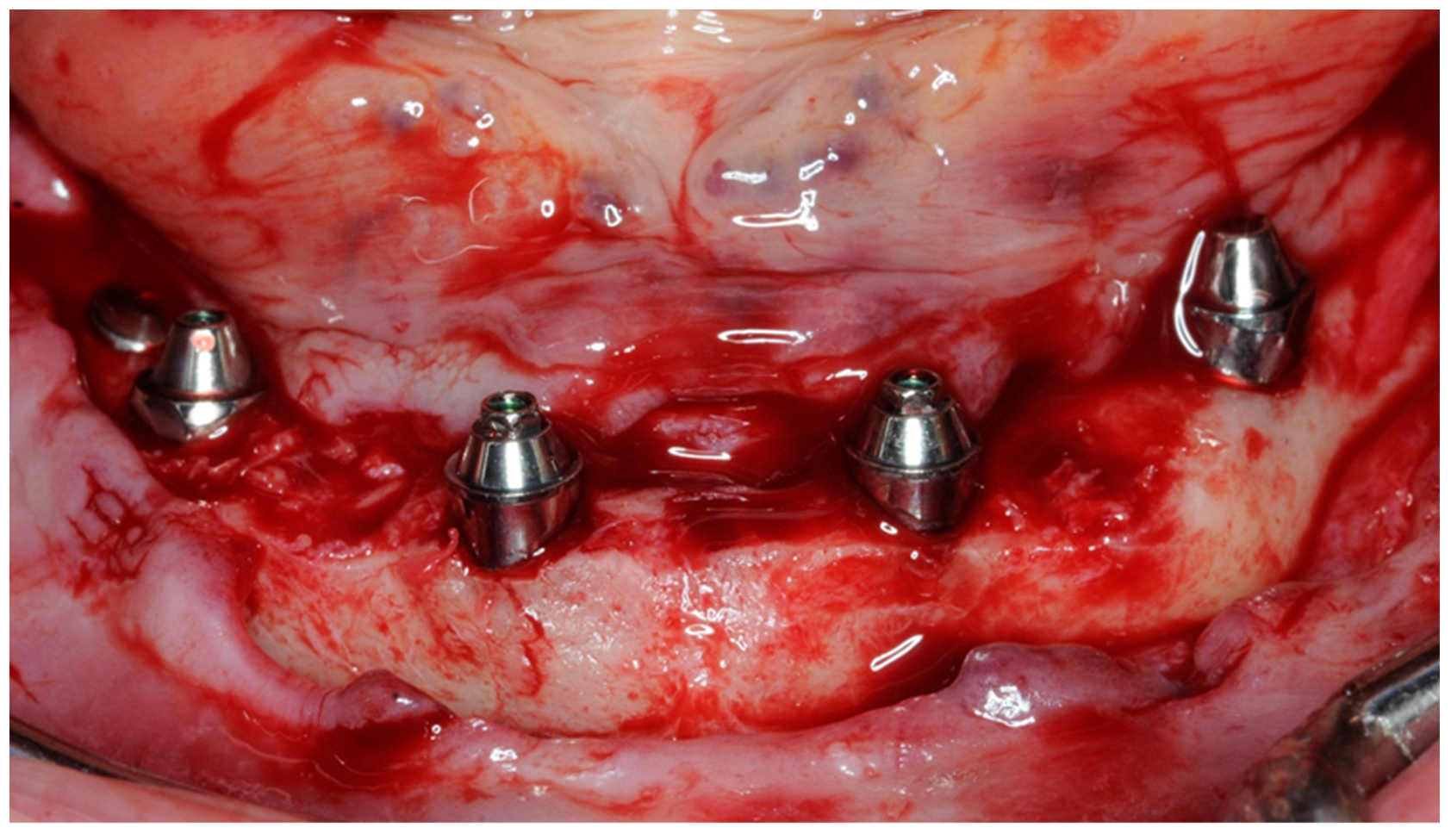
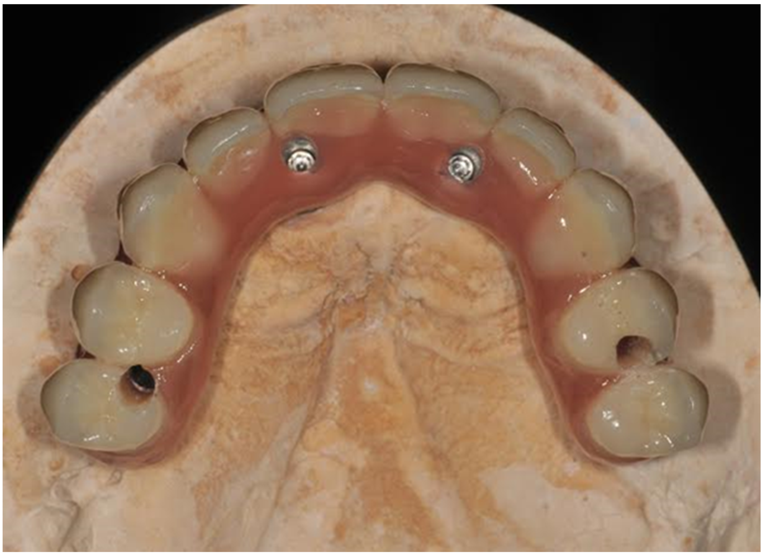
2.1. Patient Selection, Surgical and Prosthetic Procedure

To obtain updated data, only patients that showed up for a control during the last year were included. Thirty-four patients were hence included. They were treated with the All-on-4 technique in our institute (IRCCS Istituto Ortopedico Galeazzi, University of Milan). Post-extractive implants were included. The exclusion criteria for the surgery and for this study were:
  • Presence of systemic pathologies that contraindicated surgery.
  • Presence of untreated periodontal disease.
  • Smokers > 10/die.
Patients were prepared for surgery with a professional oral hygiene session at least one week before the surgical operation. Patients started mouth-rinsing three days before the intervention with a chlorhexidine 0.2% solution. Antibiotic prophylaxis was given to the patients (2 g amoxicillin + clavulanic acid). Mouth-rinsing continued for 7 days, and antibiotics continued for 6 days. A full-thickness flap was raised in order to expose the underlying bone and the anatomical structures to protect. Distal implants were placed with a 17° or 30° direction, and multi-unit abutment (MUA) with a consequent inclination was then fixed (Figure 1). To provide optimal stability for the immediate loading, implants were placed with an insertion torque of at least 35 N/Cm. An impression was then obtained, and a screw-retained provisional prosthesis was delivered within 48 h of the surgery (Figure 2). Weekly controls were carried out for the first month. Final prothesis was delivered after 6 months, and after the delivery a control was conducted every 4 months.

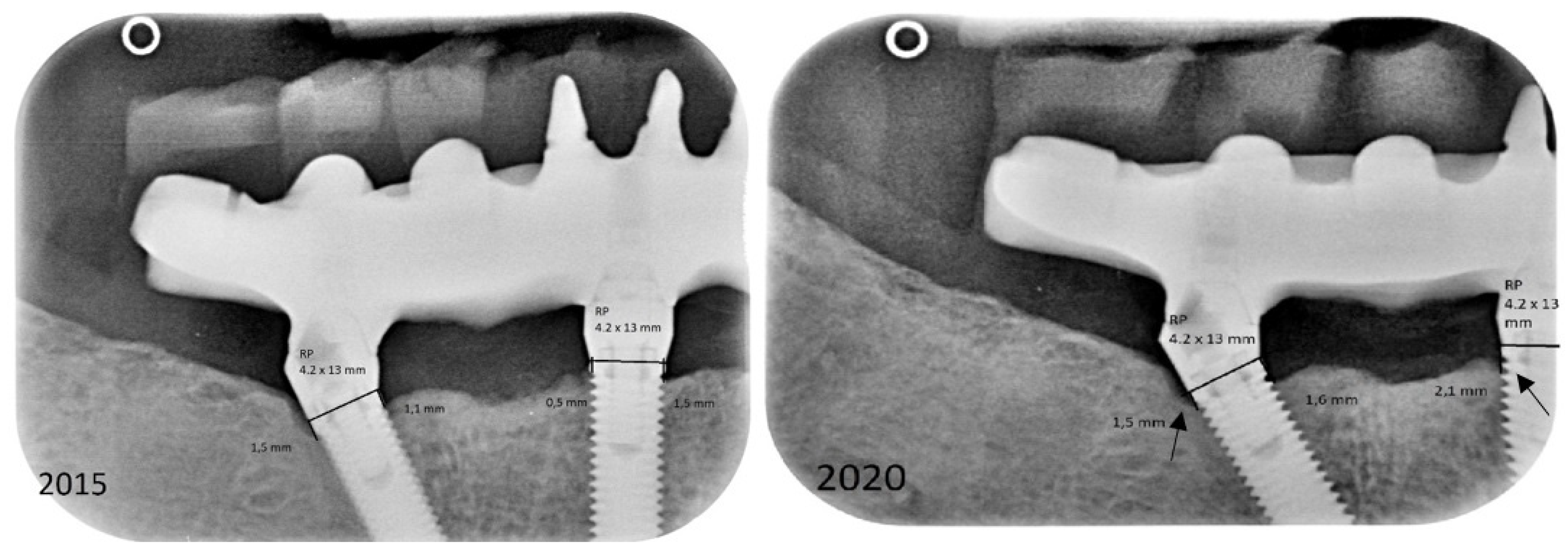
2.2. Data and Radiographic Assessment

The following data were collected on an excel table by an independent operator (F.O.) not previously involved in the treatment of the patients: follow-up years, age of patients, pathologies, smoking habits, previous diagnosis of periodontitis, implants placed in upper/lower jaw. The radiographic success rate was calculated using the Misch criteria: an implant was considered successful when stable, not painful and with less than 2 mm of MBL [12]. In addition, the presence of peri-implantitis was an exclusion criterion for success. The World Workshop on the Classification of Periodontal and Peri-Implant Diseases and Conditions in 2018 was used to define peri-implantitis [13]. Implant length was confirmed on patients’ folders and inter-thread distance was used to calibrate distances on image elaboration software (ImageJ—National Institutes of Health). Radiographs were saved with a minimum of 600 DPI quality on a personal computer. MBL was then measured on both mesial and distal aspect and a mean value was calculated. The MBL was defined as the distance in millimeters, parallel to the axis of the implant, between the implant shoulder and the first contact apically between bone and implant surface (Figure 3).

2.3. Data Analysis

Data were recorded in Excel and checked for entry errors. Analysis was conducted at patient level. Correlation tests were performed through Pearson/Spearman coefficient measures for continuous variables or Chi-square for categorical variables. A p-Value < 0.05 was considered statistically significant. Kaplan–Meyer analysis was used to evaluate survival rate and implant loss was used as the event.

3. Results

3.1. Demographic and Clinical Variables

Thirty-four patients (15 males and 19 females; mean age of 64 years) received 39 total rehabilitations (156 implants) between 2006 and 2016 and attended an average follow-up of 9 years (5–14 year range). Detailed statistics referring to group A and group B are reported in Table 1.
At the last follow-up, 152 implants were still in function, whilst 4 implants failed (2.6%), with an overall survival rate of 97.4% (Figure 4). Group A and B survival rates were 96.2% and 98.2%, respectively. One implant was removed due to lack of osseointegration and three were removed because of peri-implantitis. Thirty-five implants did not match the MBL expected by the Misch criteria, thus the overall success rate was 77.6%.
Fifteen implants suffered from peri-implantitis, distributed between group A and B.
Empirical data on plaque accumulation was obtained from the clinical folders.

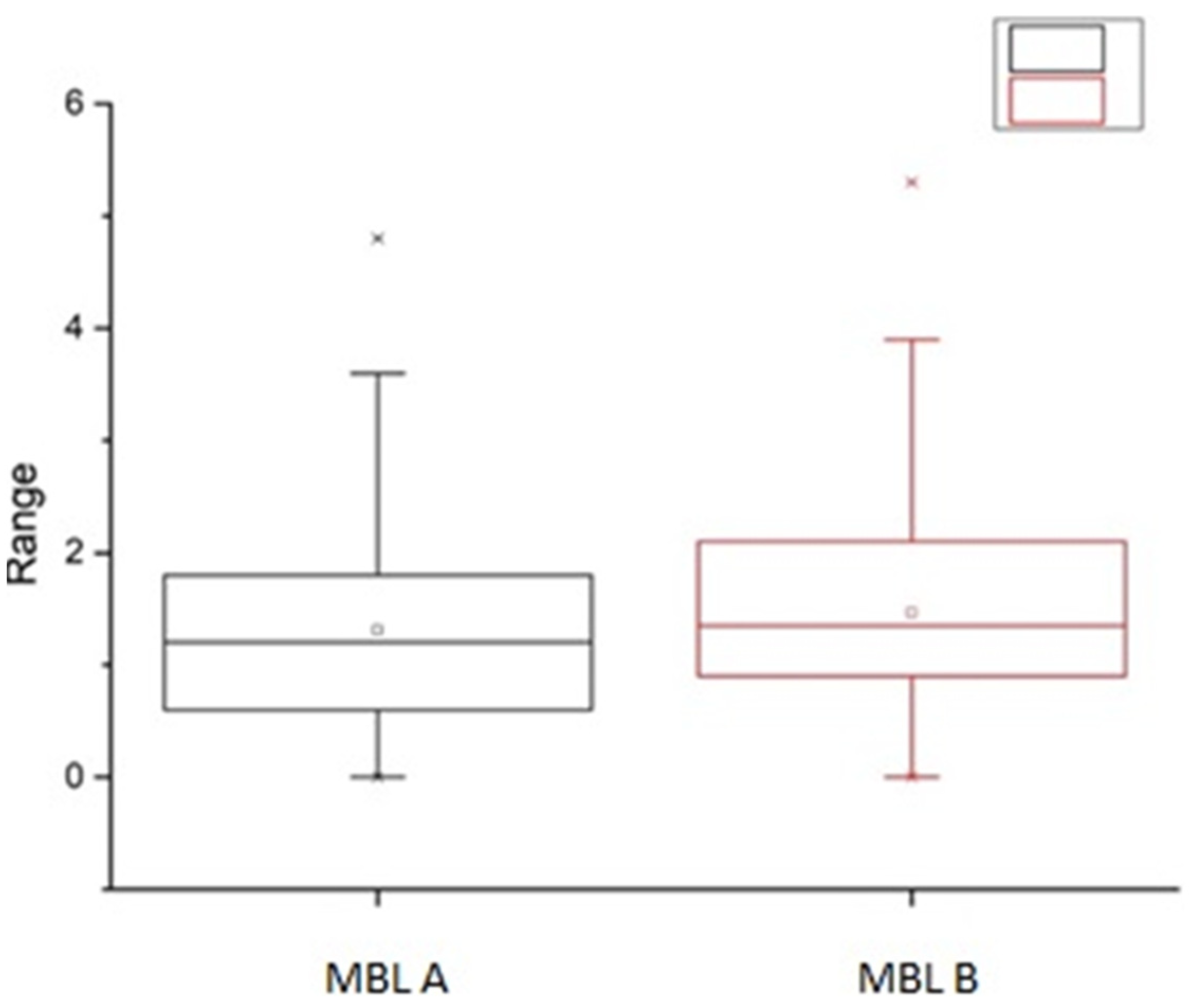
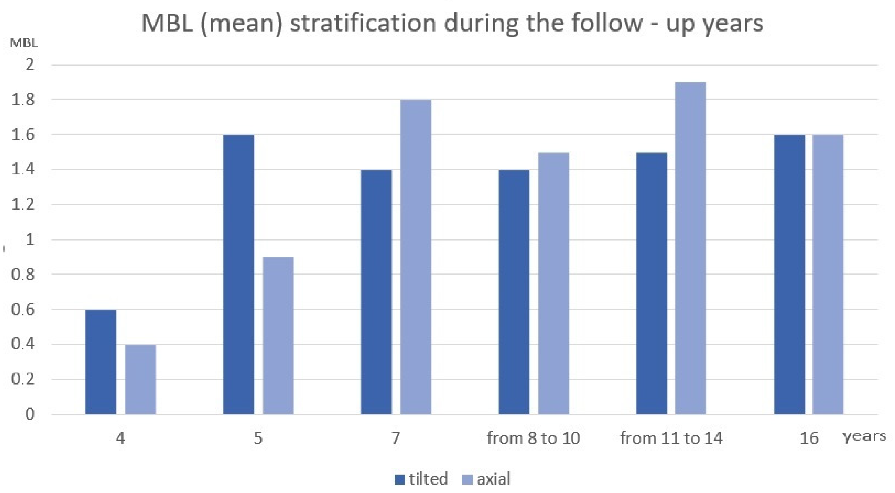
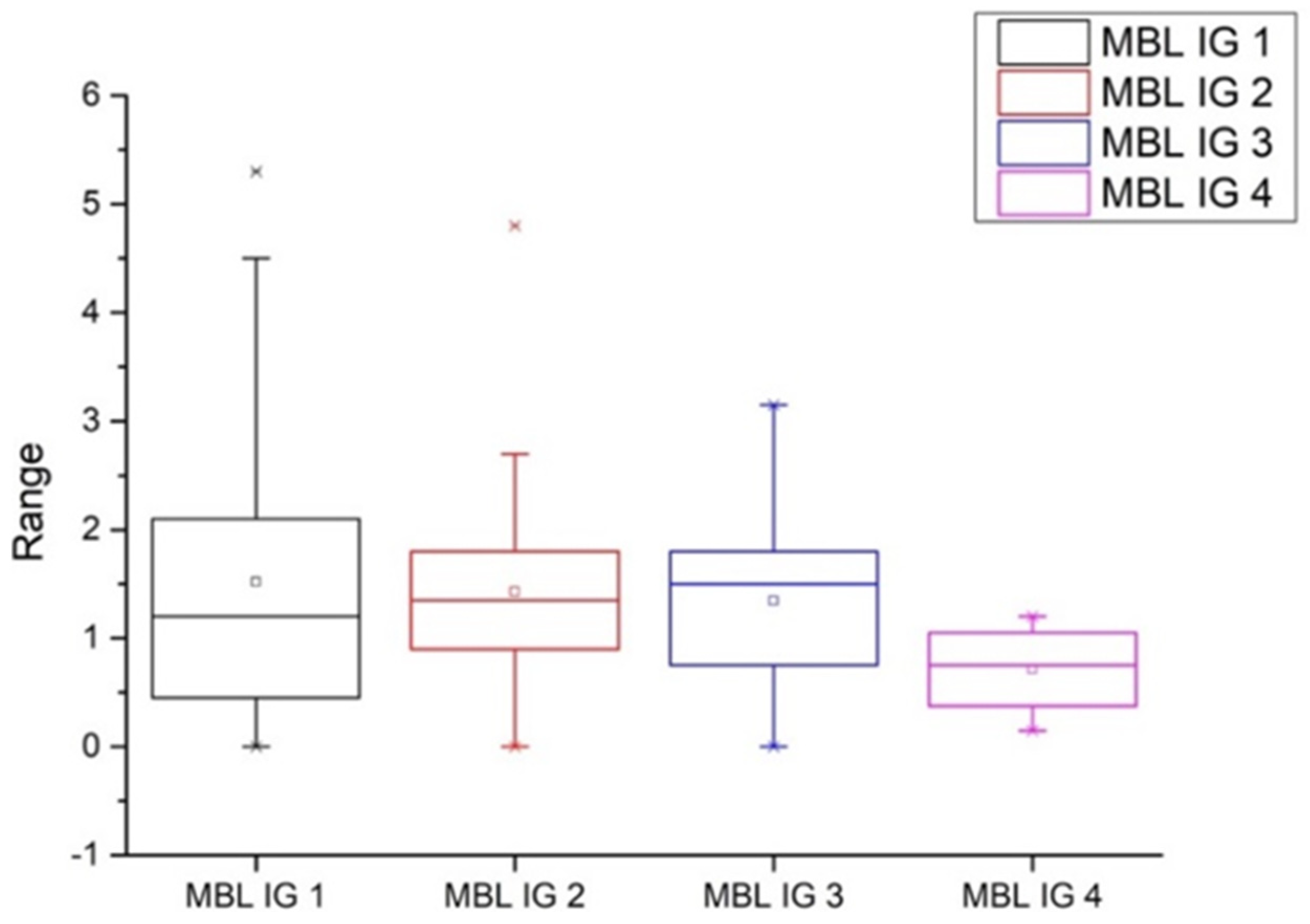
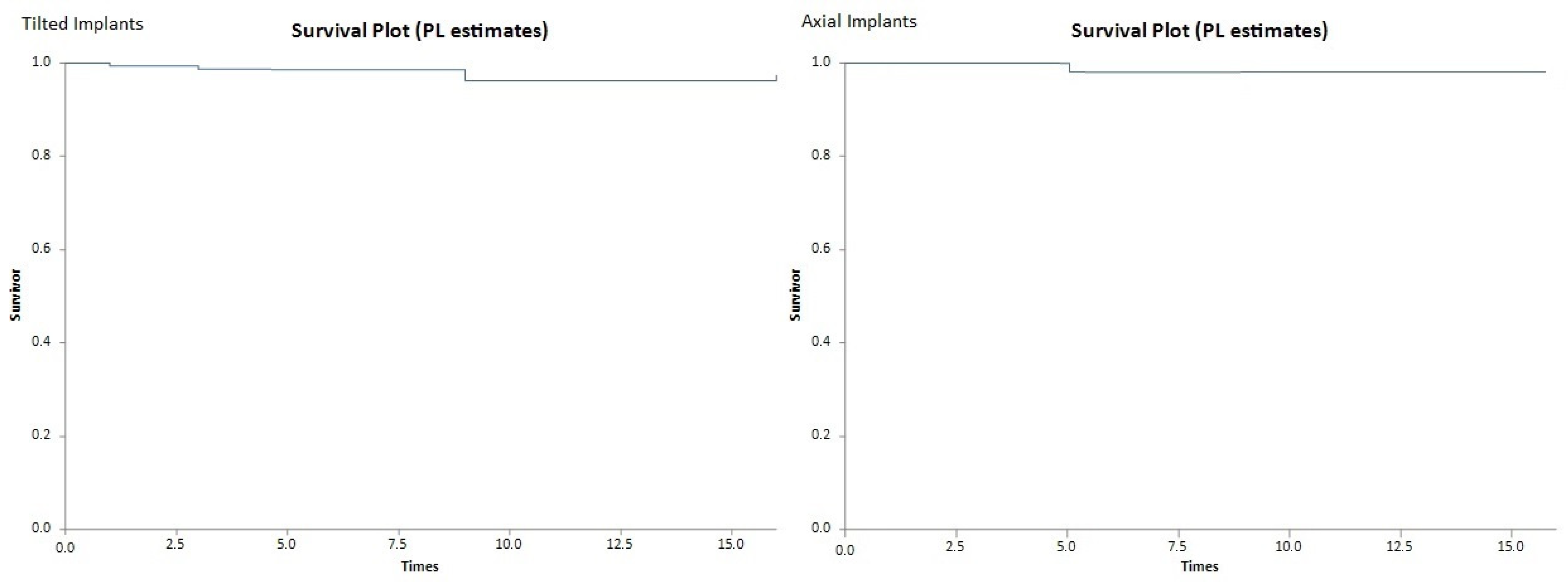
3.2. Radiographic Variables

Radiographic MBL values of group A and B, for maxillary/mandibular, expressed with their mean, interquartile range and standard deviation, are reported in Table 2. Within the rehabilitations and implant samples, there were no statistically significant differences between the levels of MBL in the Group A and B (p = 0.2) (Figure 5). MBL stratification in different follow-up years is reported in Figure 6. In addition, no statistical differences were found in MBL between implants placed in maxillary and in mandibular bone (p = 0.9). Mean MBL after overall follow-up time was 1.3 mm ± 1.06 (range of 0.1–5.3).
Different scores of oral hygiene were established in our department on an empirical basis (0 = bad, 1 = low, 2 = decent, 3 = good, 4 = optimal) (Table 3). Between the scores, no statistically significant difference in relation to MBL was observed. (Table 3) (Figure 7).

4. Discussion

This paper investigated the MBL, survival and success rate of tilted and axial implants after a long follow-up time. The idea behind this paper originates from the fact that studies on this specific topic were published first when the scientific community was triggered by the innovation of angulated implants and researchers tried to investigate their reliability; however, after this initial enthusiasm, studies with longer follow-ups diminished and are nowadays low in numbers. To better explain this concept, a meta-analysis that appeared on PubMed last September selected 10 papers published between 2007 and 2014 and only one published in 2017; furthermore, among these, only seven were eligible for an MBL analysis [14].
The All-on-4 technique has been used in our department since 2004. As previously mentioned, the use of tilted implants is well documented in the literature and has been investigated by both clinical studies and systematic reviews. The reason for these investigations lies in the original concept of placing dental implants axially, in a way that may bear occlusal stresses; thus, angulated implants were initially approached with skepticism. However, the All-on-4 technique requires the use of such angulated implants, and its reliability is reported in the literature as well [9,15,16,17].
The main aim of this study was to investigate whether tilted and axial implants showed differences in terms of MBL. In this paper, means of 1.2 and 1.4 mm of MBL were found in groups A and B, respectively. No statistically significant differences were found regarding MBL between the two groups, as shown in Figure 5. This corroborates data found in the literature in studies with shorter follow-up times and in the aforementioned meta-analysis [14,18,19]. In another systematic review published in 2018, all studies included presented “minuscule differences in MBL”. The author also reported that “greater variability was seen with mean MBL for tilted implants ranging between 0.4 and 2.0 mm and mean MBL for straight implants ranging between 0.5 and 1.9 mm” [20]. Interestingly, this quoted article also reported that “the extent of angulation for the tilted implants was significantly associated with MBL, with an additional 0.6 mm of MBL being seen for every additional 10° of implant tilting”. Such a consideration was not found in other papers on the same subject. Our group did not investigate such a variability; a further study will be needed.
As for success, the Misch criteria were chosen over Albrektsson criteria because the former gives a cut-off of 2 mm for success, which is not present in the latter; we think that this cut-off could be more useful when considering an important outcome such as the success rate. Given this explanation, thirty-five implants did not match the Misch criteria for implant success. However, it can be stated that the follow-up time of the present paper is rather long, especially when compared to the time of introduction of the All-on-4 technique, and a certain amount of bone resorption was expected. Observing MBL stratification as reported in Figure 6, the mean bone loss, even after 10 years of follow-ups, appears to be stable. Similarly, implants analyzed in other papers showed a tendency to stabilize around the fixtures, suggesting radiographical reliability even after a long time [21]. These findings are satisfying, considering the long follow-up time and the kind of prosthesis that sometimes might be challenging for patients that struggle with hygiene maneuvers.
Survival rates of both group A and B were high and consistent with the values usually provided for implants supporting single crowns of fixed bridges. In a paper published in 2018 with a very long follow-up time, straight, machined implants presented a survival rate of 97.7% [21]. These values were also consistent with data provided in the aforementioned meta-analysis.
Higher levels of bone loss were found in patients presenting peri-implantitis. Overall, 9.6% of the implants were affected by peri-implantitis and the incidence appeared to be equally distributed between group A and B. In a previous paper by our research group in 2018, peri-implantitis was assessed in 13.08% of implants after 10 years; it was also observed that after 5 years, 46.1% of the total cases of peri-implantitis were among tilted implants [17]. This minimal difference between the present paper and the paper published in 2018 can be addressed by the consistent difference of the cohorts, with the former being almost half of the latter. As reported by Alccayhuaman, there is a lack of data in the literature about the prevalence of peri-implantitis when comparing axial and tilted implants; weak deductions can thus be made [20].
The actual prevalence of this pathology is still debated in the literature, since authors consider different criteria for diagnostics, ending up with a bright range of percentages in several studies. It appears that oxidized-surface implants are more prone to present peri-implantitis, especially after 7 years [22]. Regarding plaque scores, no statistically significant difference in relation to the MBL have been observed. It must be specified, however, that the data about plaque was gathered using an index which is not strictly supported by literature. As we collected data from the clinical folders, however, it was decided to add this part of the analysis to the paper, as it was thought that it would have added interesting clinical information for clinicians. It is possible to notice an average decrease of MBL when the level of oral hygiene decreases (Figure 7). Studies including a larger cohort of patients and with a stricter plaque accumulation scale could give more information about this correlation. Particular attention should be put into this part of the treatment. We did not present any data correlation between plaque and peri-implantitis. However, the participants in this study attended a strict hygiene recall program, which consisted of a professional oral hygiene appointment every 4 months after the removal of the Toronto prosthesis. We deducted that this protocol allowed clinicians and hygienists to diagnose peri-implant inflammation before this could evolve into more serious complications. In fact, only three implants were lost due to this factor whilst the others were treated and still supported the prosthesis at the final follow-up time.

5. Conclusions

Within the limits of this study, axial and tilted implants placed according to the All-on-4 technique have similar patterns in terms of MBL, success and survival rates. The technique is a reliable choice for the treatment of both the upper and lower jaw with a long-term follow-up. More studies are needed to investigate MBL and success rate with a larger cohort of subjects.

Author Contributions

Conceptualization, F.T.; Data curation, F.T. and F.O.; Supervision, F.A., S.C. and L.A.F.; Validation, S.C.; Writing—original draft, F.T. and F.O.; Writing—review & editing, F.T. All authors have read and agreed to the published version of the manuscript.

Funding

This research received no external funding.

Institutional Review Board Statement

Not applicable.

Informed Consent Statement

Informed consent was obtained from all subjects involved in the study.

Data Availability Statement

Readers interested in original data can contact directly the corresponding author.

Conflicts of Interest

The authors declare no conflict of interest.

References

  1. Jemt, T.; Johansson, J. Implant Treatment in the Edentulous Maxillae: A 15-Year Follow-Up Study on 76 Consecutive Patients Provided with Fixed Prostheses. Clin. Implant. Dent. Relat. Res. 2006, 8, 61–69. [Google Scholar] [CrossRef] [PubMed]
  2. Åstrand, P.; Ahlqvist, J.; Gunne, J.; Nilson, H. Implant Treatment of Patients with Edentulous Jaws: A 20-Year Follow-Up. Clin. Implant. Dent. Relat. Res. 2008, 10, 207–217. [Google Scholar] [CrossRef] [PubMed]
  3. Krekmanov, L.; Kahn, M.; Rangert, B.; Lindström, H. Tilting of posterior mandibular and maxillary implants for improved prosthesis support. Int. J. Oral Maxillofac. Implant. 2000, 15, 405–414. [Google Scholar]
  4. Aparicio, C.; Perales, P.; Rangert, B. Tilted implants as an alternative to maxillary sinus grafting: A clinical, radiologic, and periotest study. Clin. Implant Dent. Relat. Res. 2001, 3, 39–49. [Google Scholar] [CrossRef]
  5. Lazarof, S. Immediate-load implant quickens integration. Dent. Today 1992, 11, 44–45. [Google Scholar]
  6. Maló, B.R.; Miguel, N. “All-on-Four” immediate-function concept with Brånemark System implants for completely edentulous mandibles: A retrospective clinical study. Clin. Implant Dent. Relat. Res. 2003, 5, 2–9. [Google Scholar] [CrossRef]
  7. Malo, P.; Rangert, B.; Nobre, M.D.A. All-on-4 Immediate-Function Concept with Branemark SystemR Implants for Completely Edentulous Maxillae: A 1-Year Retrospective Clinical Study. Clin. Implant Dent. Relat. Res. 2005, 7, s88–s94. [Google Scholar] [CrossRef]
  8. Bellini, C.M.; Romeo, D.; Galbusera, F.; Taschieri, S.; Raimondi, M.; Zampelis, A.; Francetti, L. Comparison of tilted versus nontilted implant-supported prosthetic designs for the restoration of the edentuous mandible: A biomechanical study. Int. J. Oral Maxillofac. Implant. 2009, 24, 511–517. [Google Scholar]
  9. Del Fabbro, M.; Ceresoli, V. The fate of marginal bone around axial vs. tilted implants: A systematic review. Eur. J. Oral. Implant. 2014, 7 (Suppl. 2), 171–189. [Google Scholar]
  10. Antonios, Z.; Bo, R. Lars Heijl Tilting of splinted implants for improved prosthodontic support: A two-dimensional finite element analysis. J. Prosthet. Dent. 2007, 97 (Suppl. 6), S35–S43. [Google Scholar]
  11. Erdem, K. Ozge Doganay Evaluation of Stress in Tilted Implant Concept With Variable Diameters in the Atrophic Mandible: Three-Dimensional Finite Element Analysis. J. Oral. Implantol. 2020, 46, 19–26. [Google Scholar]
  12. Misch, C.E.; Perel, M.L.; Wang, H.-L.; Sammartino, G.; Galindo-Moreno, P.; Trisi, P.; Steigmann, M.; Rebaudi, A.; Palti, A.; Pikos, M.A.; et al. Implant Success, Survival, and Failure: The International Congress of Oral Implantologists (ICOI) Pisa Consensus Conference. Implant Dent. 2008, 17, 5–15. [Google Scholar] [CrossRef] [Green Version]
  13. Caton, J.G.; Armitage, G.; Berglundh, T.; Chapple, I.L.; Jepsen, S.; Kornman, K.S.; Mealey, B.L.; Papapanou, P.N.; Sanz, M.; Tonetti, M.S. A new classification scheme for periodontal and periimplant diseases and conditions—Introduction and key changes from the 1999 classification. J. Periodontol. 2018, 89 (Suppl. 1), S1–S8. [Google Scholar] [CrossRef] [PubMed]
  14. Mehta, S.P.; Sutariya, P.V.; Pathan, M.R.; Upadhyay, H.H.; Patel, S.R.; Kantharia, N.D.G. Clinical success between tilted and axial implants in edentulous maxilla: A systematic review and meta-analysis. J. Indian Prosthodont. Soc. 2021, 21, 217–228. [Google Scholar]
  15. Agliardi, E.; Clericò, M.; Ciancio, P.; Massironi, D. Immediate loading of full-arch fixed prostheses supported by axial and tilted implants for the treatment of edentulous atrophic mandibles. Quintessence Int. 2010, 41, 285–293. [Google Scholar] [PubMed]
  16. Luca Francetti, D.; Romeo, D.; Spasari, C.; Tassera, C.; Folegatti, T.; Testori, M.; Basso, E.; Agliardi, M.; Del, F. Immediate rehabilitation of edentulous mandible by all-on-four technique. Ital. Oral. Surg. 2008, 7, 7–18. [Google Scholar]
  17. Francetti, L.; Cavalli, N.; Taschieri, S.; Corbella, S. Ten years follow-up retrospective study on implant survival rates and prevalence of peri-implantitis in implant-supported full-arch rehabilitations. Clin. Oral. Implant. Res. 2019, 30, 252–260. [Google Scholar] [CrossRef] [PubMed]
  18. Francetti, L.; Romeo, D.; Corbella, S.; Taschieri, S.; Bsc, M.D.F. Bone Level Changes Around Axial and Tilted Implants in Full-Arch Fixed Immediate Restorations. Interim Results of a Prospective Study. Clin. Implant Dent. Relat. Res. 2010, 14, 646–654. [Google Scholar] [CrossRef]
  19. Cavalli, N.; Barbaro, B.; Spasari, D.; Azzola, F.; Ciatti, A.; Francetti, L. Tilted implants for full-arch rehabilitations in completely edentulous maxilla: A retrospective study. Int. J. Dent. 2012, 2012, 180379. [Google Scholar] [CrossRef]
  20. Apaza Alccayhuaman, K.A.; Soto-Peñaloza, D.; Nakajima, Y.; Papageorgiou, S.N.; Botticelli, D.; Lang, N.P. Biological and technical complications of tilted implants in comparison with straight implants supporting fixed dental prostheses. A systematic review and meta-analysis. Clin. Oral Implant. Res. 2018, 29 (Suppl. 18), 295–308. [Google Scholar] [CrossRef]
  21. Simion, M.; Nevins, M.; Rasperini, G.; Tironi, F. A 13- to 32-Year Retrospective Study of Bone Stability for Machined Dental Implants. Int. J. Periodontics Restor. Dent. 2018, 38, 489–493. [Google Scholar] [CrossRef] [PubMed]
  22. Ferrantino, L.; Tironi, F.; Pieroni, S.; Sironi, A.; Simion, M. A Clinical and Radiographic Retrospective Study on 223 Anodized Surface Implants with a 5- to 17-Year Follow-up. Int. J. Periodontics Restor. Dent. 2019, 39, 799–807. [Google Scholar] [CrossRef] [PubMed]
Figure 1. Implant position with MUA, showing the distribution of implant emergences.
Prosthesis 04 00002 g001
Figure 2. Provisional prosthesis. Notice that during provisional rehabilitation, posterior load is characterized by the absence of distal cantilever. Teeth n. 25 was thus shortened to eliminate any load.
Prosthesis 04 00002 g002
Figure 3. Example of how marginal bone loss analysis was conducted between T0 and T1. Notice the slight bone remodeling around the fixtures (arrows).
Prosthesis 04 00002 g003
Figure 4. Kaplan–Mayer survival analysis charts for tilted and axial implants.
Prosthesis 04 00002 g004
Figure 5. Mean MBL with IQR.
Prosthesis 04 00002 g005
Figure 6. MBL distribution at different follow-up years.
Prosthesis 04 00002 g006
Figure 7. Distribution and IQR of MBL associated to plaque scores assigned.
Prosthesis 04 00002 g007
Table 1. Population Statistics.
Implants
Patients34
N. of Rehabilitations39
N. of implants156
Maxillary rehab24
Mandibular rehab15
OverallGroup AGroup B
Survival rate %97.4%96.2%98.2%
Success rate %77.6%80.8%74.4%
Failed implants n (%)4 (2.6%)31
Peri-implantitis n (%)15 (9.6%)87
Table 2. MBL comparisons between group A and B.
MBL (mm)
Group AGroup B
Implants1. (IQR 1.9–2.1)1.4 (IQR 0.6–1.8)
Maxillary1.4 (SD 0.7)1.6 (SD 0.9)
Mandibular1.3 (SD 1)1.5 (SD 1)
Table 3. Hygiene index and MBL with inter quartile ranges (IQR).
N. of ImplantsMedian (mm)
IG1561.2 (IQR 0.45–2.1)
IG2481.35 (IQR 0.26–1.12)
IG3281.5 (IQR 0.67–1.18)
IG440.75 (IQR 0.26–1.2)
Not Reported20
Publisher’s Note: MDPI stays neutral with regard to jurisdictional claims in published maps and institutional affiliations.

