Article

Organic Farm Bedded Pack System Microbiomes: A Case Study with Comparisons to Similar and Different Bedded Packs

1
Department of Plant and Soil Science, University of Vermont, Burlington, VT 05405, USA
2
Department of Animal and Veterinary Sciences, University of Vermont, Burlington, VT 05405, USA
*
Author to whom correspondence should be addressed.
Dairy 2022, 3(3), 587-607; https://doi.org/10.3390/dairy3030042
Submission received: 9 June 2022 / Revised: 20 July 2022 / Accepted: 3 August 2022 / Published: 19 August 2022
(This article belongs to the Section Dairy Farm System and Management)

Abstract

:
Animal housing and bedding materials influence cow and farm worker exposure to microbial pathogens, biocontrol agents, and/or allergens. This case study represents an effort to characterize the bacterial and fungal community of bedding systems using an amplicon sequencing approach supplemented with the ecological assessment of cultured Trichocomaceae isolates (focusing on Penicillium and Aspergillus species) and yeasts (Saccharomycetales). Bedding from five certified organic dairy farms in northern Vermont USA were sampled monthly between October 2015 and May 2016. Additional herd level samples from bulk tank milk and two bedding types were collected from two farms to collect fungal isolates for culturing and ecology. Most of the microorganisms in cattle bedding were microbial decomposers (saprophytes) or coprophiles, on account of the bedding being composed of dead plant matter, cattle feces, and urine. Composition of bacterial and fungal communities exhibited distinct patterns of ecological succession measured through time and by bedding depth. Community composition patterns were related to management practices and choice of bedding material. Aspergillus and Penicillium species exhibited niche differentiation expressed as differential substrate requirements; however, they generally exhibited traits of early colonizers of bedding substrates, typically rich in carbon and low in nitrogen. Pichia kudriavzevii was the most prevalent species cultured from milk and bedding. P. kudriavzevii produced protease and its abundance directly related to temperature. The choice of bedding and its management represent a potential opportunity to curate the microbial community of the housing environment.

1. Introduction

Dairy cattle housing and bedding systems influence animal health, reproduction, milk quality, animal well-being, productivity, and farm profitability [1]. Understanding implications of microbiome relationships between the environment and the health of animals offers a holistic perspective on the effects of farm management. Microbiomes may link bedding, teat, rumen, and milk, each of which are affected by myriad other factors including diet, season, and the milking management system. In 2014, tie-stall and free-stall housing systems were the most common housing types for lactating cows among all dairy operations in the USA [2]. Bedded pack barns are receiving increased attention as a type of open communal housing for dairy cows [3,4].
Bedded pack systems use carbon-rich substrates such as wood shavings, sawdust, straw, or wood chips to create a comfortable and clean surface on which animals move freely [5]. Fecal matter is not removed when bedding material is renewed, in contrast with other housing systems [6]. Bedded pack systems can be coarsely grouped into two management strategies. “Deep bedded pack” (DPB) is an untilled system in which strata of bedding and feces and urine accumulate throughout the bedding period [1]. Oxygen necessary for aerobic decomposition is retained in the system by the selection of bedding material and timing of application. A mechanically aerated bedded pack, commonly referred to as a “compost bedded pack” (CPB), typically uses bedding material with smaller-sized particles and relies primarily upon a tractor-drawn chisel or rotary tiller to maintain oxygen in the system and bring up partially decomposed, heat-dried bedding material to the surface to be re-used.
Bedded pack feedstocks likely contain unique microbiota [7]. The ratios of sawdust, wood chips, and straw vary greatly based on season and differences in farm management practices. In DBP, the absence of mechanical aeration results in a bedding architecture with three distinct zones: a dry top layer of hay or wood shavings, which fosters aerobic organisms, below the dry layer is a thermophilic zone, and below the thermophilic zone is a water-saturated anoxic zone (Figure 1). Aerobic and thermophilic microbes begin to decompose manure and bedding, generating heat and indirectly drying the surface layer [8]. Thus, the type and management of feedstock alters the microbial community of compost, changing rate of decomposition, and heat production [9].
While there is no standard for bedded pack construction among pasture-based dairy farms, the winter housing season in the Northeast US begins when standing forage is no longer available and temperatures decrease, typically by mid-October. Bedding and feces accumulate throughout the winter until cows can return to pasture, typically by mid-May or later. Pack depth by May ranges between 0.5 and 1.5 m. Strata vary in compaction, degree of decomposition, moisture, and temperature.
The aim of this study was to describe the basic ecology of detrital communities associated with bedded pack systems and determine whether the results could be extrapolated to other organic dairies. The study is presented in two phases. Phase 1 was part of an extensive case study that examined the microbiomes of environments around and within healthy and mastitic dairy cattle through time. To assess the generalizability of the case study, bedding samples were expanded to include additional farms, two with similarly and two with differently constructed bedded packs (Table 1, Table S1). To determine bedding microbiomes through time and between farms, amplicon sequencing enumerated the microbial community. Phase 2 used data from phase 1 to focus on the isolation and culture of two contrasting groups of fungi, Trichocomaceae and Saccharomycetales. Trichocomaceae dominate indoor environments and may impact air quality [10,11]. Furthermore, species in the family represent both the earliest decomposers and later successional communities. Many members of the family produce copious and varied exudates that impact local microbiomes [12]. Most are obligate aerobes, although hemolytic activity is noted in a few Aspergillus spp. [13,14]. Determinations on these isolates focused on environmental factors related to colonization and decomposition. In contrast, Saccharomycetales are more closely associated with the presence of cattle. Members of the order represent many facultative anaerobes and several opportunistic pathogens. Ecological characters determined for the yeasts focus on factors that could lead to an opportunistic udder infection.

2. Materials and Methods

2.1. Experimental Design

2.1.1. Micobiomes

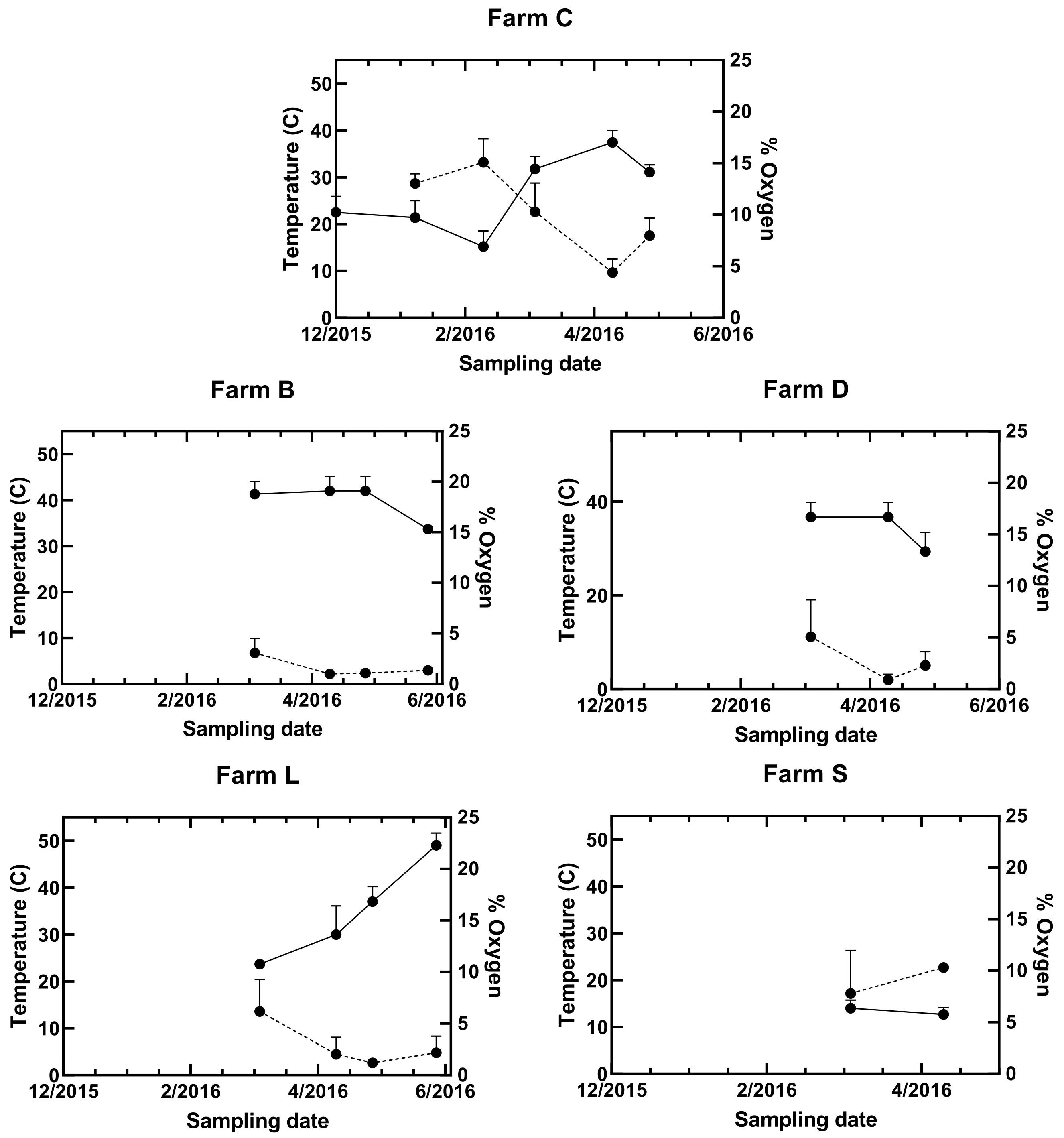
Bedding was collected from five certified organic dairy farms using a bedded pack system. The farms were located within an 80 km radius of Burlington, VT, USA (Table 1). Samples were collected between October 2015 and May 2016 when pastures were inaccessible (due to cold or wet conditions) to these pasture-based dairies. Bedding was collected on seven dates at a reference farm (Farm C). Farm C is well characterized from a previous study that compared udder microbiomes of cows that were healthy and those with mastitis [15] and is the farm presented in the greater case study. The farm was a 150-cow, certified organic, pasture-based dairy with a mix of Holstein and Jersey breeds. Bedding samples were collected at an additional four organic dairies (B, D, L, S) at six dates between November 2015 and May 2016 (on concurrent days as Farm C) to discern whether the findings could be applied more generally. Farms B and D were managed similarly to Farm C. However, Farm L used tilled wood shavings and Farm S the straw bedded pack was mesophilic and less sheltered. All bedded packs housed lactating animals except Farm S, which dried off during the winter months. Temperatures in the study region reached a mean monthly low of −12 °C in January and mean monthly high of 21 °C in May. The bedding pH in these systems ranged from 5.2 to 8.3 (Figure 2).

2.1.2. Isolations

Herd level samples from bulk tank milk and two bedding types (tie-stall and bedded pack) were collected in December 2019, January 2020, and February 2021 from two farms. One of the farms (C) was the same sampled for amplicon sequencing (Table 1). The second farm (M) relates to the student-run dairy at University of Vermont. Farm M is a tie-stall barn with sawdust on rubber mattresses, with bedding renewed daily. Farm M was chosen as an accessible source of milk for yeast isolations, not the bedding.
Figure 2. Temperature (solid line) and percent oxygen (dashed line) in the oxic zone in bedded pack at five farms. Means (± standard error) are illustrated. Farm C was the reference farm (n = 10) and four additional farms were compared (n = 3 replicate measurements per time). Some error bars cannot be seen because they are smaller than the symbols, except for the 4/2016 measurement at farm S that had only 1 sample.
Dairy 03 00042 g002

2.2. Part 1: Amplicon Sequencing

2.2.1. Sampling

Five 3.75 L samples were collected at 5-paces apart along a systematic transect with a random starting point within each bedded pack barn at each sampling date. Fresh surface hay was brushed away to sample layers that had been exposed to cows for at least three days. Samples were collected from the top layers of pack that were unsaturated and, thus, oxic (Figure 1). The top loose straw layer was separated from the compacted layer below. In untilled bedded packs, sampling the compacted layer required a mattock to penetrate. The wet anoxic layer below (fermentation layer) was not sampled. Occasionally, when the pack was less than 15 cm deep, layers were not divided into separate composite samples. Subsamples were pooled to form a composite sample of each of two layers for each barn at each sampling time. Temperature and percent oxygen were also measured at time of sampling using an OxyTempTM (Reotemp, San Diego, CA, USA) probe at the middle of the oxic zone (Figure 1). Temperature tended to be greatest at the surface 10–15 cm (35–35.5 °C) and decline progressively with depth, i.e., 32–34.5 °C at 38 cm, and 29.5–31 °C at 91.4 cm at reference farm C. Composite samples were transported to the lab in insulated containers for same-day processing. Using clean nitrile gloves, each composite sample was hand homogenized, and five subsamples removed for a total of 25 mL that were trimmed to 1 mm length pieces using ethanol flamed stainless steel shears. The pieces were again homogenized by hand and approximately 1 mL was sampled and frozen at −80 °C until DNA was extracted.

2.2.2. DNA Extraction and Sequencing

Extraction of DNA was performed using the DNeasy PowerSoil Kit (Qiagen, Germantown, MD, USA). Manufacturer’s instructions were augmented by heating bead tubes to 65 °C for 10 min, and then shaking horizontally for 2 min at maximum speed with the MoBio vortex adapter [16]. Subsamples of 0.15–0.25 g were placed into the bead tube of the PowerSoil kit. Subsample mass varied due to density differences between bedded pack samples; less dense samples filled the bead tube at a smaller weight. Extracted DNA samples were frozen at −80 °C until being shipped to the University of Colorado Next Generation Sequencing Facility (Boulder, CO, USA) for PCR amplification, sequencing, initial data filtering, and taxonomic reference mapping [17].
Detailed amplification, sequencing and taxonomic mapping methods were described previously [15]. Briefly, amplification was performed in triplicate using 515F and 806R primers targeted for the V4 region of bacterial/archaeal 16S rRNA genes and ITS1F and ITS2 primers targeting the fungal ITS1 region. Sequencing was performed on the Illumina MiSeq platform. Cleaned and quality filtered reads were dereplicated before clustering at 97% nucleotide identity into representational OTU sequences via UCLUST (version 7) [18]. Sequences were referenced to the Greengenes (bacteria) [19] and UNITE (fungi) [20] databases to produce combined Operational Taxonomic Unit (OTU) count and taxonomic information tables.
Library size (read counts per sample) was determined, and depth of sequencing relative to unique OTUs on a sample basis was compared using the rarecurve function within the vegan package for R software [21] (Figures S1 and S2). Samples with fewer than 100 sequences were removed prior to proportional scaling [22]. OTU counts within each sample were expressed as a proportion of total sample counts and multiplied by the mean of all sample counts. This scaled value was rounded to obtain a whole number, eliminating OTUs with a scaled proportion less than 1.0. Bacterial and fungal sequence counts were normalized separately. Normalizations were performed for each of two discrete subsets of data for both bacterial and fungal sequence counts: Farm C bedding (over one year, Figure S1), and all farms bedding (over four months, Figure S2). No comparisons were made between discrete datasets.

2.2.3. Statistical Analysis

OTU counts were converted to proportion of total sample counts (Relative Abundance; RA) and mean OTU RA was calculated among farms and dates. The core microbiome of Farm C was defined as OTUs that existed in 100% (bacteria) and at least 45% (fungi) of all samples collected at Farm C.
Bray–Curtis dissimilarity matrices were calculated to compare community composition among farms. Permutational multivariate analysis of variance (permanova) was used to test a three-way model describing contribution of farm, date, and depth to variation in OTUs observed at all farms and to further assess differences between sample depths at farms with similar bedding practices. Permanova post hoc tests were performed to assess pairwise differences in microbial DNA sequences between farms. Median RA of the 100 most abundant genera across dates for each of the five farms were computed (Tables S1 and S2). Statistical analyses were conducted in R version 4.02 (R Core Team, Vienna, Austria). Distance-based redundancy analysis was performed using the capscale function in vegan [21].

2.3. Part 2: Ecology

2.3.1. Culturing and Characterization by Morphology

Subsamples of bedding cultured to isolate Trichocomaceae and supplemental bulk milk samples were collected to isolate Saccharomycetales. Bulk tank milk was collected using sterile dippers (Sterilin™ Dippa™, ThermoFisher Scientific, Waltham, MA, USA). The milk was transported to the lab in a cooler. The samples were inverted 30 times for homogeneity and pipetted to 50 mL plastic conical tubes. Several handfuls of the top few inches of bedding were taken from various locations in the same barn. Bedding samples were hand-homogenized and placed in freezer bags. Subsamples of milk (2 mL) and bedding (0.5 g) were removed for culturing of isolates and functionality assays. Milk and bedding samples were frozen at −80 °C and –20 °C, respectively.
A bedding subsample (0.5 g) in 50 mL sterile milli-Q water, was macerated at 4000 rpm for 30 s in a Polytron 3100. A 1 mL aliquot of each bedding isolate culture or 100 µL milk was pipetted from each onto 11 different culture media including Czapek Yeast Autolysate Agar (CYA), blood agar media, Acidified Weak Potato Dextrose Agar with Yeast Extract (AWPY), Sabouraud Dextrose Agar (SDA), Corn Meal Agar (CMA), Caffeic Acid Agar (CAF), Brain Heart Infusion Agar (BHI), Yeast Sodium Agar (YNG), Yeast Lactose Agar (YLA), Rose Bengal Chloramphenicol (RBA), and Artificial Rumen Agar (RUM) (File S1) for yeasts. Isolates of Trichocomaceae fungi were obtained through an integrated approach of using both agar plates and natural substrates (Table 2) to identify species using current taxonomy [23,24,25]. Taxonomic identification was based on growth on different media and various macro- and micro-morphology [23,25,26]. Macromorphology characters were qualitatively based on colony texture, degree of sporulation, color of conidia, mycelia properties (abundance, texture and color), presence and color of soluble pigments and exudates, colony reverse color, growth rate, and acid production [25]. Micromorphology allowed further identification based on differences of conidial heads, stipes, hyphae, vesicles, metulae and phialides, conidia, sclerotia and hilal cells, ascomata, and ascospores. Yeast isolates were differentiated from bacteria by measuring cells under 40X magnification. Other features such as cell shape (ellipsoid, subglobose, and globose) and bud scars were also noted.

2.3.2. Carbon Utilization

Niche differentiation of 20 Aspergillus and Penicillium spp. isolates was measured as range and overlap of carbon utilization. An isolate of Hypocreales (Ascomycota) was used as a reference outlier representing another organism with a taxonomy replete with anamorphs and teleomorphs [27]. Carbon utilization profiles for each of 20 isolates were determined using Filamentous Fungal identification plates (BIOLOG FF Microplate™ Biolog Inc., Hayward, CA, USA). Instead of following manufacturer instructions, we opted to follow more specific ecological guidelines for Aspergillus and Penicillium developed in the 1980s and 1990s [23]. Briefly, each isolate was grown on malt extract agar for 4 days at 22 °C. A viscous solution of 0.3% of type II carrageenan was poured on top of the fungal colony that was gently scraped with a plastic inoculating loop to suspend the conidia uniformly. Turbidity of the suspension was measured on a spectrophotometer at OD590 and then diluted with a sterile 0.7% saline solution to achieve a final transmittance of 0.022. The final conidia solution was pipetted into the 96-well plate, adding 100 µL per well. The microplates were incubated at 22 °C and read every 24 h until sporulation was observed [23]. Optical density readings measured 24 h before sporulation were used for analysis. Response data were transformed as log (x + 0.01) and carbons were centered and standardized. A redundancy analysis was performed constrained by species as an explanatory variable and carbon metabolism activity as response variables using Canoco version 5 software (Microcomputer Power, Ithaca, NY, USA) [28].

2.3.3. Indicators of Colonization and Pathogenicity

Yeast isolates were tested for their ability to grow at 35 °C, hemolysis, protease, and two oxidases (catalase, peroxidase) as indicators of virulence/pathogenicity. Growth at 35 °C for 48 h was a measure of ability to live within a cow [29]. Yeast hemolysis was evaluated by streaking isolates on blood agar and incubated at 28 °C for 72 h. Positive hemolytic activity was indicated by the presence of a translucent halo zone around the colony. The protease assay was performed using Thermo Scientific Pierce Fluorescent Protease Assay (Waltham, MA, USA) with a negative control of 100 µL of casein tagged with fluorescein and 100 µL of suspended in buffer. Catalase and peroxidase were determined by suspending 1 mL of cells with 250 µL of ABTS (2,2′-azino-bis(3-ethylbenzothiazoline-6-sulfonic acid) and 20 µL of 3% hydrogen peroxide. Cultures were left to incubate at room temperature and checked for color change at 15 min, 1 h, and 24 h. Bubbles generated by reaction with hydrogen peroxide provided evidence of catalase activity [30]. Bubble generation and color change were evidence of peroxidase activity [31]. Raw milk was used as a positive control given it contains lactoperoxidase [32].

2.3.4. DNA Extraction, PCR and Sanger Sequencing

DNA was extracted from 20 isolates of Trichocomaceae (Ascomycota) and 46 isolates of Saccharomycetales (Ascomycota) using the Qiagen DNeasy Powermax Soil Extract Kit as described for Part 1. Extracted DNA was PCR-amplified using NS7F and ITS2R primers targeted for the first rRNA internal transcribed spacer region (ITS1). NS7F provides a longer sequence read than ITS1F, has fewer mismatches, and less bias toward Basidiomycota [33]. Reactions were held at 94 °C for 3 min to denature the DNA, with amplification proceeding for 35 cycles at 94 °C for 45 s, 54 °C for 60 s, 72 °C for 90 s, and 10 min at 72 °C, followed by a final extension of 10 min at 72 °C. Amplicons were shipped to GENEWIZ (South Plainfield, NJ, USA) for purification and Sanger Sequencing. Taxonomy was assigned to each OTU via the NCBI BLASTn database (https://blast.ncbi.nlm.nih.gov, accessed on 30 May 2022) with criteria as 0.0 E and nucleotide match of at least 97% (Table 2 and Table 3). Contigs were assembled using CAP3 software [27].

3. Results

3.1. Most Common Taxa at Farm C and Their Distribution with Season and Depth

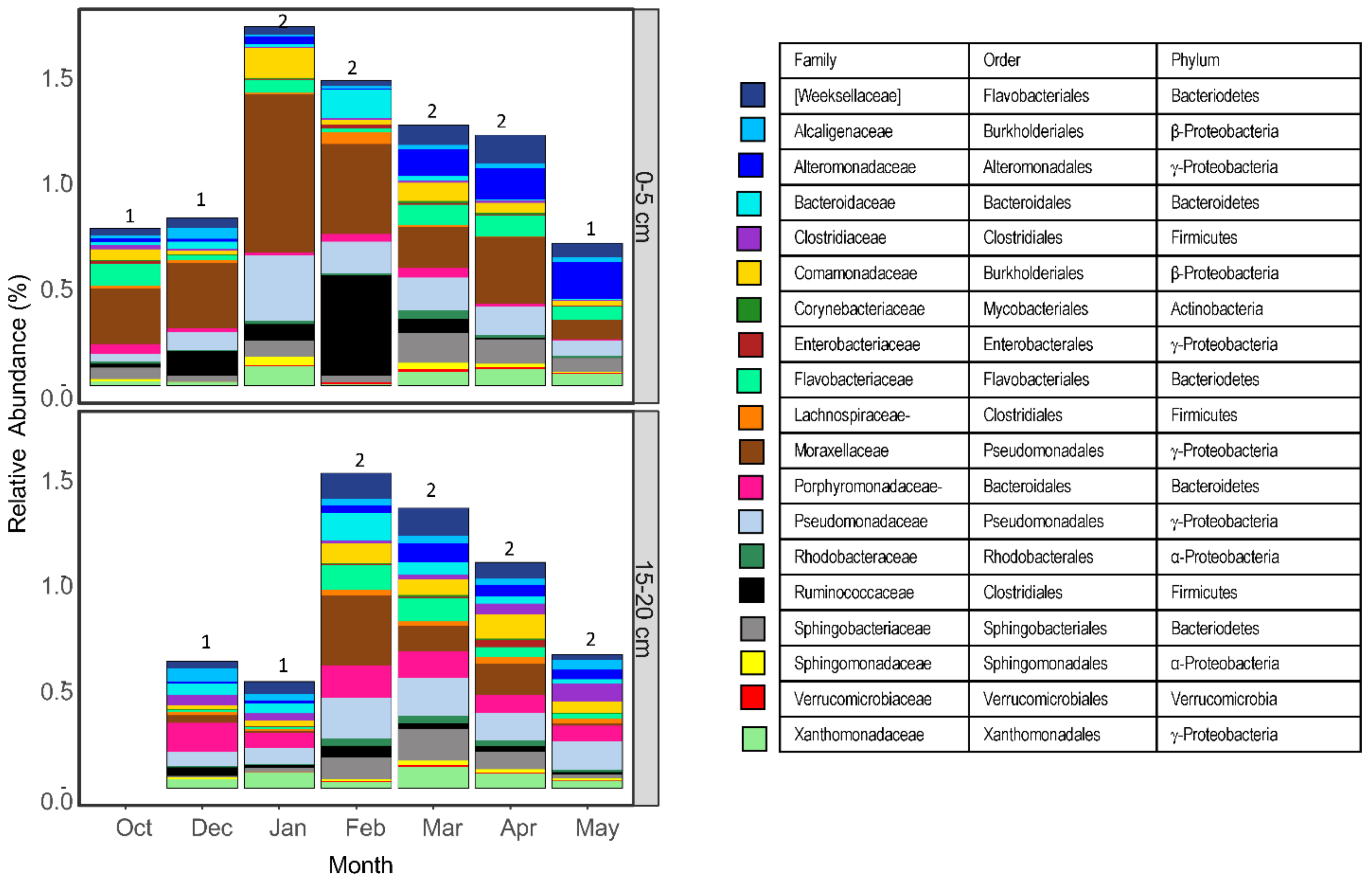
Bacteria within γ-Proteobacteria, Bacteroidetes, Firmicutes, and β-Proteobacteria dominated the bacterial community (Figure 3), whereas Ascomycota fungi represented 88% of all OTUs within the fungal community (Figure 4). Common genera within γ-Proteobacteria included Acinetobacter, Pseudomonas, and Cellvibrio, whereas Comamonas represented β-Proteobacteria (Table S2). Bacteroidetes was represented by Ruminofilibacter, Sphingobacterium and Flavobacterium. Firmicutes was represented by greater relative abundance of orders MBA08, OPB54, and Clostridiales. Fungal microbiomes were dominated by Saccharomycetales yeasts classified as Candida, the anamorph stage, associated with teleomorphs Pichiaceae (especially Pichia), Debaryomycetaceae (Yamadazyma, Debaryomyces, Kurtzmaniella, CTG clade), and Phaffomycetaceae (Wickerhamomyces, Cyberlindnera) (Table S3).
Composition of both bacterial and fungal communities at Farm C varied through time (Figure 3 and Figure 4). Bacterial communities also varied with depth but not fungal communities. For example, Moraxellaceae and Ruminoccaceae had greatest relative abundance in mid-winter of the 0–5 cm layer. Pseudomonadaceae was similarly abundant in both depths. Porphyromonadaceae was more abundant in 15–20 than 0–5 cm layers. The only time showing depth differentiation for fungal communities was in February (Figure 4). Ascobolaceae was abundant at the beginning of the bedded pack formation in October, and appeared in the 15–20 cm layer by January and persisted through May. Saccharomycetales was dominant at both depths beginning in November. Trichocomaceae (Aspergillus and Penicillium spp.) was equally abundant across depths.

3.2. Cultured Filamentous Fungi and Yeast Isolates

Filamentous fungi isolated and cultured from bedded pack samples included 36, 11, 4, and 9 isolates of Aspergillus, Penicillium, Paecilomyces, and Trichoderma, respectively. Tie-stall samples yielded 11, 9, 3, 3, 2, and 2 isolates of Aspergillus, Penicillium, Paecilomyces, Fusarium, Byssochlamys, and Trichoderma, respectively. Among the Trichocomaceae were identified three Aspergillus species and five Penicillium species (Table 2).
There was no obvious specificity of isolation media and taxonomic species of yeast from bedding (Table 3). For example, Debaryomyces hansenii, Wickerhamomyces anomalus, Diutina catenulata, and (Candida) glaebosa were isolated on at least three different media. Different species of the same genus were isolated on contrasting media. For example, D. hansenii was isolated on four media (SDA, RUM, CAF, BHI), and its congeneric species Debaryomyces coudertii was isolated on YLA. Both CAF and RUM media were more general isolation media, yielded 5 and 4 species, respectively.
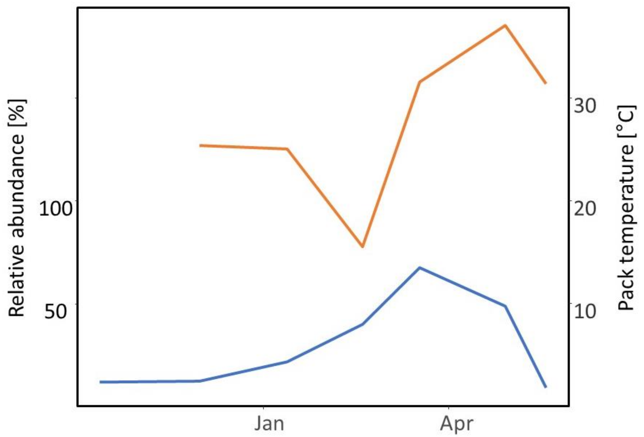
Three species of yeast cultured from milk were also present in bedding samples (Table 4). Pichia kudriavzevii was the most prevalent. Two other prevalent species were cultured from one but not both substrates. D. rugosa was present in milk but not bedding. D. hansenii (anamorph Candida famata) was present in bedding but not milk. Diutina rugosa (anamorph Candida rugosa) was isolated from milk on RBC media. A previously uncultured Tremellomycetes was isolated from bulk tank milk cultured on RBC, and from high SCC milk of a single animal cultured on CAF. W. anomalus was isolated both using AWPY and CYA. Pichia holstii (synonym Nakazawaea holstii) and P. kudriavzevii (anamorph Candida krusei) were isolated using CAF and AWPY, respectively. All milk isolates were cultured subsequently on CAF media. P. kudriavzevii relative abundance tends to be associated positively with increased temperature (Figure 5).

3.3. Indicators of Carbon Utilization and Pathogenicity

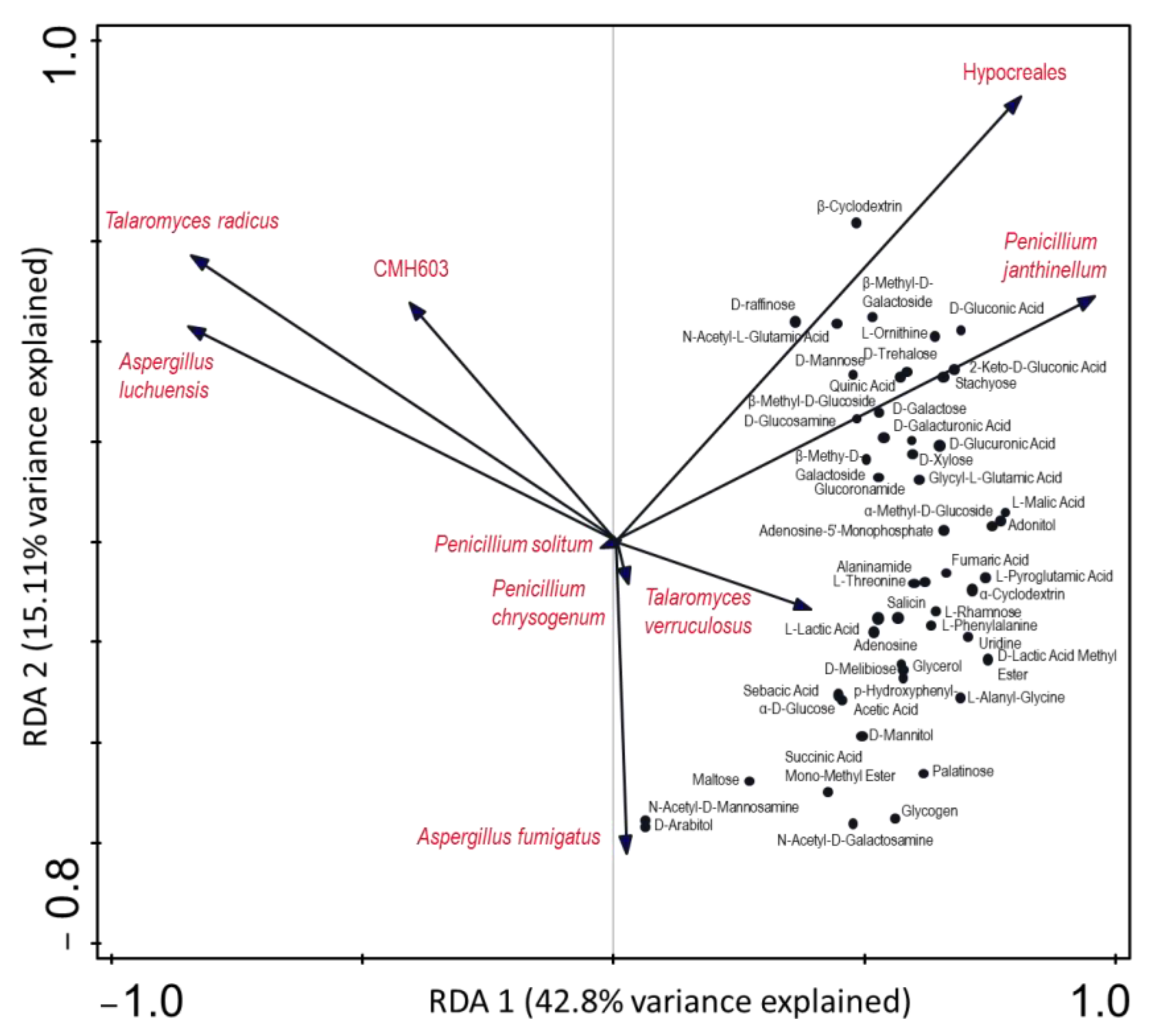
The genera Aspergillus and Penicillium and their respective species varied in substrate requirements (Figure 6). For example, A. fumigatus were associated positively with N-acetyl-D-mannosamine and D-arabitol, and A. luchuensis was associated positively with a media depletion of carbon–nitrogen substrates (Figure 6). Hypocreales and Penicillium janthinellum series were associated positively with a multitude of carbon–nitrogen substrates and were orthogonal to the other isolates.
Thirty five percent (14 of 40) of the yeast isolates did not grow at 35 °C. Twenty percent (8 of 40) of the isolates produced catalase (1 Pichia fermentans, 3 D. hansenii, 2 D. rugosa, 1 D. catenulate, 1 unknown). Variation of production of hemolytic or catalase was apparent between species of Pichia, Debaryomyces and Diutina and within species of D. hansenii, D. catenulate, D. rugosa and W. anomalus. One isolate of each D. catenulate and P. fermentans were hemolytic. All isolates produced protease with two exceptions. The single isolate of D. coudertii did not produce protease, and one of the eight W. anomalus isolates did not produce protease. None of the isolates produced peroxidase.

3.4. Comparison among Farms

Farms B and D used a DBP such as Farm C. Farm S used a DBP, but it had a lower temperature than other DBP farms. Farm L used a unique material, green pine sawdust, and was the only farm that tilled its pack. All farms except S had similar median bedding temperature and stocking density (Table 1, Figure 2). However, bedded pack farms varied in bedding replenishment rate and application of woodchips (Table S1). Farm S stocking density was the least of all the farms, and cows were not restricted to the barn: they could range into the snowy, frozen, or muddy pasture. Less fecal matter and bedding accumulated in the barn, and microbial decomposition was not well insulated from freezing temperatures, possibly explaining why the lowest bedding temperatures were observed on Farm S. Farm L and S fed cows outside the barn, whereas other farms fed cows in rings placed on the surface of the bedded pack, likely increasing cow time spent on the pack, as well as the volume and concentration of manure and urine. Composition of bacterial communities found in bedding at Farms B and D were similar to Farm C. Farms L and S communities were dissimilar (Figure 7a). Common to all farms was γ-Proteobacteria Acinetobacter and Pseudomonas. There was greater relative abundance of Clostridia (Orders MBA08 and OPB54) and Bacteroidetes (e.g., Ruminofilibacter, Sphingobacterium) at Farms C, B, D and L than Farm S (Table S2). In contrast, Farm S was enriched in different members of Bacteroidetes (Marinilabiaceae, Sphingobacteriaceae) and γ-Proteobacteria (Pseudomonadaceae) than the other farms. Bacterial community composition varied by sample depth (F = 3.417, p = 0.003) on farms that had a median bedding temperature greater than 30 °C (Table 1, Figure 2).
The composition of fungal communities found in bedding at Farms D was similar to Farm C. Farms B, L and S communities were dissimilar (Figure 7b). Most of the fungi across all farms belonged to the phylum Ascomycota (median 83.6%), whereas 13.6% belonged to the Basidiomycota (Table S3). Ascomycota order Saccharomycetales, especially Pichia spp. and Wickerhamomyces spp., were common at all farms (Table S3). Farm B was the only farm to have greater relative abundance of Thermomyces (Eurotiales) and Wallemia (Wallemiales).

4. Discussion

The focus of this study was to examine detrital microbial communities associated with organic bedded pack systems and temporal aspects of community assemblage. To further understand assemblages enumerated through DNA, the Trichocomaceae and yeasts were targeted for isolation due to their relationship to early decomposition or as opportunistic pathogens, respectively. The bedding microbiome of case study, Farm C, was most similar to the bedding microbiome of farms that used similar bedding material and managed for a warm deep bedded pack. This suggests generally that bedded pack microbial communities are similar only as far as the feedstock and management strategy create analogous environmental conditions. Culture work performed in this study suggests the fungal community is represented by two broad niches, early colonizers without specific vitamin requirements and later successional taxa that have vitamin or other nutrient requirements. Furthermore, yeasts exhibit traits associated with host colonization and pathogenicity suggesting a link between bedding microbiome and udder health.

4.1. Core Bedding Microbiome

Prevalent genera within the γ-Proteobacteria, Bacteroidia, Sphingobacteriia and Flavobacteria are reported for composts made with dairy cow feces [9,34,35], and grow well in oxic environments [36] and within temperature ranges found on compost bedded pack. Bacteroidetes classes Bacteroidia, Flavobacteriia, Sphingobacteria and Cytophagia contain gut species [37] that are common gut microbiota of cows [38].
There is increasing evidence that Bacteroidetes play a crucial role in producing polymer-degrading enzymes that metabolize a diverse array of polysaccharides, proteins, and chitin related to the cell walls of plants and fungi [37,38]. The variation in the polysaccharides that environmental Bacteroidetes can degrade is a measure of habitat breadth [37]. Bacteroidetes orders Flavobacteriales and Sphingobacteriales metabolize a variety of polysaccharides and proteins as carbon sources [39]. Spingobacterium and Flavobacterium demonstrate copiotrophic lifestyles given their ability to thrive on abundant and labile nitrogen content of dairy feces [40]. Porphyromonadaceae are also associated with copiotrophy, suggesting that consistent renewal of excess nutrients in the bedding habitat favors organisms that thrive in conditions of excess and instability [41].
Acinetobacter and Pseudomonas (γ-Proteobacteria) are also associated with copiotrophy lifestyles as created by nutrient-rich fecal material deposited on bedding. Acinetobacter guillouiae (Moraxellaceae) is common in sewage and sequesters copper and degrades microcystin [42,43]. Pseudomonas is an extremely diverse genus genetically, physiologically, and spans functions that range from pathogen to growth promoter of plants [44]. P. umsongensis metabolizes aromatic substrates [45]. Cellvibrio has the capacity to metabolize cellulose and glucose [46], and Penicillium janthinelium series is a cellulose degrader. Luteimonas spp. (Xanthomonadaceae) are mostly found in marine and freshwater environments, but one species was isolated from food waste compost [47].
OTUs from the Alcaligencaceae family exhibited a narrow range of variation through time suggesting that they do not respond rapidly to environmental conditions or availability of substrate. Instead, Alcaligenaceae and Comamonas increased abundance through time to become the dominant β-Proteobacteria in cowpats [48], behaving as copiotrophs.

4.2. Trichocomaceae

Within the filamentous fungi, the Trichocomaceae exhibit traits of early colonizers of plant material used in bedding and niche differentiation. Trichocomaceae are among the most abundant conidia found on spore traps in anthropogenic indoor environments [10,11] where animals have contact with high density of spores. However, some species of the family require specific nutrients and vitamins. This suggests assemblages from amplicon sequencing comprise a successional range of taxa [12]. Trichocomaceae are primary colonizers of substrates associated with cattle feed or bedding material such as silage, hay, and corn [49]. This suggests the taxa that colonize the surface of bedding early in the season originate from the bedding itself. The farms in this study used mostly sawdust, wood chips, and straw as bedding (Table 1), which is common [3]. Cell walls of herbaceous plants are rich with cellulose, so one would expect the microbiome to be rich with organisms that produce cellulases, e.g., Trichocomaceae genera Aspergillus and Penicillium. Cell walls of woody plants will also include lignin fibers for rigidity. Wood substrates would attract inhabitants such as jelly fungi (e.g., Tremellomycetes), and brown rot fungi (e.g., Microbotryomycetes and Agaricomycetes). These taxa produce copper-based laccases and manganous peroxidases. However, more research is necessary to demonstrate the impact of oxidative competition on opportunistic yeasts. The material used as bedding defines the matrix carbon sources which can be utilized by decomposer communities.
Conceptually, isolations attempted to select conditions that may favor later successional species by including vitamin sources such as yeast extract while using other environmental factors to limit competing isolates including submerged and unsaturated enrichment with sodium chloride and sucrose solutions at 23 °C and 35 °C. Morphological consistencies of the isolates and growth characters indicate that all isolates were early successional species. All Aspergillus and Penicillium isolates obtained from bedding, grow on minimal media without specific vitamin or amino acid requirements. The isolates reduced nitrate to nitrite by producing the enzyme nitrate reductase [50]. Nitrate utilization is significant due to being somewhat uncommon in decomposer communities. These genera can produce structural proteins and enzymes in environments high in carbon, but scarce of other nitrogen sources, which is the case for cattle bedding. Class Eurotiomycetes (enriched in farms B, D, and L) consisted mostly of family Trichocomaceae within order Eurotiales. However, Farm D had an elevated abundance of Aspergillus. Both Aspergillus and Penicillium of Trichocomaceae are early ecological colonizers in decomposition environments, with a large breadth of carbon and nitrogen utilization [36]. In addition, Aspergillus and Penicillium exude antifungals, enzymes, organic acids, immunosuppressive compounds, and mycotoxins [51,52,53]. These extrolites allow Aspergillus and Penicillium to be highly competitive in their environments due to the inhibition of other organisms [36,54] and may structurally impact community composition.
Studies investigating their ecological role in animal bedding decomposition are underrepresented although these genera are researched extensively due to their applications in food safety, fermentation, pharmaceuticals, and bioremediation [55]. Further studies should explore the relationship between barn air quality parameters and growth of species associated with allergic hypersensitivities. Surface growth, in culture, indicates that most isolates are obligate aerobes and likely detected as transient conidia in milk by DNA methods. However, Aspergillus can co-occur on the skin with Alternaria and Cryptococcus [15]. Results from this study suggest that early colonizers in the family Trichocomaceae partition their ecological niche chemically and the ability to utilize nitrate N versus requirements of amino acids and vitamins separate the Trichocomaceae into colonizers and later successional niches.

4.3. Yeasts

Fungal yeasts are prevalent in bedding and milk [15]. Initial BLAST results returned fungal microbiomes dominated by yeasts classified as Candida, the anamorph stage. However, recent studies link anamorphs and teleomorphs and resulted in their reclassification as Pichia, Wickerhamomyces, Cyberlindnera, Nakazawaea, Yamadazyma, Debaryomyces, Barnettozyma, and Kurtzmaniella. The anamorph Candida (associated with teleomorphs Debaryomyces, Diutina, Pichia and Wickerhamomyces) is a commensal yeast that can become pathogenic when host defense breaks down [56], and has been isolated from cases of bovine mastitis [57]. Yeasts did not dominate the fungal microbiome until about one month of cow presence (Figure 4). This suggests that prevalence of yeast is related to cumulative effects of cow presence on bedding. Two yeasts found in the bedding environment were capable of hemolysis that suggest they may be competent opportunists in the mammary environment. Debaryomyces is a facultative aerobe with optimal temperature of 22 °C yet tolerates freezing conditions and metabolizes diverse carbon substrates [58,59,60]. Wickerhamomyces has diverse strain morphology and can survive a wide range of environmental conditions. Both Wickerhamomyces and Debaryomyces produce mycotoxins to compete with other organisms [58]. Debaryomyces hansenii was only from bedding in this study, but also isolated from milk isolated in other studies [15,61] that isolated from bulk tank milk in contrast to individual animals [62]. D. hansenii is a facultative aerobe and known as an extremophilic yeast that metabolizes diverse carbon substrates [59]. Strains of D. hansenii produce myocins that suppress in vitro growth of common pathogens including Staphylococcus aureus, Escherichia coli, Klebsiella pneumoniae, and Streptococcus pyogenes [63].
Diutina rugosa and Pichia kudriavzevii have known associations with bovine dairies [64,65] and cases of mastitis [61]. D. rugosa has been isolated from dung of a scouring cow, soil, cheese, and considered a rare human pathogen linked to invasive medical procedures or previous exposure to broad spectrum antibiotics [64]. P. kudriavzevii is thermotolerant [66] and exhibits antibacterial activity against pathogens including Staphylococcus aureus [67]. It follows that P. kudriavzevii is prevalent in mastitic milk samples [65]. P. holstii demonstrates antifungal activity with applications in the wine industry [68]. The Tremellomycetes isolate had not been cultivated prior to this study; however, sequences of this taxon have been associated with milk from organic dairies [69] and detected in air samples from five dairy farms using high-throughput sequencing [70].

4.4. Indicators of Pathogenicity

Hemolysins are virulence factors which help pathogens to survive and persist in the host [14]. A hemolysis test is an indicator used to determine whether a yeast isolate is potentially pathogenic [61]. Hemolysins are often defined as extracellularly produced mycocins and are understudied in fungi compared to bacteria [13]. Hemolytic activity in Candida and Cryptococcus species are associated with iron uptake and regulation required for a variety of mechanisms including phenotype switching, metabolism, and enzyme production [13,71]. Notable iron containing pathogenic enzymes produced by yeasts are peroxidase and catalase which were evaluated in this study where P. fermentans and Diutina catenulate isolates exhibited hemolytic properties, but not D. rugosa. This contrasts with another study that demonstrated that D. rugosa exhibited hemolytic properties [72]. Related species P. kudriavzevii and D. hansenii have been implicated in hemolysis [61].
Proteases are hydrolytic enzymes that are produced by organisms to degrade proteins and can be implemented to break down free intercellular or intracellular proteins for metabolic purposes. Proteases can also be produced pathogenically or defensively to degrade proteins which maintain the structure of cell membranes [26]. Most isolates in this study produced proteases, which supports reports of D. hansenii, W. anomalus, D. rugosa, P. holstii and P. fermentans all producing proteases in consumable food products [73,74,75,76,77]. Yeast mycocins are being touted as having potential applications in promoting health as antimicrobials, with its main mechanism of action being the inhibition of β-glucan synthesis in the cell wall of sensitive strains [78].
Extracellular oxidases are deployed by both bacteria and fungi to mitigate the toxicity of phenolic molecules and metal ions, and aid in antimicrobial defense [79]. Oxidative enzymes are a basic fungal response to hostile chemical conditions [80]. Peroxidases play a key role in the defense against oxidative stress in bacteria by catalyzing the decomposition of hydrogen peroxide [30] with peroxidases [31]. In this study, catalase oxidases were apparent but not peroxidases capable of reacting with ABTS. However, in pure culture, these genes might not be up-regulated due to an absence of oxidative stress brought on by competition. Many of the wood substrates inhabitants in bedding, particularly Basidiomycetes, produce laccases and manganous peroxidases. However, more research is necessary to demonstrate the impact of oxidative competition on opportunistic yeasts. Finally, none of the isolates produced both hemolytic and peroxidase capabilities in pure culture.

4.5. Source of Yeasts

Teat surface is a transmission point of butyric acid bacteria (BAB) originating in silage to the mammary gland and then to bulk tank milk [81,82]. Similarly, the same genotype of Pichia kudriavzevii was present in bulk tank milk, teat surface, and the feed of sheep [83]. Furthermore, both BAB and P. kudriavzevii are normal flora of the healthy rumen [53,84]. In this study, P. kudriavzevii was cultured from both milk and bedding, suggesting a link between bedding, teat, rumen, and milk microbiomes. A longitudinal study of bulk tank milk microbiota suggests that milk microbiomes respond more to weather and feeding than milking system, number of cows and quality of milk [85]. One can hypothesize that in pasture-based dairies, management response to seasonal changes, for example, feeding preserved hay in the winter versus fresh grass in the summer and use of seasonal winter bedding changes the environmental microbial population, impacting exposure and transmission of potential cow symbionts such as P. kudriavzevii.

4.6. Bedding Management and the Microbiome

The bacterial and fungal microbiome of the bedding was related to farm management practice. Management factors including bedding replenishment rate, stocking density, tillage, and cow time spent on pack likely impact microbial habitat and the introduction of organisms [4,7]. For example, tillage homogenizes bedding material and creates a physical zone of aeration, whereas an untilled bedded pack has intact fecal pats that may remain anoxic throughout the bedding period. Farm L was the only farm that mechanically aerated bedding. Thermomyces lanuginosus, a thermophilic hemicellulose degrader [86], was greater in farms B and L than farm C, all of which were thermophilic packs. Farm B replenished the bedding most frequently and this indicates that both mechanical aeration and bedding rates promote elevated temperature [4]. Temperature, moisture and oxygen are intercorrelated and impact microbial community composition [4,39]. The microbiome on farms with warmest bedded packs varied between the slightly used (0–5 cm) and compacted strata (15–20 cm) of the oxic bedding zone. Vertical gradients create environments for niche differentiation, with deeper levels being older and more compacted and/or anoxic, representing a later stage of ecological succession.

4.7. Bedding Material and the Microbiome

A unique feature of the bedded pack habitat is constant replenishment with bedding, fresh cattle feces, and urine [3]. Feces and urine contribute a large amount of nitrogen to bedding environment, which varies based on what the cows are fed [87].
Aspects of community structure appeared more dependent on bedding material than management. While bedding management practices differed between Farm S and Farms C, B and D, all four farms used cellulose-based bedding material. Farms that used cellulose bedding had the greatest abundance of Cellvibrio. Cellvibrio is an aerobic bacterial species with the capacity to degrade cellulose. Cellvibrio japonicus is a model organism for utilizing nearly all components of plant cell wall polysaccharides [46]. The cellulose-to-lignin ratio of straw is typically 50% greater than wood [88,89]. Likewise, relative abundance of Basidiomycota was more prevalent in Farm L, which used primarily wood sawdust bedding. Basidiomycota contain white-rot fungi known for their ability to produce oxidative enzymes that degrade lignin [79].
Wallemia is a xerophilic mold that has been isolated from hay and some species are associated with “Farmers Lung Disease” [90]. Farm L, which did not use dried grass bedding, was void in this genus, suggesting that Wallemia is a bedding related taxon. However, divergent abundance among farms primarily using hay and straw bedding suggests that Wallemia is also sensitive to conditions related to management practices.

5. Conclusions

In the case study, the bedded pack contained a temporally dynamic detrital microbial community structured by temperature and bedding architecture. Comparison to other bedded packs suggested that similar pack management and bedding materials yield similar microbial communities among farms, though more detailed environmental measurements and a review of management practices would be required to assess specific questions related to producer decision making. Bedding may contain a mixture of pathogens and biological control organisms that directly affect the microbiomes of cow udders and indirectly impact health status. Understanding whether the greater microbiome can mediate organisms that impact mammary gland infections is critical to managing mastitis. The choice of bedding and its management represent a potential opportunity to curate the microbial community of the housing environment. There is need for further investigation of how management practices affect the transmission of microbes between the bedding and mammary gland. Some bedding OTUs were associated with key ruminal organisms, raising questions about the relationship between bedding and the gut microbiomes and feeding management.

Supplementary Materials

The following supporting information can be downloaded at: https://www.mdpi.com/article/10.3390/dairy3030042/s1, File S1: Detailed methods for isolation and culture of Trichocomaceae and yeasts. File S2: Figure S1: Normalization curves for bacterial amplicons; Figure S2: Normalization curves for fungal amplicons; Table S1: Farm Management Practices; Table S2: Most abundant bacterial genera; Table S3: Most abundant fungal genera.

Author Contributions

Conceptualization, D.A.N., T.R.W. and J.W.B.; Data curation, D.A.N. and T.D.A.; Formal analysis, D.A.N., T.D.A. and T.R.W.; Funding acquisition, D.A.N. and J.W.B.; Investigation, D.A.N., T.R.W. and A.H.; Methodology, D.A.N., T.R.W. and A.H.; Project administration, D.A.N.; Supervision, D.A.N. and J.W.B.; Visualization, T.D.A.; Writing—original draft, D.A.N. and T.D.A.; Writing—review and editing, D.A.N., T.D.A., T.R.W., A.H. and J.W.B. All authors have read and agreed to the published version of the manuscript.

Funding

This work was supported by a University of Vermont, College of Agriculture and Life Sciences, Dairy Center of Excellence, USDA HATCH experiment station award (VT-H02203) to J.W.B. and D.A.N., Vermont Multistate Hatch award (VT-H02601) to D.A.N., and United States Department of Agriculture-National Institute of Food and Agriculture-2018-51300-28561 Organic Research Extension Initiative to J.W.B. and D.A.N.

Informed Consent Statement

Not applicable.

Data Availability Statement

The datasets generated during and/or analyzed during the current study are available in at https://doi.org/10.6084/m9.figshare.17854808.v1 (accessed on 2 August 2022).

Acknowledgments

Many thanks to the cooperating farmers for the opportunity to study their herd, and thanks to Robert Mugabi for technical assistance collecting and processing samples, and thanks to Noah Fierer (University of Colorado-Boulder) for assistance with amplicon sequencing.

Conflicts of Interest

The authors declare no conflict of interest.

References

  1. Bewley, J.M.; Robertson, L.M.; Eckelkamp, E.A. A 100-Year Review: Lactating dairy cattle housing management. J. Dairy Sci. 2017, 100, 10418–10431. [Google Scholar] [CrossRef] [PubMed]
  2. USDA. Dairy Cattle Management Practices in the United States. 2014. Available online: https://www.aphis.usda.gov/animal_health/nahms/dairy/downloads/dairy14/Dairy14_dr_PartI_1.pdf (accessed on 2 August 2022).
  3. Leso, L.; Barbari, M.; Lopes, M.A.; Damasceno, F.A.; Galama, P.; Taraba, J.L.; Kuipers, A. Invited review: Compost-bedded pack barns for dairy cows. J. Dairy Sci. 2020, 103, 1072–1099. [Google Scholar] [CrossRef] [PubMed]
  4. Black, R.A.; Taraba, J.L.; Day, G.B.; Damasceno, F.A.; Bewley, J.M. Compost bedded pack dairy barn management, performance, and producer satisfaction. J. Dairy Sci. 2013, 96, 8060–8074. [Google Scholar] [CrossRef] [PubMed]
  5. Barberg, A.E.; Endres, M.I.; Salfer, J.A.; Reneau, J.K. Performance and welfare of dairy cows in an alternative housing system in Minnesota. J. Dairy Sci. 2007, 90, 1575–1583. [Google Scholar] [CrossRef]
  6. Benson, A.F. Consider Deep Pack Barns for Cow Comfort and Manure Management. Available online: https://smallfarms.cornell.edu/2012/04/consider-deep-pack-barns-for-cow-comfort-and-manure-management/ (accessed on 2 August 2022).
  7. Ray, T.; Gaire, T.N.; Dean, C.J.; Rowe, S.; Godden, S.M.; Noyes, N.R. The microbiome of common bedding materials before and after use on commercial dairy farms. Anim. Microbiome 2022, 4, 18. [Google Scholar] [CrossRef]
  8. Bewley, J.M.; Taraba, J.; Day, G.; Black, R.A.; Damasceno, F. Compost Bedded Pack Barn Design: Features and Management Considerations. Available online: http://www2.ca.uky.edu/agcomm/pubs/ID/ID206/ID206.pdf (accessed on 2 August 2022).
  9. Neher, D.A.; Weicht, T.R.; Bates, S.T.; Leff, J.W.; Fierer, N. Changes in bacterial and fungal communities across compost recipes, preparation methods, and composting times. PLoS ONE 2013, 8, e79512. [Google Scholar] [CrossRef]
  10. Jurjevic, Z.; Peterson, S.W.; Solfrizzo, M.; Peraica, M. Sterigmatocystin production by nine newly described Aspergillus species in section Versicolores grown on two different media. Mycotoxin Res. 2013, 29, 141–145. [Google Scholar] [CrossRef]
  11. Stein, R.A.; Bulboacӑ, A.E. Mycotoxins. In Foodborne Diseases; Dodd, C.E.R., Aldsworth, T., Stein, R.A., Cliver, D.O., Riemann, H.P., Eds.; Academic Press: Cambridge, MA, USA, 2017; pp. 407–446. [Google Scholar]
  12. Frisvad, J.C.; Larsen, T.O. Extrolites of Aspergillus fumigatus and other pathogenic species in Aspergillus section Fumigati. Front. Microbiol. 2015, 6, 1485. [Google Scholar] [CrossRef]
  13. Nayak, A.P.; Green, B.J.; Beezhold, D.H. Fungal hemolysins. Med. Mycol. 2013, 51, 1–16. [Google Scholar] [CrossRef]
  14. Wan, L.; Luo, G.; Lu, H.; Xuan, D.; Cao, H.; Zhang, J. Changes in the hemolytic activity of Candida species by common electrolytes. BMC Microbiol. 2015, 15, 171. [Google Scholar] [CrossRef]
  15. Andrews, T.; Neher, D.A.; Weicht, T.R.; Barlow, J.W. Mammary microbiome of lactating organic dairy cows varies by time, tissue site, and infection status. PLoS ONE 2019, 14, e0225001. [Google Scholar] [CrossRef] [PubMed]
  16. Lauber, C.L.; Hamady, M.; Knight, R.; Fierer, N. Pyrosequencing-based assessment of soil pH as a predictor of soil bacterial community structure at the continental scale. Appl. Environ. Microb. 2009, 75, 5111–5120. [Google Scholar] [CrossRef] [PubMed]
  17. Emerson, J.B.; Keady, P.B.; Brewer, T.E.; Clements, N.; Morgan, E.E.; Awerbuch, J.; Miller, S.L.; Fierer, N. Impacts of flood damage on airborne bacteria and fungi in homes after the 2013 Colorado Front Range flood. Environ. Sci. Technol. 2015, 49, 2675–2684. [Google Scholar] [CrossRef] [PubMed]
  18. Edgar, R.C. Search and clustering orders of magnitude faster than BLAST. Bioinformatics 2010, 26, 2460–2461. [Google Scholar] [CrossRef]
  19. McDonald, D.; Price, M.N.; Goodrich, J.; Nawrocki, E.P.; DeSantis, T.Z.; Probst, A.; Andersen, G.L.; Knight, R.; Hugenholtz, P. An improved Greengenes taxonomy with explicit ranks for ecological and evolutionary analyses of bacteria and archaea. ISME J. 2012, 6, 610–618. [Google Scholar] [CrossRef]
  20. Kõljalg, U.; Nilsson, R.H.; Abarenkov, K.; Tedersoo, L.; Taylor, A.F.S.; Bahram, M.; Bates, S.T.; Bruns, T.D.; Bengtsson-Palme, J.; Callaghan, T.M. Towards a Unified Paradigm for Sequence-Based Identification of Fungi; Wiley Online Library, 2013; Available online: https://onlinelibrary.wiley.com/doi/10.1111/mec.12481 (accessed on 2 August 2022).
  21. Oksanen, J.; Blanchet, F.G.; Friendly, M.; Kindt, R.; Legendre, P.; McGlinn, D.; Minchin, P.R.; O’Hara, R.B.; Simpson, G.L.; Solymos, P.; et al. Vegan: Community Ecology Package: Ordination Methods, Diversity Analysis and Other Functions for Community and Vegetation Ecologists. Ver. 2.5-7. Available online: https://cran.r-project.org/web/packages/vegan/index.html (accessed on 2 August 2022).
  22. McMurdie, P.J.; Holmes, S. Waste not, want not: Why rarefying microbiome data is inadmissible. PLoS Comput. Biol. 2014, 10, e1003531. [Google Scholar] [CrossRef]
  23. Samson, R.A.; Pitt, J.I. Integration of Modern Taxonomic Methods for Penicillium and Aspergillus Classification; Harwood Academic: Amsterdam, The Netherlands, 2000. [Google Scholar]
  24. Frisvad, J.C.; Samson, R.A. Polyphasic taxonomy of Penicillium subgenus Penicillium—A guide to identification of food and air-borne terverticillate Penicillia and their mycotoxins. Stud. Mycol. 2004, 49, 1–173. [Google Scholar]
  25. Visagie, C.M.; Houbraken, J.; Frisvad, J.C.; Hong, S.-B.; Klaassen, C.H.W.; Perrone, G.; Seifert, K.A.; Varga, J.; Yaguchi, T.; Samson, R.A. Identification and nomenclature of the genus Penicillium. Stud. Mycol. 2005, 53, 53–62. [Google Scholar] [CrossRef]
  26. Hecht, K.A.; O’Donnell, A.F.; Brodsky, J.L. The proteolytic landscape of the yeast vacuole. Cell Logist. 2014, 4, e28023. [Google Scholar] [CrossRef]
  27. Huang, X.; Madan, A. CAP3: A DNA sequence assembly program. Genome. Res. 1999, 9, 868–877. [Google Scholar] [CrossRef]
  28. Ter Braak, C.J.F.; Šmilauer, P. Canoco Reference Manual and User’s Guide. Software for Ordination, Version 5; Microcomputer Power: Ithaca, NY, USA, 2012. [Google Scholar]
  29. Metzner, M.; Sauter-Louis, C.; Seemueller, A.; Petzl, W.; Klee, W. Infrared thermography of the udder surface of dairy cattle: Characteristics, methods, and correlation with rectal temperature. Vet. J. 2014, 199, 57–62. [Google Scholar] [CrossRef] [PubMed]
  30. Yuan, F.; Yin, S.; Xu, Y.; Xiang, L.; Wang, H.; Li, Z.; Fan, K.; Pan, G. The richness and diversity of catalases in bacteria. Front. Microbiol. 2021, 12, 645477. [Google Scholar] [CrossRef] [PubMed]
  31. Cuellar-Cruz, M.; Briones-Martin-del-Campo, M.; Canas-Villamar, I.; Montalvo-Arredondo, J.; Riego-Ruiz, L.; Castano, I.; De Las Penas, A. High resistance to oxidative stress in the fungal pathogen Candida glabrata is mediated by a single catalase, Cta1p, and is controlled by the transcription factors Yap1p, Skn7p, Msn2p, and Msn4p. Eukaryot. Cell 2008, 7, 814–825. [Google Scholar] [CrossRef] [PubMed]
  32. Wolfson, L.M.; Sumner, S.S. Antibacterial activity of the lactoperoxidase system: A review. J. Food Prot. 1993, 56, 887–892. [Google Scholar] [CrossRef]
  33. Bellemain, E.; Carlsen, T.; Brochmann, C.; Coissac, E.; Taberlet, P.; Kauserud, H. ITS as an environmental DNA barcode for fungi: An in silico approach reveals potential PCR biases. BMC Microbiol. 2010, 10, 189. [Google Scholar] [CrossRef]
  34. Sakamoto, M. The family Porphyromonadaceae. In The Prokaryotes: Other Major Lineages of Bacteria and the Archaea; Rosenberg, E., DeLong, E.F., Lory, S., Stackebrandt, E., Thompson, F., Eds.; Springer: New York, NY, USA, 2014. [Google Scholar]
  35. Júnior, J.C.R.; De Oliveira, A.M.; de Silva, F.G.; Tamanini, R.; de Oliveira, A.L.M.; Beloti, V. The main spoilage-related psychrotrophic bacteria in refrigerated raw milk. J. Dairy Sci. 2018, 101, 75–83. [Google Scholar] [CrossRef]
  36. Trujillo, M.E.; Dedysh, S.; DeVos, P.; Hedlund, B.; Kämpfer, P.; Rainey, F.A.; Whitman, W.B. Bergey’s Manual of Systematics of Archaea and Bacteria; Whitman, W.B., Ed.; Wiley Online Library: Hoboken, NJ, USA, 2015. [Google Scholar] [CrossRef]
  37. Thomas, F.; Hehemann, J.H.; Rebuffet, E.; Czjzek, M.; Michel, G. Environmental and gut Bacteroidetes: The food connection. Front. Microbiol. 2011, 2, 93. [Google Scholar] [CrossRef]
  38. Cheng, Y.; Wang, Y.; Li, Y.; Zhang, Y.; Liu, T.; Wang, Y.; Sharpton, T.J.; Zhu, W. Progressive colonization of bacteria and degradation of rice straw in the rumen by Illumina sequencing. Front. Microbiol. 2017, 8, 2165. [Google Scholar] [CrossRef]
  39. Sun, L.; Han, X.; Li, J.; Zhao, Z.; Liu, Y.; Xi, Q.; Guo, X.; Gun, S. Microbial community and its association with physicochemical factors during compost bedding for dairy cows. Front. Microbiol. 2020, 11, 254. [Google Scholar] [CrossRef]
  40. Neher, D.A.; Limoges, M.A.; Weicht, T.R.; Sharma, M.; Millner, P.D.; Donnelly, C. Bacterial community dynamics distinguish poultry compost from dairy compost and non-amended soils planted with spinach. Microorganisms 2020, 8, 1601. [Google Scholar] [CrossRef]
  41. Ho, A.; Di Lonardo, D.P.; Bodelier, P.L. Revisiting life strategy concepts in environmental microbial ecology. FEMS Microbiol. Ecol. 2017, 93, fix006. [Google Scholar] [CrossRef] [PubMed]
  42. Yi, Y.L.; Yu, X.B.; Zhang, C.; Wang, G.X. Growth inhibition and microcystin degradation effects of Acinetobacter guillouiae A2 on Microcystis Aeruginosa. Res. Microbiol. 2015, 166, 93–101. [Google Scholar] [CrossRef]
  43. Majumder, S.; Gangadhar, G.; Raghuvanshi, S.; Gupta, S. A comprehensive study on the behavior of a novel bacterial strain Acinetobacter guillouiae for bioremediation of divalent copper. Bioprocess. Biosyst. Eng. 2015, 38, 1749–1760. [Google Scholar] [CrossRef]
  44. Spiers, A.J.; Buckling, A.; Rainey, P.B. The causes of Pseudomonas diversity. Microbiology (Reading) 2000, 146, 2345–2350. [Google Scholar] [CrossRef]
  45. Narancic, T.; Salvador, M.; Hughes, G.M.; Beagan, N.; Abdulmutalib, U.; Kenny, S.T.; Wu, H.H.; Saccomanno, M.; Um, J.; O’Connor, K.E.; et al. Genome analysis of the metabolically versatile Pseudomonas umsongensis GO16: The genetic basis for PET monomer upcycling into polyhydroxyalkanoates. Microb. Biotechnol. 2021, 14, 2463–2480. [Google Scholar] [CrossRef] [PubMed]
  46. Attia, M.A.; Nelson, C.E.; Offen, W.A.; Jain, N.; Davies, G.J.; Gardner, J.G.; Brumer, H. In vitro and in vivo characterization of three Cellvibrio japonicus glycoside hydrolase family 5 members reveals potent xyloglucan backbone-cleaving functions. Biotechnol. Biofuels 2018, 11, 45. [Google Scholar] [CrossRef] [PubMed]
  47. Young, C.C.; Kampfer, P.; Chen, W.M.; Yen, W.S.; Arun, A.B.; Lai, W.A.; Shen, F.T.; Rekha, P.D.; Lin, K.Y.; Chou, J.H. Luteimonas composti sp. Nov., a moderately thermophilic bacterium isolated from food waste. Int. J. Syst. Evol. Microbiol. 2007, 57, 741–744. [Google Scholar] [CrossRef]
  48. Wong, K.; Shaw, T.I.; Oladeinde, A.; Glenn, T.C.; Oakley, B.; Molina, M. Rapid microbiome changes in freshly deposited cow feces under field conditions. Front. Microbiol. 2016, 7, 500. [Google Scholar] [CrossRef]
  49. Bonner, R.D.; Fergus, C.L. The fungus flora of cattle feeds. Mycologia 1959, 51, 855–863. [Google Scholar] [CrossRef]
  50. Morton, A.G. A study of nitrate reduction in mould fungi. J. Exp. Bot. 1956, 7, 97–112. [Google Scholar] [CrossRef]
  51. Xu, J.; Song, Y.C.; Guo, Y.; Mei, Y.N.; Tan, R.X. Fumigaclavines D-H, new ergot alkaloids from endophytic Aspergillus fumigatus. Planta. Med. 2014, 80, 1131–1137. [Google Scholar] [CrossRef]
  52. Heo, Y.M.; Lee, H.; Kim, K.; Kwon, S.L.; Park, M.Y.; Kang, J.E.; Kim, G.H.; Kim, B.S.; Kim, J.J. Fungal diversity in intertidal mudflats and abandoned solar salterns as a source for biological resources. Mar. Drugs 2019, 17, 601. [Google Scholar] [CrossRef]
  53. Fernandes, T.; Carvalho, B.F.; Mantovani, H.C.; Schwan, R.F.; Avila, C.L.S. Identification and characterization of yeasts from bovine rumen for potential use as probiotics. J. Appl. Microbiol. 2019, 127, 845–855. [Google Scholar] [CrossRef] [PubMed]
  54. Losada, L.; Ajayi, O.; Frisvad, J.C.; Yu, J.; Nierman, W.C. Effect of competition on the production and activity of secondary metabolites in Aspergillus species. Med. Mycol. 2009, 47 (Suppl. 1), S88–S96. [Google Scholar] [CrossRef]
  55. Magan, N. Fungal colonization and decomposition of cereal straw. Int. Biodeterior. 1988, 24, 435–443. [Google Scholar] [CrossRef]
  56. Karapetsa, M.; Tsolaki, V.; Arabatzis, M.; Petinaki, E.; Velegraki, A.; Zakynthinos, E. Septic shock due to Candida famata (Debaryomyces hansenii) candidemia in an ICU immunocompetent trauma-patient. J. Infect. Public Health 2019, 12, 594–597. [Google Scholar] [CrossRef] [PubMed]
  57. Watts, J.L. Etiological agents of bovine mastitis. Vet. Microbiol. 1988, 16, 41–66. [Google Scholar] [CrossRef]
  58. Banjara, N.; Nickerson, K.W.; Suhr, M.J.; Hallen-Adams, H.E. Killer toxin from several food-derived Debaryomyces hansenii strains effective against pathogenic Candida yeasts. Int. J. Food Microbiol. 2016, 222, 23–29. [Google Scholar] [CrossRef]
  59. Breuer, U.; Harms, H. Debaryomyces hansenii—an extremophilic yeast with biotechnological potential. Yeast 2006, 23, 415–437. [Google Scholar] [CrossRef]
  60. Dworecka-Kaszak, B.; Krutkiewicz, A.; Szopa, D.; Kleczkowski, M.; Bieganska, M. High prevalence of Candida yeast in milk samples from cows suffering from mastitis in Poland. Sci. World J. 2012, 2012, 196347. [Google Scholar] [CrossRef]
  61. Moravkova, M.; Huvarova, V.; Vlkova, H.; Kostovova, I.; Bacova, R. Raw bovine milk as a reservoir of yeast with virulence factors and decreased susceptibility to antifungal agents. Med. Mycol. 2021, 59, 1032–1040. [Google Scholar] [CrossRef] [PubMed]
  62. Andrews, T.D. Ecology of Composted Bedded Pack and Its Impact on the Udder Microbiome with an Emphasis on Mastitis Epidemiology. Master’s Thesis, University of Vermont, Burlington, VT, USA, 2018. [Google Scholar]
  63. Al-Qaysi, S.A.S.; Al-Haideri, H.; Thabit, Z.A.; Al-Kubaisy, W.H.A.A.; Ibrahim, J.A.A. Production, characterization, and antimicrobial activity of mycocin produced by Debaryomyces hansenii DSMZ70238. Int. J. Microbiol. 2017, 2017, 2605382. [Google Scholar] [CrossRef] [PubMed]
  64. Padovan, A.C.; Melo, A.S.; Colombo, A.L. Systematic review and new insights into the molecular characterization of the Candida rugosa species complex. Fungal Genet. Biol 2013, 61, 33–41. [Google Scholar] [CrossRef] [PubMed]
  65. Hayashi, T.; Sugita, T.; Hata, E.; Katsuda, K.; Zhang, E.; Kiku, Y.; Sugawara, K.; Ozawa, T.; Matsubara, T.; Ando, T.; et al. Molecular-based identification of yeasts isolated from bovine clinical mastitis in Japan. J. Vet. Med. Sci. 2013, 75, 387–390. [Google Scholar] [CrossRef] [PubMed]
  66. Pongcharoen, P. The ability of Pichia kudriavzevii to tolerate multiple stresses makes it promising for developing improved bioethanol production processes. Lett. Appl. Microbiol. 2022, 75, 36–44. [Google Scholar] [CrossRef]
  67. Bajaj, B.K.; Raina, S.; Singh, S. Killer toxin from a novel killer yeast Pichia kudriavzevii RY55 with idiosyncratic antibacterial activity. J. Basic Microbiol. 2013, 53, 645–656. [Google Scholar] [CrossRef]
  68. Antunes, J.; Aguiar, C. Search for killer phenotypes with potential for biological control. Ann. Microbiol. 2012, 62, 427–433. [Google Scholar] [CrossRef]
  69. Gomes, S.I.F.; van Bodegom, P.M.; van Agtmaal, M.; Soudzilovskaia, N.A.; Bestman, M.; Duijm, E.; Speksnijder, A.; van Eekeren, N. Microbiota in dung and milk differ between organic and conventional dairy farms. Front. Microbiol. 2020, 11, 1746. [Google Scholar] [CrossRef]
  70. Mbareche, H.; Veillette, M.; Bilodeau, G.J.; Duchaine, C. Fungal aerosols at dairy farms using molecular and culture techniques. Sci. Total Environ. 2019, 653, 253–263. [Google Scholar] [CrossRef]
  71. Fourie, R.; Kuloyo, O.O.; Mochochoko, B.M.; Albertyn, J.; Pohl, C.H. Iron at the centre of Candida albicans interactions. Front. Cell Infect. Microbiol. 2018, 8, 185. [Google Scholar] [CrossRef]
  72. Luo, G.; Samaranayake, L.P.; Yau, J.Y. Candida species exhibit differential in vitro hemolytic activities. J. Clin. Microbiol. 2001, 39, 2971–2974. [Google Scholar] [CrossRef] [PubMed]
  73. Schlander, M.; Distler, U.; Tenzer, S.; Thines, E.; Claus, H. Purification and properties of yeast proteases secreted by Wickerhamomyces anomalus 227 and Metschnikovia pulcherrima 446 during growth in a white grape juice. Fermentation 2016, 3, 2. [Google Scholar] [CrossRef]
  74. Montoya, A.M.; Luna-Rodriguez, C.E.; Gracia-Robles, G.; Rojas, O.C.; Trevino-Rangel, R.J.; Gonzalez, G.M. In vitro virulence determinants, comparative pathogenicity of Diutina (Candida) mesorugosa clinical isolates and literature review of the D. rugosa complex. Mycologia 2019, 111, 395–407. [Google Scholar] [CrossRef] [PubMed]
  75. Cadez, N.; Raspor, P.; Turchetti, B.; Cardinali, G.; Ciafardini, G.; Veneziani, G.; Peter, G. Candida adriatica sp. nov. and Candida molendinolei sp. nov., two yeast species isolated from olive oil and its by-products. Int. J. Syst. Evol. Microbiol. 2012, 62, 2296–2302. [Google Scholar] [CrossRef]
  76. Lagace, L.S.; Bisson, L.F. Survey of yeast acid proteases for effectiveness of wine haze reduction. Am. J. Enol. Viticult. 1990, 41, 147–155. [Google Scholar]
  77. Bolumar, T.; Sanz, Y.; Aristoy, M.C.; Toldra, F. Purification and characterisation of proteases A and D from Debaryomyces hansenii. Int. J. Food Microbiol. 2008, 124, 135–141. [Google Scholar] [CrossRef]
  78. Nascimento, B.L.; Delabeneta, M.F.; Rosseto, L.R.B.; Junges, D.S.B.; Paris, A.P.; Persel, C.; Gandra, R.F. Yeast mycocins: A great potential for application in health. FEMS Yeast Res. 2020, 20, foaa016. [Google Scholar] [CrossRef]
  79. Sinsabaugh, R.L. Phenol oxidase, peroxidase and organic matter dynamics of soil. Soil Biol. Biochem. 2010, 42, 391–404. [Google Scholar] [CrossRef]
  80. Janusz, G.; Pawlik, A.; Swiderska-Burek, U.; Polak, J.; Sulej, J.; Jarosz-Wilkolazka, A.; Paszczynski, A. Laccase properties, physiological functions, and evolution. Int. J. Mol. Sci. 2020, 21, 966. [Google Scholar] [CrossRef]
  81. Vissers, M.M.; Driehuis, F.; Te Giffel, M.C.; De Jong, P.; Lankveld, J.M. Concentrations of butyric acid bacteria spores in silage and relationships with aerobic deterioration. J. Dairy Sci. 2007, 90, 928–936. [Google Scholar] [CrossRef]
  82. Vissers, M.M.; Driehuis, F.; Te Giffel, M.C.; De Jong, P.; Lankveld, J.M. Short communication: Quantification of the transmission of microorganisms to milk via dirt attached to the exterior of teats. J. Dairy Sci. 2007, 90, 3579–3582. [Google Scholar] [CrossRef] [PubMed]
  83. Quintana, A.R.; Perea, J.M.; Garcia-Bejar, B.; Jimenez, L.; Garzon, A.; Arias, R. Dominant yeast community in raw sheep’s milk and potential transfers of yeast species in relation to farming practices. Animal 2020, 10, 906. [Google Scholar] [CrossRef] [PubMed]
  84. Stergiadis, S.; Cabeza-Luna, I.; Mora-Ortiz, M.; Stewart, R.D.; Dewhurst, R.J.; Humphries, D.J.; Watson, M.; Roehe, R.; Auffret, M.D. Unravelling the role of rumen microbial communities, genes, and activities on milk fatty acid profile using a combination of omics approaches. Front. Microbiol. 2021, 11, 590441. [Google Scholar] [CrossRef] [PubMed]
  85. Porcellato, D.; Smistad, M.; Bombelli, A.; Abdelghani, A.; Jorgensen, H.J.; Skeie, S.B. Longitudinal study of the bulk tank milk microbiota reveals major temporal shifts in composition. Front. Microbiol. 2021, 12, 616429. [Google Scholar] [CrossRef]
  86. Zhang, L.L.; Ma, H.X.; Zhang, H.Q.; Xun, L.Y.; Chen, G.J.; Wang, L.S. Thermomyces lanuginosus is the dominant fungus in maize straw composts. Bioresour. Technol. 2015, 197, 266–275. [Google Scholar] [CrossRef]
  87. Yan, T.; Frost, J.P.; Agnew, R.E.; Binnie, R.C.; Mayne, C.S. Relationships among manure nitrogen output and dietary and animal factors in lactating dairy cows. J. Dairy Sci. 2006, 89, 3981–3991. [Google Scholar] [CrossRef]
  88. Lawther, J.M.; Sun, R.C.; Banks, W.B. Extraction, fractionation, and characterization of structural polysaccharides from wheat-straw. J. Agr Food Chem 1995, 43, 667–675. [Google Scholar] [CrossRef]
  89. Tullus, A.; Mandre, M.; Soo, T.; Tullus, H. Relationships between cellulose, lignin and nutrients in the stemwood of hybrid aspen in Estonian plantations. Cell Chem.Technol. 2010, 44, 101–109. [Google Scholar]
  90. Roussel, S.; Reboux, G.; Dalphin, J.C.; Bardonnet, K.; Millon, L.; Piarroux, R. Microbiological evolution of hay and relapse in patients with farmer’s lung. Occup Environ. Med. 2004, 61, e3. [Google Scholar]
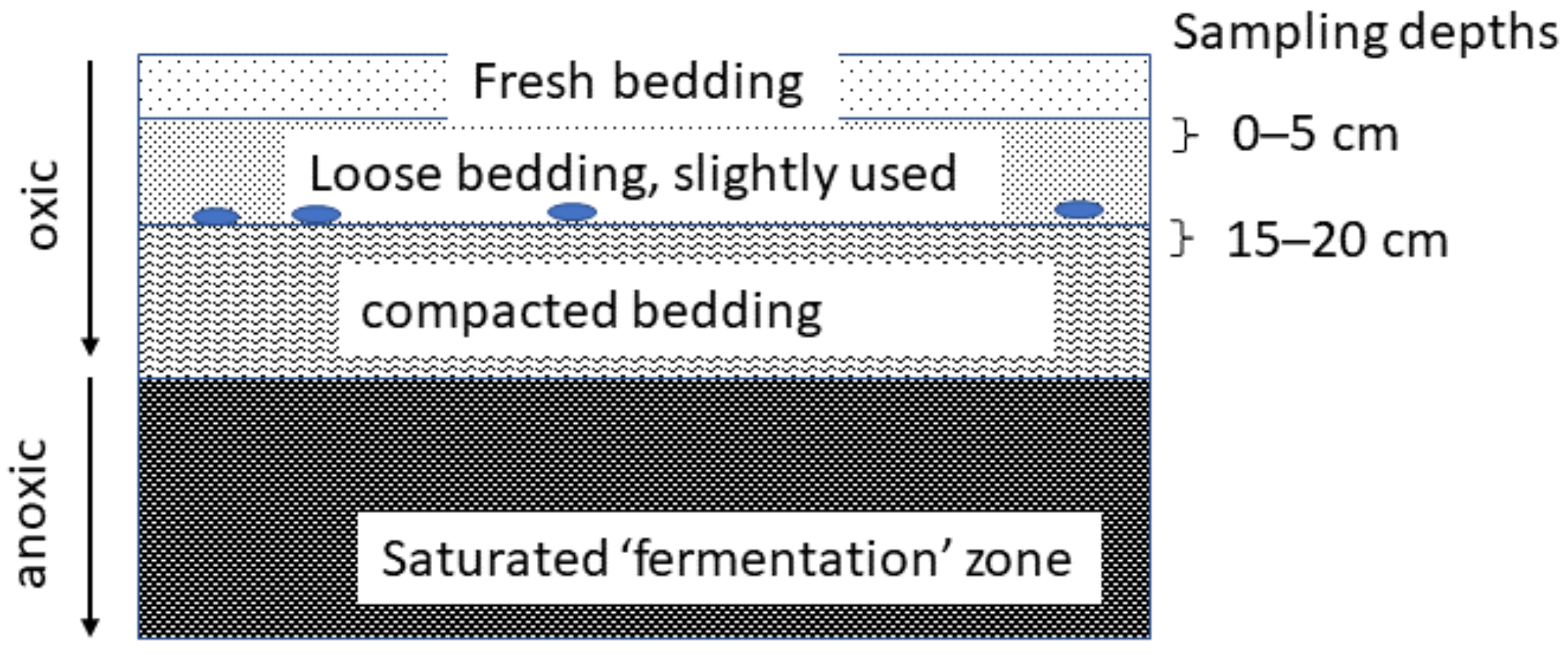
Figure 1. Vertical stratification of compost bedded pack. Loose fresh bedding was removed and two layers of oxic conditions were sampled. The upper (0–5 cm) layer was a relatively dry zone with slightly used, loose bedding. The second layer (15–20 cm) layer was compacted and thermophilic. Temperature and oxygen measurements (ovals) were taken at the interface between two oxic layers, not the saturated ‘fermentation’ zone.
Dairy 03 00042 g001
Figure 3. Relative abundance of bacterial taxa (out of 220) that are represented in all 100% samples split by depth and month at Farm C. The first date (October) is just the top layer because it is the beginning of the pack building. Sample size is illustrated above each bar. Microbiome varied by time (pseudo-F = 1.57, p = 0.002) and depth (pseudo-F = 1.55, p = 0.031).
Dairy 03 00042 g003
Figure 4. Relative abundance of fungal taxa (out of 82) that are represented in 45% of all samples split by depth and month at Farm C. The first date (October) is just the top layer because it is the beginning of the pack building. Sample size is illustrated above each bar. Microbiome varied by time (pseudo-F = 1.27, p = 0.003).
Dairy 03 00042 g004
Figure 5. Mean monthly relative abundance of Pichia kudriavzevii (blue line, left y-axis) as a function of bedding temperature (orange line, right y-axis) in the oxic zone through time at Farm C.
Dairy 03 00042 g005
Figure 6. Redundancy Analysis biplot of carbon utilization by Trichocomaceae species constrained by genus. Points represent 50 best-fitting (out of 95) carbon substrates shown using data compiled from 40 isolates from the results of the Filamentous Fungal identification plates. Response values are transformed as log (x + 0.01) and carbons were centered and standardized. An uncultured Hypocreaceae is included as a reference outlier. Eigenvalues are 0.2458, 0.0921, 0.0731, and 0.0405 for the x-, y-, third, and fourth axes, respectively (pseudo-F = 2.2, p = 0.004). Analysis performed with Canoco Ver 5 software.
Dairy 03 00042 g006
Figure 7. Distance-based Redundancy Analysis biplot of Bray–Curtis dissimilarity of bacterial (a) and fungal (b) communities constrained by farm (tan: Farm B, blue: Farm C, red: Farm D, green: Farm L, yellow: Farm S). Points represent monthly samples for each farm. Bacterial microbiome varied by farm (pseudo-F = 4.09, p = 0.001), time (pseudo-F = 1.72, p = 0.001), and depth (pseudo-F = 2.37, p = 0.001). Fungal microbiome varied by farm (pseudo-F = 2.52, p = 0.001), and time (pseudo-F = 1.55, p = 0.001). Post hoc comparison among farms: C and D were similar, and B and D were similar, but all other comparisons were different for both bacteria (p fdr < 0.03) and fungi (p fdr < 0.04). Ordination calculated via capscale and model terms compared via adonis2 in vegan package for R software version 4.0.2 (R Core Team, Vienna, Austria).
Dairy 03 00042 g007
Table 1. Bedding material, stocking density and median temperature and oxygen for each farm. Wood shavings were dried compressed pine, sawdust was freshly milled pine. Wood chips were unidentified type, except Farm C used hardwood chips. Hay was farm-grown. Straw was purchased (Farm S) or farm-grown (B). Primary bedding represents approximately 75% of bedding, whereas secondary bedding represents 25%. Bedding style is either deep bedded pack (DBP) or compost bedded pack (CBP). More management details for each farm are available in Table S1.
Management PracticeFarm Name
CBDLS
Bedding StyleDBPDBPDBPCBPDBP
Primary Beddinghayhayhaysawduststraw
Secondary Beddingwood chipswood chips, strawwood chipshaywood shavings
BreedHolstein—Jersey crossJerseyJerseyHolstein—Jersey crossJersey
Stocking Density (m2/cow)8.187.438.239.312.01
Bedding (kg/cow/day) a4.142.4512.475.335.02
Median Temperature °C30.5 (n = 12)41 (n = 4)36 (n = 3)33.5 (n = 4)13 (n = 2)
Median Oxygen (%)8.66 (n = 9)1.23 (n = 4)2.3 (n = 3)2.10 (n = 4)7.8 (n = 1)
a Farmer reported data.
Table 2. Trichocomaceae isolates from bedding material collected at Farm C between October 2015 and May 2016. Species were identified by Sanger sequencing of the amplicon generated by NS7-F and ITS2-R primers and contigs assembled using CAP3 [27]. Nucleotide sequences were blasted in NCBI, all yielding E values of 0. Percentage nucleotide matching for the assembled contig sequence is illustrated.
Isolate Contig Sample Source aIsolation Media bCulture Temperature (°C)Species% Match
1BPCZA22Aspergillus niger99.67
5BPCZA22Penicillium solitum strain 20-0199.83
6BPCZA22Penicillium solitum strain 20-0199.83
9TSCYA35Aspergillus fumigatus strain cy01899.65
11TSCYA35Uncultured fungus CMH60399.78
14BPUnknown22Penicillium solitum strain 20-0199.82
16TSCYA22Penicillium solitum strain 20-0199.82
17TSCYA22Penicillium solitum strain 20-0199.82
19TSCZA22Penicillium solitum strain 20-0199.83
21BPDG1822Penicillium solitum strain 20-0199.83
27BPYE20S22Penicillium chrysogenum strain ZJ-T299.65
32BPYE20S22Aspergillus fumigatus strain cy01899.65
33TSDG1822Penicillium janthinellum series99.65
37BPDG1822Penicillium solitum strain 20-0195.65
39BPHumidity Chamber22Penicillium solitum strain 20-0199.82
41TSCZA22Hypocreales uncultured99.26
47TSCYA22Talaromyces radicus (Penicillium radicum)96.91
95BPCMA22Aspergillus luchuensis100
129BPHumidity Chamber34Talaromyces verruculosus (Penicillium verruculosum)99.13
CG48BPYES34Aspergillus fumigatus strain cy01899.82
CH4TSDG1835Aspergillus fumigatus strain cy01899.83
a Source (BP: bedded pack, TS; tie-stall); b CMA: Cornmeal Agar, CYA: Czapek Yeast Autolysate agar, CZA: Czapek-Dox Agar, DG18: Dichloran Glycerol 18% agar, MEA: Blakeslee Malt Extract Autolysate agar, YES: Yeast extract sucrose agar, YE20S: Yeast Extract Sucrose 20% Salt agar.
Table 3. Sanger Sequencing results for cultured yeast isolates and their respective physiological characteristics. Samples collected in December 2019, January 2020, and February 2020 at Farms M (milk only) and C (milk and bedding). Species were identified by Sanger sequencing of the amplicon generated by NS7-F and ITS2-R primers and contigs assembled using CAP3 [27]. Nucleotide sequences were blasted in NCBI, all yielding E values of 0. Percentage nucleotide matching for the assembled contig sequence is illustrated.
Isolate ContigFarmSample Source aIsolation Media bSpecies% MatchProteaseHemolytic35 °CCatalasePeroxidase
1MBTMRBCDiutina rugosa95.710110
2MBTMRBCDiutina rugosa95.710110
3MBTMRBCDiutina rugosa95.710110
5MBTMRBCUncultured Tremellomycetes98.910000
6CHigh SCCCAFUncultured Tremellomycetes99.810000
7CBTMCAFPichia holstii97.210100
8CBTMAWPYPichia kudriavzevii99.810100
9CBTMAWPYWickerhamomyces anomalus99.800100
10CBTMCYAWickerhamomyces anomalus82.410100
11CBTMCYAWickerhamomyces anomalus99.710100
12CQMBlood AgarDiutina rugosa96.010100
13CBPBHIDiutina catenulata100.011100
14CBPBHIWickerhamomyces anomalus99.710100
15CBPRUMPichia fermentans99.811100
16CBPRUMPichia fermentans99.610010
17CBPCAFPichia fermentans99.610100
22CBPYLA(Candida) glaebosa99.310000
23CBPBHIWickerhamomyces anomalus100.010000
25CBPRUM(Candida) glaebosa99.010000
26CBPCAFHyphopichia pseudoburtonii99.610000
27CTSSDA(Candida) glaebosa98.710000
28CTSSDA(Candida) glaebosa98.410100
29CTSSDADebaryomyces hansenii100.010000
30CTSRUMDebaryomyces hansenii99.810010
31CTSRUMDebaryomyces hansenii100.010010
32CTSRUMHyphopichia burtonii99.810100
33CTSCAFDiutina catenulata99.410110
34CTSCAFDebaryomyces hansenii100.010010
35CTSBHIDebaryomyces hansenii100.010000
36CTSBHIDebaryomyces hansenii99.810000
37CTSBHIDiutina catenulata100.010100
38CTSBHIDebaryomyces hansenii99.710100
39CTSYLAWickerhamomyces anomalus99.710100
40CTSYNGHyphopichia burtonii99.810100
41CBPSDADebaryomyces hansenii99.710100
42CBPCAFWickerhamomyces anomalus100.010100
43CBPYLADebaryomyces coudertii99.500100
44CBPBHIDiutina catenulata100.010100
45CBPYLAWickerhamomyces anomalus99.710100
46CBPSDADiutina catenulata100.010100
a Source (BTM: bulk tank milk, QM: quarter milk, BP: bedded pack, TS: tie-stall, High SCC: high somatic cell count); b Media (AWPY: Acidified Weak Potato Dextrose Agar with Yeast Extract, BHI: Brain Heart Infusion Agar, CAF: Caffeic Acid Agar, CYA: Czapek Yeast Autolysate Agar, RBC: Rose Bengal Chloramphenicol, RUM: Artificial Rumen Agar, SDA: Sabouraud Dextrose Agar, YLA: Yeast Lactose Agar, YNG: Yeast Sodium Agar).
Table 4. Milk isolates cultured from milk also present in bedded pack samples from each of the five farms in phase 1 of the case study (n = 40). Bedding samples were estimated by amplicon sequencing [15]. Sequence counts were converted to a proportion of total within-sample counts (relative abundance) and median relative abundance of OTUs with matching binomial epithets were calculated on a species basis among samples in which the matching taxa were present.
Pichia kudriavzeviiPichia holstiiWickerhamomyces anomalus
Samples in which isolate is present90%8%3%
Median relative abundance among samples in which isolate is present12.2%0.1%0.9%
Publisher’s Note: MDPI stays neutral with regard to jurisdictional claims in published maps and institutional affiliations.