Article

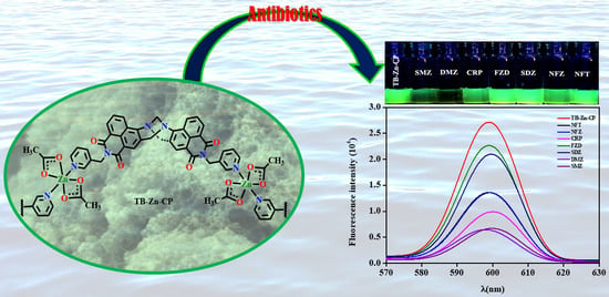
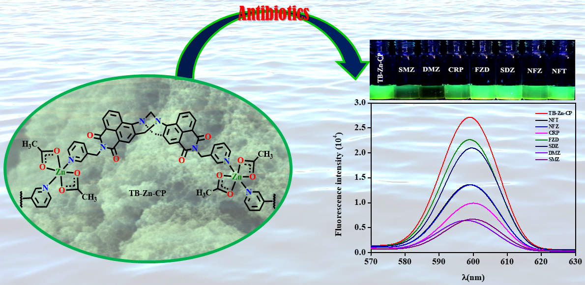
Differential Fluorescent Chemosensing of Antibiotics Using a Luminescent Zn(II) Coordination Polymer Based on a 4-Amino-1,8-naphthalimide Tröger’s Base Fluorophore

by
Purti Patel
,
Mannanthara Kunhumon Noushija
and
Sankarasekaran Shanmugaraju
*
Department of Chemistry, Indian Institute of Technology Palakkad, Palakkad 678557, Kerala, India
*
Author to whom correspondence should be addressed.
These authors contributed equally to this work.
Chemistry 2024, 6(1), 237-248; https://doi.org/10.3390/chemistry6010011
Submission received: 22 December 2023 / Revised: 1 February 2024 / Accepted: 5 February 2024 / Published: 7 February 2024
(This article belongs to the Section Supramolecular Chemistry)

Abstract

:
The design and development of useful chemosensors for the ultra-trace detection of environmental pollutants and contaminants is a topical area of research. Herein, we report a new nanoscale emissive Zn(II) coordination polymer (TB-Zn-CP) for differential fluorescence sensing of various antibiotics in water. TB-Zn-CP was synthesized using a unique V-shaped green emitting 4-amino-1,8-naphthalimide Tröger’s base (TBNap) fluorophore. The structural and morphological features of TB-Zn-CP were characterized by various standard spectroscopic and microscopy techniques. The fluorescence titration studies in water demonstrated a remarkable sensitivity and differential fluorescence sensing properties of TB-Zn-CP for the fast detection of different antibiotics. Among different antibiotics, chloramphenicol (CRP), 1,2-dimethyl-5-nitroimidazole (DMZ), and sulfamethazine (SMZ) displayed the highest fluorescence-quenching efficiency and superior sensitivity in their detection. The differential sensing capability of TB-Zn-CP was also indicated by visualizable color changes. The Stern–Volmer quenching constant KSV was determined to be in the order of 103–104 M−1, and the sensitivity was shown to be at a nanomolar (10−9 M) level. All these results confirm that TB-Zn-CP can be a potential and practically useful polymeric sensor for differential fluorescence and visual detection of different antibiotics in water.

Graphical Abstract

1. Introduction

Since the 1940s, various bacterial infections and illnesses have been treated by different antibiotics [1]. Today, antibiotics have become a cornerstone in treating various infections in humans as well as in animals, experiencing widespread use in healthcare and animal farming industries [2,3]. Additionally, in the natural environment, antibiotics serve as vital tools for the survival and competitive advantage of organisms, often referred to as the weapons of producer microorganisms [4]. These microorganisms employ antibiotics to compete with other microbes, eliminating rival bacteria and establishing dominance in the microbial community [5]. However, the extensive use of antibiotics by living systems has led to their large accumulation in water streams and ecosystems [6]. The environmental contamination due to antibiotics stems from their excessive consumption, improper excretion practices, and inadequate disposal methods, posing risks to human health and environmental well-being [7]. Even though they are important in both animal and human life, the indiscriminate and excessive use of antibiotics considerably contributes to the emergence of resistant strains [1]. According to the World Health Organization (WHO), “antibiotic resistance” poses a severe economic and sociological risk to public health [2,3]. Due to their lengthy persistence in the natural environment and the harm that they do to living things, several antibiotics have been identified as potential environmental pollutants, including penicillin, fluoroquinolones, lactams, tetracyclines, and so on [8]. For instance, continuous intake of antibiotics can weaken immunity and lead to hepatotoxicity, allergic reactions, gastrointestinal disturbances, genetic problems, and various diseases [9,10]. Over the next 35 years, 300 million premature deaths are predicted worldwide if the current trend holds [11,12]. Therefore, addressing the overuse and inappropriate disposal of antibiotics is crucial to mitigating the growing threats of antibiotic resistance and safeguarding both human health and the environment. Dose modification and trace-level antibiotic monitoring are highly recommended to reduce the prevalence of antibiotic-resistant bacteria [13]. For the well-being of human health and to protect the environment, it is essential to find an efficient and feasible detection method with high selectivity for the sensitive analysis and identification of antibiotics.
Several analytical techniques have been used so far for precise and trustworthy monitoring of antibiotic residues in natural resources, including liquid chromatography–mass spectrometry (LC-MS), high-performance liquid chromatography (HPLC), electro-kinetic capillary chromatography (EC), and surface-enhanced Raman spectroscopy (SERS) [14,15,16,17]. All of these techniques, meanwhile, are time-consuming and expensive and call for sophisticated machinery and skilled labor [16,17]. Additionally, despite the development of numerous techniques, including photolysis, hydrolysis, thermolysis, technical oxidation processes, and biodegradation, all of which are based on chemical treatment, technologies for the efficient detection and removal of antibiotics are still in their infancy. In recent years, researchers have been very concerned with developing portable, trustworthy, and affordable methodologies and technologies for selective sensing and determining antibiotic contaminant concentrations in water bodies [18,19,20]. Luminescence-based detection and surface adsorption methods are promising for antibiotic sensing and removal due to their simplicity, high sensitivity, and enhanced adsorption efficiency [21,22]. However, the challenge lies in finding a suitable material that can exhibit highly selective and superior sensitivity for the detection of particular types of antibiotics [23,24,25]. Coordination polymers (CPs), an emerging class of organic–inorganic hybrid materials, are regarded as an advantageous platform for detection, sensing [26,27], and adsorption [28,29] applications due to their unique electronic and optical properties, high surface area, easily adaptable structures, and functions.
On the other hand, fluorescence-based sensing is known for its high sensitivity and straightforward visualization and has become a promising and cost-effective sensing method for the detection of environmental pollutants [30,31,32,33,34]. In light of this, in the past years, luminescent CPs have gained special research attention for their potential in detecting antibiotics [35,36,37,38,39,40,41,42]. The fluorescence properties and sensing capabilities of CPs can be easily tailored by selecting and combining pre-designed multidentate organic linkers and metal ions, or by incorporating suitable fluorescent signal molecules [43,44,45,46]. Reducing the particle size of bulk crystalline coordination polymers (CPs) to the micro- and nanoscale is demonstrated to enhance their solution processibility in aqueous environments, elevate their interaction with analytes, and improve their sensing capacity [47]. However, only a limited number of luminous nanoscale coordination polymers have been developed to date and utilized as potential fluorescence sensors for antibiotic detection. To enhance the selectivity of CPs in detecting antibiotics, a proposed method involves incorporating Lewis base functional groups, such as amines and pyridines. These groups can potentially detect antibiotics through intermolecular H-bonding interactions with the hydrogen bond donor groups like hydroxy and amine groups of antibiotics [23,48,49,50,51,52].
Considering these aspects, herein, we designed and synthesized a new fluorescent nanoscale Zn(II) coordination polymer, TB-Zn-CP, built from a green-emitting 3-picolyl-4-amino-1,8-naphthalimide Tröger’s base (TBNap) fluorophore [53,54,55,56,57]. The intrinsic chirality of Tröger’s base moiety favors the unusual V-shaped configuration of two 4-amino-1,8-naphthalimides, which facilitates the guest binding through numerous supramolecular interactions and demonstrates the enhanced sensing capability for analyte detection. We foresaw that Tröger’s base’s Lewis base properties would boost the selectivity of TB-Zn-CP for the differential detection of antibiotics via intermolecular hydrogen-bonding interactions [58]. Indeed, TB-Zn-CP has demonstrated remarkable sensitivity and differential sensing capabilities for the rapid detection of antibiotics in an aqueous medium. Fluorescent titration studies have revealed that TB-Zn-CP exhibits preferential binding of Chloramphenicol (CRP), 1,2-Dimethyl-5-nitroimidazole (DMZ), and Sulfamethazine (SMZ) antibiotics, displaying superior sensitivity in their detection. Notably, other antibiotics have shown minimal to moderate binding affinity and, thus, fluorescence emission quenching. The differential fluorescence-sensing ability of TB-Zn-CP towards different antibiotics was also seen by sharp color changes before and after the mixing of antibiotics in water.

2. Experimental

2.1. Materials

All reagents, solvents, and starting precursors were procured from Sigma-Aldrich (Bangalore, India) and utilized without further purification. For the synthesis of TBNap and TB-Zn-CP, commercially available reagent-grade chemicals were employed. Spectroscopy-grade solvents from Merck were used without additional purification. 4-nitro-1,8-naphthalic anhydride, 3-picolylamine, palladium on carbon (10 wt.% loadings), paraformaldehyde, and trifluoroacetic acid (TFA) were sourced from Sigma-Aldrich and used in their as-received state. Reagent-grade antibiotics analytes, essential for sensing studies, were obtained from the Tokyo Chemical Industry (Hyderabad, India) and used as purchased. Deuterated solvent (CD3)2SO, employed for NMR analyses of the monomer, was acquired from Sigma-Aldrich. The synthesis of organic linker TBNap followed the procedure outlined in the literature [59].

2.2. Instrumentations

The as-synthesized TB-Zn-CP was dried under a high vacuum to remove the trapped solvents before its characterization. The FT-IR spectrums of polymer TB-Zn-CP and ligand TBNap were obtained using the Shimadzu Scientific Instruments (IR Tracer 100) (Science Park, Singapore, Singapore) equipped with an ATR sampler. Thermogravimetric analysis (TGA) of TB-Zn-CP was conducted on the PerkinElmer STA 8000 analyzer (Thane, India), which featured an ultra-microbalance with a sensitivity of 0.1 mg. The analysis spanned a temperature range from 30 °C to 1000 °C, with a scan rate of 10 °C min−1 under N2 purge. Powder X-ray diffraction analysis of TB-Zn-CP and TBNap was carried out on a Rigaku (XRD Smart Lab) diffractometer operating at 9 kW (Bangalore, India). Dynamic Light Scattering (DLS) analysis of TB-Zn-CP was carried out using Malvern Panalytical Zetasizer (Bangalore, India) at PSG Institute of Advanced Studies, Coimbatore. The particle size analysis was performed by keeping the sample temperature at 25 °C. The surface morphology of TB-Zn-CP was visualized using field emission scanning electron microscopy (FE-SEM) on a Carl Zeiss (Gemini SEM 300) microscope. Sample preparation involved drop-casting the aqueous suspension of the as-synthesized polymer onto a stub, followed by coating with Au and drying under vacuum before imaging. The UV–visible absorption titration studies were carried out using a Thermo Fisher scientific absorption spectrometer in a 1 cm quartz cuvette, with baseline correction applied to all measurements (Waltham, MA, USA). The fluorescence titration studies were performed on a PerkinElmer FL-8500 fluorescence spectrophotometer (Waltham, MA, USA), which featured a Xenon arc (150 W) light source. The measurements were conducted at a constant temperature of 298 K, utilizing a quartz cuvette and a scan rate of 1200 nm min−1. The results obtained were graphically represented using Origin Pro 8.5 software.

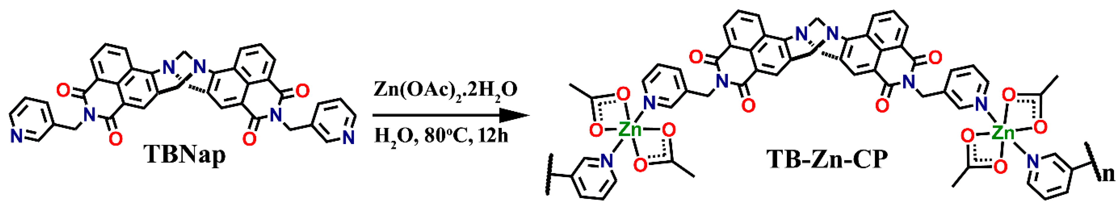
2.3. Synthesis and Characterization of Coordination Polymer TB-Zn-CP

The organic linker TBNap (10 mg, 0.016 mmol, 1.0 eq.) was taken in a glass vial along with 2 mL of water and then sonication to achieve a well-dispersed suspension. Subsequently, an aqueous solution of Zn(OAc)2·2H2O (3.5 mg, 0.016 mmol, 1.0 eq.) was added dropwise under constant stirring. After a few minutes of stirring at room temperature, the reaction mixture turned turbid. The reaction mixture was further stirred overnight at 80 °C. After cooling to room temperature, the reaction mixture was centrifuged, and the resulting precipitate was washed with water (2 × 10 mL) and then dried in an oven for 1 h to isolate the targeted product coordination polymer TB-Zn-CP as a yellow powder in 50% yield (calculated based on the organic linker’s molecular weight). The formation of TB-Zn-CP was fully characterized by using various spectroscopy and microscopy techniques.

2.4. Fluorescence Sensing Studies

To prepare the stock solution, 2 mg of TB-Zn-CP was dispersed in 10 mL of deionized water. The resulting suspension was sonicated for 30 min and then allowed to age for one day to form a uniformly dispersed colloidal suspension. Similarly, 1 mM stock solutions of different antibiotics of each were freshly prepared in water. A 50 μL aliquot of the TB-Zn-CP colloidal suspension was taken with 1950 μL of water in a 3 mL cuvette for the fluorescence sensing studies. Subsequently, 1 mM stock solutions of individual antibiotics (20 μL at once) were incrementally added to the cuvette containing TB-Zn-CP colloidal suspension. The fluorescence emission intensity of TB-Zn-CP before and after the addition of antibiotics was measured. For all the fluorescence titration experiments, the sample was excited at λ = 300 nm, and emission was monitored in the range of λ = 550 to 650 nm at 298 K. For all the measurements, the slit width was kept at 10 nm.
The percentage of fluorescence-quenching efficiency of TB-Zn-CP towards antibiotics was determined using the following equation:
Quenching efficiency (%) = (I0 − I)/I0 × 100
where I0 is the initial fluorescence emission intensity of TB-Zn-CP in water, and I is the fluorescence emission intensity after the addition of antibiotic solutions.
The relative changes (I0/I) in fluorescence emission intensity of TB-Zn-CP colloidal suspension with increasing the concentration [Q] of antibiotics was demonstrated by the following the Stern–Volmer equation:
(I0/I) = 1 + KSV[Q]
The Stern–Volmer quenching constant (KSV) was determined from the slope of the linear plot of I0/I vs. Q.
The limit of detection (LoD) was calculated from the fluorescence titration profile using the following equation:
Detection limit = 3σ/K
where σ is the standard deviation of the initial fluorescence emission intensity in the absence of the antibiotic analytes, and K is the slope of the linear calibration curve.

3. Results and Discussion

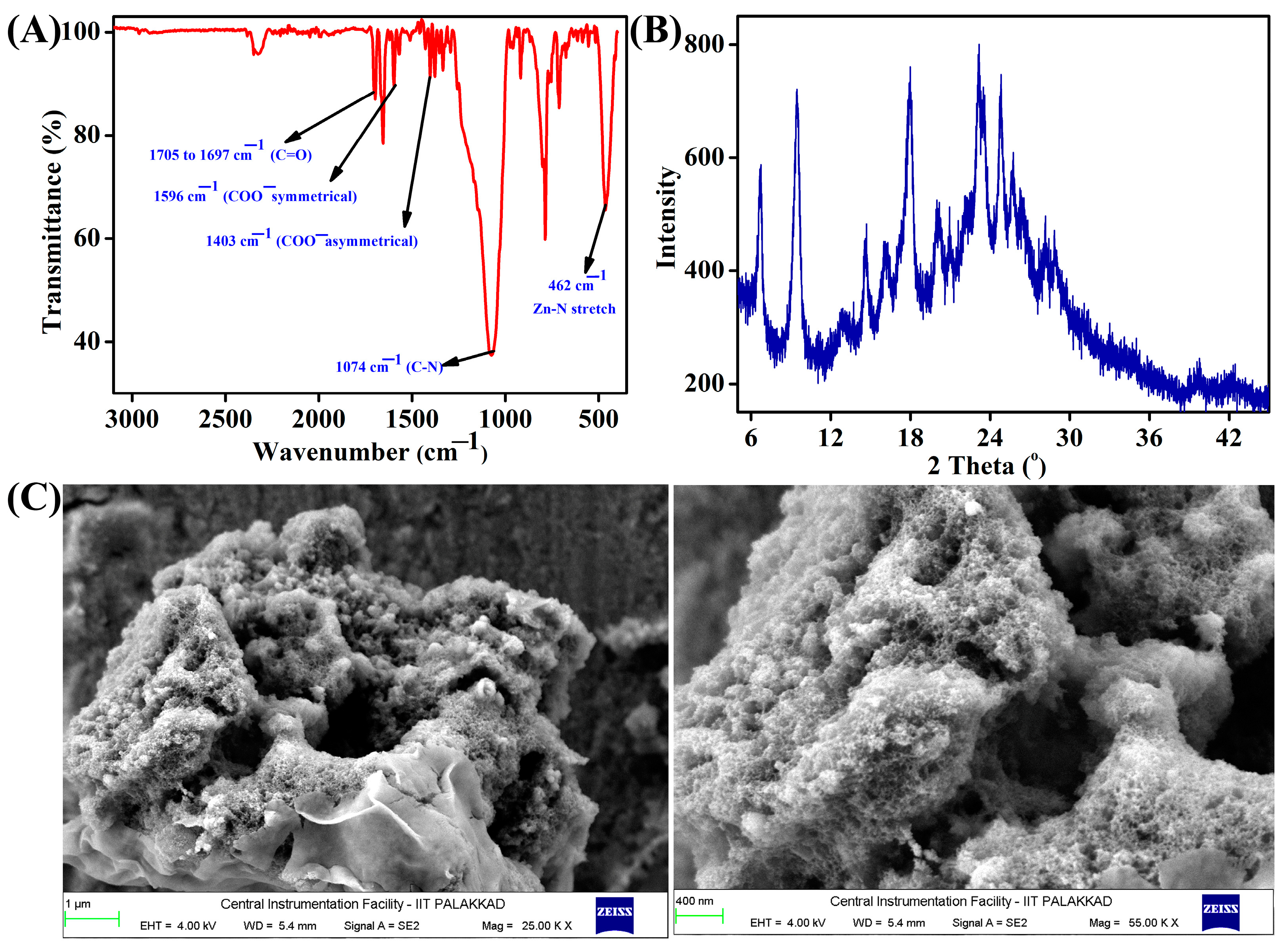
3.1. Synthesis and Characterization of TB-Zn-CP

The organic linker TBNap was freshly synthesized following the procedure reported in the literature [59]. The commercially purchased precursor, 4-nitro-1,8-naphthalic anhydride, was condensed with 3-picolylamine to isolate N-(3-picolyl)-4-nitro-1,8-naphthalimide, which was subsequently reduced to its corresponding amine derivative. N-(3-picolyl)-4-amino-1,8-naphthalimide was then reacted with paraformaldehyde in the presence of trifluoroacetic acid to yield TBNap in quantitative yield. As shown in Scheme 1, the Zn(II) coordination polymer TB-Zn-CP was obtained by stirring Zn(OAc)2·2H2O (1.0 eq.) and the organic ligand TBNap (1.0 eq.) in H2O (4 mL) at 80 °C for 12 h. The well-dispersed TBNap aqueous suspension was changed to a yellow colloidal suspension after the addition of Zn(OAc)2·2H2O, which evidences the formation of the infinite coordination polymer. The heavy precipitate that formed after cooling to room temperature was collected through centrifugation, and the precipitate was washed with H2O to isolate the coordination polymer TB-Zn-CP as a yellow powder in 50% yield. TB-Zn-CP was characterized by using various standard spectroscopy and microscopy techniques.
As shown in Figure 1A, the FTIR spectrum of TB-Zn-CP showed two sharp peaks at 1596 cm−1 (for symmetrical) and 1403 cm−1 (for asymmetrical), corresponding to the coordinated acetate (-CH3COO-) anions; these peaks were absent in the FTIR spectrum of TBNap (see Figure S1, Supplementary Materials). Two sharp bands in the range of 1705 to 1697 cm−1 were assigned to the carbonyl (-C=O-) stretching frequencies, and a strong band appeared at 462 cm−1, which was ascribed to the Zn−N coordination bond. The appearance of an intense band at 1074 cm−1 confirmed the presence of Tröger’s base unit in the isolated solid material. The X-ray powder diffraction analysis of TB-Zn-CP showed relatively broader peaks, indicating that the isolated coordination polymer is microcrystalline (Figure 1B). The particle size of the well-dispersed colloidal suspension of TB-Zn-CP was measured by Dynamic Light Scattering (DLS) analysis. The average particle size of TB-Zn-CP in an aqueous medium is ~81 nm, confirming that TB-Zn-CP is a nanoscale polymeric material (see Figure S2, Supplementary Materials). Under a nitrogen atmosphere, the thermal gravimetric analysis (TGA) of TB-Zn-CP demonstrated thermal stability up to 400 °C, with approximately 20% of its initial mass retained at 700 °C. Notably, a marginal weight gain (~7%) was observed in the initial phase, likely due to the adsorption of N2 gas within the pores of the coordination polymer (Figure S3, Supplementary Materials). To determine the morphological feature, scanning electron microscopy (SEM) was employed, which revealed a nano-porous sponge-like morphology, as depicted in Figure 1C. Furthermore, the energy-dispersive X-ray spectroscopy (EDX) confirmed the presence of Zn, C, O, and N through distinctive peaks (Figure S4A, Supplementary Materials). Additionally, Quanta mapping highlighted a uniform distribution of Zn, C, O, and N across the entire sample of TB-Zn-CP (Figure S4B, Supplementary Materials).

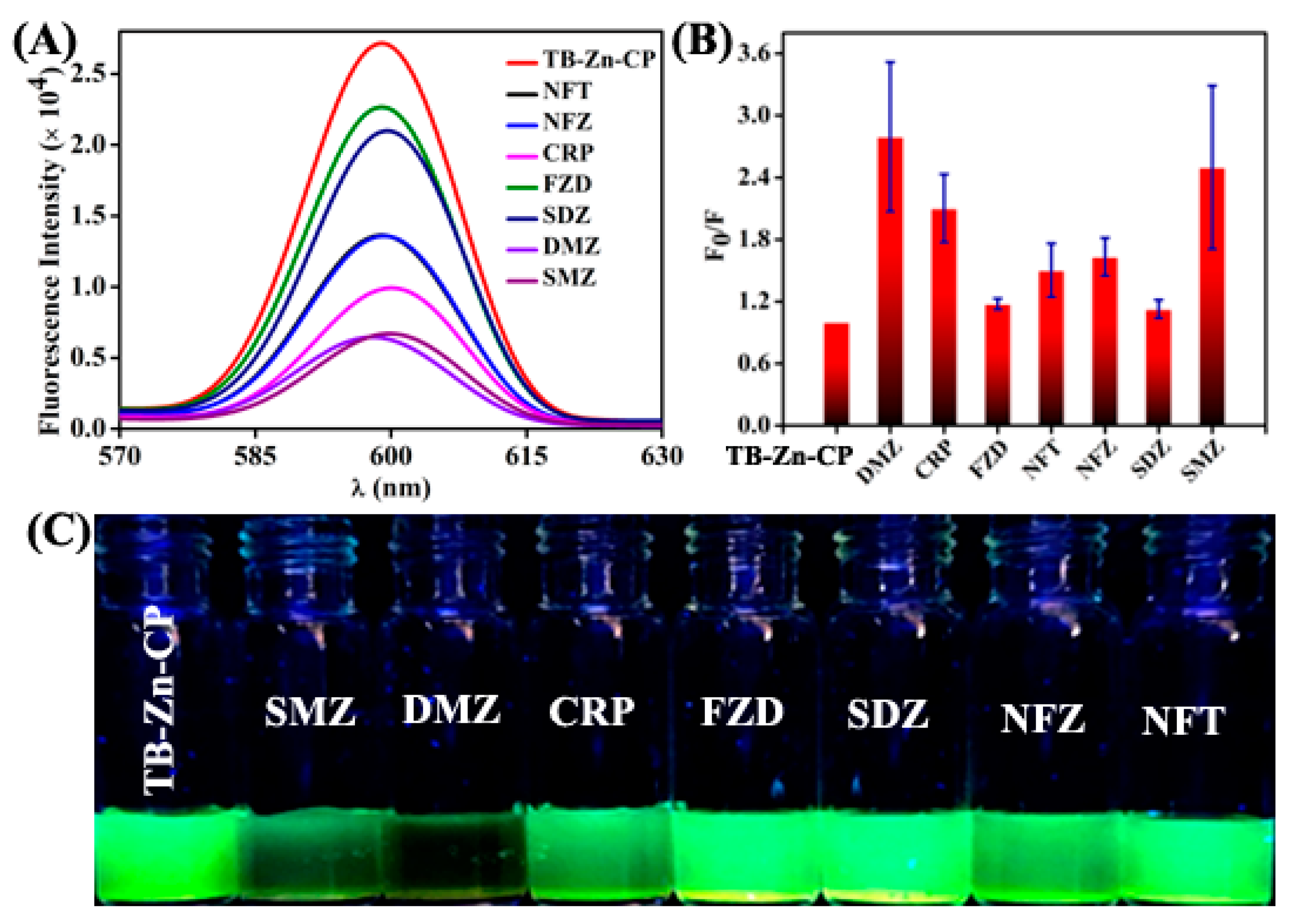
3.2. Fluorescence Sensing of Antibiotics

Amino-1,8-naphthalimide derivatives (Naps) are well-known classical fluorophores that are strongly colored and absorbed, and which emit in the visible region of the spectrum. Naps are highly emissive due to the excited state internal charge transfer (ICT) transition from the electron-rich amino group to the electron-deficient imide unit [53,54,55]. On the other hand, Tröger’s base is a unique V-shaped and C2-symmetric organic scaffold with a large internal hydrophobic cavity that can be exploited for host–guest chemistry [56,57]. Given this, over the past several years, we have developed a plethora of amino-1,8-naphthalimide Tröger’s bases (TBNaps) and employed them as a bifunctional organic scaffold for the development of multifaceted structures, functional materials, and porous organic polymers for their application in environmental-related studies and biomedicine [46,59]. Coordination polymers based on electronically inert Zn(II) metal ions and fluorescence organic linkers are shown to display ligand-based strong photoluminescence properties [46]. Further, it has been shown that the coordination linking of fluorescence organic ligands into a coordination network emits fluorescence emissions at an enhanced intensity, and this is ascribed to the long-range electronic communications between the adjacent ligands [45]. Herein, we combined the fluorescence Tröger’s base linker TBNap with Zn(II) ion to generate the nanoscale coordination polymer TB-Zn-CP and explored its fluorescence-sensing properties towards various antibiotics in a water medium. TB-Zn-CP was taken in Milli-Q water and sonicated to obtain a well-dispersed aqueous colloidal suspension, which was subsequently used for fluorescence sensing studies. The UV-visible absorption of TB-Zn-CP displayed the typical spectrum of a high-energy band at λ = 352 nm, corresponding to the π-π* transition, and a low-energy absorption band at λ = 419 nm due to the internal charge transfer (ICT) transition (Figure S5, Supplementary Materials). The fluorescence emission spectrum of TB-Zn-CP showed a broad emission band centered at λ = 599 nm due to the “push–pull”-based ICT transition (Figure S6, Supplementary Materials).
Inspired by the strong fluorescence emission, we next assessed the fluorescence-sensing property of TB-Zn-CP towards various antibiotics in water mediums. We chose different environmentally hazardous antibiotics, including Nitrofurantoin (NFT), Nitrofurazone (NFZ), Chloramphenicol (CRP), Furazolidone (FZD), Sulfadiazine (SDZ), 1,2-Dimethyl-5-nitroimidazole (DMZ), and Sulfamethazine (SMZ). First, a 1 mM stock solution of antibiotics was freshly prepared in Milli-Q water. To corroborate the selectivity of TB-Zn-CP in terms of sensing different antibiotics, we first measured the fluorescence emission intensity of TB-Zn-CP before and after the addition of various antibiotics. As depicted in Figure 2A,B, the coordination polymer TB-Zn-CP exhibited differential fluorescence-quenching responses for different antibiotics. Among the tested antibiotics, the DMZ, SMZ, and CRP antibiotics showed the largest fluorescence-quenching efficiency, while other antibiotics, NFT, NFZ, FZD, and SDZ, displayed moderate to poor fluorescence-quenching effects. The observed differential fluorescence-quenching responses for various antibiotics were also seen by sharp visual color changes (Figure 2C). Notably, the addition of SMZ and DMZ antibiotics changed the color of TB-Zn-CP from a bright greenish-yellow to colorless, but the mixing of other tested antibiotics showed no observable color changes.
We next performed fluorescence titration studies to probe the binding between TB-Zn-CP and the antibiotics DMZ, SMZ, and CRP in an aqueous medium. The initial fluorescence emission intensity of TB-Zn-CP was gradually decreased with the gradual increases in the concentrations (0.0 to 99.9 μM) of antibiotics (Figure 3A), and the quenching efficiency was found to be 60% for DMZ, 53% for SMZ, and 50% for CRP, indicating the strong interactions between sensor TB-Zn-CP and antibiotics. Except for the attenuation of fluorescence emission intensity, no other spectral changes, such as a shift in the emission maxima or the appearance of any peaks, were observed. In all cases, a linear Stern–Volmer plot was obtained, and the Stern–Volmer quenching constant (KSV) was determined to be 2.86 × 10⁴ M−1 for DMZ, 2.61 × 10⁴ M−1 for SMZ, and 2.85 × 10⁴ M−1 for CRP (Figure 3B). The calculated KSV values for different antibiotics from the fluorescence titration are summarized in Table 1. These values are comparable to those reported for other luminescence coordination polymer-based fluorescence sensors for antibiotics detection [60,61]. The high binding constant and sensing in a water medium make TB-Zn-CP a practically useful fluorescence sensor system for antibiotics detection.
To further investigate the binding between TB-Zn-CP and antibiotics, a UV–visible absorption titration study was performed using DMZ as representative antibiotics. Upon the addition of DMZ from 0.0 to 90.9 μM, the peak at λ = 419 nm decreased in its intensity, which was accompanied by an increase in intensity at λ = 317 nm and also a clear isosbestic point at λ = 387 nm, indicating the equilibrium ground state charge transfer (CT) complexation between TB-Zn-CP and DMZ (Figure 4A) [62]. A linear Stern–Volmer plot, significant changes in the absorption spectrum, and sharp visual color changes for TB-Zn-CP with the addition of DMZ antibiotic demonstrate that the observed fluorescence-quenching follows a static quenching mechanism via the formation of a ground-state non-fluorescent charge transfer complex between TB-Zn-CP and the antibiotics. The observed highest fluorescence-quenching for DMZ antibiotics, compared with the others, is presumably due to its more electron-deficient nature, caused by the electron-withdrawing -NO2 group. To check the sensitivity of TB-Zn-CP, further fluorescence titration studies were performed by taking the antibiotics DMZ, SMZ, and CRP at nanomolar (nM) concentrations. Interestingly, it was found that TB-Zn-CP can sense antibiotics at the nM level of concentrations. The limit of detection (LoD) values for DMZ, SMZ, and CRP were determined to be 9.3 nM, 12.9 nM, and 25.5 nM, respectively. This level of sensitivity is much lower than that reported for several other fluorescence sensors and within the allowed concentration level [60].

4. Conclusions

In summary, we have developed a new nitrogen-rich luminescent Zn(II) coordination polymer, TB-Zn-CP, based on 4-amino-1,8-naphthalimide Tröger’s base (TBNap) structural motif for differential fluorescence sensing of antibiotics in water mediums. The characterization using various spectroscopy and microscopy techniques indicated that TB-Zn-CP is a microcrystalline powder with sponge-like morphological features and exhibits high thermal stability. Owing to the strong ICT-based fluorescence emission, TB-Zn-CP was employed as a potential fluorescence chemosensor for antibiotic detection. TB-Zn-CP demonstrated differential fluorescence-sensing properties for different antibiotics, with high quenching efficiency and preferred sensing ability for antibiotics such as SMZ, DMZ, and CRP. The differential sensing ability of TB-Zn-CP was also seen by marked color changes, and fluorescence titration studies also conveyed that TB-Zn-CP can sense antibiotics at a nanomolar level of concentrations. In conclusion, all the studies discussed herein demonstrate that TB-Zn-CP can be a potential and practically useful fluorescence and visual sensor for the differential, quick, and ultra-sensitive sensing of antibiotic contaminants in water. Further work is in progress to design highly porous coordination polymer-based TBNap scaffolds and explore their applications as molecular adsorbents for the efficient adsorption and removal of emerging and persistent organic contaminants in water.

Supplementary Materials

The following supporting information can be downloaded at: https://www.mdpi.com/article/10.3390/chemistry6010011/s1, Figure S1: The FTIR spectrum of ligand TBNap; Figure S2: The particle size distribution data was obtained by DLS measurement for the polymer TB-Zn-CP in an aqueous medium; Figure S3: Thermogravimetric analysis (TGA) spectrum of TB-Zn-CP; Figure S4: (A) Energy dispersive X-ray spectroscopy (EDX) data and (B) Quanta mapping images for TB-Zn-CP; Figure S5: UV-visible absorption spectrum of TB-Zn-CP measured in water; Figure S6: The fluorescence emission spectrum of TB-Zn-CP was recorded in water.

Author Contributions

Investigation, M.K.N. and P.P.; writing—original draft preparation, M.K.N., P.P. and S.S.; writing—review and editing, M.K.N. and S.S.; supervision, S.S.; project administration, S.S.; funding acquisition, S.S. All authors have read and agreed to the published version of the manuscript.

Funding

Indian Institute of Technology Palakkad (ERG research grant 2023-168-CHY-SHS-ERG-SP to S.S.) and Science and Engineering Research Board, India (EMEQ research grant EEQ/2023/000386 to S.S.).

Data Availability Statement

The raw data supporting the conclusions of this article will be made available by the authors on request.

Acknowledgments

The authors thank the Central Instrumentation Facility (CIF) at IIT Palakkad for its research facilities. We gratefully acknowledge the Indian Institute of Technology Palakkad (ERG research grant 2023-168-CHY-SHS-ERG-SP to S.S.) and the Science and Engineering Research Board (EMEQ research grant EEQ/2023/000386 to S.S.), India, for the financial support.

Conflicts of Interest

The authors declare no conflicts of interest.

References

  1. Mesak, L.R.; Miao, V.; Davies, J. Effects of Subinhibitory Concentrations of Antibiotics on SOS and DNA Repair Gene Expression in Staphylococcus aureus. Antimicrob. Agents Chemother. 2008, 52, 3394–3397. [Google Scholar] [CrossRef]
  2. Yi, H.; Huang, D.; Qin, L.; Zeng, G.; Lai, C.; Cheng, M.; Ye, S.; Song, B.; Ren, X.; Guo, X. Selective Prepared Carbon Nanomaterials for Advanced Photocatalytic Application in Environmental Pollutant Treatment and Hydrogen Production. Appl. Catal. B Environ. 2018, 239, 408–424. [Google Scholar] [CrossRef]
  3. Paul, M.; Shani, V.; Muchtar, E.; Kariv, G.; Robenshtok, E.; Leibovici, L. Systematic Review and Meta-Analysis of the Efficacy of Appropriate Empiric Antibiotic Therapy for Sepsis. Antimicrob. Agents Chemother. 2010, 54, 4851–4863. [Google Scholar] [CrossRef]
  4. Huang, D.; Wang, X.; Zhang, C.; Zeng, G.; Peng, Z.; Zhou, J.; Cheng, M.; Wang, R.; Hu, Z.; Qin, X. Sorptive Removal of Ionizable Antibiotic Sulfamethazine from Aqueous Solution by Graphene Oxide-Coated Biochar Nanocomposites: Influencing Factors and Mechanism. Chemosphere 2017, 186, 414–421. [Google Scholar] [CrossRef]
  5. Davies, J. What Are Antibiotics? Archaic Functions for Modern Activities. Mol. Microbiol. 1990, 4, 1227–1232. [Google Scholar] [CrossRef]
  6. Zhang, C.; Lai, C.; Zeng, G.; Huang, D.; Yang, C.; Wang, Y.; Zhou, Y.; Cheng, M. Efficacy of Carbonaceous Nanocomposites for Sorbing Ionizable Antibiotic Sulfamethazine from Aqueous Solution. Water Res. 2016, 95, 103–112. [Google Scholar] [CrossRef]
  7. Klein, E.Y.; Van Boeckel, T.P.; Martinez, E.M.; Pant, S.; Gandra, S.; Levin, S.A.; Goossens, H.; Laxminarayan, R. Global Increase and Geographic Convergence in Antibiotic Consumption between 2000 and 2015. Proc. Natl. Acad. Sci. USA 2018, 115, E3463–E3470. [Google Scholar] [CrossRef]
  8. Morrison, L.; Zembower, T.R. Antimicrobial Resistance. Gastrointest. Endosc. Clin. 2020, 30, 619–635. [Google Scholar] [CrossRef]
  9. Zhao, D.; Liu, X.-H.; Zhao, Y.; Wang, P.; Liu, Y.; Azam, M.; Al-Resayes, S.I.; Lu, Y.; Sun, W.-Y. Luminescent Cd (II)–Organic Frameworks with Chelating NH 2 Sites for Selective Detection of Fe (III) and Antibiotics. J. Mater. Chem. A 2017, 5, 15797–15807. [Google Scholar] [CrossRef]
  10. Zhu, X.-D.; Zhang, K.; Wang, Y.; Long, W.-W.; Sa, R.-J.; Liu, T.-F.; Lv, J. Fluorescent Metal–Organic Framework (MOF) as a Highly Sensitive and Quickly Responsive Chemical Sensor for the Detection of Antibiotics in Simulated Wastewater. Inorg. Chem. 2018, 57, 1060–1065. [Google Scholar] [CrossRef]
  11. Xu, S.; Shi, J.-J.; Ding, B.; Liu, Z.-Y.; Wang, X.-G.; Zhao, X.-J.; Yang, E.-C. A Heterometallic Sodium (i)–Europium (Iii)-Organic Layer Exhibiting Dual-Responsive Luminescent Sensing for Nitrofuran Antibiotics, Cr2O72− and MnO4 Anions. Dalton Trans. 2019, 48, 1823–1834. [Google Scholar] [CrossRef]
  12. Liu, X.-G.; Tao, C.-L.; Yu, H.-Q.; Chen, B.; Liu, Z.; Zhu, G.-P.; Zhao, Z.; Shen, L.; Tang, B.Z. A New Luminescent Metal–Organic Framework Based on Dicarboxyl-Substituted Tetraphenylethene for Efficient Detection of Nitro-Containing Explosives and Antibiotics in Aqueous Media. J. Mater. Chem. C 2018, 6, 2983–2988. [Google Scholar] [CrossRef]
  13. Mungroo, N.A.; Neethirajan, S. Biosensors for the Detection of Antibiotics in Poultry Industry—A Review. Biosensors 2014, 4, 472–493. [Google Scholar] [CrossRef]
  14. Li, Y.-T.; Qu, L.-L.; Li, D.-W.; Song, Q.-X.; Fathi, F.; Long, Y.-T. Rapid and Sensitive In-Situ Detection of Polar Antibiotics in Water Using a Disposable Ag–Graphene Sensor Based on Electrophoretic Preconcentration and Surface-Enhanced Raman Spectroscopy. Biosens. Bioelectron. 2013, 43, 94–100. [Google Scholar] [CrossRef]
  15. Cinquina, A.L.; Longo, F.; Anastasi, G.; Giannetti, L.; Cozzani, R. Validation of a High-Performance Liquid Chromatography Method for the Determination of Oxytetracycline, Tetracycline, Chlortetracycline and Doxycycline in Bovine Milk and Muscle. J. Chromatogr. A 2003, 987, 227–233. [Google Scholar] [CrossRef]
  16. Wickramanayake, P.U.; Tran, T.C.; Hughes, J.G.; Macka, M.; Simpson, N.; Marriott, P.J. Simultaneous Separation of Nitrofuran Antibiotics and Their Metabolites by Using Micellar Electrokinetic Capillary Chromatography. Electrophoresis 2006, 27, 4069–4077. [Google Scholar] [CrossRef]
  17. Aguilera-Luiz, M.M.; Vidal, J.L.M.; Romero-Gonzalez, R.; Frenich, A.G. Multi-Residue Determination of Veterinary Drugs in Milk by Ultra-High-Pressure Liquid Chromatography–Tandem Mass Spectrometry. J. Chromatogr. A 2008, 1205, 10–16. [Google Scholar] [CrossRef]
  18. Paul, T.; Miller, P.L.; Strathmann, T.J. Visible-Light-Mediated TiO2 Photocatalysis of Fluoroquinolone Antibacterial Agents. Environ. Sci. Technol. 2007, 41, 4720–4727. [Google Scholar] [CrossRef]
  19. González, O.; Sans, C.; Esplugas, S. Sulfamethoxazole Abatement by Photo-Fenton: Toxicity, Inhibition and Biodegradability Assessment of Intermediates. J. Hazard. Mater. 2007, 146, 459–464. [Google Scholar] [CrossRef]
  20. Li, D.; Yang, M.; Hu, J.; Zhang, Y.; Chang, H.; Jin, F. Determination of Penicillin G and Its Degradation Products in a Penicillin Production Wastewater Treatment Plant and the Receiving River. Water Res. 2008, 42, 307–317. [Google Scholar] [CrossRef]
  21. Germain, M.E.; Knapp, M.J. Optical Explosives Detection: From Color Changes to Fluorescence Turn-On. Chem. Soc. Rev. 2009, 38, 2543–2555. [Google Scholar] [CrossRef]
  22. Ahmaruzzaman, M. Adsorption of Phenolic Compounds on Low-Cost Adsorbents: A Review. Adv. Colloid Interface Sci. 2008, 143, 48–67. [Google Scholar] [CrossRef]
  23. Wanderley, M.M.; Wang, C.; Wu, C.-D.; Lin, W. A Chiral Porous Metal–Organic Framework for Highly Sensitive and Enantioselective Fluorescence Sensing of Amino Alcohols. J. Am. Chem. Soc. 2012, 134, 9050–9053. [Google Scholar] [CrossRef]
  24. Nagarkar, S.S.; Desai, A.V.; Ghosh, S.K. A Fluorescent Metal–Organic Framework for Highly Selective Detection of Nitro Explosives in the Aqueous Phase. Chem. Commun. 2014, 50, 8915–8918. [Google Scholar] [CrossRef]
  25. Lin, X.; Hong, Y.; Zhang, C.; Huang, R.; Wang, C.; Lin, W. Pre-Concentration and Energy Transfer Enable the Efficient Luminescence Sensing of Transition Metal Ions by Metal–Organic Frameworks. Chem. Commun. 2015, 51, 16996–16999. [Google Scholar] [CrossRef]
  26. Cui, Y.; Yue, Y.; Qian, G.; Chen, B. Luminescent Functional Metal–Organic Frameworks. Chem. Rev. 2012, 112, 1126–1162. [Google Scholar] [CrossRef]
  27. Kreno, L.E.; Leong, K.; Farha, O.K.; Allendorf, M.; Van Duyne, R.P.; Hupp, J.T. Metal–Organic Framework Materials as Chemical Sensors. Chem. Rev. 2012, 112, 1105–1125. [Google Scholar] [CrossRef]
  28. Van de Voorde, B.; Bueken, B.; Denayer, J.; De Vos, D. Adsorptive Separation on Metal–Organic Frameworks in the Liquid Phase. Chem. Soc. Rev. 2014, 43, 5766–5788. [Google Scholar] [CrossRef]
  29. Li, B.; Wen, H.-M.; Wang, H.; Wu, H.; Tyagi, M.; Yildirim, T.; Zhou, W.; Chen, B. A Porous Metal–Organic Framework with Dynamic Pyrimidine Groups Exhibiting Record High Methane Storage Working Capacity. J. Am. Chem. Soc. 2014, 136, 6207–6210. [Google Scholar] [CrossRef]
  30. Wu, D.; Sedgwick, A.C.; Gunnlaugsson, T.; Akkaya, E.U.; Yoon, J.; James, T.D. Fluorescent Chemosensors: The Past, Present and Future. Chem. Soc. Rev. 2017, 46, 7105–7123. [Google Scholar] [CrossRef]
  31. Shanmugaraju, S.; Mukherjee, P.S. π-Electron Rich Small Molecule Sensors for the Recognition of Nitroaromatics. Chem. Commun. 2015, 51, 16014–16032. [Google Scholar] [CrossRef]
  32. Sun, X.; Wang, Y.; Lei, Y. Fluorescence Based Explosive Detection: From Mechanisms to Sensory Materials. Chem. Soc. Rev. 2015, 44, 8019–8061. [Google Scholar] [CrossRef]
  33. Shanmugaraju, S.; Mukherjee, P.S. Self-assembled Discrete Molecules for Sensing Nitroaromatics. Chem. Eur. J. 2015, 21, 6656–6666. [Google Scholar] [CrossRef]
  34. Erbas-Cakmak, S.; Kolemen, S.; Sedgwick, A.C.; Gunnlaugsson, T.; James, T.D.; Yoon, J.; Akkaya, E.U. Molecular Logic Gates: The Past, Present and Future. Chem. Soc. Rev. 2018, 47, 2228–2248. [Google Scholar] [CrossRef]
  35. Suh, M.P.; Park, H.J.; Prasad, T.K.; Lim, D.-W. Hydrogen Storage in Metal–Organic Frameworks. Chem. Rev. 2012, 112, 782–835. [Google Scholar] [CrossRef]
  36. Wang, B.; Lv, X.-L.; Feng, D.; Xie, L.-H.; Zhang, J.; Li, M.; Xie, Y.; Li, J.-R.; Zhou, H.-C. Highly Stable Zr (IV)-Based Metal–Organic Frameworks for the Detection and Removal of Antibiotics and Organic Explosives in Water. J. Am. Chem. Soc. 2016, 138, 6204–6216. [Google Scholar] [CrossRef]
  37. Lustig, W.P.; Mukherjee, S.; Rudd, N.D.; Desai, A.V.; Li, J.; Ghosh, S.K. Metal–Organic Frameworks: Functional Luminescent and Photonic Materials for Sensing Applications. Chem. Soc. Rev. 2017, 46, 3242–3285. [Google Scholar] [CrossRef]
  38. Chen, Z.; Jiang, H.; Li, M.; O’Keeffe, M.; Eddaoudi, M. Reticular Chemistry 3.2: Typical Minimal Edge-Transitive Derived and Related Nets for the Design and Synthesis of Metal–Organic Frameworks. Chem. Rev. 2020, 120, 8039–8065. [Google Scholar] [CrossRef]
  39. Wu, S.; Zhu, M.; Zhang, Y.; Kosinova, M.; Fedin, V.P.; Gao, E. A Water-stable Lanthanide Coordination Polymer as Multicenter Platform for Ratiometric Luminescent Sensing Antibiotics. Chem. Eur. J. 2020, 26, 3137–3144. [Google Scholar] [CrossRef]
  40. Liu, S.; Bai, J.; Huo, Y.; Ning, B.; Peng, Y.; Li, S.; Han, D.; Kang, W.; Gao, Z. A Zirconium-Porphyrin MOF-Based Ratiometric Fluorescent Biosensor for Rapid and Ultrasensitive Detection of Chloramphenicol. Biosens. Bioelectron. 2020, 149, 111801. [Google Scholar] [CrossRef]
  41. Zhu, K.; Fan, R.; Zheng, X.; Wang, P.; Chen, W.; Sun, T.; Gai, S.; Zhou, X.; Yang, Y. Dual-Emitting Dye-CDs@ MOFs for Selective and Sensitive Identification of Antibiotics and MnO4 in Water. J. Mater. Chem. C 2019, 7, 15057–15065. [Google Scholar] [CrossRef]
  42. Zhu, Q.-Q.; Zhou, Q.-S.; Zhang, H.-W.; Zhang, W.-W.; Lu, D.-Q.; Guo, M.-T.; Yuan, Y.; Sun, F.; He, H. Design and Construction of a Metal–Organic Framework as an Efficient Luminescent Sensor for Detecting Antibiotics. Inorg. Chem. 2020, 59, 1323–1331. [Google Scholar] [CrossRef]
  43. Liu, Y.; Xie, X.-Y.; Cheng, C.; Shao, Z.-S.; Wang, H.-S. Strategies to Fabricate Metal–Organic Framework (MOF)-Based Luminescent Sensing Platforms. J. Mater. Chem. C 2019, 7, 10743–10763. [Google Scholar] [CrossRef]
  44. Hawes, C.S.; Máille, G.M.Ó.; Byrne, K.; Schmitt, W.; Gunnlaugsson, T. Tetraarylpyrrolo[3,2-b]Pyrroles as Versatile and Responsive Fluorescent Linkers in Metal–Organic Frameworks. Dalton Trans. 2018, 47, 10080–10092. [Google Scholar] [CrossRef]
  45. Shanmugaraju, S.; Dabadie, C.; Byrne, K.; Savyasachi, A.J.; Umadevi, D.; Schmitt, W.; Kitchen, J.A.; Gunnlaugsson, T. A Supramolecular Tröger’s Base Derived Coordination Zinc Polymer for Fluorescent Sensing of Phenolic-Nitroaromatic Explosives in Water. Chem. Sci. 2017, 8, 1535–1546. [Google Scholar] [CrossRef]
  46. Rudd, N.D.; Wang, H.; Fuentes-Fernandez, E.M.A.; Teat, S.J.; Chen, F.; Hall, G.; Chabal, Y.J.; Li, J. Highly Efficient Luminescent Metal–Organic Framework for the Simultaneous Detection and Removal of Heavy Metals from Water. ACS Appl. Mater. Interfaces 2016, 8, 30294–30303. [Google Scholar] [CrossRef]
  47. Li, Y.; Liu, K.; Li, W.-J.; Guo, A.; Zhao, F.-Y.; Liu, H.; Ruan, W.-J. Coordination Polymer Nanoarchitecture for Nitroaromatic Sensing by Static Quenching Mechanism. J. Phys. Chem. C 2015, 119, 28544–28550. [Google Scholar] [CrossRef]
  48. Desai, A.V.; Inamdar, A.I.; Ghosh, S.K.; Mukherjee, S.; Manna, B. Exploitation of Guest Accessible Aliphatic Amine Functionality of a Metal Organic Framework for Selective Detection of 2,4,6-Trinitrophenol (TNP) in Water. Cryst. Growth Des. 2015, 15, 4627–4634. [Google Scholar]
  49. Joarder, B.; Desai, A.V.; Samanta, P.; Mukherjee, S.; Ghosh, S.K. Selective and Sensitive Aqueous-phase Detection of 2,4,6-trinitrophenol (TNP) by an Amine-functionalized Metal–Organic Framework. Chem. Eur. J. 2015, 21, 965–969. [Google Scholar] [CrossRef]
  50. Ivy, M.A.; Gallagher, L.T.; Ellington, A.D.; Anslyn, E. V Exploration of Plasticizer and Plastic Explosive Detection and Differentiation with Serum Albumin Cross-Reactive Arrays. Chem. Sci. 2012, 3, 1773–1779. [Google Scholar] [CrossRef]
  51. Song, B.-Q.; Qin, C.; Zhang, Y.-T.; Wu, X.-S.; Yang, L.; Shao, K.-Z.; Su, Z.-M. Spontaneous Chiral Resolution of a Rare 3D Self-Penetration Coordination Polymer for Sensitive Aqueous-Phase Detection of Picric Acid. Dalton Trans. 2015, 44, 18386–18394. [Google Scholar] [CrossRef]
  52. Nagarkar, S.S.; Desai, A.V.; Samanta, P.; Ghosh, S.K. Aqueous Phase Selective Detection of 2,4,6-Trinitrophenol Using a Fluorescent Metal–Organic Framework with a Pendant Recognition Site. Dalton Trans. 2015, 44, 15175–15180. [Google Scholar] [CrossRef]
  53. Banerjee, S.; Bright, S.A.; Smith, J.A.; Burgeat, J.; Martinez-Calvo, M.; Williams, D.C.; Kelly, J.M.; Gunnlaugsson, T. Supramolecular Approach to Enantioselective DNA Recognition Using Enantiomerically Resolved Cationic 4-Amino-1,8-naphthalimide-Based Tröger’s Bases. J. Org. Chem. 2014, 79, 9272–9283. [Google Scholar] [CrossRef]
  54. Veale, E.B.; Gunnlaugsson, T. Synthesis, Photophysical, and DNA Binding Studies of Fluorescent Troger’s Base Derived 4-Amino-1,8-naphthalimide Supramolecular Clefts. J. Org. Chem. 2010, 75, 5513–5525. [Google Scholar] [CrossRef]
  55. Murphy, S.; Bright, S.A.; Poynton, F.E.; McCabe, T.; Kitchen, J.A.; Veale, E.B.; Williams, D.C.; Gunnlaugsson, T. Synthesis, Photophysical and Cytotoxicity Evaluations of DNA Targeting Agents Based on 3-Amino-1,8-naphthalimide Derived Tröger’s Bases. Org. Biomol. Chem. 2014, 12, 6610–6623. [Google Scholar] [CrossRef]
  56. Banerjee, S.; Kitchen, J.A.; Bright, S.A.; O’Brien, J.E.; Williams, D.C.; Kelly, J.M.; Gunnlaugsson, T. Synthesis, Spectroscopic and Biological Studies of a Fluorescent Pt (II)(Terpy) Based 1,8-Naphthalimide Conjugate as a DNA Targeting Agent. Chem. Commun. 2013, 49, 8522–8524. [Google Scholar] [CrossRef]
  57. Veale, E.B.; Frimannsson, D.O.; Lawler, M.; Gunnlaugsson, T. 4-Amino-1,8-Naphthalimide-Based Troger’s Bases as High Affinity DNA Targeting Fluorescent Supramolecular Scaffolds. Org. Lett. 2009, 11, 4040–4043. [Google Scholar] [CrossRef]
  58. Xu, Y.; Li, B.; Li, W.; Zhao, J.; Sun, S.; Pang, Y. “ICT-Not-Quenching” near Infrared Ratiometric Fluorescent Detection of Picric Acid in Aqueous Media. Chem. Commun. 2013, 49, 4764–4766. [Google Scholar] [CrossRef]
  59. Lovitt, J.I.; Umadevi, D.; Raja Lakshmi, P.; Twamley, B.; Gunnlaugsson, T.; Shanmugaraju, S. Synthesis, Structural Characterization, Antibiotics Sensing and Coordination Chemistry of a Fluorescent 4-Amino-1,8-Naphthalimide Tröger’s Base Supramolecular Scaffold. Supramol. Chem. 2020, 32, 620–633. [Google Scholar] [CrossRef]
  60. Lakshmi, P.R.; Nanjan, P.; Kannan, S.; Shanmugaraju, S. Recent Advances in Luminescent Metal–Organic Frameworks (LMOFs) Based Fluorescent Sensors for Antibiotics. Coord. Chem. Rev. 2021, 435, 213793. [Google Scholar] [CrossRef]
  61. Ghosh, S.; Rana, A.; Biswas, S. Metal–Organic Framework-Based Fluorescent Sensors for the Detection of Pharmaceutically Active Compounds. Chem. Mater. 2024, 36, 99–131. [Google Scholar] [CrossRef]
  62. Shanmughan, A.; Lakshmi, P.R.; Umadevi, D.; Shanmugaraju, S. Discriminative Fluorescent Sensing of Nitro-Antibiotics at Ppb Level Using N-Phenyl-Amino-1,8-Naphthalimides Chemosensors. Results Chem. 2022, 4, 100546. [Google Scholar] [CrossRef]
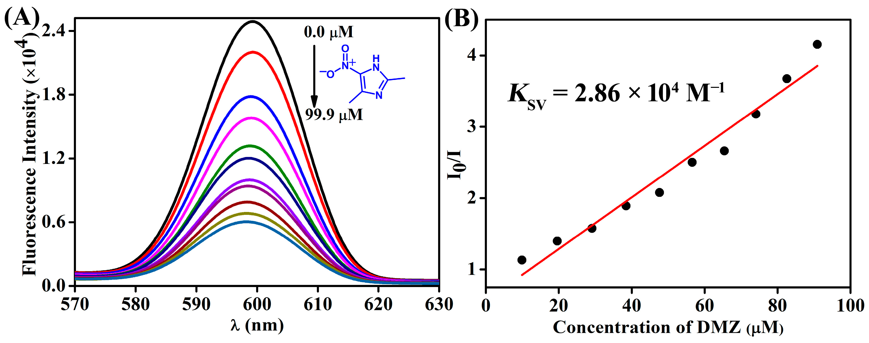
Scheme 1. Schematic representation of the synthesis of Zn(II) coordination polymer TB-Zn-CP from TBNap.
Chemistry 06 00011 sch001
Figure 1. (A) The FTIR spectrum, (B) powder X-ray diffraction pattern, and (C) SEM images at different magnifications for TB-Zn-CP.
Chemistry 06 00011 g001
Figure 2. (A) The fluorescence emission spectra of TB-Zn-CP in the presence of different antibiotics and (B) corresponding bar diagram with error bar. (C) Observed visual color changes for TB-Zn-CP in the presence of antibiotics in water, imaged under UV light irradiation (Nitrofurantoin, NFT; Nitrofurazone, NFZ; Chloramphenicol, CRP; Furazolidone, FZD; Sulfadiazine, SDZ; 1,2-Dimethyl-5-nitroimidazole, DMZ; and Sulfamethazine, SMZ).
Chemistry 06 00011 g002
Figure 3. (A) The observed fluorescence emission-quenching of TB-Zn-CP with the gradual addition of 5-nitroimidazole (DMZ) antibiotic in water and (B) its corresponding Stern–Volmer plot.
Chemistry 06 00011 g003
Figure 4. (A) The changes in the UV–visible absorption spectrum of TB-Zn-CP dispersed in water with the incremental addition of DMZ (from 0.0 to 90.9 μM). (B) The sensitivity plot of changes in emission intensity of TB-Zn-CP upon increasing the concentration of 5-nitroimidazole (DMZ) at the nM level.
Chemistry 06 00011 g004
Table 1. The calculated Stern–Volmer quenching constant (KSV) for different antibiotics.
AntibioticsKSV (×104 M−1)
DMZ2.86
SMZ2.61
CRP2.85
NFT0.58
NFZ0.14
SDZ0.57
FZD0.42
Disclaimer/Publisher’s Note: The statements, opinions and data contained in all publications are solely those of the individual author(s) and contributor(s) and not of MDPI and/or the editor(s). MDPI and/or the editor(s) disclaim responsibility for any injury to people or property resulting from any ideas, methods, instructions or products referred to in the content.

