
New Functionalized Chitosan with Thio-Thiadiazole Derivative with Enhanced Inhibition of Pathogenic Bacteria, Plant Threatening Fungi, and Improvement of Seed Germination

 ,
,  ,
,  ,
, 
Abstract
1. Introduction
2. Materials and Methods
2.1. Materials
2.2. Methods
2.2.1. Synthesis of 5-Amino-1,3,4-thiadiazole-2-thiol (Compound 1)
2.2.2. Synthesis of 5-(Methylthio)-1,3,4-thiadiazol-2-amine (Compound 2)
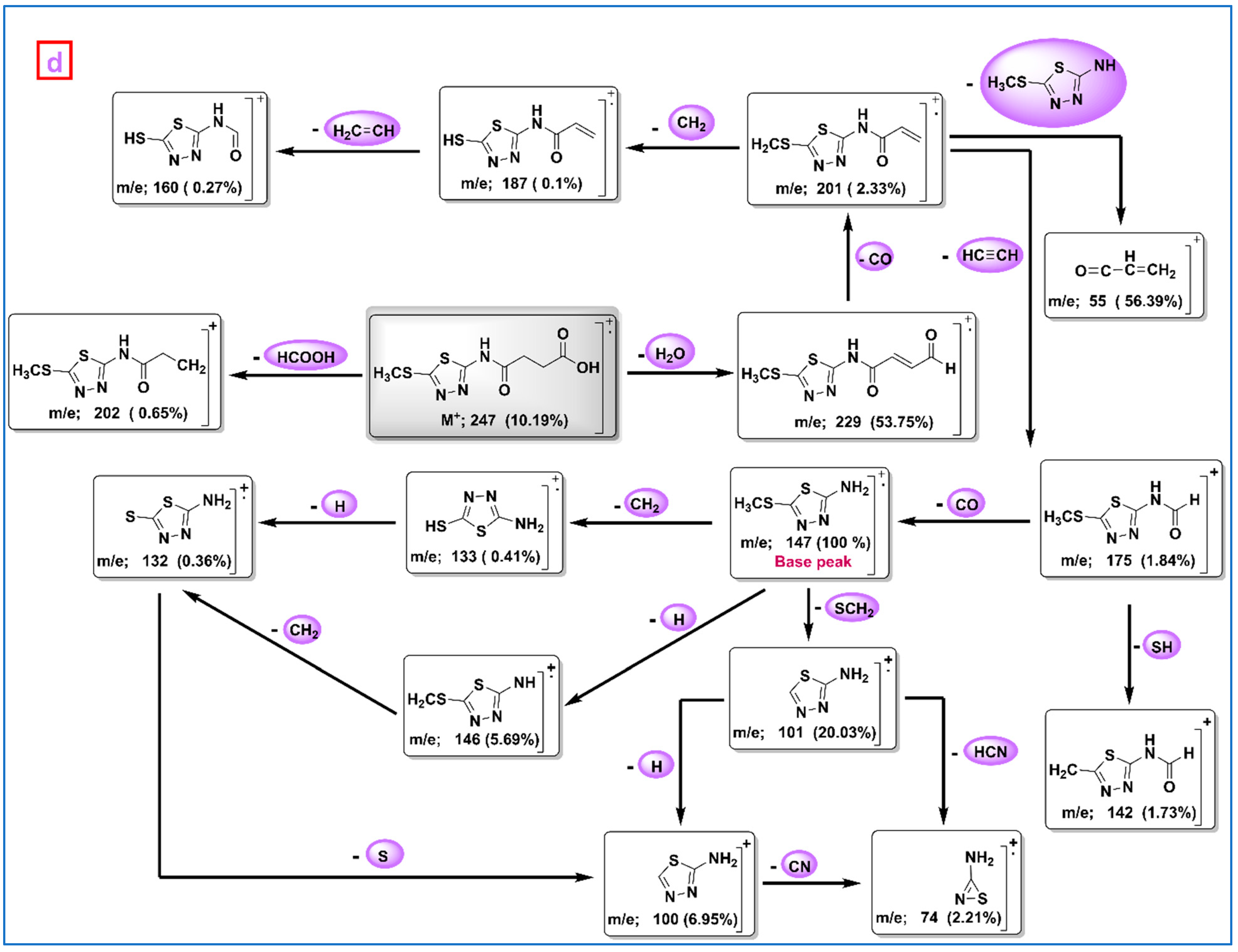
2.2.3. Synthesis of 4-((5-(Methylthio)-1,3,4-thiadiazol-2-yl)amino)-4-oxobutanoic Acid (Compound 3)
2.2.4. Synthesis of Chitosan Derivative (Chito-TZ)
2.3. Instrument Specifications
2.4. Biological Analysis
2.4.1. Antimicrobial Activity
2.4.2. Activity of Chito-TZ and Chito against Phytopathogen
2.4.3. Seed Germination
2.4.4. Statistical Analysis
3. Results and Discussion
3.1. Synthesis of the Chitosan Conjugate
3.2. Spectral Analysis
3.3. Elemental Analysis
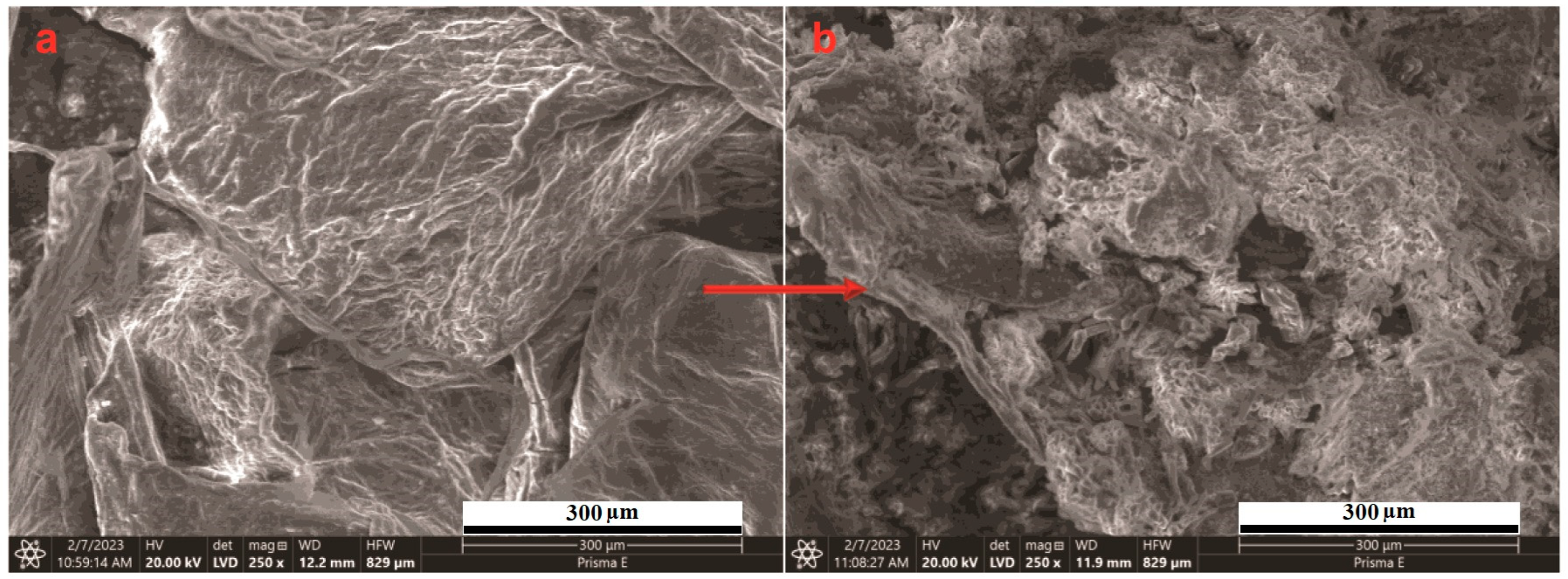
3.4. SEM Investigation
3.5. Thermal Properties
3.6. XRD Analysis
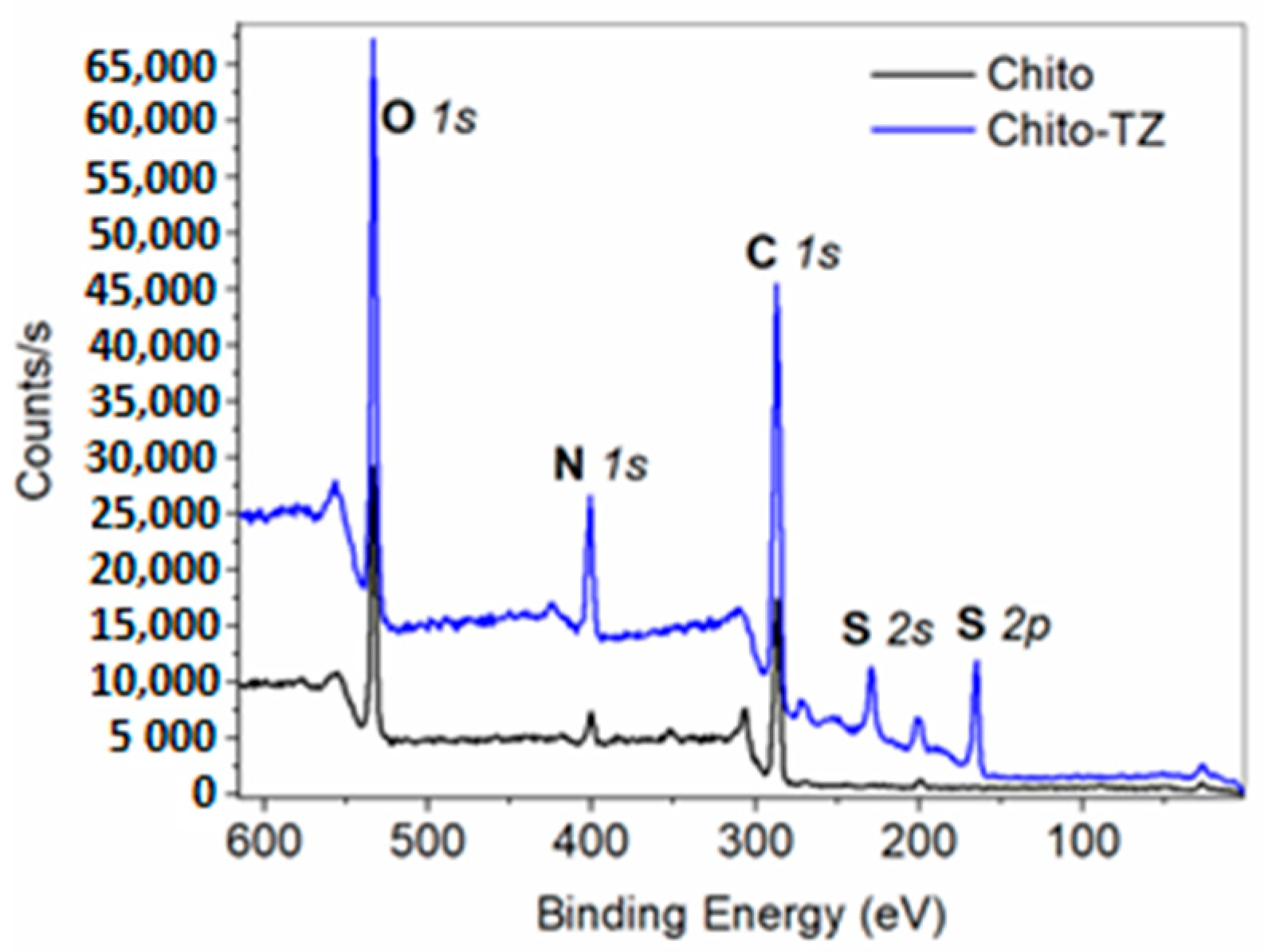
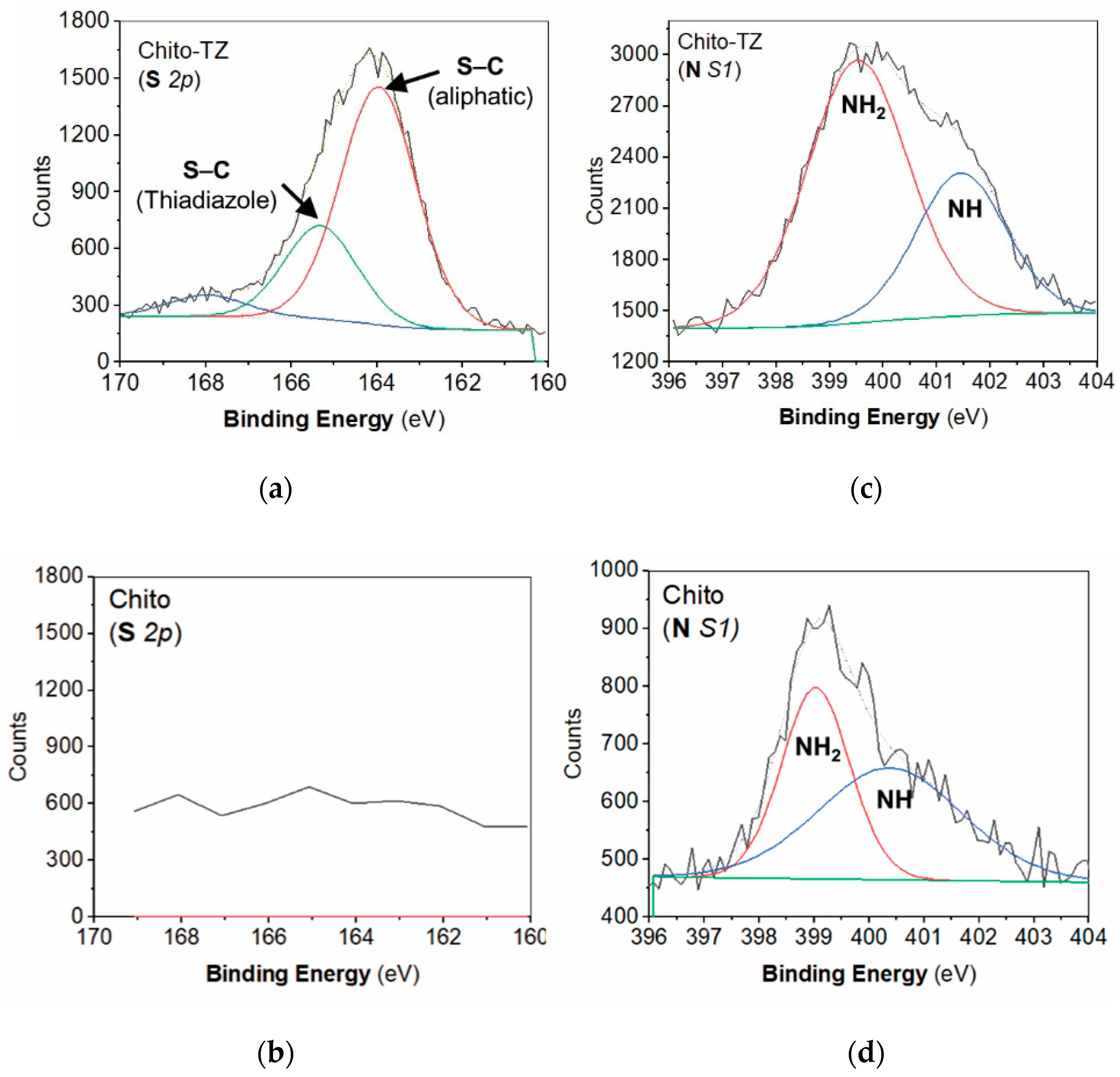
3.7. XPS Analysis
3.8. Biological Activity
3.8.1. Antimicrobial Activity
3.8.2. Biocontrol of Phytopathogens
3.8.3. Seed Germination
4. Conclusions
Author Contributions
Funding
Institutional Review Board Statement
Informed Consent Statement
Data Availability Statement
Acknowledgments
Conflicts of Interest
References
- Mohamed, A.E.; Elgammal, W.E.; Eid, A.M.; Dawaba, A.M.; Ibrahim, A.G.; Fouda, A.; Hassan, S.M. Synthesis and characterization of new functionalized chitosan and its antimicrobial and in-vitro release behavior from topical gel. Int. J. Biol. Macromol. 2022, 207, 242–253. [Google Scholar] [CrossRef] [PubMed]
- Muñoz-Nuñez, C.; Cuervo-Rodríguez, R.; Echeverría, C.; Fernández-García, M.; Muñoz-Bonilla, A. Synthesis and characterization of thiazolium chitosan derivative with enhanced antimicrobial properties and its use as component of chitosan based films. Carbohydr. Polym. 2023, 302, 120438. [Google Scholar] [CrossRef] [PubMed]
- Ibrahim, A.G.; Saleh, A.S.; Elsharma, E.M.; Metwally, E.; Siyam, T. Chitosan-g-maleic acid for effective removal of copper and nickel ions from their solutions. Int. J. Biol. Macromol. 2019, 121, 1287–1294. [Google Scholar] [CrossRef]
- Zhang, W.; Hadidi, M.; Karaca, A.C.; Hedayati, S.; Tarahi, M.; Assadpour, E.; Jafari, S.M. Chitosan-grafted phenolic acids as an efficient biopolymer for food packaging films/coatings. Carbohydr. Polym. 2023, 314, 120901. [Google Scholar] [CrossRef]
- Wang, L.; Xu, Z.; Zhang, H.; Yao, C. A review on chitosan-based biomaterial as carrier in tissue engineering and medical applications. Eur. Polym. J. 2023, 191, 112059. [Google Scholar] [CrossRef]
- Hamza, M.F.; Hamad, N.A.; Hamad, D.M.; Khalafalla, M.S.; Abdel-Rahman, A.A.H.; Zeid, I.F.; Wei, Y.; Hessien, M.M.; Fouda, A.; Salem, W.M. Synthesis of Eco-Friendly Biopolymer, Alginate-Chitosan Composite to Adsorb the Heavy Metals, Cd(II) and Pb(II) from Contaminated Effluents. Materials 2021, 14, 2189. [Google Scholar] [CrossRef]
- Hamza, M.F.; Hamad, D.M.; Hamad, N.A.; Abdel-Rahman, A.A.H.; Fouda, A.; Wei, Y.; Guibal, E.; El-Etrawy, A.-A.S. Functionalization of magnetic chitosan microparticles for high-performance removal of chromate from aqueous solutions and tannery effluent. Chem. Eng. J. 2022, 428, 131775. [Google Scholar] [CrossRef]
- Hamza, M.F.; Abdel-Rahman, A.A.H.; Negm, A.S.; Hamad, D.M.; Khalafalla, M.S.; Fouda, A.; Wei, Y.; Amer, H.H.; Alotaibi, S.H.; Goda, A.E.S. Grafting of Thiazole Derivative on Chitosan Magnetite Nanoparticles for Cadmium Removal—Application for Groundwater Treatment. Polymers 2022, 14, 1240. [Google Scholar] [CrossRef]
- Saleh, A.S.; Ibrahim, A.G.; Abdelhai, F.; Elsharma, E.M.; Metwally, E.; Siyam, T. Preparation of poly (chitosan-acrylamide) flocculant using gamma radiation for adsorption of Cu (II) and Ni (II) ions. Radiat. Phys. Chem. 2017, 134, 33–39. [Google Scholar] [CrossRef]
- Anand, M.; Sathyapriya, P.; Maruthupandy, M.; Beevi, A.H. Synthesis of chitosan nanoparticles by TPP and their potential mosquito larvicidal application. Front. Lab. Med. 2018, 2, 72–78. [Google Scholar] [CrossRef]
- Gamal, A.; Ibrahim, A.G.; Eliwa, E.M.; El-Zomrawy, A.H.; El-Bahy, S.M. Synthesis and characterization of a novel benzothiazole functionalized chitosan and its use for effective adsorption of Cu (II). Int. J. Biol. Macromol. 2021, 183, 1283–1292. [Google Scholar] [CrossRef]
- Hamza, M.F.; Goda, A.E.S.; Ning, S.; Mira, H.I.; Abdel-Rahman, A.A.H.; Wei, Y.; Fujita, T.; Amer, H.H.; Alotaibi, S.H.; Fouda, A. Photocatalytic Efficacy of Heterocyclic Base Grafted Chitosan Magnetite Nanoparticles on Sorption of Pb(II); Application on Mining Effluent. Catalysts 2022, 12, 330. [Google Scholar] [CrossRef]
- Tan, W.; Li, Q.; Dong, F.; Zhang, J.; Luan, F.; Wei, L.; Chen, Y.; Guo, Z. Novel cationic chitosan derivative bearing 1, 2, 3-triazolium and pyridinium: Synthesis, characterization, and antifungal property. Carbohydr. Polym. 2018, 182, 180–187. [Google Scholar] [CrossRef] [PubMed]
- Qin, Y.; Liu, S.; Xing, R.; Li, K.; Yu, H.; Li, P. Synthesis and antifungal evaluation of (1, 2, 3-triazol-4-yl) methyl nicotinate chitosan. Int. J. Biol. Macromol. 2013, 61, 58–62. [Google Scholar] [CrossRef] [PubMed]
- Ali, M.A.; Musthafa, S.A.; Munuswamy-Ramanujam, G.; Jaisankar, V. 3-Formylindole-based chitosan Schiff base polymer: Antioxidant and in vitro cytotoxicity studies on THP-1 cells. Carbohydr. Polym. 2022, 290, 119501. [Google Scholar] [CrossRef] [PubMed]
- Huang, C.; Liao, H.; Liu, X.; Xiao, M.; Liao, S.; Gong, S.; Yang, F.; Shu, X.; Zhou, X. Preparation and characterization of vanillin-chitosan Schiff base zinc complex for a novel Zn2+ sustained released system. Int. J. Biol. Macromol. 2022, 194, 611–618. [Google Scholar] [CrossRef]
- Přichystalová, H.; Almonasy, N.; Abdel-Mohsen, A.M.; Abdel-Rahman, R.M.; Fouda, M.M.G.; Vojtova, L.; Kobera, L.; Spotz, Z.; Burgert, L.; Jancar, J. Synthesis, characterization and antibacterial activity of new fluorescent chitosan derivatives. Int. J. Biol. Macromol. 2014, 65, 234–240. [Google Scholar] [CrossRef]
- Aytekin, A.O.; Morimura, S.; Kida, K. Synthesis of chitosan–caffeic acid derivatives and evaluation of their antioxidant activities. J. Biosci. Bioeng. 2011, 111, 212–216. [Google Scholar] [CrossRef]
- Huang, C.; Liao, H.; Ma, X.; Xiao, M.; Liu, X.; Gong, S.; Shu, X.; Zhou, X. Adsorption performance of chitosan Schiff base towards anionic dyes: Electrostatic interaction effects. Chem. Phys. Lett. 2021, 780, 138958. [Google Scholar] [CrossRef]
- Tamer, T.M.; Hassan, M.A.; Omer, A.M.; Baset, W.M.A.; Hassan, M.E.; El-Shafeey, M.E.A.; Eldin, M.S.M. Synthesis, characterization and antimicrobial evaluation of two aromatic chitosan Schiff base derivatives. Process Biochem. 2016, 51, 1721–1730. [Google Scholar] [CrossRef]
- Li, Q.; Ren, J.; Dong, F.; Feng, Y.; Gu, G.; Guo, Z. Synthesis and antifungal activity of thiadiazole-functionalized chitosan derivatives. Carbohydr. Res. 2013, 373, 103–107. [Google Scholar] [CrossRef] [PubMed]
- Zhang, J.; Tan, W.; Wei, L.; Chen, Y.; Mi, Y.; Sun, X.; Li, Q.; Dong, F.; Guo, Z. Synthesis of urea-functionalized chitosan derivatives for potential antifungal and antioxidant applications. Carbohydr. Polym. 2019, 215, 108–118. [Google Scholar] [CrossRef] [PubMed]
- Mohamed, A.E.; Elgammal, W.E.; Dawaba, A.M.; Ibrahim, A.G.; Fouda, A.; Hassan, S.M. A novel 1,3,4-thiadiazole modified chitosan: Synthesis, characterization, antimicrobial activity, and release study from film dressings. Appl. Biol. Chem. 2022, 65, 54. [Google Scholar] [CrossRef]
- Aghcheli, A.; Toolabi, M.; Ayati, A.; Moghimi, S.; Firoozpour, L.; Bakhshaiesh, T.O.; Nazeri, E.; Norouzbahari, M.; Esmaeili, R.; Foroumadi, A. Design, synthesis, and biological evaluation of 1-(5-(benzylthio)-1, 3, 4-thiadiazol-2-yl)-3-phenylurea derivatives as anticancer agents. Med. Chem. Res. 2020, 29, 2000–2010. [Google Scholar] [CrossRef]
- Humphries Romney, M.; Ambler, J.; Mitchell Stephanie, L.; Castanheira, M.; Dingle, T.; Hindler Janet, A.; Koeth, L.; Sei, K. CLSI Methods Development and Standardization Working Group Best Practices for Evaluation of Antimicrobial Susceptibility Tests. J. Clin. Microbiol. 2018, 56, e01934-17. [Google Scholar] [CrossRef]
- Fouda, A.; Eid, A.M.; Abdel-Rahman, M.A.; El-Belely, E.F.; Awad, M.A.; Hassan, S.E.; Al-Faifi, Z.E.; Hamza, M.F. Enhanced Antimicrobial, Cytotoxicity, Larvicidal, and Repellence Activities of Brown Algae, Cystoseira crinita-Mediated Green Synthesis of Magnesium Oxide Nanoparticles. Front. Bioeng. Biotechnol. 2022, 10, 849921. [Google Scholar] [CrossRef]
- Fouda, A.; Hassan, S.E.-D.; Eid, A.M.; Awad, M.A.; Althumayri, K.; Badr, N.F.; Hamza, M.F. Endophytic bacterial strain, Brevibacillus brevis-mediated green synthesis of copper oxide nanoparticles, characterization, antifungal, in vitro cytotoxicity, and larvicidal activity. Green Process. Synth. 2022, 11, 931–950. [Google Scholar] [CrossRef]
- Sathiyabama, M.; Parthasarathy, R. Biological preparation of chitosan nanoparticles and its in vitro antifungal efficacy against some phytopathogenic fungi. Carbohydr. Polym. 2016, 151, 321–325. [Google Scholar] [CrossRef]
- Redayan, M.; Ali, W.; Mohammed, A. Synthesis, Characterization and Antibacterial Evaluation of some Novel Benzimidazole Derivatives Containing 1,3,4-Thiadiazole Moiety. Orient. J. Chem. 2017, 33, 3138–3143. [Google Scholar] [CrossRef]
- Hamza, M.F.; Guibal, E.; Althumayri, K.; Wei, Y.; Eid, A.M.; Fouda, A. Poly-condensation of N-(2-acetamido)-2-aminoethanesulfonic acid with formaldehyde for the synthesis of a highly efficient sorbent for Cs (I). Chem. Eng. J. 2023, 454, 140155. [Google Scholar] [CrossRef]
- Li, Q.; Tan, W.; Zhang, C.; Gu, G.; Guo, Z. Novel triazolyl-functionalized chitosan derivatives with different chain lengths of aliphatic alcohol substituent: Design, synthesis, and antifungal activity. Carbohydr. Res. 2015, 418, 44–49. [Google Scholar] [CrossRef] [PubMed]
- Elzamly, R.A.; Mohamed, H.M.; Mohamed, M.I.; Zaky, H.T.; Harding, D.R.K.; Kandile, N.G. New sustainable chemically modified chitosan derivatives for different applications: Synthesis and characterization. Arab. J. Chem. 2021, 14, 103255. [Google Scholar] [CrossRef]
- Hamza, M.F.; Guibal, E.; Wei, Y.; Fouda, A.; Althumayri, K.; Abu Khoziem, H.A.; Mashaal, N.M. Functionalization of thiosemicarbazide/gellan gum for the enhancement of cadmium removal from aqueous solutions—Grafting of tributyl phosphate derivative. J. Water Process Eng. 2023, 54, 103928. [Google Scholar] [CrossRef]
- Hu, L.; Meng, X.; Xing, R.; Liu, S.; Chen, X.; Qin, Y.; Yu, H.; Li, P. Design, synthesis and antimicrobial activity of 6-N-substituted chitosan derivatives. Bioorg. Med. Chem. Lett. 2016, 26, 4548–4551. [Google Scholar] [CrossRef] [PubMed]
- Rahmouni, N.; Tahri, W.; Sbihi, H.M.; Nehdi, I.A.; Desbrieres, J.; Besbes-Hentati, S. Improvement of chitosan solubility and bactericidity by synthesis of N-benzimidazole-O-acetyl-chitosan and its electrodeposition. Int. J. Biol. Macromol. 2018, 113, 623–630. [Google Scholar] [CrossRef]
- Baran, T.; Menteş, A. Cu(II) and Pd(II) complexes of water soluble O-carboxymethyl chitosan Schiff bases: Synthesis, characterization. Int. J. Biol. Macromol. 2015, 79, 542–554. [Google Scholar] [CrossRef] [PubMed]
- Gonçalves, F.J.; Gurgel, L.V.A.; Soares, L.C.; Teodoro, F.S.; Ferreira, G.M.D.; Coelho, Y.L.; da Silva, L.H.M.; Prim, D.; Gil, L.F. Application of pyridine-modified chitosan derivative for simultaneous adsorption of Cu (II) and oxyanions of Cr (VI) from aqueous solution. J. Environ. Manag. 2021, 282, 111939. [Google Scholar] [CrossRef]
- Salama, H.E.; Saad, G.R.; Sabaa, M.W. Synthesis, characterization and biological activity of Schiff bases based on chitosan and arylpyrazole moiety. Int. J. Biol. Macromol. 2015, 79, 996–1003. [Google Scholar] [CrossRef]
- Mohammadi, B.; Pirsa, S.; Alizadeh, M. Preparing chitosan–polyaniline nanocomposite film and examining its mechanical, electrical, and antimicrobial properties. Polym. Polym. Compos. 2019, 27, 507–517. [Google Scholar] [CrossRef]
- Acosta-Ferreira, S.; Castillo, O.S.; Madera-Santana, J.T.; Mendoza-García, D.A.; Núñez-Colín, C.A.; Grijalva-Verdugo, C.; Villa-Lerma, A.G.; Morales-Vargas, A.T.; Rodríguez-Núñez, J.R. Production and physicochemical characterization of chitosan for the harvesting of wild microalgae consortia. Biotechnol. Rep. 2020, 28, e00554. [Google Scholar] [CrossRef]
- Franca, E.F.; Lins, R.D.; Freitas, L.C.G.; Straatsma, T.P. Characterization of chitin and chitosan molecular structure in aqueous solution. J. Chem. Theory Comput. 2008, 4, 2141–2149. [Google Scholar] [CrossRef] [PubMed]
- Pandit, A.H.; Mazumdar, N.; Imtiyaz, K.; Alam Rizvi, M.M.; Ahmad, S. Self-healing and injectable hydrogels for anticancer drug delivery: A study with multialdehyde gum arabic and succinic anhydride chitosan. ACS Appl. Bio Mater. 2020, 3, 8460–8470. [Google Scholar] [CrossRef] [PubMed]
- Fouda, A.; Awad, M.A.; Eid, A.M.; Saied, E.; Barghoth, M.G.; Hamza, M.F.; Awad, M.F.; Abdelbary, S.; Hassan, S.E. An Eco-Friendly Approach to the Control of Pathogenic Microbes and Anopheles stephensi Malarial Vector Using Magnesium Oxide Nanoparticles (Mg-NPs) Fabricated by Penicillium chrysogenum. Int. J. Mol. Sci. 2021, 22, 5096. [Google Scholar] [CrossRef] [PubMed]
- Frost, I.; Kapoor, G.; Craig, J.; Liu, D.; Laxminarayan, R. Status, challenges and gaps in antimicrobial resistance surveillance around the world. J. Glob. Antimicrob. Resist. 2021, 25, 222–226. [Google Scholar] [CrossRef]
- Ventola, C.L. The antibiotic resistance crisis: Part 1: Causes and threats. P T Peer Rev. J. Formul. Manag. 2015, 40, 277–283. [Google Scholar]
- Li, J.; Zhuang, S. Antibacterial activity of chitosan and its derivatives and their interaction mechanism with bacteria: Current state and perspectives. Eur. Polym. J. 2020, 138, 109984. [Google Scholar] [CrossRef]
- Hamza, M.F.; Fouda, A.; Wei, Y.; El Aassy, I.E.; Alotaibi, S.H.; Guibal, E.; Mashaal, N.M. Functionalized biobased composite for metal decontamination—Insight on uranium and application to water samples collected from wells in mining areas (Sinai, Egypt). Chem. Eng. J. 2022, 431, 133967. [Google Scholar] [CrossRef]
- Guarnieri, A.; Triunfo, M.; Scieuzo, C.; Ianniciello, D.; Tafi, E.; Hahn, T.; Zibek, S.; Salvia, R.; De Bonis, A.; Falabella, P. Antimicrobial properties of chitosan from different developmental stages of the bioconverter insect Hermetia illucens. Sci. Rep. 2022, 12, 8084. [Google Scholar] [CrossRef]
- Zahra, M.H.; Hamza, M.F.; El-Habibi, G.; Abdel-Rahman, A.A.H.; Mira, H.I.; Wei, Y.; Alotaibi, S.H.; Amer, H.H.; Goda, A.E.S.; Hamad, N.A. Synthesis of a Novel Adsorbent Based on Chitosan Magnetite Nanoparticles for the High Sorption of Cr (VI) Ions: A Study of Photocatalysis and Recovery on Tannery Effluents. Catalysts 2022, 12, 678. [Google Scholar] [CrossRef]
- Devlieghere, F.; Vermeulen, A.; Debevere, J. Chitosan: Antimicrobial activity, interactions with food components and applicability as a coating on fruit and vegetables. Food Microbiol. 2004, 21, 703–714. [Google Scholar] [CrossRef]
- Fernandez-Saiz, P.; Lagarón, J.M.; Ocio, M.J. Optimization of the film-forming and storage conditions of chitosan as an antimicrobial agent. J. Agric. Food Chem. 2009, 57, 3298–3307. [Google Scholar] [CrossRef]
- Hosseinnejad, M.; Jafari, S.M. Evaluation of different factors affecting antimicrobial properties of chitosan. Int. J. Biol. Macromol. 2016, 85, 467–475. [Google Scholar] [CrossRef]
- Chien, R.C.; Yen, M.T.; Mau, J.L. Antimicrobial and antitumor activities of chitosan from shiitake stipes, compared to commercial chitosan from crab shells. Carbohydr. Polym. 2016, 138, 259–264. [Google Scholar] [CrossRef] [PubMed]
- Hamza, M.F.; Abdel-Rahman, A.A.H.; Hawata, M.A.; El Araby, R.; Guibal, E.; Fouda, A.; Wei, Y.; Hamad, N.A. Functionalization of magnetic chitosan microparticles—Comparison of trione and trithione grafting for enhanced silver sorption and application to metal recovery from waste X-ray photographic films. J. Environ. Chem. Eng. 2022, 10, 107939. [Google Scholar] [CrossRef]
- Li, Z.; Yang, F.; Yang, R. Synthesis and characterization of chitosan derivatives with dual-antibacterial functional groups. Int. J. Biol. Macromol. 2015, 75, 378–387. [Google Scholar] [CrossRef]
- Raafat, D.; Von Bargen, K.; Haas, A.; Sahl, H.-G. Insights into the mode of action of chitosan as an antibacterial compound. Appl. Environ. Microbiol. 2008, 74, 3764–3773. [Google Scholar] [CrossRef]
- Yilmaz Atay, H. Antibacterial Activity of Chitosan-Based Systems. In Functional Chitosan: Drug Delivery and Biomedical Applications; Jana, S., Jana, S., Eds.; Springer: Singapore, 2019; pp. 457–489. [Google Scholar]
- El-tahlawy, K.F.; El-bendary, M.A.; Elhendawy, A.G.; Hudson, S.M. The antimicrobial activity of cotton fabrics treated with different crosslinking agents and chitosan. Carbohydr. Polym. 2005, 60, 421–430. [Google Scholar] [CrossRef]
- Kowalska-Krochmal, B.; Dudek-Wicher, R. The Minimum Inhibitory Concentration of Antibiotics: Methods, Interpretation, Clinical Relevance. Pathogens 2021, 10, 165. [Google Scholar] [CrossRef]
- Ibrahim, A.G.; Fouda, A.; Elgammal, W.E.; Eid, A.M.; Elsenety, M.M.; Mohamed, A.E.; Hassan, S.M. New thiadiazole modified chitosan derivative to control the growth of human pathogenic microbes and cancer cell lines. Sci. Rep. 2022, 12, 21423. [Google Scholar] [CrossRef]
- Maluin, F.N.; Hussein, M.Z. Chitosan-Based Agronanochemicals as a Sustainable Alternative in Crop Protection. Molecules 2020, 25, 1611. [Google Scholar] [CrossRef]
- Sajomsang, W.; Gonil, P.; Saesoo, S.; Ovatlarnporn, C. Antifungal property of quaternized chitosan and its derivatives. Int. J. Biol. Macromol. 2012, 50, 263–269. [Google Scholar] [CrossRef] [PubMed]
- Qin, Y.; Li, P.; Guo, Z. Cationic chitosan derivatives as potential antifungals: A review of structural optimization and applications. Carbohydr. Polym. 2020, 236, 116002. [Google Scholar] [CrossRef] [PubMed]
- Tan, W.; Li, Q.; Dong, F.; Qiu, S.; Zhang, J.; Guo, Z. Novel 1,2,3-triazolium-functionalized starch derivatives: Synthesis, characterization, and evaluation of antifungal property. Carbohydr. Polym. 2017, 160, 163–171. [Google Scholar] [CrossRef] [PubMed]
- Confederat, L.G.; Tuchilus, C.G.; Dragan, M.; Sha’at, M.; Dragostin, O.M. Preparation and Antimicrobial Activity of Chitosan and Its Derivatives: A Concise Review. Molecules 2021, 26, 3694. [Google Scholar] [CrossRef]
- Suarez-Fernandez, M.; Marhuenda-Egea, F.C.; Lopez-Moya, F.; Arnao, M.B.; Cabrera-Escribano, F.; Nueda, M.J.; Gunsé, B.; Lopez-Llorca, L.V. Chitosan Induces Plant Hormones and Defenses in Tomato Root Exudates. Front. Plant Sci. 2020, 11, 572087. [Google Scholar] [CrossRef]
- Liu, H.; Zheng, Z.; Han, X.; Zhang, C.; Li, H.; Wu, M. Chitosan Soaking Improves Seed Germination of Platycodon Grandiflorus and Enhances Its Growth, Photosynthesis, Resistance, Yield, and Quality. Horticulturae 2022, 8, 943. [Google Scholar] [CrossRef]
- Pasquariello, M.S.; Di Patre, D.; Mastrobuoni, F.; Zampella, L.; Scortichini, M.; Petriccione, M. Influence of postharvest chitosan treatment on enzymatic browning and antioxidant enzyme activity in sweet cherry fruit. Postharvest Biol. Technol. 2015, 109, 45–56. [Google Scholar] [CrossRef]
- Ngo, D.-H.; Kim, S.-K. Chapter Two—Antioxidant Effects of Chitin, Chitosan, and Their Derivatives. In Advances in Food and Nutrition Research; Kim, S.-K., Ed.; Academic Press: Cambridge, MA, USA, 2014; Volume 73, pp. 15–31. [Google Scholar]


| Instrument | Description |
|---|---|
| Elemental analyzer | AnalyzerVario El M, Germany, Elements C-H-N-S, |
| FT-IR | Schimadzu, Nicolet iS10FT IR (Thermo Fisher Scientific, Waltham, MA, USA), resolution 16, scanning range 4000–400 cm−1, KBr plates, wavenumber scale (cm−1). |
| 1H/13C-NMR | JNM-ECA 500 II/JEOL-JAPAN instrument, solvent DMSO-d6, at 500 MHz for 1H and 125 MHz for 13C), ẟ scale (ppm) downfield from TMS. |
| Mass analysis | Shimadzu Japan’s GC–2010 |
| SEM | SEM (FEI inspect 5 with an EDX unit, Holland) with an operating voltage of 20–30 KV. |
| XPS | K-ALPHA (Thermo Fisher Scientific, Waltham, MA, USA), monochromatic x-ray Al K-Alpha radiation from −10 to 1350 e.v. Spot size 400 micro m at pressure 10–9 mbar with full spectrum pass energy 200 e.v. and at narrow spectrum 50 e.v. |
| TGA | Discovery SDT 650-Simultaneous DSC-TGA/DTA Instruments, New Castle, DE, USA, N2 atmosphere, temperature range 0 to 500 °C, heating rate 10 °C/min. |
| XRD | X’Pert powder PAN analytical, Cu-radiation (l = 1.542 Å), 45 K.V., 35 M.A. scanning speed 0.02°/sec. |
| Treatment | Decomposition Stages | Decomposition Temp. Range | Wt. Loss (%) | a T10 (°C) | b T20 (°C) | Ea (KJ mol−1) | R2 |
|---|---|---|---|---|---|---|---|
| Chito | 1 | 266.04–310.16 | 29.77 | 133.8 | 276.5 | 95.57 | 0.991 |
| Chito-TZ | 3 | 141.9–233.2 | 14.69 | 178.5 | 226.2 | 24.70 | 0.966 |
| 233.2–245.3 | 8.87 | 86.90 | 0.995 | ||||
| 245.3 -332.6 | 16.00 | 18.62 | 0.963 |
| Concentration | Treatment | Germination Percentages (%) | Shoot Length (cm) | Root Length (cm) | Shoot Weight (mg) | Root Weight (mg) | SVI * |
|---|---|---|---|---|---|---|---|
| Control | H2O | 87.0 ± 0.7 | 11.7 ± 0.8 | 11.1 ± 1.5 | 1080.0 ± 5.6 | 711.0 ± 6.6 | 1018 |
| 100 µg mL−1 | Chito | 90.0 ± 0.5 | 15.3 ± 1.5 | 12.7 ± 11.2 | 1186.7 ± 22.7 | 761.0 ± 16.1 | 1377 |
| Chito-TZ | 92.0 ± 0.6 | 13.3 ± 0.6 | 10.9 ± 0.6 | 1104.0 ± 7.6 | 726.0 ± 4.6 | 1224 | |
| 200 µg mL−1 | Chito | 96.0 ± 1.1 | 17.2 ± 0.3 | 15.0 ± 1.0 | 2337.7 ± 26.1 | 997.0 ± 22.3 | 1651 |
| Chito-TZ | 94.0 ± 0.9 | 14.8 ± 0.3 | 12.7 ± 0.7 | 1362.7 ± 9.5 | 814.7 ± 18.8 | 1391 | |
| 300 µg mL−1 | Chito | 90.0 ± 0.5 | 14.0 ± 1.3 | 14.7 ± 0.6 | 1807.7 ± 16.4 | 980.7 ± 20.8 | 1260 |
| Chito-TZ | 90.0 ± 0.5 | 13.3 ± 0.6 | 11.2 ± 1.5 | 1215.3 ± 22.5 | 733.3 ± 20.1 | 1197 |
Disclaimer/Publisher’s Note: The statements, opinions and data contained in all publications are solely those of the individual author(s) and contributor(s) and not of MDPI and/or the editor(s). MDPI and/or the editor(s) disclaim responsibility for any injury to people or property resulting from any ideas, methods, instructions or products referred to in the content. |
© 2023 by the authors. Licensee MDPI, Basel, Switzerland. This article is an open access article distributed under the terms and conditions of the Creative Commons Attribution (CC BY) license (https://creativecommons.org/licenses/by/4.0/).
Share and Cite
Ibrahim, A.G.; Elgammal, W.E.; Eid, A.M.; Alharbi, M.; Mohamed, A.E.; Alayafi, A.A.M.; Hassan, S.M.; Fouda, A. New Functionalized Chitosan with Thio-Thiadiazole Derivative with Enhanced Inhibition of Pathogenic Bacteria, Plant Threatening Fungi, and Improvement of Seed Germination. Chemistry 2023, 5, 1722-1744. https://doi.org/10.3390/chemistry5030118
Ibrahim AG, Elgammal WE, Eid AM, Alharbi M, Mohamed AE, Alayafi AAM, Hassan SM, Fouda A. New Functionalized Chitosan with Thio-Thiadiazole Derivative with Enhanced Inhibition of Pathogenic Bacteria, Plant Threatening Fungi, and Improvement of Seed Germination. Chemistry. 2023; 5(3):1722-1744. https://doi.org/10.3390/chemistry5030118
Chicago/Turabian StyleIbrahim, Ahmed G., Walid E. Elgammal, Ahmed M. Eid, Maha Alharbi, Ahmad E. Mohamed, Aisha A. M. Alayafi, Saber M. Hassan, and Amr Fouda. 2023. "New Functionalized Chitosan with Thio-Thiadiazole Derivative with Enhanced Inhibition of Pathogenic Bacteria, Plant Threatening Fungi, and Improvement of Seed Germination" Chemistry 5, no. 3: 1722-1744. https://doi.org/10.3390/chemistry5030118
APA StyleIbrahim, A. G., Elgammal, W. E., Eid, A. M., Alharbi, M., Mohamed, A. E., Alayafi, A. A. M., Hassan, S. M., & Fouda, A. (2023). New Functionalized Chitosan with Thio-Thiadiazole Derivative with Enhanced Inhibition of Pathogenic Bacteria, Plant Threatening Fungi, and Improvement of Seed Germination. Chemistry, 5(3), 1722-1744. https://doi.org/10.3390/chemistry5030118