Abstract
Tetracyclines are a group of common antibiotics, but owing to their toxicity, most of them are only used in animal husbandry and veterinary medicine. A DNA aptamer for tetracyclines has recently been reported. Upon aptamer binding, the fluorescence of tetracyclines was enhanced. This unique fluorescence enhancement was used to selectively detect the tetracyclines. The purpose of this study was to use graphene oxide (GO) to suppress the background fluorescence for enhanced detection. First, the adsorption of doxycycline on GO was studied. At pH 8.0, 82.7% of doxycycline was adsorbed by GO, and adding 2 µM aptamer desorbed 55.4% of doxycycline. With GO, the signal increase was comparable from pH 6 to 8, whereas without GO, the increase was significantly lower at pH 8. Under optimized condition, a detection limit of 1.6 nM doxycycline was achieved at pH 8.0 in the presence of GO, whereas without GO, the detection limit was 18.9 nM. This is an interesting example of the use of nanomaterials to enhance the performance of aptamer-based biosensors.
1. Introduction
Antibiotics have been used to selectively kill bacteria or slow their growth to prevent them from multiplying, and they include a range of potent life-saving drugs. Tetracyclines are broad-spectrum antibiotics, including doxycycline, tetracycline, and hyoscine, among others [1]. Tetracyclines enter bacterial cells by passive diffusion and inhibit bacterial growth by interfering with protein synthesis or by disrupting cell membranes. Due to their toxicity and side effects in humans, such as teeth staining in children, and stomach problems, many of the early discovered tetracyclines, such as tetracycline and oxytetracycline, are mainly used in animal husbandry and the veterinary industry. Other compounds, such as doxycycline, are still used in humans [2].
While various methods are available for the detection of tetracyclines, aptamers are particularly attractive for this purpose [3,4,5,6]. Aptamers are single-stranded oligonucleotides that fold into three dimensional structures upon binding to a target molecule [7,8,9,10,11]. Compared to antibodies, aptamers are more stable and easier to modify at a lower cost. Aptamer-based biosensors allow the use of portable devices for signal quantification and have obvious advantages over traditional instrumentation analysis methods such as HPLC in terms of portability [12].
Tetracyclines are known to bind to various RNA structures, such as the 30S ribosomal RNA and tRNA [6]. Therefore, some early studies selected RNA aptamers for tetracycline. For example, the aptamer reported by Berens et al. in 2001 has a Kd of approximately 100 nM [6]. After optimizing the aptamer sequence, the Kd value was further lowered [13]. Based on this RNA aptamer, Tickner et al. performed a re-selection experiment and obtained a highly selective RNA aptamer for doxycycline [5]. For biosensor applications, DNA aptamers are more attractive than RNA aptamers for stability and cost considerations. Niazi et al. reported the first DNA aptamers for oxytetracycline in 2008, and the best aptamer had a reported Kd of 10 nM [3]. Niazi et al. also selected another DNA aptamer using a mixture of magnetic beads coated with tetracycline, oxytetracycline, and doxycycline. The authors reported that the resulting aptamer was able to bind all three tetracyclines [4].
All these aptamers were obtained by immobilization of target molecules. Our lab recently selected a DNA aptamer named OTC5 that can bind tetracycline antibiotics using the library immobilization method [14]. OTC5 has a high affinity for tetracyclines with a dissociation constant (Kd) of about 100 nM. Tetracyclines are fluorescent and the fluorescence intensity is enhanced upon binding by aptamers [13,14,15]. We then developed a unique sensing mechanism based on this aptamer-dependent fluorescence enhancement [14]. Such sensors would work better if the background fluorescence from tetracyclines can be suppressed.
Graphene oxide (GO) is a widely used substrate to adsorb biomolecules, and it is known for its fluorescence quenching properties [16]. A large number of oxygen atoms in the form of epoxy, hydroxyl, and carboxyl groups are present on the surface of GO. Given the structure of tetracycline antibiotics, we expected that GO can adsorb them. In the literature, GO adsorption of tetracycline was previously reported [17]. At the same time, GO can also adsorb DNA [18,19], but its adsorption of aptamer/target complex is slower and weaker [20,21]. Therefore, we aimed to use GO to enhance the performance of such sensors by suppressing the background fluorescence.
Among various tetracycline antibiotics, we chose to use doxycycline in this work due to its higher stability [22]. Doxycycline is a tetracycline antibiotic used primarily for the treatment and prevention of diseases caused by bacterial infections, such as gonorrhea, syphilis, pneumonia, respiratory infections, Rocky Mountain spotted fever, and acne vulgaris. Because it is less toxic than other tetracyclines to humans, doxycycline may be a safe alternative to tetracycline when appropriate [23]. In this work, we used doxycycline as a model target to study the effect of GO on its aptamer-based detection.
2. Materials and Methods
2.1. Chemicals
All of the DNA oligonucleotides were purchased from Integrated DNA Technologies (Coralville, IA, USA). Tetracycline, oxytetracycline, doxycycline, ampicillin, and chloramphenicol were purchased from Sigma-Aldrich. Doxycycline has a higher stability and a stock solution was prepared at 10 mM concentration and stored at −20 °C. It was used for up to a week. Tetracycline and oxytetracycline were freshly prepared from their powders each time due to their lower stability. All the buffers, including 2-(N-morpholino)ethanesulfonic acid (MES), piperazine-N,N′-bis(2-ethanesulfonic acid) (PIPES), tris(hydroxymethyl)aminomethane (Tris), and salts (NaCl, MgCl2), were purchased from Mandel Scientific (Guelph, ON, Canada). Graphene oxide (GO) flakes were purchased from ACS Material (Pasadena, CA, USA) and they were dispersed in water with the assistance of ultrasonication at a concentration of 1 mg/mL. Milli-Q water was used to prepare all the buffers and solutions.
2.2. Fluorescence Spectroscopy
Fluorescence spectroscopy experiments were performed on a Tecan Spark microplate reader with excitation wavelength set at 370 nm and emission at 530 nm. For OTC5 aptamer detection of doxycycline-dependent fluorescence, 200 nM or 500 nM of doxycycline was dissolved in various buffers (10 mM pH 6.0 MES, 10 mM pH 7.0 PIPES, or 10 mM pH 8.0 Tris) containing 2 mM MgCl2, 0 or 2 μM OTC5 aptamer, 20 μg/mL GO, and equilibrated for 30 min before assay after each dissolution. The same method was used for 0 or 2 μM OTC5 aptamer to detect tetracycline, oxytetracycline, ampicillin, and chloramphenicol dependent fluorescence.
2.3. Adsorption Kinetics
The adsorption kinetics used an Agilent eclipse fluorometer at an excitation wavelength of 370 nm and emission wavelength of 530 nm, with a setting time of 10 min and a detection interval of 0.2 min. To achieve sufficient fluorescence, 2 μM doxycycline was used and its fluorescence was monitored for 3 min followed by the addition of 2 μM OTC5 in the absence of GO. In another experiment, kinetic curves of 2 μM doxycycline adsorption by 20 µg/mL GO followed. Then, 2 µM OTC5 was added. The experiments were performed in 10 mM buffer with pH 8.0.
2.4. TEM
Transmission electron microscopy (TEM) images were captured with a Philips CM10 100 kV microscope. A copper TEM grid that was lacey carbon-supported was placed on a filter paper to prepare the TEM sample. The TEM grid was then coated with 10 mL of a GO solution (250 g/mL) and left to dry at room temperature for the next day.
3. Results and Discussion
3.1. Biosensor Design
The structure of doxycycline is shown in Figure 1A, and its extensively conjugate structure may allow favorable adsorption by GO. Doxycycline has three pKa values at 3.0, 8.0, and 9.2, respectively [24]. At pH 6.0, it is nearly charge neutral, whereas at pH 8.0, half of it carries one negative charge. The secondary structure of the OTC5 aptamer is shown in Figure 1B [14], which has two short stems along with three loops. The important target binding regions are in the two loops. Using 370 nm excitation, we monitored the fluorescence of doxycycline at 530 nm. Upon mixing doxycycline with the aptamer, the fluorescence of doxycycline was increased around seven-fold (Figure 1C). We previously designed a biosensor for the detection of tetracycline antibiotics based on this unique fluorescence enhancement (Figure 1D). The fluorescence intensity of other molecules was not changed by the aptamer, regardless of whether those molecules were fluorescent or not.

Figure 1.
(A) The structure of doxycycline. (B) The secondary structure of the OTC5 aptamer. (C) The fluorescence intensity changes before and after the reaction of 200 nM doxycycline with 2 µM of the OTC5 aptamer at pH 6.0. (D) Scheme showing the previous sensor design, where enhanced doxycycline fluorescence was achieved upon aptamer binding. (E) Schematic diagram of the current sensor design, where doxycycline adsorption by GO results in quenched fluorescence, and subsequent binding to the OTC5 aptamer leads to enhanced fluorescence.
For a more sensitive sensor, it is desirable to have a lower background fluorescence before the addition of the aptamer (the first bar in Figure 1C). At the same time, the signal after adding the aptamer should remain the same or even stronger (the second bar in Figure 1C). In this work, we aimed to suppress the background fluorescence of doxycycline or other molecules in the sample using GO to adsorb doxycycline. Since doxycycline/aptamer complex may not be favorably adsorbed by GO, a strong final fluorescence may still be anticipated. The new design is shown in Figure 1E, where the sample is first treated by GO to achieve adsorption and suppression of background fluorescence. Upon the addition of the aptamer, aptamer binding may lead to selective desorption of doxycycline and enhanced fluorescence.
3.2. Adsorption of Doxycycline to GO
Figure 2A shows a TEM micrograph of the GO sheets used in this study, and they appeared to be well dispersed. For this sensor design to work, GO needs to be able to adsorb doxycycline and quench its fluorescence. For adsorption on GO, pH and salt concentration are important variables. Both GO and doxycycline have groups that can be protonated at close to neutral pH. Thus, we tested three pH values close to neutral of 6.0, 7.0, and 8.0, respectively. In this pH range, doxycycline is either charge neutral or carries a partial negative charge. GO is negatively charged due to its surface carboxyl groups. Since DNA is negatively charged, its adsorption to GO requires the presence of metal ions [25]. In addition, metal ions can also affect target binding of this aptamer [15]. Therefore, we included Na+ and Mg2+ in the buffers.
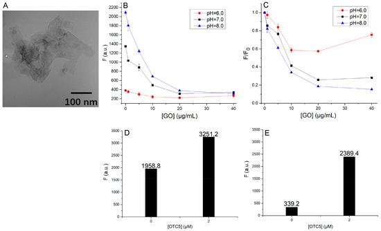
Figure 2.
(A) A TEM micrograph of the GO used in this study. (B) The quenching of doxycycline background fluorescence by various concentrations of GO in the presence of 2 mM Mg2+ and 100 mM NaCl at pH of 6.0, 7.0 and 8.0. (C) The same plot as (B) but the initial fluorescence was normalized. The fluorescence intensity of doxycycline before and after adding 2 µM OTC5 aptamer (D) in the absence of GO and (E) in the presence of 20 µg/mL GO at pH 8.0.
First, at the three pH values, we gradually added GO to doxycycline solutions, and we noticed that the background fluorescence was higher when the pH was raised (Figure 2B), which was consistent with our previous observations [15]. Then, significant quenching was observed with increasing concentration of GO at all the three pH values. To better observe the fluorescence quenching, we plotted the normalized fluorescence in Figure 2C. Regardless of the pH, the background fluorescence intensity of doxycycline started to decrease after the addition of GO and reached the maximum at a GO concentration of 20 μg/mL (Figure 2C). The quenched fluorescence was indicative of the adsorption of doxycycline by GO. The phenomenon became more obvious with the increase in pH value, and GO was able to bring the background fluorescence of doxycycline at different pH to a similar level (Figure 2B). Based on the results, it appears that GO had the highest effect in adsorbing doxycycline at pH 8, both in terms of the absolute fluorescence drop (Figure 2B) and in terms of the relative fluorescence drop (Figure 2C).
In a previous paper, the pH-dependent adsorption of tetracycline antibiotics by GO was systematically studied, and it reported a better adsorption at lower pH [17]. This is reasonable since a lower pH can help to reduce charge repulsion between the antibiotics and GO. In our work, however, we observed the opposite. We attributed the difference to the presence of metal ions. The previous work was performed in water, whereas our buffer contained 2 mM Mg2+. It is likely that Mg2+ can bridge negatively charged GO and doxycycline, leading to more efficient adsorption. The effect of metal ions in such cases has been well documented [25,26].
3.3. Suppression of Background Fluorescence by GO
After understanding the adsorption of doxycycline by GO, we then turned our attention to sensing, which relied on the enhanced fluorescence upon aptamer binding. The background fluorescence intensity of doxycycline without GO was 1959 units at pH 8.0, while adding the OTC5 aptamer could increase the fluorescence intensity of doxycycline to 3251 units, i.e., a 1.66-fold increase (Figure 2D). After GO addition, the background fluorescence dropped to 339 units, attributable to the adsorption by GO. After adding the OTC5 aptamer, the fluorescence still reached 2389 units, a seven-fold increase. Therefore, adding GO has drastically improved the signal-to-background ratio at pH 8.0.
Based on this data, we can also calculate the amount of desorbed doxycycline from GO due to binding by the aptamer. Assuming that the fluorescence intensity of doxycycline adsorbed by GO is 0, the remaining fluorescence intensity of 339 units was the background fluorescence due to free doxycycline not adsorbed by GO. Thus, only 17.3% of doxycycline was not adsorbed and 82.7% doxycycline was being adsorbed. After the addition of 2 µM OTC5, the fluorescence intensity of the sample solution rose to 2389 units (Figure 2E). Thus, the fluorescence due to aptamer-induced doxycycline desorption was 2389.4 − 339.2 × 1.66 = 1826.3. Thus, the fraction of desorbed doxycycline was (1826.3/1.66)/1985.8 = 55.4%. Therefore, adding the aptamer did not fully desorb doxycycline, and the 55.4% desorption was already sufficient to boost the signal enhancement. It is likely that adding a higher concentration of OTC5 would desorb more doxycycline to further increase the sensitivity, although this would increase the cost of the sensor.
3.4. Optimization of Sensing Conditions
The above experiments suggested the feasibility of this sensing strategy. We then optimized the sensing conditions. First, we tested the sensor at pH 6.0, 7.0 and 8.0, respectively. In the samples without GO (Figure 3A), the background fluorescence intensity of doxycycline was higher at higher pH, as expected. After adding the OTC5 aptamer, the fluorescence intensities were similar for all the three pH’s. Therefore, the best enhancement caused by the aptamer was at pH 6, reaching nearly seven-fold. However, at pH 8, the enhancement was only two-fold (Figure 3C). This pH-dependent variation in sensor performance may cause problems in calibration curves when the pH of a sample is unknown or unstable.
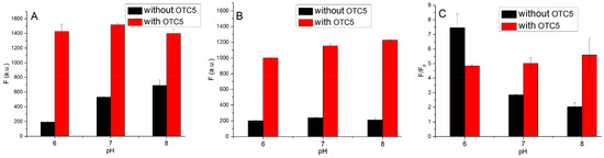
Figure 3.
Fluorescence intensity of 200 nM doxycycline before and after adding 2 µM OTC5 aptamer at different pH values (A) in the absence of GO and (B) in the presence of 20 µg/mL GO. (C) Effect of GO on the signal-to-background ratio of the sensor at different pH values.
We then tested the sensor in the presence of GO, which brought all background fluorescence intensities of doxycycline to a similarly low level (close to pH 6 of the GO-free sample, see the black bars in Figure 3B). Interestingly, the increased fluorescence intensity after the addition of OTC5 was not affected by pH, all reaching a similarly high level (Figure 3B, red bars). Therefore, all the three pH values resulted in a similar five- to six-fold enhancement. The effect of GO on signal-to-background ratio is plotted in Figure 3C, where the red bars all have the same height, whereas the black bars dropped at a higher pH.
We then optimized the concentration of GO by testing it up to 40 μg/mL (Figure 4A). The fold of fluorescence enhancement increased with GO concentration. After reaching a peak value at 20 μg/mL GO, a further increase in GO concentration even dropped the enhancement. We reason that when the concentration of GO is too high, a great amount of free GO surface is available, and it can also adsorb the added aptamer and thus decrease the target binding efficiency of the aptamer. Therefore, for 200 nM doxycycline and 2 µM OTC5 aptamer, 20 μg/mL GO was an optimal concentration.
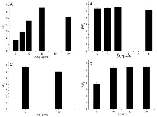
Figure 4.
(A) Effect of the concentrations of GO on the fold of fluorescence enhancement upon adding the aptamer. (B) Effect of different concentrations of Mg2+. The NaCl concentration in this experiment was 0. (C) Effect of the presence of Na+ on fluorescence intensity. The Mg2+ concentration in this experiment was 2 mM. (D) Effect of reaction time after adding OTC5.
The effect of Mg2+ concentration was then examined up to 5 mM, and the optimal Mg2+ concentration was determined to be 2 mM (Figure 4B). When the Mg2+ concentration was too low, the aptamer cannot bind since we previously demonstrated that OTC5 required Mg2+ for binding [14]. In addition, we also demonstrated above that Mg2+ was required to adsorb doxycycline to GO. When the Mg2+ concentration was too high, it may push the aptamer/doxycycline complex to adsorb [27]. In the presence of 2 mM Mg2+, Na+ was tested at 0 and 100 mM. As shown in Figure 4C, Na+ had little effect. Therefore, Mg2+ was a key metal ion in this system.
All previous experiments were measured within 2 min of the addition of OTC5. Here, the reaction time after the addition of OTC5 was optimized. The kinetics after the addition of OTC5 was followed for a duration of 40 min, and the fluorescence intensity increased immediately but did not change significantly in the first 40 min (Figure 4D).
To understand the reaction kinetics more precisely, we then followed the reaction kinetics in a fluorometer. As shown in Figure 5A, the fluorescence of doxycycline was monitored for 3 min (no GO) and then OTC5 aptamer was added. An immediate fluorescence increase was observed due to the aptamer binding, indicating that aptamer binding was a fast reaction. In a separate experiment, we added GO to doxycycline and a fluorescence drop occurred in 1 min, indicating fast adsorption. At 5 min, 2 µM OTC5 aptamer was added, and the fluorescence of doxycycline immediately increased followed by a slow increase phase (Figure 5B). We reason that the initial rapid increase was mainly from the binding of the free doxycycline by the aptamer, whereas the subsequent slower phase was due to binding to the desorbed doxycycline. Since the fluorometer had a lower sensitivity than our microplate reader, we had to use a higher doxycycline concentration here.
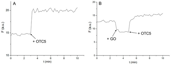
Figure 5.
(A) Kinetic trace of 2 μM doxycycline fluorescence for 3 min followed by the addition of 2 μM OTC5 in the absence of GO. The immediate fluorescence increase indicates a rapid binding reaction. (B) Kinetic curves of 2 μM doxycycline adsorption by 20 µg/mL GO. At 5 min, doxycycline desorption by 2 µM OTC5 from GO. The experiments were performed in 10 mM buffer with pH 8.0.
3.5. Detection of Doxycycline
After optimization of the conditions, we then tested the performance of the sensor without and with GO. To measure sensitivity, doxycycline concentration varied from 0 to 2 μM. At pH 8, the fluorescence intensity of doxycycline was slightly increased upon adding 2 µM OTC5 aptamer (Figure 6A). We also plotted the difference in the fluorescence intensity after adding OTC5 (Figure 6B), which can be considered as the calibration curve of the sensor. A roughly linear increase was observed up to 1.5 µM doxycycline. Based on the 3σ/slope calculation, the limit of detection (LOD) was calculated to be 18.9 nM. In the presence of GO (Figure 6C), the background was suppressed at all the tested doxycycline concentrations and the difference was also larger (Figure 6D). The linear range reached 2.0 µM doxycycline. The slope of the curve in Figure 6D was 1.98-fold that in Figure 6C. The LOD of the sensor with GO was calculated to be 2.9 nM. Therefore, the sensor in the presence of GO was more sensitive and had a lower LOD.

Figure 6.
(A) Effect of OTC5 on a 0–2 μM concentration gradient of doxycycline in the absence of GO. (B) The difference in fluorescence intensity of the sample solution before and after the addition of OTC5. The R2 value of the fitting is 0.881. (C) Effect of OTC5 on a 0–2 μM concentration gradient of doxycycline in the presence of GO. (D) The difference in fluorescence intensity of the sample solution before and after the addition of OTC5. The R2 value of the fitting is 0.991.
3.6. Selectivity Test
To test the selectivity, this experiment was conducted for the adsorption experiments of oxytetracycline (OTC) and tetracycline (TC), which are also tetracycline antibiotics, and two other non-tetracycline antibiotics, chloramphenicol (CAP) and ampicillin (AMP), under the same conditions. In the absence of GO, OTC5 resulted in a significant increase in the fluorescence intensity of the OTC and TC sample solutions, while there was no significant change in the fluorescence intensity of the CAP and AMP sample solutions (Figure 7A). In the presence of GO, fluorescence quenching of the antibiotics by GO still occurred in the OTC and TC sample solutions, but the same fluorescence quenching was not found in the CAP and AMP sample solutions (Figure 7B).

Figure 7.
(A) Adsorption of four antibiotics, OTC, TC, CAP, and AMP, in sample solution at a concentration of 0.5 μM with OTC5 aptamer in the absence of GO. (B) Effect of GO on the adsorption of four antibiotics, OTC, TC, CAP, and AMP, with OTC5 aptamer at a concentration of 0.5 μM in the sample solution.
It can be concluded that since OTC and TC are tetracycline antibiotics, they also bind to OTC5 and cause background fluorescence enhancement. This is consistent with our previous observations. The structure of CAP and AMP is very different from that of tetracycline antibiotics such as doxycycline, which itself has no fluorescence intensity and does not produce adsorption and binding phenomena with OTC5 and GO. Hence, no obvious phenomena occurred for CAP and AMP.
3.7. Further Discussion
For DNA aptamer and GO-based biosensors, most of the previous work focused on the adsorption of a fluorophore-labeled aptamer followed by target-induced aptamer desorption [16,21]. For such sensors, a covalently labeled aptamer is required and this increases the cost. In this work, by taking advantage of the intrinsic fluorescence of the target analyte, we first adsorbed the analyte and demonstrated that adding its aptamer can also desorb the analyte. This is a label-free sensor, and no extra dyes were added. This work showed that aptamer can also induce desorption of target analytes from GO.
Nanomaterials can help DNA-based sensors and devices in many different ways [28,29,30]. Research has been focused on the adsorption of aptamers. In a few studies, we noticed the importance of target adsorption, especially on gold nanoparticles [31] and metal oxides [32]. In those cases, the proposed sensing mechanisms could be changed by considering target adsorption. In this case, we intentionally used target adsorption to improve the sensor.
4. Conclusions
In this work, we studied the adsorption of doxycycline by GO from pH 6 to 8 in the presence of 2 mM Mg2+ and found that a higher adsorption was achieved at a higher pH. Using this observation, we were able to suppress the background fluorescence of doxycycline for its aptamer-based detection. Doxycycline’s fluorescence can be enhanced by the OTC5 aptamer, which is a unique way of detecting doxycycline and other tetracycline antibiotics. By adding GO, the signal-to-background ratio was independent of pH, whereas the signal-to-background ratio was much lower at a higher pH in the absence of GO. Most previous GO and aptamer-based biosensors adsorbed a fluorophore-labeled aptamer and relied on target binding to induce aptamer desorption [16,33]. In this case, we adsorbed the target molecule and relied on aptamer binding to generate the signal. This is a unique way of sensing taking advantage of the intrinsic fluorescence properties of the target.
Author Contributions
Conceptualization, J.L.; methodology, Y.Z. and J.L.; investigation, Y.Z. and J.L.; data curation, Y.Z.; writing—original draft preparation, Y.Z. and J.L.; writing—review and editing, J.L.; supervision, J.L.; funding acquisition, J.L. All authors have read and agreed to the published version of the manuscript.
Funding
This research was funded by the Natural Sciences and Engineering Research Council of Canada (NSERC).
Institutional Review Board Statement
Not applicable.
Informed Consent Statement
Not applicable.
Data Availability Statement
Data is available upon reasonable request from the authors.
Conflicts of Interest
The authors declare no conflict of interest.
References
- Chopra, I.; Roberts, M. Tetracycline Antibiotics: Mode of Action, Applications, Molecular Biology, and Epidemiology of Bacterial Resistance. Microbiol. Mol. Biol. Rev. 2001, 65, 232–260. [Google Scholar] [CrossRef] [PubMed]
- Schnappinger, D.; Hillen, W. Tetracyclines: Antibiotic action, uptake, and resistance mechanisms. Arch. Microbiol. 1996, 165, 359–369. [Google Scholar] [CrossRef] [PubMed]
- Niazi, J.H.; Lee, S.J.; Kim, Y.S.; Gu, M.B. ssDNA aptamers that selectively bind oxytetracycline. Biorg. Med. Chem. 2008, 16, 1254–1261. [Google Scholar] [CrossRef] [PubMed]
- Niazi, J.H.; Lee, S.J.; Gu, M.B. Single-stranded DNA aptamers specific for antibiotics tetracyclines. Biorg. Med. Chem. 2008, 16, 7245–7253. [Google Scholar] [CrossRef] [PubMed]
- Tickner, Z.J.; Zhong, G.; Sheptack, K.R.; Farzan, M. Selection of High-Affinity RNA Aptamers That Distinguish between Doxycycline and Tetracycline. Biochemistry 2020, 59, 3473–3486. [Google Scholar] [CrossRef]
- Berens, C.; Thain, A.; Schroeder, R. A tetracycline-binding RNA aptamer. Biorg. Med. Chem. 2001, 9, 2549–2556. [Google Scholar] [CrossRef]
- Yu, H.; Alkhamis, O.; Canoura, J.; Liu, Y.; Xiao, Y. Advances and Challenges in Small-Molecule DNA Aptamer Isolation, Characterization, and Sensor Development. Angew. Chem. Int. Ed. 2021, 60, 16800–16823. [Google Scholar] [CrossRef]
- Wu, L.; Wang, Y.; Xu, X.; Liu, Y.; Lin, B.; Zhang, M.; Zhang, J.; Wan, S.; Yang, C.; Tan, W. Aptamer-Based Detection of Circulating Targets for Precision Medicine. Chem. Rev. 2021, 121, 12035–12105. [Google Scholar] [CrossRef]
- He, L.; Huang, R.; Xiao, P.; Liu, Y.; Jin, L.; Liu, H.; Li, S.; Deng, Y.; Chen, Z.; Li, Z.; et al. Current signal amplification strategies in aptamer-based electrochemical biosensor: A review. Chin. Chem. Lett. 2021, 32, 1593–1602. [Google Scholar] [CrossRef]
- Li, Y.; Gao, H.; Qi, Z.; Huang, Z.; Ma, L.; Liu, J. Freezing-Assisted Conjugation of Unmodified Diblock DNA to Hydrogel Nanoparticles and Monoliths for DNA and Hg2+ Sensing. Angew. Chem. Int. Ed. 2021, 60, 12985–12991. [Google Scholar] [CrossRef]
- McConnell, E.M.; Nguyen, J.; Li, Y. Aptamer-Based Biosensors for Environmental Monitoring. Front. Chem. 2020, 8, 434. [Google Scholar] [CrossRef]
- Zhao, Y.; Yavari, K.; Wang, Y.; Pi, K.; Van Cappellen, P.; Liu, J. Deployment of functional DNA-based biosensors for environmental water analysis. TrAC Trends Anal. Chem. 2022, 153, 116639. [Google Scholar] [CrossRef]
- Müller, M.; Weigand, J.E.; Weichenrieder, O.; Suess, B. Thermodynamic characterization of an engineered tetracycline-binding riboswitch. Nucleic Acids Res. 2006, 34, 2607–2617. [Google Scholar] [CrossRef]
- Zhao, Y.; Ong, S.; Chen, Y.; Jimmy Huang, P.-J.; Liu, J. Label-free and Dye-free Fluorescent Sensing of Tetracyclines Using a Capture-Selected DNA Aptamer. Anal. Chem. 2022, 94, 10175–10182. [Google Scholar] [CrossRef]
- Zhao, Y.; Gao, B.; Sun, P.; Liu, J.; Liu, J. Metal and pH-Dependent Aptamer Binding of Tetracyclines Enabling Highly Sensitive Fluorescence Sensing. Biosensors 2022, 12, 717. [Google Scholar] [CrossRef]
- Lu, C.H.; Yang, H.H.; Zhu, C.L.; Chen, X.; Chen, G.N. A Graphene Platform for Sensing Biomolecules. Angew. Chem. Int. Ed. 2009, 48, 4785–4787. [Google Scholar] [CrossRef]
- Gao, Y.; Li, Y.; Zhang, L.; Huang, H.; Hu, J.; Shah, S.M.; Su, X. Adsorption and removal of tetracycline antibiotics from aqueous solution by graphene oxide. J. Colloid Interface Sci. 2012, 368, 540–546. [Google Scholar] [CrossRef]
- Liu, B.; Salgado, S.; Maheshwari, V.; Liu, J. DNA adsorbed on graphene and graphene oxide: Fundamental interactions, desorption and applications. Curr. Opin. Colloid Interface Sci. 2016, 26, 41–49. [Google Scholar] [CrossRef]
- Liu, X.Q.; Aizen, R.; Freeman, R.; Yehezkeli, O.; Willner, I. Multiplexed Aptasensors and Amplified DNA Sensors Using Functionalized Graphene Oxide: Application for Logic Gate Operations. ACS Nano 2012, 6, 3553–3563. [Google Scholar] [CrossRef]
- He, S.J.; Song, B.; Li, D.; Zhu, C.F.; Qi, W.P.; Wen, Y.Q.; Wang, L.H.; Song, S.P.; Fang, H.P.; Fan, C.H. A Graphene Nanoprobe for Rapid, Sensitive, and Multicolor Fluorescent DNA Analysis. Adv. Funct. Mater. 2010, 20, 453–459. [Google Scholar] [CrossRef]
- Lopez, A.; Liu, J. Covalent and Noncovalent Functionalization of Graphene Oxide with DNA for Smart Sensing. Adv. Intell. Syst. 2020, 2, 2000123. [Google Scholar] [CrossRef]
- Hassani, M.; Lázaro, R.; Pérez, C.; Condón, S.; Pagán, R. Thermostability of Oxytetracycline, Tetracycline, and Doxycycline at Ultrahigh Temperatures. J. Agric. Food. Chem. 2008, 56, 2676–2680. [Google Scholar] [CrossRef] [PubMed]
- Heaton, P.C.; Fenwick, S.R.; Brewer, D.E. Association between tetracycline or doxycycline and hepatotoxicity: A population based case–control study1. J. Clin. Pharm. Ther. 2007, 32, 483–487. [Google Scholar] [CrossRef] [PubMed]
- Kogawa, A.C.; Salgado, H.R.N. Quantification of Doxycycline Hyclate in Tablets by HPLC–UV Method. J. Chromatogr. Sci. 2012, 51, 919–925. [Google Scholar] [CrossRef]
- Kushalkar, M.P.; Liu, B.; Liu, J. Promoting DNA Adsorption by Acids and Polyvalent Cations: Beyond Charge Screening. Langmuir 2020, 36, 11183–11195. [Google Scholar] [CrossRef]
- Wang, Z.; Huang, Z.; Han, J.; Xie, G.; Liu, J. Polyvalent Metal Ion Promoted Adsorption of DNA Oligonucleotides by Montmorillonite. Langmuir 2021, 37, 1037–1044. [Google Scholar] [CrossRef]
- Huang, P.-J.J.; Liu, J. Signaling Kinetics of DNA and Aptamer Biosensors Revealing Graphene Oxide Surface Heterogeneity. J. Anal. Test. 2022, 6, 20–27. [Google Scholar] [CrossRef]
- Katz, E.; Willner, I. Nanobiotechnology: Integrated nanoparticle-biomolecule hybrid systems: Synthesis, properties, and applications. Angew. Chem. Int. Ed. 2004, 43, 6042–6108. [Google Scholar] [CrossRef]
- Rosi, N.L.; Mirkin, C.A. Nanostructures in Biodiagnostics. Chem. Rev. 2005, 105, 1547–1562. [Google Scholar] [CrossRef]
- Li, L.L.; Xing, H.; Zhang, J.J.; Lu, Y. Functional DNA Molecules Enable Selective and Stimuli-Responsive Nanoparticles for Biomedical Applications. Acc. Chem. Res. 2019, 52, 2415–2426. [Google Scholar] [CrossRef]
- Zhang, F.; Liu, J. Label-Free Colorimetric Biosensors Based on Aptamers and Gold Nanoparticles: A Critical Review. Anal. Sens. 2021, 1, 30–43. [Google Scholar] [CrossRef]
- Lopez, A.; Liu, J. Nanomaterial and Aptamer-Based Sensing: Target Binding versus Target Adsorption Illustrated by the Detection of Adenosine and ATP on Metal Oxides and Graphene Oxide. Anal. Chem. 2021, 93, 3018–3025. [Google Scholar] [CrossRef]
- Li, F.; Huang, Y.; Yang, Q.; Zhong, Z.T.; Li, D.; Wang, L.H.; Song, S.P.; Fan, C.H. A graphene-enhanced molecular beacon for homogeneous DNA detection. Nanoscale 2010, 2, 1021–1026. [Google Scholar] [CrossRef]


Disclaimer/Publisher’s Note: The statements, opinions and data contained in all publications are solely those of the individual author(s) and contributor(s) and not of MDPI and/or the editor(s). MDPI and/or the editor(s) disclaim responsibility for any injury to people or property resulting from any ideas, methods, instructions or products referred to in the content. |
© 2023 by the authors. Licensee MDPI, Basel, Switzerland. This article is an open access article distributed under the terms and conditions of the Creative Commons Attribution (CC BY) license (https://creativecommons.org/licenses/by/4.0/).