Opinion

From Traditional Nanoparticles to Cluster-Triggered Emission Polymers for the Generation of Smart Nanotheranostics in Cancer Treatment

by
Cristina Blasco-Navarro
1,
Carlos Alonso-Moreno
2 and
Iván Bravo
1,*
1
Unidad nanoDrug, Facultad de Farmacia de Albacete, Departamento de Química Física, Universidad de Castilla-La Mancha, 02071 Albacete, Spain
2
Unidad nanoDrug, Facultad de Farmacia-Centro de Innovación en Química Avanzada (ORFEO-CINQA), Departamento de Química Inorgánica, Orgánica y Bioquímica, Universidad de Castilla-La Mancha, 02071 Albacete, Spain
*
Author to whom correspondence should be addressed.
J. Nanotheranostics 2025, 6(1), 3; https://doi.org/10.3390/jnt6010003
Submission received: 8 December 2024 / Revised: 10 January 2025 / Accepted: 20 January 2025 / Published: 22 January 2025

Abstract

:
Nanotheranostics integrates diagnostic and therapeutic functionalities using nanoscale materials, advancing personalized medicine by enhancing treatment precision and reducing adverse effects. Key materials for nanotheranostics include metallic nanoparticles, quantum dots, carbon dots, lipid nanoparticles and polymer-based nanocarriers, each offering unique benefits alongside specific challenges. Polymer-based nanocarriers, including hybrid and superparamagnetic nanoparticles, improve stability and functionality but are complex to manufacture. Polymeric nanoparticles with aggregation-induced emission (AIE) present promising theranostic potential for cancer detection and treatment. However, challenges such as translating the AIE concept to living systems, addressing toxicity concerns, overcoming deep-tissue imaging limitations, or ensuring biocompatibility remain to be resolved. Recently, cluster-triggered emission (CTE) polymers have emerged as innovative materials in nanotheranostics, offering enhanced fluorescence and biocompatibility. These polymers exhibit increased fluorescence intensity upon aggregation, making them highly sensitive for imaging and therapeutic applications. CTE nanoparticles, crafted from biodegradable polymers, represent a safer alternative to traditional nanotheranostics that rely on embedding conventional fluorophores or metal-based agents. This advancement significantly reduces potential toxicity while enhancing biocompatibility. The intrinsic fluorescence allows real-time monitoring of drug distribution and activity, optimizing therapeutic efficacy. Despite their potential, these systems face challenges such as maintaining stability under physiological conditions and addressing the need for comprehensive safety and efficacy studies to meet clinical and regulatory standards. Nevertheless, their unique properties position CTE nanoparticles as promising candidates for advancing theranostic strategies in personalized medicine, bridging diagnostic and therapeutic functionalities in innovative ways.

Theranostics is a synergistic field that combines diagnostics with treatment. So, the term “nanotheranostics” refers to the practice of using and improving nanomedicine strategies for advanced theranostic purposes [1]. It utilizes nanoparticles or molecules at the nanoscale to simultaneously identify, monitor, and treat diseases. In oncology, it facilitates early cancer detection by enabling the design of smart nanoparticles that selectively target and bind to specific cancer cells. Nanotheranostics significantly advances personalized medicine by enabling the development of tailored treatments based on the patient’s unique biomarkers. This approach optimizes therapeutic efficacy while minimizing adverse effects. Furthermore, it offers the capability to monitor the body’s real-time response to treatment, allowing for precise adjustments in dosage to enhance outcomes. Therefore, the benefits of nanotheranostics include enhanced precision in disease localization and treatment, improved efficiency by integrating diagnosis and therapy into a single intervention, personalized treatments tailored to each patient’s unique needs, and reduced side effects though targeted delivery exclusively to affected cells [2].
Nanotheranostics are complex systems designed to integrate diagnostic and therapeutic functionalities within a single platform. At their core, the diagnostic agent, often metallic nanoparticles like quantum dots or gold, provides capabilities such as imaging or molecular detection. These are typically encapsulated or functionalized with a construction material, such as biodegradable polymers, lipids, or hybrid structures like carbon dot–silica composites, that enhances biocompatibility, stability, and targeted delivery. The therapeutic agent can vary from drugs or gene therapy molecules, which are either loaded onto these carriers or conjugated to their surface, to modalities like photothermal therapy, leveraging the unique properties of metallic nanoparticles [3,4]. This strategic integration enables precise imaging and effective treatment, while the choice of construction material ensures enhanced biodistribution, minimized toxicity, and versatile functionality. Figure 1A showcases the different types of nanotheranostic systems employed, highlighting their key characteristics.
Among all of these systems, inorganic nanoparticles are the most widely used for theranostic applications, with gold nanoparticles standing out prominently. One of their key advantages lies in their highly modifiable surface, which enables straightforward conjugation with drugs or imaging contrast agents [5]. Furthermore, gold nanoparticles have been widely explored for their applications in photothermal therapy, utilizing their ability to absorb incident light and convert it into heat, leading to the destruction of tumor cells. These features render them indispensable in theranostics, where molecular detection, biological imaging, and targeted cancer therapy converge with photothermal treatment to enhance precision and therapeutic efficacy [6]. Additionally, they can be easily conjugated with antitumor drugs, such as antisense DNA, through simple covalent bonding [7]. However, despite their biocompatibility and low toxicity, challenges persist related to their toxicological profile and biodistribution when administered in their non-functionalized form. Recent strategies to overcome these challenges involve coating gold nanoparticles with biodegradable polymers or functionalizing them with tumor-targeting ligands, which improves both their biodistribution and specificity [3,8].
Quantum dots (QDs) are another notable category of nanotheranostics, which are renowned for their photochemical stability and robust fluorescence emission. These properties make QDs excellent candidates as imaging agents offering high-resolution, multiplexed imaging for biological applications [9]. Recently, QDs have been explored as drug delivery vehicles through surface modification and conjugation. However, concerns persist regarding the potential toxicity of heavy metals such as cadmium and lead, which are commonly found in their composition [10]. On the other hand, diagnostic nanomaterials like carbon dots (CDs) have garnered attention due to their optical emissive properties, high quantum yield, photostability, and low toxicity. To further enhance their theranostic potential, hybrid carbon dot–silica nanostructures have been developed. In these systems, carbon dots provide fluorescence, while silica contributes porosity and a large surface area. This combination allows for functionalization, as well as the loading and controlled release of therapeutic agents, making them promising candidates for both diagnostic and therapeutic applications [11].
Lipid nanoparticles are another widely studied material for nanotheranostics, primarily as nanocarriers in cancer therapy. Liposomes, capable of encapsulating hydrophilic molecules within their core and hydrophobic molecules in their lipid bilayer, are especially prominent. These versatile nanoparticles are the most investigated systems for drug delivery in clinical trials and even approved therapies [12]. Despite their versatility in encapsulating diverse molecules, liposomes face limitations regarding stability. In addition, as theranostic systems, they often require the encapsulation of a fluorescent drug or probe for tracking purposes [13].
Polymer-based nanocarriers, including dendrimers, have emerged as a significant focus in nanomedicine due to their versatility in enhancing nanoparticle stability, biocompatibility, and functionality. Conventional FDA-approved polymers typically lack the inherent characteristics necessary for imaging or tracking, highlighting the need for innovative designs and modifications to fully realize their potential. According to S.M. Hosseini et al. [1], the design of polymeric theranostic nanoparticles depends on their primary function. These nanoparticles can be engineered for a variety of roles, such as delivering anticancer drugs or light-sensitive agents, improving imaging contrast, or integrating both therapeutic and imaging capabilities within a single system. However, nanoparticles that combine both imaging and drug delivery capabilities face challenges related to functionalization and loading capacity. As a result, this type of nanotheranostic is very difficult to find in the clinic because it is a complex system that is not easy to manufacture or reach the final stages of clinical trials. An example of the strategies studied for the creation of polymer-based theranostic nanoparticles involves superparamagnetic nanoparticles, which have been applied in numerous areas ranging from cancer treatment through magnetic hyperthermia, contrast agents, and MRI-guided drug delivery. Superparamagnetic iron oxide nanoparticles (SPIONs) are particularly notable in these applications [14,15]. Another promising approach in polymer-based nanotheranostics is the use of polymeric nanoparticles as radionuclide carriers. These nanocarriers can improve the efficacy of radiotherapy by facilitating the targeted delivery of radiosensitizers or radionuclides to tumor-affected organs. The use of radioisotopes allows the treatment to be monitored through imaging techniques such as positron emission tomography (PET) or Cerenkov luminescence (CL) [16,17,18]. However, these systems still have the disadvantage of containing metals, which can be toxic, and therefore the biocompatibility of the nanosystem is not fully solved.
To address these challenges, hybrid systems such as polymer–lipid, lipid–metal, or liposome–QD (quantum dot) combinations have been developed. These systems aim to overcome the stability issues while enhancing their functionality for specific theranostic applications. These hybrid materials aim to combine the advantages of each component, such as the biocompatibility and drug delivery capabilities of liposomes, the structural stability of polymers, the enhanced imaging potential of quantum dots, and the unique properties of metallic nanoparticles. By tailoring the properties of these hybrid systems, researchers are able to improve the stability, targeting efficiency, and therapeutic efficacy of nanotheranostics, thus broadening their potential for clinical use [19,20,21]. The complexity of intricate nanosystems can obscure key aspects, such as in vivo nano–bio interactions and the functional outcomes at the tissue, cellular, and molecular levels, which are often overlooked in the pursuit of novel designs. In this context, simpler nanosystems that demonstrate clear evidence of their mechanisms within these biological environments are preferable to developing numerous new and complicated systems [22,23].
In the search for this simplicity, recent years have seen the investigation of fluorescent polymeric nanoparticles as innovative theranostic agents for the simultaneous detection and treatment of cancer. These nanoparticles are typically fabricated using diagnostic agents such as fluorescent proteins, inorganic QDs, or fluorescent chromophores, while also encapsulating the therapeutic drug [21]. The use of a biocompatible polymeric matrix in these designs enhances their suitability for biomedical applications. However, the inclusion of chromophores or QDs can introduce potential toxicity concerns, which may impact their long-term biocompatibility and clinical viability. To overcome these challenges, AIE polymers have emerged as a transformative solution. Incorporating aggregation-induced emission (AIE) units into polymer structures offers significant advantages for nanotheranostic applications, including enhanced luminescence efficiency and sensitivity in aggregated states, which bypasses issues like aggregation-caused quenching (ACQ). These properties make AIE polymers ideal for bioimaging, biosensing, and drug delivery. However, challenges remain, such as the complexity of translating AIE properties to living systems, where controlling aggregation in a complex media remains a significant challenge [24,25,26]. Additionally, short-wavelength emissions and broad spectra limit deep-tissue imaging due to autofluorescence and light penetration issues. The development of near-infrared (NIR-II) AIEgens with advanced functionalization is necessary to address these limitations. Furthermore, toxicity concerns arise from incorporating AIE units into polymer chains, necessitating thorough biocompatibility and safety assessments for clinical translation [26,27].
Recent research efforts are focusing on the development of fluorescent theranostic agents only based on biocompatible and safer polymers, aiming to balance imaging capabilities with minimal toxicity. Cluster-triggered emission (CTE) polymers represent a promising advancement in the field of nanotheranostics [28,29,30,31]. Cluster-triggered emission (CTE) is a phenomenon where the fluorescence intensity of polymers significantly increases upon the aggregation of different subunits within the polymer chain, similar to AIE systems. However, AIE and CTE are distinct chemical mechanisms with different requirements, underlying principles, and applications. AIE involves luminophores that are weakly emissive or non-emissive in solution but become highly emissive upon aggregation due to restricted molecular motions (e.g., rotation or vibration), which suppress non-radiative decay pathways. In contrast, CTE arises from the formation of non-covalent molecular clusters, usually made up of non-conjugated molecules containing subunits with n and/or π electrons, such as C=O, NH2, C=N, etc. (see Figure 1B). Fluorescence in CTE systems results from electronic coupling, intermolecular interactions, or hydrogen bonding, based on the through-space interaction (TSI) model. Notably, CTE does not require π-conjugation and can occur in small or simple molecules that are individually non-fluorescent [32,33,34,35].
CTE polymers offer significant advantages for biomedical applications, particularly due to their biodegradable nature, making them safer and more sustainable. These polymers are often derived from FDA-approved polyesters, such as PLA and PLGA, which, upon degradation, produce non-toxic metabolites that can be easily eliminated by the body. This biodegradation minimizes the risk of long-term accumulation and side effects, thus enhancing their potential in drug delivery and theranostic systems [36]. However, their application in nanotheranostic remains limited and few examples can be found in the literature. For instance, Shao et al. (2019) developed a non-conjugated PLA-based polyethylenimine copolymer for CTE nanoparticles, which were used in both in vitro and in vivo bioimaging [37]. In addition, Peng et al. (2024) introduced multicolor CTE polymers for the theranostics of osteoarthritis, based on polycysteine [38]. More recently, CTE nanoparticles have also been described in the context of photothermal imaging [31] and as photosensitizers in antimicrobial photodynamic therapy [39]. In addition, these nanoparticles could overcome a key limitation of traditional fluorophores: the phenomenon of fluorescence quenching that occurs at high concentrations. In CTE nanoparticles, the fluorescence is not only preserved but is actually enhanced when the nanoparticles aggregate. In this sense and as an example, Wang et al., in 2021, developed coassembled chitosan–hyaluronic acid CTE nanoparticles as theranostic agents targeting Alzheimer’s β-Amyloid, which exhibited increased red fluorescence upon interaction with amyloid-beta oligomers and fibrils [40]. This property results in clearer and more reliable fluorescence signals, making them particularly advantageous for diagnostic applications. The enhanced emission in aggregated states improves the sensitivity and accuracy of imaging techniques, such as fluorescence imaging and fluorescence lifetime imaging microscopy (FLIM), allowing for the more precise detection of biomarkers or cellular targets in theranostic systems. Additionally, integration with other imaging modalities, such as positron emission tomography (PET), can be explored through the encapsulation or conjugation of a radiotracer to the CTE nanoparticle. Another notable advantage is the ability to tune their optical properties by modifying the polymer composition and structure. This tunability enables the precise control of the nanoparticle’s fluorescence characteristics, which can be tailored for specific diagnostic and therapeutic applications. By adjusting the polymer’s composition, the fluorescence emission wavelength, intensity, and quantum yield can be optimized, enhancing their suitability for various imaging techniques, such as fluorescence microscopy and in vivo imaging. Since these polymers can form nanoparticles, they could have great potential for controlled drug encapsulation and release, as well as for conjugation with antibodies or other molecules due to their large and modifiable surface area. Moreover, their intrinsic fluorescence allows real-time monitoring of the drug’s distribution and activity within the body, optimizing therapeutic efficacy [40,41]. An example that combines nanotheranostics with drug delivery is the work of Saha et al. (2019), who designed a CTE polymer through the copolymerization of PEG and maleimide, enabling both bioimaging and the controlled release of chlorambucil for monitoring cancer treatment [42]. This combination of controllable drug delivery and fluorescence-based tracking makes CTE nanoparticles highly promising for advancing theranostic applications in personalized medicine.
Despite the promising potential of CTE polymers for nanotheranostics, significant challenges hinder their widespread application. These include limited studies on their stability under physiological conditions, raising concerns about their durability in vivo. Crucial factors such as in vivo nano–bio interactions, colloidal stability, stealth properties, tissue-specific targeting, and cellular uptake remain inadequately understood. Addressing these challenges requires rigorous evaluation of their pharmacokinetics, biodistribution, and functional performance at the tissue, cellular, and molecular levels. Additionally, enhancing their design for better physiological compatibility and controlled functional outcomes is essential to facilitate their transition from experimental models to clinical applications [22,23]. To enhance the functionality and clinical translation of CTE nanoparticles, it is crucial to prioritize biocompatibility by using biodegradable polymers like polyesters, reducing toxicity risks and ensuring safe degradation. Optical properties must be optimized by designing systems with red or near-infrared (NIR) emissions for deeper tissue imaging and better contrast. Incorporating stimuli-responsive features, such as pH or enzyme sensitivity, can enable precise targeting and controlled drug release. Multifunctional systems combining imaging and therapy should be developed to expand biomedical applications like photodynamic therapy. Preclinical studies are essential to evaluate pharmacokinetics, long-term safety, and efficacy, while scalable manufacturing methods must ensure reproducibility. Collaborations between researchers, clinicians, and industry experts are vital to streamline clinical adoption and regulatory approval processes.
Nonetheless, CTE nanoparticles hold significant promise in nanotheranostics, offering straightforward platforms capable of controlled drug delivery, real-time imaging, and potentially minimal side effects. Their simplicity and biocompatibility make them attractive candidates for integrating diagnostic and therapeutic functions. As research addresses existing challenges such as stability, tissue targeting, and long-term safety, CTE nanoparticles are poised to play a pivotal role in personalized medicine. This could lead to more precise, effective treatments for cancer and other complex diseases, paving the way for their clinical application in advanced therapeutic strategies.

Author Contributions

Conceptualization and writing: C.B.-N., C.A.-M. and I.B.; Reviewing and editing C.B.-N., C.A.-M. and I.B. All authors have read and agreed to the published version of the manuscript.

Funding

We gratefully acknowledge the financial support from the Ministerio de Ciencia e Innovación y Agencia Estatal de Investigación, Spain: CPP2021-008597, PID2020-117788RB-I00, RED2022-134287-T; European Union NextGenerationEU/PRTR: MICIU/AEI/10.13039/501100011033; Junta de Comunidades de Castilla La Mancha: SBPLY/21/180501/000050 and SBPLY/23/180502/000013; Universidad de Castilla-La Mancha: 2022-GRIN-34143; and ACEPAIN Foundation.

Data Availability Statement

Not applicable.

Conflicts of Interest

The authors declare no conflicts of interest.

References

  1. Hosseini, S.; Mohammadnejad, J.; Salamat, S.; Zadeh, Z.B.; Tanhaei, M.; Ramakrishna, S. Theranostic polymeric nanoparticles as a new approach in cancer therapy and diagnosis: A review. Mater. Today Chem. 2023, 29, 101400. [Google Scholar] [CrossRef]
  2. Tiwari, H.; Rai, N.; Singh, S.; Gupta, P.; Verma, A.; Singh, A.K.; Kajal; Salvi, P.; Singh, S.K.; Gautam, V. Recent Advances in Nanomaterials-Based Targeted Drug Delivery for Preclinical Cancer Diagnosis and Therapeutics. Bioengineering 2023, 10, 760. [Google Scholar] [CrossRef] [PubMed]
  3. Al-Thani, A.N.; Jan, A.G.; Abbas, M.; Geetha, M.; Sadasivuni, K.K. Nanoparticles in cancer theragnostic and drug delivery: A comprehensive review. Life Sci. 2024, 352, 122899. [Google Scholar] [CrossRef]
  4. Oehler, J.B.; Rajapaksha, W.; Albrecht, H. Emerging Applications of Nanoparticles in the Diagnosis and Treatment of Breast Cancer. J. Pers. Med. 2024, 14, 723. [Google Scholar] [CrossRef] [PubMed]
  5. Shabatina, T.I.; Vernaya, O.I.; Shimanovskiy, N.L.; Melnikov, M.Y. Metal and Metal Oxides Nanoparticles and Nanosystems in Anticancer and Antiviral Theragnostic Agents. Pharmaceutics 2023, 15, 1181. [Google Scholar] [CrossRef] [PubMed]
  6. Kesharwani, P.; Ma, R.; Sang, L.; Fatima, M.; Sheikh, A.; Abourehab, M.A.S.; Gupta, N.; Chen, Z.-S.; Zhou, Y. Gold nanoparticles and gold nanorods in the landscape of cancer therapy. Mol. Cancer 2023, 22, 98. [Google Scholar] [CrossRef] [PubMed]
  7. Wang, X.; Yang, T.; Yu, Z.; Liu, T.; Jin, R.; Weng, L.; Bai, Y.; Gooding, J.J.; Zhang, Y.; Chen, X. Intelligent Gold Nanoparticles with Oncogenic MicroRNA-Dependent Activities to Manipulate Tumorigenic Environments for Synergistic Tumor Therapy. Adv. Mater. 2022, 34, 2110219. Available online: https://pubmed.ncbi.nlm.nih.gov/35170096/ (accessed on 3 December 2024). [CrossRef]
  8. Liong, M.; Lu, J.; Kovochich, M.; Xia, T.; Ruehm, S.G.; Nel, A.E.; Tamanoi, F.; Zink, J.I. Multifunctional inorganic nanoparticles for imaging, targeting, and drug delivery. ACS Nano 2008, 2, 889–896. [Google Scholar] [CrossRef]
  9. Debnath, M.; Sarkar, S.; Debnath, S.K.; Dkhar, D.S.; Kumari, R.; Vaskuri, G.S.S.J.; Srivastava, A.; Chandra, P.; Prasad, R.; Srivastava, R. Photothermally Active Quantum Dots in Cancer Imaging and Therapeutics: Nanotheranostics Perspective. ACS Appl. Bio Mater. 2024, 7, 8126–8148. [Google Scholar] [CrossRef]
  10. Gaur, M.; Misra, C.; Yadav, A.B.; Swaroop, S.; Maolmhuaidh, F.; Bechelany, M.; Barhoum, A. Biomedical Applications of Carbon Nanomaterials: Fullerenes, Quantum Dots, Nanotubes, Nanofibers, and Graphene. Materials 2021, 14, 5978. [Google Scholar] [CrossRef]
  11. Ornelas-Hernández, L.F.; Garduno-Robles, A.; Zepeda-Moreno, A. A Brief Review of Carbon Dots–Silica Nanoparticles Synthesis and their Potential Use as Biosensing and Theragnostic Applications. Nanoscale Res. Lett. 2022, 17, 56. [Google Scholar] [CrossRef]
  12. Liu, P.; Chen, G.; Zhang, J. A Review of Liposomes as a Drug Delivery System: Current Status of Approved Products, Regulatory Environments, and Future Perspectives. Molecules 2022, 27, 1372. [Google Scholar] [CrossRef] [PubMed]
  13. Islam, R.; Patel, J.; Back, P.I.; Shmeeda, H.; Adamsky, K.; Yang, H.; Alvarez, C.; Gabizon, A.A.; La-Beck, N.M. Comparative effects of free doxorubicin, liposome encapsulated doxorubicin and liposome co-encapsulated alendronate and doxorubicin (PLAD) on the tumor immunologic milieu in a mouse fibrosarcoma model. Nanotheranostics 2022, 6, 451. [Google Scholar] [CrossRef] [PubMed]
  14. Ulbrich, K.; Holá, K.; Šubr, V.; Bakandritsos, A.; Tuček, J.; Zbořil, R. Targeted Drug Delivery with Polymers and Magnetic Nanoparticles: Covalent and Noncovalent Approaches, Release Control, and Clinical Studies. Chem. Rev. 2016, 116, 5338–5431. [Google Scholar] [CrossRef] [PubMed]
  15. Huang, Y.; Mao, K.; Zhang, B.; Zhao, Y. Superparamagnetic iron oxide nanoparticles conjugated with folic acid for dual target-specific drug delivery and MRI in cancer theranostics. Mater. Sci. Eng. C 2017, 70, 763–771. [Google Scholar] [CrossRef]
  16. Indoria, S.; Singh, V.; Hsieh, M.F. Recent advances in theranostic polymeric nanoparticles for cancer treatment: A review. Int. J. Pharm. 2020, 582, 119314. [Google Scholar] [CrossRef]
  17. Luk, B.T.; Zhang, L. Current advances in polymer-based nanotheranostics for cancer treatment and diagnosis. ACS Appl. Mater. Interfaces 2014, 6, 21859–21873. [Google Scholar] [CrossRef]
  18. Pant, K.; Sedláček, O.; Nadar, R.A.; Hrubý, M.; Stephan, H. Radiolabelled Polymeric Materials for Imaging and Treatment of Cancer: Quo Vadis? Adv. Healthc. Mater. 2017, 6, 1601115. [Google Scholar] [CrossRef]
  19. Gajbhiye, K.R.; Salve, R.; Narwade, M.; Sheikh, A.; Kesharwani, P.; Gajbhiye, V. Lipid polymer hybrid nanoparticles: A custom-tailored next-generation approach for cancer therapeutics. Mol. Cancer 2023, 22, 160. [Google Scholar] [CrossRef]
  20. Liu, X.; Yang, Y.; Wang, X.; Liu, X.; Cheng, H.; Wang, P.; Shen, Y.; Xie, A.; Zhu, M. Self-assembled Au4Cu4/Au25NCs@liposome tumor nanotheranostics with PT/fluorescence imaging-guided synergetic PTT/PDT. J. Mater. Chem. B 2021, 9, 6396–6405. [Google Scholar] [CrossRef]
  21. Sonali; Singh, R.P.; Sharma, G.; Kumari, L.; Koch, B.; Singh, S.; Bharti, S.; Rajinikanth, P.S.; Pandey, B.L.; Muthu, M.S. RGD-TPGS decorated theranostic liposomes for brain targeted delivery. Colloids Surf. B Biointerfaces 2016, 147, 129–141. [Google Scholar] [CrossRef] [PubMed]
  22. Wen, P.; Ke, W.; Dirisala, A.; Toh, K.; Tanaka, M.; Li, J. Stealth and pseudo-stealth nanocarriers. Adv. Drug Deliv. Rev. 2023, 198, 114895. [Google Scholar] [CrossRef] [PubMed]
  23. Li, J.; Kataoka, K. Chemo-physical Strategies to Advance the in Vivo Functionality of Targeted Nanomedicine: The Next Generation. J. Am. Chem. Soc. 2021, 143, 538–559. [Google Scholar] [CrossRef] [PubMed]
  24. Pacheco-Liñán, P.J.; Alonso-Moreno, C.; Ocaña, A.; Ripoll, C.; García-Gil, E.; Garzón-Ruíz, A.; Herrera-Ochoa, D.; Blas-Gómez, S.; Cohen, B.; Bravo, I. Formation of Highly Emissive Anthracene Excimers for Aggregation-Induced Emission/Self-Assembly Directed (Bio)imaging. ACS Appl. Mater. Interfaces 2023, 15, 44786–44795. [Google Scholar] [CrossRef] [PubMed]
  25. Ripoll, C.; del Campo-Balguerías, A.; Alonso-Moreno, C.; Herrera-Ochoa, D.; Ocaña, A.; Martín, C.; Garzón-Ruíz, A.; Bravo, I. Fluorescence lifetime nanothermometer based on the equilibrium formation of anthracene AIE-excimers in living cells. J. Colloid. Interface Sci. 2024, 674, 186–193. [Google Scholar] [CrossRef]
  26. Hu, R.; Qin, A.; Tang, B.Z. AIE polymers: Synthesis and applications. Prog. Polym. Sci. 2020, 100, 101176. [Google Scholar] [CrossRef]
  27. Chowdhury, P.; Banerjee, A.; Saha, B.; Bauri, K.; De, P. Stimuli-Responsive Aggregation-Induced Emission (AIE)-Active Polymers for Biomedical Applications. ACS Biomater. Sci. Eng. 2022, 8, 4207–4229. [Google Scholar] [CrossRef]
  28. Li, C.; Shi, X.; Zhang, X. Clustering-Triggered Emission of EPS-605 Nanoparticles and Their Application in Biosensing. Polymers 2022, 14, 4050. [Google Scholar] [CrossRef]
  29. Scholtz, L.; Eckert, J.G.; Elahi, T.; Lübkemann, F.; Hübner, O.; Bigall, N.C.; Resch-Genger, U. Luminescence encoding of polymer microbeads with organic dyes and semiconductor quantum dots during polymerization. Sci. Rep. 2022, 12, 12061. [Google Scholar] [CrossRef]
  30. Hu, J.; Lu, K.; Gu, C.; Heng, X.; Shan, F.; Chen, G. Synthetic Sugar-Only Polymers with Double-Shoulder Task: Bioactivity and Imaging. Biomacromolecules 2022, 23, 1075–1082. [Google Scholar] [CrossRef]
  31. de la Cruz-Martínez, F.; Bresolí-Obach, R.; Bravo, I.; Alonso-Moreno, C.; Hermida-Merino, D.; Hofkens, J.; Lara-Sánchez, A.; Castro-Osma, J.A.; Martín, C. Unexpected luminescence of non-conjugated biomass-based polymers: New approach in photothermal imaging. J. Mater. Chem. B 2023, 11, 316–324. [Google Scholar] [CrossRef] [PubMed]
  32. Epstein, S.T.; Rosenthal, C.M. The Hohenberg–Kohn theorem. J. Chem. Phys. 1976, 64, 247–249. [Google Scholar] [CrossRef]
  33. Wang, S.; Wu, D.; Yang, S.; Lin, Z.; Ling, Q. Regulation of clusterization-triggered phosphorescence from a non-conjugated amorphous polymer: A platform for colorful afterglow. Mater. Chem. Front. 2020, 4, 1198–1205. [Google Scholar] [CrossRef]
  34. Wang, Z.; Zhang, H.; Li, S.; Lei, D.; Tang, B.Z.; Ye, R. Recent Advances in Clusteroluminescence. Top. Curr. Chem. 2021, 379, 43–64. [Google Scholar] [CrossRef]
  35. Bresolí-Obach, R.; Castro-Osma, J.A.; Nonell, S.; Lara-Sánchez, A.; Martín, C. Polymers showing cluster triggered emission as potential materials in biophotonic applications. J. Photochem. Photobiol. C Photochem. Rev. 2024, 58, 100653. [Google Scholar] [CrossRef]
  36. Longobardi, G.; Moore, T.L.; Conte, C.; Ungaro, F.; Satchi-Fainaro, R.; Quaglia, F. Polyester nanoparticles delivering chemotherapeutics: Learning from the past and looking to the future to enhance their clinical impact in tumor therapy. Wiley Interdiscip. Rev. Nanomed. Nanobiotechnol. 2024, 16, e1990. [Google Scholar] [CrossRef]
  37. Shao, L.; Wan, K.; Wang, H.; Cui, Y.; Zhao, C.; Lu, J.; Li, X.; Chen, L.; Cui, X.; Wang, X.; et al. A non-conjugated polyethylenimine copolymer-based unorthodox nanoprobe for bioimaging and related mechanism exploration. Biomater. Sci. 2019, 7, 3016–3024. [Google Scholar] [CrossRef]
  38. Peng, C.; Zhu, Y.; Zhang, K.; Wang, Y.; Zheng, Y.; Liu, Y.; Fu, W.; Tan, H.; Fu, Q.; Ding, M. Redox-switchable multicolor luminescent polymers for theragnosis of osteoarthritis. Nat. Commun. 2024, 15, 10078. [Google Scholar] [CrossRef]
  39. Dueñas-Parro, K.; Gulias, O.; Agut, M.; de la de la Cruz-Martínez, F.; Lara-Sánchez, A.; Castro-Osma, J.A.; García-Reyes, J.F.; Sánchez-Ruiz, A.; Martín, C.; Nonell, S.; et al. Cluster and Kill: The Use of Clustering-Triggered Emission Materials for Singlet Oxygen Photosensitization in Antimicrobial Photodynamic Therapy. Adv. Opt. Mater. 2024, 2402179. [Google Scholar] [CrossRef]
  40. Wang, W.; Liu, M.; Gao, W.; Sun, Y.; Dong, X. Coassembled Chitosan-Hyaluronic Acid Nanoparticles as a Theranostic Agent Targeting Alzheimer’s β-Amyloid. ACS Appl. Mater. Interfaces 2021, 13, 55879–55889. [Google Scholar] [CrossRef]
  41. Wan, Q.; Jiang, R.; Guo, L.; Yu, S.; Liu, M.; Tian, J.; Liu, G.; Deng, F.; Zhang, X.; Wei, Y. Novel Strategy toward AIE-Active Fluorescent Polymeric Nanoparticles from Polysaccharides: Preparation and Cell Imaging. ACS Sustain. Chem. Eng. 2017, 5, 9955–9964. [Google Scholar] [CrossRef]
  42. Saha, B.; Choudhury, N.; Seal, S.; Ruidas, B.; De, P. Aromatic Nitrogen Mustard-Based Autofluorescent Amphiphilic Brush Copolymer as pH-Responsive Drug Delivery Vehicle. Biomacromolecules 2019, 20, 546–557. [Google Scholar] [CrossRef] [PubMed]
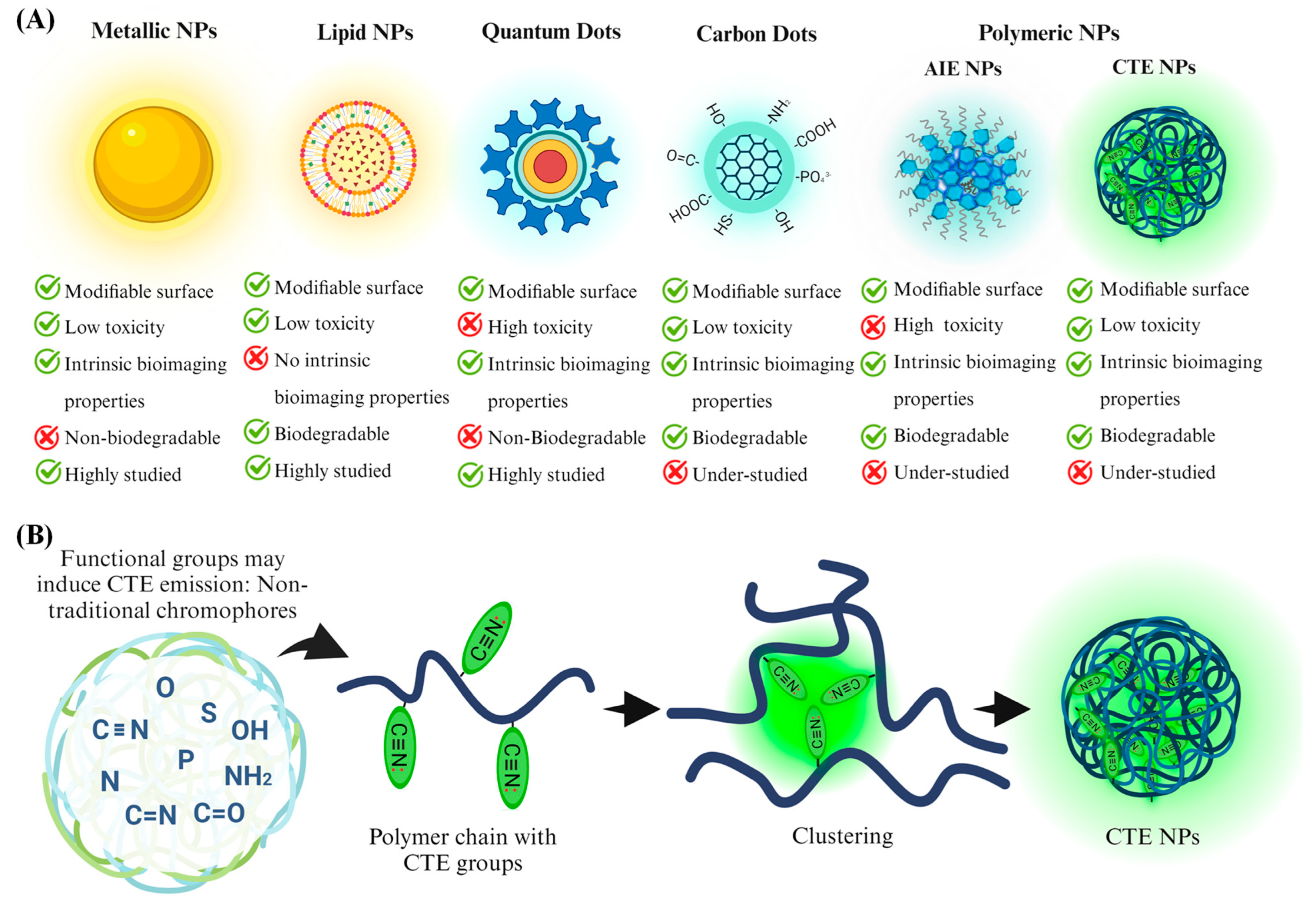
Figure 1. (A) Different types of nanotheranostics: key advantages and disadvantages. (B) Scheme of CTE mechanism and nanoparticle formation.
Jnt 06 00003 g001
Disclaimer/Publisher’s Note: The statements, opinions and data contained in all publications are solely those of the individual author(s) and contributor(s) and not of MDPI and/or the editor(s). MDPI and/or the editor(s) disclaim responsibility for any injury to people or property resulting from any ideas, methods, instructions or products referred to in the content.