Article

Ultrastructural Evidence of Interactions Between Eosinophils and Mast Cells in Gastric Cancer: Considerations in AllergoOncology Research

1
Department of Human Pathology in Adult and Developmental Age “G. Barresi”, University of Messina, 98123 Messina, Italy
2
Department of Neuroscience, University of Camerino, 62032 Camerino, Italy
*
Author to whom correspondence should be addressed.
Gastrointest. Disord. 2025, 7(3), 41; https://doi.org/10.3390/gidisord7030041
Submission received: 1 May 2025 / Revised: 5 June 2025 / Accepted: 11 June 2025 / Published: 20 June 2025

Abstract

:
Background/Objectives: AllergoOncology is a new field of study that investigates the relationship between allergic inflammation and cancer. Mast cells and eosinophils are two critical players in allergy reactions, where they can interact and release bioactive granules. The electron microscope is an indispensable tool for analyzing membrane contacts and degranulation patterns in mast cells and eosinophils. The aim of the present ultrastructural study is to analyze the interactions between tumor-associated eosinophils and mast cells (TATEM) in nine cases of gastric cancer. Methods: Seventy-two gastric cancer samples were analyzed using light microscopy, and nine cases exhibiting TATEM were selected for additional examination by transmission electron microscopy. Results: In seven cases, there was direct interaction between non-activated eosinophils and mast cells demonstrating piecemeal degranulation and/or exocytosis. In cases 8 and 9, both cell types showed more advanced stages of degranulation. Mast cells exhibited either massive degranulation (anaphylactic type) or signs of recovery, while eosinophils displayed cytolysis, with or without extracellular trap formation (ETosis). The concurrent activation of both cell types may indicate a collaborative immune response that could affect tumor behavior. There was a trend toward an association with low-stage (I-II) gastric cancer in patients with TATEM, but this difference was not statistically significant (p = 0.06). Conclusions: This work is the first investigation to present ultrastructural evidence of the intimate relationship between degranulating mast cells and cytolytic eosinophils, with or without ETosis, in gastric cancer. These findings support the emerging field of AllergoOncology, which examines the role of allergy-like immune responses in tumor immunity.

1. Introduction

Mast cells and eosinophils are cellular protagonists in allergic diseases, and they cooperate bidirectionally to increase an inflammatory response [1,2,3]. This interaction results in the designation of the “allergic effector unit”, which characterizes their reciprocal activation, mediated by surface molecules such as CD48 and 2B4, as well as by the release of cytokines and chemokines [4,5]. Eosinophil–mast cell interactions may be important not only in allergic diseases but also in many neoplastic disorders [6]. Mast cells and eosinophils are frequently found in the tumor stroma, where their ability to release immunomodulatory granules may shape local immune responses [4,5,6]. In particular, inflammatory pathways associated with allergic reactions may influence tumor development, with pro- or anti-tumor effects depending on the tumor microenvironment [6]. These data are the focus of AllergoOncology, a recently developed interdisciplinary area that investigates the possible correlations between allergies and cancer [7,8,9].
Electron microscopy is essential for the analysis of cellular interactions between mast cells and eosinophils in the tumor stroma of neoplastic disorders [10]. This technique can identify three main patterns of eosinophil degranulation: exocytosis, piecemeal degranulation, and cytolysis (extensively reviewed by Melo et al. [11]). Cytolysis is distinguished by heterochromatin decondensation and focal disruption of the plasma membrane, which results in the release of free extracellular granules (FEGs) [11]. Extracellular trap cell death (ETosis), a variant of eosinophil cytolysis, has recently been documented [12]. It is defined by rapid and extensive degranulation, which includes nuclear envelope disintegration, plasma membrane rupture, and the extracellular release of filamentous chromatin structures (extracellular traps), FEGs, and, in certain instances, Charcot–Leyden crystals [11,12]. Only a small number of studies have investigated the ultrastructural characteristics of eosinophil ETosis in vivo, despite its potential importance. Consequently, it may be particularly relevant to examine eosinophil ETosis in the context of mast cell activation and degranulation in human gastric cancer.
In our previous work, we identified aggregates of mast cells and eosinophils in gastric cancer samples, suggesting the presence of an allergy-like immune reaction within the tumor microenvironment [10]. Building on these findings, the present study focuses on the ultrastructural analysis of tumor-associated tissue eosinophils and mast cells (TATEM) in gastric cancer, aiming to clarify their interaction patterns and degranulation processes, as well as to investigate their potential role in tumor immunity.

2. Results

2.1. Clinicopathological Findings

Out of the 72 gastric cancer specimens initially examined, only nine (12.5%) exhibited TATEM. The comparison of clinicopathological features of gastric carcinomas with and without TATEM is summarized in Table 1. No statistically significant differences were observed between patients with and without TATEM in terms of age (median 65 vs. 70 years, p = 0.39) or tumor size (median 5.0 cm in both groups, p = 0.43), as assessed by the Mann–Whitney U test. Fisher’s exact test indicated a trend toward a greater proportion of low-stage (I–II) gastric cancers in patients with TATEM; however, this difference was not statistically significant (p = 0.06). There were no significant differences with regard to other variables such as gender, anatomical site, depth of invasion (pT stage), Lauren classification [13], and lymph node metastasis.

2.2. Light Microscopy

Mast cells were easily identifiable in Toluidine Blue-stained semithin sections due to the presence of characteristic granules. They typically appeared as individual cells or formed clusters, establishing heterotypic aggregates with eosinophils (Figure 1). In all nine cases, mast cell–eosinophil aggregates ranged from three to fifteen.

2.3. Electron Microscopy

Ultrastructural analysis demonstrated that solitary mast cells possessed a monolobed nucleus characterized by peripheral heterochromatin, surface membrane folds, and cytoplasmic granules (Figure 2). The granules appeared as particulate or electron-dense, but scroll-like granules were detected only sporadically (Figure 2). In other instances, mast cells exhibited a hypogranular morphology, indicative of immaturity or granule depletion resulting from previous degranulation events (Figure 3). In all nine cases of gastric cancer, intact, non-degranulated mast cells were absent from the tumor stroma.

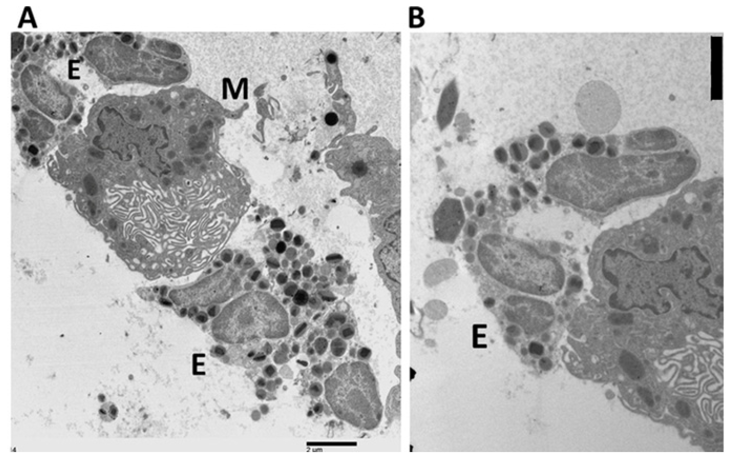
2.4. Ultrastructural Evidence of Mast Cell–Eosinophil Interactions

Heterotypic interactions between mast cells and eosinophils were a consistent ultrastructural feature in cases 1–7. Non-activated eosinophils interacting with these mast cells exhibited intact specific granules, lacking ultrastructural evidence of degranulation (Figure 4). Mast cells frequently displayed granules concentrated in the cytoplasmic region adjacent to eosinophils, with features of mild piecemeal degranulation (Figure 4). Some mast cells demonstrated anaphylactic degranulation, characterized by the release of granules outside the cell and the presence of labyrinthine canalicular structures (Figure 5A).

2.5. Ultrastructural Pattern of Degranulation and Cytolysis

In case 8, heterotypic aggregates of mast cells and eosinophils exhibited pronounced degranulation in both cell types. Eosinophils showed cytolysis without extracellular trap cell death (ETosis), while mast cells presented either anaphylactic degranulation or signs of recovery from it (Figure 5B). In some mast cells, recovery from anaphylactic degranulation coexisted with mild piecemeal degranulation, as shown by the presence of intracytoplasmic canaliculi and granules with semilunar profiles (Figure 6 and Figure 7).
In case 9, mast cells in recovery from anaphylactic degranulation were observed in proximity to several eosinophils undergoing ETotic cytolysis (Figure 8A), along with extracellular hexagonal crystals resembling Charcot–Leyden crystals (Figure 8B). Additional observations revealed close contact between mast cells and ETotic eosinophils (Figure 9A), which displayed extracellular deposition of specific granules and decondensed nuclear chromatin (Figure 9B).
Figure 10 presents a simplified schematic representation of the essential ultrastructural characteristics, emphasizing the spatial proximity between a mast cell in the recovery phase of anaphylactic degranulation and an eosinophil undergoing ETosis, as reported in case 9.

3. Discussion

Our ultrastructural analysis of the initial seven cases identified heterotypic aggregations of eosinophils and mast cells in the tumor stroma. The Levi-Schaffer research group has identified analogous relationships, employing immunohistochemistry to demonstrate the co-localization of mast cells and eosinophils in bronchial samples from asthmatic patients [2]. In vitro ultrastructural investigations confirmed direct associations between mast cells and non-activated eosinophils, consequently reinforcing these conclusions [3]. Moreover, our data indicate that the mast cells within the tumor stroma were activated, exhibiting piecemeal and/or anaphylactic degranulation. The findings align with the experimental research conducted by Mihlan et al. [14], which indicated that during passive cutaneous anaphylaxis in mice, eosinophils clustered around degranulating mast cells. Our ultrastructural data suggest that the interactions between eosinophils and mast cells in the tumor stroma of gastric carcinomas may be considered as an allergic-like immune response.
In cases 8 and 9, heterotypic aggregations of mast cells and eosinophils exhibited greater degrees of degranulation in both cell types. Mast cells demonstrated anaphylactic degranulation followed by signs of recovery from this process. Eosinophils displayed two different degranulation patterns upon interaction with mast cells: cytolysis without extracellular trap cell death (ETosis) in case 8 and cytolysis with extracellular trap cell death (ETosis) in case 9. These degranulation patterns are interpreted using well-established ultrastructural classifications [11,12,15,16]. In case 8, eosinophils exhibited cytolysis without ETosis, characterized by nuclear envelope dilatation, cytoplasmic vacuolization, focal disruption of the plasma membrane, and the release of free extracellular granules (FEGs) without nuclear chromatin extrusion [11,15]. Conversely, the eosinophils in case 9 demonstrated extracellular trap cell death (ETosis), characterized by chromatin decondensation, disintegration of the nuclear envelope and plasma membrane, and the release of DNA traps and granules [11,12,15]. In case 9, this degranulation pattern was observed in conjunction with extracellular deposits of Charcot–Leyden crystals, which are commonly associated with eosinophil ETosis [17].
For the first time, these ultrastructural data reveal a close association between degranulating mast cells and cytolytic eosinophils, with or without ETosis, in two cases of gastric cancer. The results suggest that mast cells may be involved in the induction of eosinophil cytolysis, but further studies are needed to confirm this morphological hypothesis.
The concomitant presence of cytolytic eosinophils and mast cells undergoing anaphylactic degranulation suggests the coordinated release of cytotoxic mediators with potential anti-tumoral activity. Mast cell granules contain bioactive compounds such as TNF-alpha, which may exert significant anticancer effects [18]. Free extracellular granules (FEGs) can operate as autonomous organelles that can trigger an active secretory response [19,20]. They are released during cytolysis, with or without extracellular trap cell death (ETosis) [21]. Eosinophils produce cytotoxic proteins, such as major basic protein, eosinophil cationic protein, eosinophil peroxidase, and eosinophil-derived neurotoxin, to destroy tumor cells [22,23,24,25,26,27]. Moreover, extracellular histones that accumulate in eosinophil DNA traps generated during ETosis have demonstrated considerable cytotoxic effects on host cells [28,29,30]. According to these results, mast cells and eosinophils can collaborate to generate cytotoxic mediators that create an anticancer microenvironment. However, whereas these interactions can suggest an anti-tumoral immune response, they could additionally promote fibrosis, angiogenesis, or other tumor-supporting processes [6,23].
Fisher’s exact test disclosed a trend toward an association between TATEM and early-stage gastric carcinoma (stage I–II; p = 0.06). Although suggestive, this result does not meet the conventional threshold for statistical significance (p < 0.05) and therefore cannot be interpreted as a definitive association. To determine if these immune aggregates are genuinely enriched in early-stage tumors or occur solely under particular tumor microenvironmental conditions, larger and more adequately powered studies are required.
Our study has some limitations that merit discussion. This study is a retrospective analysis of surgical specimens collected between 1998 and 2005. The findings may not accurately represent contemporary clinical practices due to advancements in diagnostic techniques and treatment procedures over recent decades. Considering the relatively low prevalence of TATEM (12.5%), further research on larger gastric cancer cohorts is necessary to confirm our findings and assess their broader application. The lack of molecular or functional evidence precludes a deeper exploration of the immunological mechanisms regulating these interactions and their potential prognostic implications. Consequently, these results should be considered hypothesis-generating and necessitate further investigation in larger cohorts and through integrated experimental methodologies, including in vitro co-culture, cytokine profiling, and gene expression analysis.

4. Methods

4.1. Tissue Sample

A total of 72 surgically resected gastric cancer specimens were collected between 1998 and 2005 and processed according to established protocols for both light and electron microscopy, the latter being an ancillary technique that may be routinely applied in diagnostic histopathology. None of the patients had undergone preoperative radiotherapy or immunochemotherapy. Cases were included consecutively and were not pre-screened for mast cells or eosinophils. Tumor tissue was divided into two portions. One portion was processed for standard paraffin embedding, which included additional tissue samples obtained from the tumor, surgical margins, and regional lymph nodes. Sections were stained with hematoxylin and eosin. The second portion was minced into small fragments and immediately fixed in 3% phosphate-buffered glutaraldehyde (pH 7.4), followed by post-fixation in 1% osmium tetroxide for electron microscopy. Semithin sections embedded in Araldite were stained for one minute on a hot plate preheated to 80 °C using a 1% Toluidine Blue solution (FLUKA, catalog number 89640, Honeywell International Inc., Morris Plains, NJ, USA), prepared by dissolving 1 g of Toluidine Blue and 1 g of sodium tetraborate in 100 mL of distilled water. Ultrathin sections were then double-stained with uranyl acetate and lead citrate and examined using a JEOL 1200 electron microscope (JEOL, Tokyo, Japan).

4.2. Definition of TATEM

The presence of paraffin and Araldite-embedded tissue blocks, together with the corresponding surgical reports, enabled the retrospective selection of 72 gastric carcinoma cases from the pathology archives of the Department of Human Pathology in Adult and Developmental Age at the University of Messina, Italy. Tumor-associated tissue eosinophils and mast cells (TATEM) have been categorized as a binary variable (present or absent).
For each patient, at least 10 Araldite blocks (range: 8–12) were obtained, and the corresponding semithin sections, stained with Toluidine Blue, were carefully examined under a light microscope (Zeiss Axioplan microscope; Carl Zeiss Microscopy GmbH, Jena, Germany) at ×4 objective magnification (×40 total). Positive cases were confirmed at higher magnification using a ×40 objective (×400 total magnification). Eosinophils were recognized by their two-lobed nuclei and abundant cytoplasmic granules, whereas mast cells were identified by their single-lobed nuclei and cytoplasmic granules exhibiting red/purple metachromasia with Toluidine Blue staining. From the initial cohort of 72 cases, 63 were excluded due to the absence of TATEM, leaving 9 cases eligible for electron microscopy analysis.

4.3. Ultrastructural Characteristics of Mast Cell Degranulation

According to Dvorak et al. [31,32] and Andersson et al. [33], mast cell degranulation is classified into three types: mild piecemeal degranulation, which involves a limited number of granules losing structural integrity and electron density, sometimes resulting in semilunar margination of residual granule material; advanced piecemeal degranulation, characterized by granules that are largely or completely emptied, accompanied by numerous secretory vesicles and without fusion with the plasma membrane; and anaphylactic degranulation, distinguished by granule swelling, fusion, the formation of degranulation channels, and the release of extracellular granules. The presence of multiple intracytoplasmic canaliculi, showing internalized narrow folds and lacking altered granule matrix materials, indicates recovery from anaphylactic degranulation.

4.4. Ultrastructural Characteristics of Eosinophil Degranulation

The classification of eosinophil degranulation adhered to the criteria established by Melo et al. [11], which distinguish between piecemeal degranulation, classic and compound exocytosis, and cytolysis, with or without extracellular trap cell death (ETosis). The morphological categories were utilized to identify the distinct degranulation patterns found using transmission electron microscopy in the tumor microenvironment.

4.5. Statistical Analyses

Histograms were used to assess the distribution of continuous variables, such as age and tumor size. As the data were not normally distributed, the findings were presented as medians, and the Mann–Whitney U test was used to compare groups. Categorical variables were subdivided into two or three groups as follows: gender (male vs. female), tumor location (cardia/fundus vs. corpus vs. antrum), Lauren histological classification [13] (intestinal vs. diffuse), depth of invasion (T1–T2 vs. T3–T4), lymph node metastasis (negative vs. positive), and disease stage (I–II vs. III–IV) [34]. Fisher’s exact test was used to compare categorical variables. Statistical analyses were conducted using STATA software (version 18.0; StataCorp, College Station, TX, USA). All variables with a p-value < 0.05 were considered statistically significant.

5. Conclusions

In conclusion, this ultrastructural study has, for the first time, revealed a close association between degranulating mast cells and cytolytic eosinophils, with or without ETosis, in the tumor tissue of gastric carcinomas. Ultrastructural findings indicate that immune responses resembling those seen in allergies may influence the tumor microenvironment, thus supporting the concept of AllergoOncology.

Author Contributions

R.C.: conceptualization, investigation, writing—original draft, writing—review and editing, supervision. V.C. and L.R.: investigation, visualization, writing—review and editing. All authors have read and agreed to the published version of the manuscript.

Funding

This research received no external funding.

Institutional Review Board Statement

The study was conducted in accordance with the Declaration of Helsinki. It was based exclusively on histological and ultrastructural analyses of archived tissue samples collected between 1998 and 2005 for routine diagnostic purposes. All investigations were performed on anonymized, pre-existing material, with no impact on patient care. According to Italian law, ethics committee approval is required only for interventional clinical trials involving medicinal products for human use. As specified in Articles 6 and 9 of Legislative Decree No. 211/2003, this entirely non-interventional study, which did not involve any medicinal products, was therefore exempt from ethical review.

Informed Consent Statement

Written informed consent for research use of anonymized biological material was obtained from all patients at the time of surgery (1998–2005). The need for additional consent was waived due to the retrospective nature of the study.

Data Availability Statement

The raw data supporting the conclusions of this article will be made available by the authors on request.

Conflicts of Interest

The authors declare no conflicts of interest.

Abbreviations

The following abbreviations are used in this manuscript:
FEGsFree extracellular granules
ETosisExtracellular trap cell death
TATEMTumor-associated tissue eosinophils and mast cells

References

  1. Minai-Fleminger, Y.; Levi-Schaffer, F. Mast Cells and Eosinophils: The Two Key Effector Cells in Allergic Inflammation. Inflamm. Res. 2009, 58, 631–638. [Google Scholar] [CrossRef] [PubMed]
  2. Elishmereni, M.; Alenius, H.T.; Bradding, P.; Mizrahi, S.; Shikotra, A.; Minai-Fleminger, Y.; Mankuta, D.; Eliashar, R.; Zabucchi, G.; Levi-Schaffer, F. Physical Interactions between Mast Cells and Eosinophils: A Novel Mechanism Enhancing Eosinophil Survival In Vitro. Allergy 2011, 66, 376–385. [Google Scholar] [CrossRef] [PubMed]
  3. Minai-Fleminger, Y.; Elishmereni, M.; Vita, F.; Soranzo, M.R.; Mankuta, D.; Zabucchi, G.; Levi-Schaffer, F. Ultrastructural Evidence for Human Mast Cell-Eosinophil Interactions In Vitro. Cell Tissue Res. 2010, 341, 405–415. [Google Scholar] [CrossRef] [PubMed]
  4. Gangwar, R.S.; Levi-Schaffer, F. Eosinophils Interaction with Mast Cells: The Allergic Effector Unit. Methods Mol. Biol. 2014, 1178, 231–249. [Google Scholar] [CrossRef]
  5. Pahima, H.; Puzzovio, P.G.; Levi-Schaffer, F. 2B4 and CD48: A Powerful Couple of the Immune System. Clin. Immunol. 2019, 204, 64–68. [Google Scholar] [CrossRef]
  6. Galdiero, M.R.; Varricchi, G.; Seaf, M.; Marone, G.; Levi-Schaffer, F.; Marone, G. Bidirectional Mast Cell-Eosinophil Interactions in Inflammatory Disorders and Cancer. Front. Med. 2017, 4, 103. [Google Scholar] [CrossRef]
  7. Jensen-Jarolim, E.; Achatz, G.; Turner, M.C.; Karagiannis, S.; Legrand, F.; Capron, M.; Penichet, M.L.; Rodríguez, J.A.; Siccardi, A.G.; Vangelista, L.; et al. AllergoOncology: The Role of IgE-Mediated Allergy in Cancer. Allergy 2008, 63, 1255–1266. [Google Scholar] [CrossRef]
  8. Della Valle, L.; Gatta, A.; Farinelli, A.; Scarano, G.; Lumaca, A.; Tinari, N.; Cipollone, F.; Paganelli, R.; Di Gioacchino, M. Allergooncology: An Expanding Research Area. J. Biol. Regul. Homeost. Agents 2020, 34, 319–326. [Google Scholar] [CrossRef]
  9. Pascal, M.; Bax, H.J.; Bergmann, C.; Bianchini, R.; Castells, M.; Chauhan, J.; De Las Vecillas, L.; Hartmann, K.; Álvarez, E.I.; Jappe, U.; et al. Granulocytes and Mast Cells in AllergoOncology—Bridging Allergy to Cancer: An EAACI Position Paper. Allergy 2024, 79, 2319–2345. [Google Scholar] [CrossRef]
  10. Caruso, R.A.; Fedele, F.; Parisi, A.; Paparo, D.; Bonanno, A.; Finocchiaro, G.; Branca, G.; Scardigno, M.; Rigoli, L. Chronic Allergic-Like Inflammation in the Tumor Stroma of Human Gastric Carcinomas: An Ultrastructural Study. Ultrastruct. Pathol. 2012, 36, 139–144. [Google Scholar] [CrossRef]
  11. Melo, R.C.N.; Dvorak, A.M.; Weller, P.F. Eosinophil Ultrastructure. In Atlas of Eosinophil Cell Biology and Pathology; Elsevier: London, UK, 2022; pp. 61–105. [Google Scholar]
  12. Ueki, S.; Melo, R.C.N.; Ghiran, I.; Spencer, L.A.; Dvorak, A.M.; Weller, P.F. Eosinophil Extracellular DNA Trap Cell Death Mediates Lytic Release of Free Secretion-Competent Eosinophil Granules in Humans. Blood 2013, 121, 2074–2083. [Google Scholar] [CrossRef] [PubMed]
  13. Lauren, P. The Two Histological Main Types of Gastric Carcinoma: Diffuse and So-Called Intestinal-Type Carcinoma. An Attempt at a Histo-Clinical Classification. Acta Pathol. Microbiol. Scand. 1965, 64, 31–49. [Google Scholar] [CrossRef] [PubMed]
  14. Mihlan, M.; Wissmann, S.; Gavrilov, A.; Kaltenbach, L.; Britz, M.; Franke, K.; Hummel, B.; Imle, A.; Suzuki, R.; Stecher, M.; et al. Neutrophil Trapping and Nexocytosis, Mast Cell-Mediated Processes for Inflammatory Signal Relay. Cell 2024, 187, 5316–5335.e28. [Google Scholar] [CrossRef] [PubMed]
  15. Neves, V.H.; Palazzi, C.; Bonjour, K.; Ueki, S.; Weller, P.F.; Melo, R.C.N. In Vivo ETosis of Human Eosinophils: The Ultrastructural Signature Captured by TEM in Eosinophilic Diseases. Front. Immunol. 2022, 13, 938691. [Google Scholar] [CrossRef]
  16. Neves, V.H.; Palazzi, C.; Malta, K.K.; Bonjour, K.; Kneip, F.; Dias, F.F.; Neves, J.S.; Weller, P.F.; Melo, R.C.N. Extracellular Sombrero Vesicles Are Hallmarks of Eosinophilic Cytolytic Degranulation in Tissue Sites of Human Diseases. J. Leukoc. Biol. 2024, 116, 398–408. [Google Scholar] [CrossRef]
  17. Ueki, S.; Tokunaga, T.; Melo, R.C.N.; Saito, H.; Honda, K.; Fukuchi, M.; Konno, Y.; Takeda, M.; Yamamoto, Y.; Hirokawa, M.; et al. Charcot-Leyden Crystal Formation Is Closely Associated with Eosinophil Extracellular Trap Cell Death. Blood 2018, 132, 2183–2187. [Google Scholar] [CrossRef]
  18. Fereydouni, M.; Ahani, E.; Desai, P.; Motaghed, M.; Dellinger, A.; Metcalfe, D.D.; Yin, Y.; Lee, S.H.; Kafri, T.; Bhatt, A.P.; et al. Human Tumor Targeted Cytotoxic Mast Cells for Cancer Immunotherapy. Front. Oncol. 2022, 12, 871390. [Google Scholar] [CrossRef]
  19. Neves, J.S.; Perez, S.A.; Spencer, L.A.; Melo, R.C.N.; Reynolds, L.; Ghiran, I.; Mahmudi-Azer, S.; Odemuyiwa, S.O.; Dvorak, A.M.; Moqbel, R.; et al. Eosinophil Granules Function Extracellularly as Receptor-Mediated Secretory Organelles. Proc. Natl. Acad. Sci. USA 2008, 105, 18478–18483. [Google Scholar] [CrossRef]
  20. Muniz, V.S.; Baptista-Dos-Reis, R.; Neves, J.S. Functional Extracellular Eosinophil Granules: A Bomb Caught in a Trap. Int. Arch. Allergy Immunol. 2013, 162, 276–282. [Google Scholar] [CrossRef]
  21. Melo, R.C.N.; Weller, P.F. Contemporary Understanding of the Secretory Granules in Human Eosinophils. J. Leukoc. Biol. 2018, 104, 85–93. [Google Scholar] [CrossRef]
  22. Andreone, S.; Spadaro, F.; Buccione, C.; Mancini, J.; Tinari, A.; Sestili, P.; Gambardella, A.R.; Lucarini, V.; Ziccheddu, G.; Parolini, I.; et al. IL-33 Promotes CD11b/CD18-Mediated Adhesion of Eosinophils to Cancer Cells and Synapse-Polarized Degranulation Leading to Tumor Cell Killing. Cancers 2019, 11, 1664. [Google Scholar] [CrossRef] [PubMed]
  23. Mattei, F.; Andreone, S.; Marone, G.; Gambardella, A.R.; Loffredo, S.; Varricchi, G.; Schiavoni, G. Eosinophils in the Tumor Microenvironment. Adv. Exp. Med. Biol. 2020, 273, 1–28. [Google Scholar] [CrossRef]
  24. Kubo, H.; Loegering, D.A.; Adolphson, C.R.; Gleich, G.J. Cytotoxic Properties of Eosinophil Granule Major Basic Protein for Tumor Cells. Int. Arch. Allergy Immunol. 1999, 118, 426–428. [Google Scholar] [CrossRef] [PubMed]
  25. Young, J.D.; Peterson, C.G.; Venge, P.; Cohn, Z.A. Mechanism of Membrane Damage Mediated by Human Eosinophil Cationic Protein. Nature 1986, 321, 613–616. [Google Scholar] [CrossRef]
  26. Navarro, S.; Aleu, J.; Jiménez, M.; Boix, E.; Cuchillo, C.M.; Nogués, M.V. The Cytotoxicity of Eosinophil Cationic Protein/Ribonuclease 3 on Eukaryotic Cell Lines Takes Place through Its Aggregation on the Cell Membrane. Cell. Mol. Life Sci. 2008, 65, 324–337. [Google Scholar] [CrossRef]
  27. Agosti, J.M.; Altman, L.C.; Ayars, G.H.; Loegering, D.A.; Gleich, G.J.; Klebanoff, S.J. The Injurious Effect of Eosinophil Peroxidase, Hydrogen Peroxide, and Halides on Pneumocytes In Vitro. J. Allergy Clin. Immunol. 1987, 79, 496–504. [Google Scholar] [CrossRef]
  28. Li, X.; Ye, Y.; Peng, K.; Zeng, Z.; Chen, L.; Zeng, Y. Histones: The Critical Players in Innate Immunity. Front. Immunol. 2022, 13, 1030610. [Google Scholar] [CrossRef]
  29. Aoki, A.; Hirahara, K.; Kiuchi, M.; Nakayama, T. Eosinophils: Cells Known for Over 140 Years with Broad and New Functions. Allergol. Int. 2021, 70, 3–8. [Google Scholar] [CrossRef]
  30. Thompson-Souza, G.A.; Vasconcelos, C.R.I.; Neves, J.S. Eosinophils: Focus on DNA Extracellular Traps. Life Sci. 2022, 311, 121191. [Google Scholar] [CrossRef]
  31. Dvorak, A.M. Ultrastructural Studies of Human Basophils and Mast Cells. J. Histochem. Cytochem. 2005, 53, 1043–1070. [Google Scholar] [CrossRef]
  32. Dvorak, A.M.; Tepper, R.I.; Weller, P.F.; Morgan, E.S.; Estrella, P.; Monahan-Earley, R.A.; Galli, S.J. Piecemeal Degranulation of Mast Cells in the Inflammatory Eyelid Lesions of Interleukin-4 Transgenic Mice. Blood 1994, 83, 3600–3612. [Google Scholar] [CrossRef]
  33. Andersson, C.K.; Mori, M.; Bjermer, L.; Löfdahl, C.G.; Erjefält, J.S. Alterations in Lung Mast Cell Populations in Patients with Chronic Obstructive Pulmonary Disease. Am. J. Respir. Crit. Care Med. 2010, 181, 206–217. [Google Scholar] [CrossRef]
  34. WHO Classification of Tumors Editorial Board. Tumors of the Stomach. In WHO Classification of Tumors: Digestive System Tumors, 5th ed.; IARC Press: Lyon, France, 2019; pp. 59–110. [Google Scholar]
Figure 1. Semithin section of a diffuse-type gastric carcinoma. The tumor stroma contains numerous eosinophils, either isolated or located in close proximity to a mast cell (black arrow). The mast cell shows a limited number of metachromatic granules in its cytoplasm. A blood vessel is also present in the stromal area.
Gastrointestdisord 07 00041 g001
Figure 2. Ultrastructural image of a solitary mast cell (M) displaying thin surface folds and numerous cytoplasmic granules containing electron-dense material and scroll-like structures (curved arrow). Several granules exhibit partially emptied chambers (black arrows), consistent with mild piecemeal degranulation. Scale bar (right, horizontal): 6 µm.
Gastrointestdisord 07 00041 g002
Figure 3. Ultrastructural image of a small mast cell (M) with a limited number of electron-dense secretory granules localized at the periphery of the cytoplasm. Scale bar (right, vertical): 2 µm.
Gastrointestdisord 07 00041 g003
Figure 4. Ultrastructural image showing a heterotypic interaction between a single eosinophil (E) and a mast cell (M). The mast cell exhibits narrow plasmalemmal folds establishing close contact with the adjacent eosinophil. Secretory granules are concentrated in the cytoplasmic region near the eosinophil, and some of them exhibit mild piecemeal degranulation (black arrow). Scale bar (right, horizontal): 2 µm.
Gastrointestdisord 07 00041 g004
Figure 5. (A) Ultrastructural features consistent with mast cell (M) anaphylactic degranulation. Extracellular deposition of granules (black arrow) is observed in association with a labyrinthine canalicular system. The adjacent eosinophil (E) shows no ultrastructural signs of degranulation. Scale bar (left, vertical): 2 µm. (B) Interaction between a cytolytic eosinophil (E) and a mast cell (M) undergoing anaphylactic degranulation. A mast cell granule is visible within a large intracellular degranulation chamber opening to the extracellular space (black arrow). The eosinophil exhibits localized plasma membrane disruption, nuclear envelope dilation, cytoplasmic vacuolization, and extracellular release of specific granules (curved arrow). Scale bar (right, horizontal): 5 µm.
Gastrointestdisord 07 00041 g005
Figure 6. Ultrastructural image showing a mast cell (M) in close proximity to two cytolytic eosinophils (E) and several free extracellular specific eosinophil granules (curved arrow). The mast cell contains granules with a characteristic semilunar profile (black arrow), consistent with mild piecemeal degranulation. Intracytoplasmic cana-liculi with internalized narrow folds are also visible; they lack altered matrix material, suggesting recovery from anaphylactic degranulation (pentagonal arrow). Scale bar (right, horizontal): 5 µm.
Gastrointestdisord 07 00041 g006
Figure 7. Ultrastructural image showing two mast cells (M) adjacent to a cytolytic eosinophil (E). Both mast cells contain granules with characteristic semilunar profiles (black arrows), indicative of mild piecemeal degranulation. One of the mast cells also displays large intracytoplasmic canaliculi, consistent with a recovery phase following anaphylactic degranulation. Scale bar (right, horizontal): 2 µm.
Gastrointestdisord 07 00041 g007
Figure 8. (A) Ultrastructural image showing the interaction between mast cells (M) and cytolyt-ic eosinophils (E). A prominent labyrinthine canalicular structure is visible within the mast cell cytoplasm, suggesting a recovery phase following anaphylactic degranulation. Scale bar (right, horizontal): 2 µm. (B) Extracellular hexagonal Charcot–Leyden crystals located near eosinophils (E), indicative of ETotic cytolysis. Scale bar (right, vertical): 3 µm.
Gastrointestdisord 07 00041 g008
Figure 9. (A) Ultrastructural image of a mast cell (M) exhibiting a motile phenotype in contact with two ETotic eosinophils (E). The mast cell displays peripheral heterochromatin, surface membrane folds, and large immature granules. Scale bar (right, horizontal): 2 µm. (B) Detailed view of the interaction between an ETotic eosinophil (E) and a mast cell (M). The eosinophil shows early extracellular release of specific granules and decondensed nuclear chromatin with focal areas of cytoplasmic loss (curved arrow). Immature mast cell granules display irregular loss of internal density (black arrows). A solitary granule (pentagonal arrow) exhibits a characteristic semilunar profile. Scale bar (right, horizontal): 3 µm.
Gastrointestdisord 07 00041 g009
Figure 10. Diagram of a mast cell (M) in the recovery phase of anaphylactic degranulation (A), located next to an eosinophil (E). The eosinophil exhibits characteristics of ETosis, such as localized plasma membrane rupture, initial extrusion of nuclear chromatin (black arrow), and the discharge of free extracellular granules (curved arrow). Charcot–Leyden crystals (C) are located in the extracellular space and are associated with eosinophil degranulation.
Gastrointestdisord 07 00041 g010
Table 1. Comparison of Clinicopathologic Characteristics between TATEM-Positive and TATEM-Negative Gastric Cancers.
PatientsPatients with TATEM n = 9 (%)Patients without TATEM n = 63 (%)p-Value *
Age (median)65 (50–78)70 (41–83)0.39
Tumor size (median)5 cm (3–8)5 cm (2–9)0.43
Gender
Male6 (67)37 (59)
Female3 (33)26 (41)0.73
Location
Upper + Middle third3 (33)33 (52)
Lower third6 (67)30 (48)0.47
Depth of invasion
T1–T23 (33)23 (37)
T3–T46 (67)40 (63)1.00
Lauren classification
Intestinal6 (67)44 (70)
Diffuse3 (33)19 (30)1.00
Lymph node metastasis
Negative4 (44)18 (29)
Positive5 (56)45 (71)0.44
Stage of disease
I–II8 (89)33 (52)
III–IV1 (11)30 (48)0.06
* Mann–Whitney U-test for age and tumor size; otherwise, Fisher’s exact test.
Disclaimer/Publisher’s Note: The statements, opinions and data contained in all publications are solely those of the individual author(s) and contributor(s) and not of MDPI and/or the editor(s). MDPI and/or the editor(s) disclaim responsibility for any injury to people or property resulting from any ideas, methods, instructions or products referred to in the content.

