Article

Pre- and Postoperative Risk Factors for Hirschsprung-Associated Enterocolitis in Vietnamese Children

by
Hoang Tran Viet
1,2,
Tuan Huynh Minh
2,
Nhan Vu Truong
3,
Anh Huynh Thi Phuong
4,
Bich-Uyen Nguyen
2,
Hao Chung The
5,
Cong Phi Dang
6,* and
Linh Truong Nguyen Uy
2
1
Department of Surgery, Faculty of Medicine, Can Tho University of Medicine and Pharmacy, Can Tho 90000, Vietnam
2
Faculty of Medicine, University of Medicine and Pharmacy at Ho Chi Minh City, Ho Chi Minh City 70000, Vietnam
3
Department of Pediatric Surgery, Children’s Hospital 2, Ho Chi Minh City 70000, Vietnam
4
Department of Pediatric Surgery, Children’s Hospital 1, Ho Chi Minh City 70000, Vietnam
5
Oxford University Clinical Research Unit, Ho Chi Minh City 70000, Vietnam
6
Department of Medical Laboratory Science, Faculty of Medicine, Da Nang University of Medical Technology and Pharmacy, Da Nang 55000, Vietnam
*
Author to whom correspondence should be addressed.
Gastrointest. Disord. 2025, 7(1), 17; https://doi.org/10.3390/gidisord7010017
Submission received: 14 January 2025 / Revised: 14 February 2025 / Accepted: 16 February 2025 / Published: 20 February 2025

Abstract

:
Background/Objective: Hirschsprung-associated enterocolitis (HAEC) can occur before and after surgery, increasing the complication rates, hospital stay, and treatment costs. This study aims to determine the incidence of preoperative and postoperative Hirschsprung-associated enterocolitis HAEC and the related risk factors. Methods: This study is a prospective cohort study of Hirschsprung’s disease patients under 16 years of age at two Children’s Hospitals in Ho Chi Minh City, Vietnam from December 2022 to June 2024. The postoperative follow-up is 12 months. Results: We enrolled 84 pediatric patients, with a male-to-female ratio of 5.4/1 and an average age of 7.2 ± 1.07 months. Of the patients, 25% had preoperative enterocolitis at the time of admission. Statistical analysis revealed that risk factors for preoperative enterocolitis at admission included a history of enterocolitis (p = 0.024), low weight in hospital (p = 0.001), and lack of preoperative treatment (p = 0.01). Postoperative enterocolitis occurred in 14 patients (16.7%) at an average of 4.03 months postoperatively. Multiple logistic regression of postoperative enterocolitis was associated with a history of preoperative enterocolitis (p < 0.001), anastomotic stricture (p = 0.002), and the length of the aganglionic segment (p = 0.031). No statistically significant association was found between the surgical method and the risk of postoperative enterocolitis. Conclusions: A history of preoperative enterocolitis, low weight, and anastomotic stricture are significant risk factors for postoperative enterocolitis in patients with Hirschsprung’s disease. Therefore, preoperative medical treatment is recommended for the patients who have not yet undergone a definitive surgical procedure.

1. Introduction

Congenital megacolon, first reported by Harald Hirschsprung at a pediatric conference in Berlin in 1886 with two cases of “constipation in infants leading to colonic dilation”, is now known as Hirschsprung’s disease (HSCR) [1,2]. Hirschsprung-associated enterocolitis (HAEC) can occur both before and after surgical repair, leading to an approximately 2.5-fold increase in both hospital length of stay and treatment costs [3]. Recurrent enterocolitis can also impact the child’s social blending [4]. The incidence of preoperative enterocolitis varies across studies in the world, generally around 25% [5,6,7]. The diagnosis of enterocolitis primarily relies on clinical assessment, resulting in significant variability between studies [4].
The pathogenesis of HAEC remains unknown to date, and there is no specific therapy for treating HAEC. Treatment for enterocolitis before and after surgery is similar with the general aim to alleviate symptoms, which includes resuscitation, fluid and electrolyte replacement, and colonic decompression. Recent studies established guidelines for treatment based on the severity degree of clinical symptoms [1,8]. It is reported that early diagnosis and proper treatment can reduce later complications [1,2]. However, very little is known about potential risk factors for prevention of HAEC recurrence. Certain factors including age and body weight at diagnosis and the length of the aganglionic segments observed during operation should be investigated for predicting postoperative complications.
Herein, our study aims to determine the incidence of preoperative and postoperative enterocolitis in HSCR pediatric patients and identify risky factors of HAEC recurrence in these participants. The diagnosis and classification of HAEC for our cohort was assessed using the Pastor score, the diagnostic score of 16 criteria created by Delphi expert consensus [9]. This study contributes better understandings to the limited volume of evidence for HAEC prognosis and prevention. Records of clinical parameters, including both pre- and postoperative risk factors, were analyzed to predict HAEC recurrence. Moreover, this study will discuss treatment approaches for HSCR patients with risky factors.

2. Materials and Methods

2.1. Patients

This study was designed as a prospective cohort study. All pediatric patients under 16 years old with a confirmed histological diagnosis of Hirschsprung’s disease at Children’s Hospital 1 (from December 2022 to June 2024) and Children’s Hospital 2 (from December 2023 to June 2024), were included in the study. The patients were diagnosed with Hirschsprung’s disease based on suspected symptoms, including delayed meconium passage, constipation, abdominal distension, high-pressure rectal decompression (the ‘blast sign’), and apparent findings on a colon X-ray. All patients presenting suspicious symptoms will undergo a biopsy, and the pathological results must confirm Hirschsprung’s disease. Patients without a postoperative histologic diagnosis of Hirschsprung’s disease were excluded.
Demographics data including age, sex, and preoperative weight using the WHO Anthro software were recorded at recruitment [10]. Clinical characteristics including symptoms, history of preoperative enterocolitis, types of surgical procedures, length of the aganglionic segment, and postoperative complications were recorded upon treatment and follow-up. Particularly, enterocolitis severity was assessed using the Pastor score system to evaluate a collection of 16 diagnostic components including patient history, clinical symptoms, and laboratory exams [9]. Each criterion is assigned to a specific point value, and the total score allows clinicians to determine the severity of HAEC with a cutoff threshold of ≥4. Assessment through the Pastor score allow us to determine the classification of HAEC, including HAEC I, mild cases with fever, mild abdominal distension, and diarrhea; HAEC II, moderate cases with some severe symptoms such as explosive diarrhea, fever, and lethargy; and HAEC III, severe vases with signs of shock such as poor perfusion and altered mental status. The outcome variables were the incidence of enterocolitis before and after surgery. The history of HAEC and preoperative medical care such as enemas or laxative drugs were defined by the recorded documents. Anastomosis stricture and length of aganglionosis were measured visually and intraoperatively and confirmed by postoperative histological examination.

2.2. Data Analysis

Data were analyzed using SPSS 20.0 and R software (version 4.2.1). The relationship between categorical variables will be assessed using chi-square tests, and continuous variables will be analyzed using t-tests or Wilcoxon tests with a significance level of p < 0.05. Odds ratios and 95% confidence intervals will be calculated based on frequency tables between categorical variables and outcome variables. For the performance of HAEC risk score, we applied the survival package in R with the Kaplan–Meier estimation method. Cox proportional hazards regression was used to calculate the hazard ratio and assess the significance of survival differences between groups.

3. Results

During the 18 months from December 2022 to June 2024, we enrolled 84 cases of Hirschsprung’s disease patients meeting the inclusion criteria.

3.1. General Characteristics

There were 71 (84.5%) male patients and 13 (15.5%) female patients, with a male-to-female ratio of 5.4:1. The average age at diagnosis was 7.2 ± 10.7 months, with an interquartile range of 1.5–8 months. Four patients (4.8%) had a family history of Hirschsprung’s disease, primarily occurring in parents. Associated congenital anomalies were present in 14.4% of cases, including cardiac defects in seven cases (8.5%), genitourinary defects in four cases (4.8%), and Down syndrome in one case (1.2%). The majority (96.4%) were born at term. Clinical symptoms included delayed meconium passage after 24 h in 86% of cases, abdominal distension in 92.9% of cases, and high rectal decompression in 45.2% of cases (Table 1).

3.2. Preoperative Enterocolitis

Among the 84 patients, there were 21 patients (25%) diagnosed with enterocolitis at the time of inclusion in the study. The average Pastor’s score was 8.52 ± 1.2. Patients diagnosed with HAEC I accounted for 2.4%, with 2 cases, and HAEC II accounted for 22.6%, with 19 cases (Figure 1). No case was diagnosed with HAEC III. The clinical characteristics of enterocolitis were assessed with the use of the Pastor score. The most prevalent signs of HAEC were abdominal distension and foul-smell diarrhea, which were present in all patients. The other common signs found in HAEC included diarrhea with explosive stool (95.2%), dilated loops of bowel (95.2%), and lethargy (76.2%). Meanwhile, the rectosigmoid cutoff sign was rarely present, with 4.1%.
We then investigated risk factors of preoperative HAEC by comparing the 21 patients with HAEC with their counterparts (Table 2). Those in the underweight group, defined by ratio of weight and age, had a significantly higher risk of developing HAEC (p = 0.001). Additionally, we identified other risk factors for preoperative enterocolitis, including a history of HAEC (OR = 3.53, 95% CI 1.26–10.3, p = 0.024) and no preoperative medical treatment (OR = 0.18, 95% CI 0.04–0.61, p = 0.001). No statistically significant association was found between preoperative enterocolitis and radiologic length of the aganglionic segment (OR = 1.23, 95% CI 0.16–35.1, p = 1.000).

3.3. Postoperative Enterocolitis

All 84 patients underwent surgical repair with pathological examination showing the presence of ganglia at the anastomosis site. The prevalence of surgical procedures for HSCR treatment [11] in our study were as follows: Swenson-type (Swenson-like) (66.7%), TEPT (16.7%), and Swenson-type combined with laparoscopic or open techniques at the umbilicus, each accounting for 8.3%. The average surgery time was 102.32 ± 38.77 min. The average length of the excised aganglionic segment was 7.20 ± 5.30 cm, and the average length of resected bowel was 17.89 ± 6.52 cm. Postoperatively, 66.7% of patients had no complications. Anal ulceration complications were seen in 32.1% of patients amongst 84 patients. There was one case of mortality (1.2%) from anastomotic leak.
We then followed up the 83 patients postoperatively. Enterocolitis was seen in 14 cases (16.7%). Of the total cases, HAEC I accounted for 10.8%, with nine cases; HAEC II for 4.8%, with four cases; and HAEC III for 1.2%, with one case (Figure 2). The average time to develop postoperative enterocolitis was 4.46 ± 4.21 months, with the greatest incidence occurring at 1 month and 1.5 months post-surgery.
The average time from onset of symptoms to admission for postoperative HAEC was 2.01 ± 0.53 days. The clinical characteristics of postoperative HAEC, assessed using the Pastor score, were as follows: diarrhea with foul-smelling stool (100%), abdominal distension (100%), history of enterocolitis (78.6%), fever (78.6%), diarrhea with explosive stool (71.4%), and lethargy (71.4%). Leukocytosis ranked last with 42.9% (Figure 2). The average Pastor score after surgery was 7.5 ± 1.4.
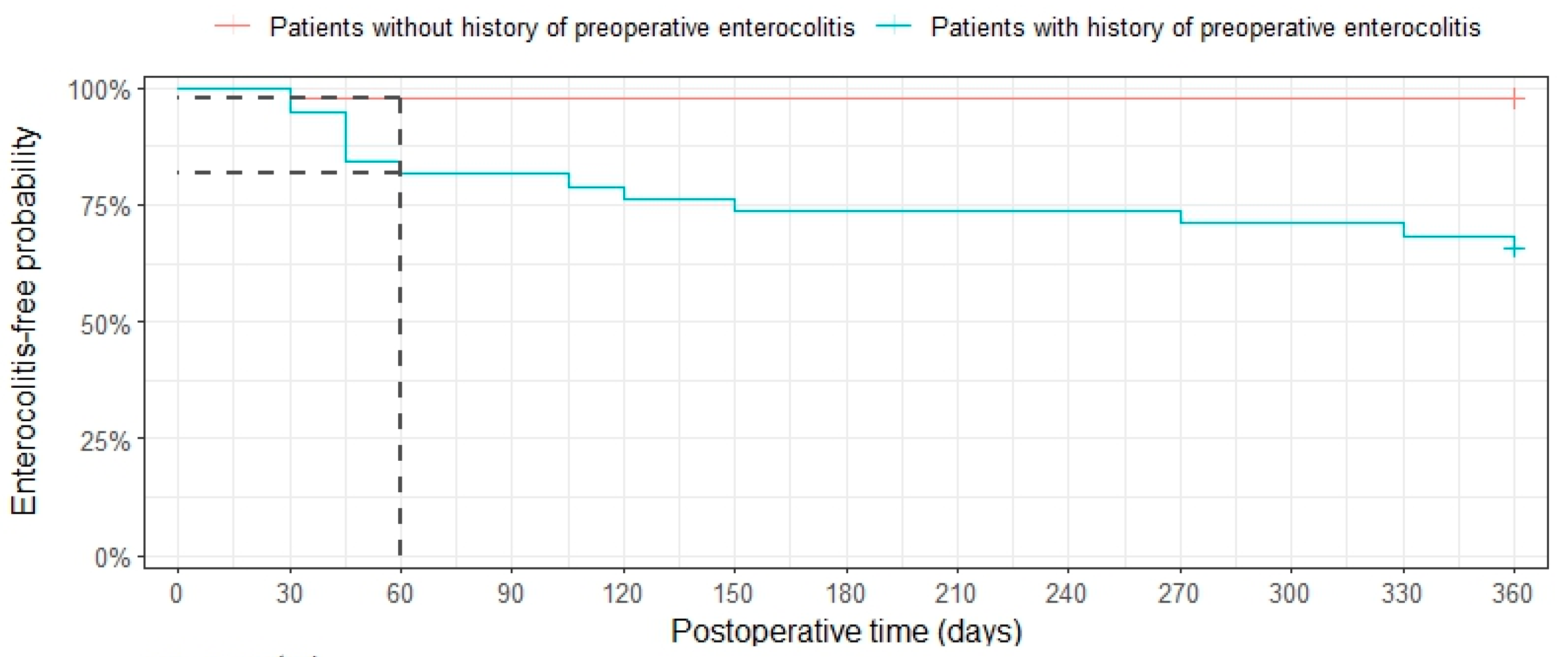
The analysis of risk factors associated with postoperative HAEC identified two risk factors for postoperative HAEC: a history of preoperative enterocolitis (OR = 9.74, p < 0.001) and anastomotic stricture (OR = 23.3, p = 0.002), and aganglionic length (OR = 1.11, p = 0.031). No significant associations were found between preoperative hemoglobin levels, blood loss, or surgical method and postoperative HAEC (Table 3). Interestingly, comparing the relative survival rate between patients with and those without a history of preoperative HAEC by the Kaplan–Meier plot, we found that patients with a history of preoperative HAEC had more probability of postoperative HAEC with a hazard ratio of 18.5 (Figure 3).

4. Discussion

Hirschsprung’s disease has an incidence rate of 1 in 5000 live births, with a higher prevalence in males compared to females. Additionally, 4.5% to 16% of Hirschsprung’s disease cases are associated with Down syndrome [2]. In recent years, Hirschsprung’s disease-associated enterocolitis (HAEC) has garnered increasing attention due to its impact on extending hospital stays, increasing complications, and raising treatment costs by approximately 2.5 times. Recurrent enterocolitis can also affect the child’s social reintegration [4]. According to Gosain (2017), the incidence of preoperative enterocolitis can greatly vary, ranging from 6% to 60% [1]. Pruitt (2020) et al. found that the incidence of preoperative enterocolitis in the US was approximately 3% to 50% [6]. Due to the wide variety in incidence of HAEC, large-scale studies across diverse populations and regions are warranted. In our study, the rate of preoperative HAEC in Vietnam is 25%, requiring heightened awareness of this illness among gastroenterologists. In Vietnam and other regions with similar resource-limited settings, early identification of high-risk patients (e.g., those with a history of HAEC or anastomotic stricture) is crucial for optimizing preoperative and postoperative management. Our findings highlight the need for targeted nutritional interventions, routine postoperative monitoring for anastomotic strictures, and patient education on early HAEC symptoms to improve outcomes.
The symptoms of enterocolitis exhibit wide variation, with signs such as abdominal distension and large stool volume always present (100%) in enterocolitis cases. Additionally, findings of colonic dilation on X-ray are very common (95.2%), and elevated white blood cell count is observed in 61.9% of cases in our study. According to Dicky (2019), the most reliable symptoms of preoperative enterocolitis are abdominal distension (100%), large stool volume (100%), and colonic dilation (100%) [6]. Gunadi listed the characteristic symptoms of enterocolitis according to the Pastor score in the following order: abdominal distension (100%), bowel dilation (100%), elevated white blood cell count (78.6%), fatigue (71.4%), absence of rectal gas (71.4%), and left shift in white blood cells (71.4%) [12]. In HAEC, both enterocolitis and colonic dilation are symptoms that almost always occur.
Congenital abnormalities could be of risk factors for HAEC. Down syndrome is identified as an independent risk factor for enterocolitis [2]. Patients with Down syndrome have a risk of HAEC ranging from 45% to 50%. In comparison, only 19% to 29% of patients with Hirschsprung’s disease who do not have Down syndrome experience enterocolitis [13]. Additionally, preoperative enterocolitis is recognized as a risk factor for postoperative enterocolitis. Patients with preoperative enterocolitis have a 1.5-fold increased risk of developing enterocolitis after surgery [6]. We observed one patient with Down syndrome who developed preoperative enterocolitis and also experienced complications and mortality after surgery. Additionally, a history of enterocolitis or poor nutritional status before surgery can be risk factors for developing enterocolitis, leading to a challenging treatment [14,15].
A long segment of aganglionosis in Hirschsprung’s disease is considered a risk factor for preoperative enterocolitis [2,4]. According to Yuliand Dicky (2019), there is no difference in preoperative enterocolitis between long-segment and short-segment aganglionosis (OR = 1.36, p = 1.0) [16]. According to J. Hager (2021), a meta-analysis on the risk of enterocolitis in Hirschsprung’s disease patients concluded that the length of aganglionosis is a risk factor for preoperative enterocolitis, but it is not a risk factor for postoperative enterocolitis [17]. We found no association between long-segment and short-segment aganglionosis with preoperative HAEC.
Patients have been followed up with for up to 16 months after surgery. Postoperative enterocolitis in our study accounted for 16.7%. The average duration of postoperative enterocolitis was 4.03 months, with the highest incidence occurring within the first 2 months after surgery. Postoperative enterocolitis typically occurs within the first 2 months after surgery. Clinical symptoms do not differ significantly from those of preoperative HAEC. However, the incidence of HAEC II decreased markedly postoperatively, likely due to the surgical intervention addressing the underlying cause of fecal stasis. In our study, patients were operated on using four different surgical methods. Although the rates of enterocolitis varied among these methods, the differences were not statistically significant (p = 0.581). Xintao Zhang and colleagues (2023), in a meta-analysis study, also found no significant differences between different methods of transanal colonic decompression, with differences only noted for the group undergoing abdominal surgery [15].
Regarding risk factors for HAEC, we observe that patients with a history of enterocolitis have a significantly higher risk of postoperative enterocolitis. The hypothesis proposed is that patients with a history of enterocolitis experience changes in the colonic mucosa, short-chain fatty acids, and gut microbiota, even after undergoing definitive surgery [18]. Patients with a history of HAEV can experience recurrent colitis. Therefore, preoperative medical treatment should be administered to the patients who have not yet undergone definitive surgical procedures.
Additionally, the analysis of the length of enterocolitis shows that a longer aganglionic segment is associated with an increased risk of postoperative enterocolitis. Regression analysis in our study indicates that if the length of the aganglionic segment measured during surgery exceeds 10 cm, the risk of enterocolitis significantly increases. Results from a meta-analysis also suggest that an aganglionic segment length greater than 30 cm is strongly associated with postoperative enterocolitis [15]. Sakurai T (2021) also concluded that a longer aganglionic segment is a risk factor for postoperative enterocolitis [19]. Most studies indicate that postoperative obstruction, specifically anastomotic stricture, significantly impacts postoperative enterocolitis, as it can cause retention after the removal of aganglionic segment [4,15,20].
Finally, our findings on pre- and postoperative risk factors in HAEC patients encourage several preventive strategies: (i) Preoperative nutrition should be improved to mitigate the impact of malnutrition on HAEC risk; (ii) routine screening and early dilation for anastomotic strictures is recommended; and (iii) prophylactic bowel irrigation or targeted antibiotic therapy should be considered in high-risk patients. Further studies are needed to evaluate the effectiveness of these interventions. This study, however, has some limitations: the research being conducted at two centers with treatment selection differences and variable patient populations, our exclusion of patients with preoperative HAEC with colostomy and short follow-up, and the subjectivity in evaluating the disease segment length that may bias the sample selection and the analyses.

5. Conclusions

To conclude, our study identifies risk factors for preoperative enterocolitis, including a history of enterocolitis recorded, being underweight, and a lack of preoperative medical treatment. Meanwhile, the length of the aganglionic segment, anastomotic stricture, and a history of enterocolitis present as three significant risk factors after surgery. This understanding can improve the prediction and diagnosis of patients with postoperative HAEC, contributing to reducing the under-diagnosis of HAEC and preventing unnecessary treatments for misdiagnosed cases.

Author Contributions

Conceptualization, H.T.V., L.T.N.U. and T.H.M.; methodology, H.T.V. and B.-U.N.; formal analysis, H.T.V. and H.C.T.; investigation, A.H.T.P. and N.V.T.; data curation, H.T.V. and C.P.D.; writing—review and editing, H.T.V., B.-U.N. and C.P.D.; supervision, L.T.N.U., T.H.M. and C.P.D. All authors have read and agreed to the published version of the manuscript.

Funding

This research received no external funding.

Institutional Review Board Statement

The study was conducted in accordance with the Declaration of Helsinki and approved by the institutional Human Research Bioethics Committee of Children’s Hospital 1 (protocol code 265/GCN-BVNĐ1 and date of approval 3 October 2022) and Children’s Hospital 2 (protocol code 772/GCN-BVND2 and date of approval 12 December 2023) at Ho Chi Minh city, Viet Nam.

Informed Consent Statement

Informed consent was obtained from all subjects involved in the study.

Data Availability Statement

The original contributions are presented in this study article. Further inquiries can be directed to the corresponding author.

Acknowledgments

The authors thank all participants and the Surgery Department of Children’s Hospital 1 and 2, University of Medicine and Pharmacy at Ho Chi Minh City, and Can Tho University of Medicine and Pharmacy. We thank Ai-Xuan Holterman for support in revising our manuscript. We also thank the Biocodex Microbiota Foudation (Henri Boulard’s Award) for the support in our study.

Conflicts of Interest

The authors declare no conflicts of interest.

Abbreviations

The following abbreviations are used in this manuscript:
CIConfidence intervals
HAECHirschsprung-associated enterocolitis
HbHemoglobin
HSCRHirschsprung’s disease
OROdds ratio
TEPTTransanal endorectal pull-through
WHOWorld Health Organization

References

  1. Gosain, A.; Frykman, P.K.; Cowles, R.A.; Horton, J.; Levitt, M.; Rothstein, D.H.; Langer, J.C.; Goldstein, A.M. Guidelines for the diagnosis and management of Hirschsprung-associated enterocolitis. Pediatr. Surg. Int. 2017, 33, 517–521. [Google Scholar] [CrossRef] [PubMed]
  2. Puri, P.; Holscheneider, A.M. Hirschsprung’s Disease and Allied Disorder, 4th ed.; Springer: Berlin/Heidelberg, Germany, 2019. [Google Scholar]
  3. Pecoraro, A.R.; Hunter, C.E.; Bennett, W.E.; Markel, T.A. Factors Affecting Higher Readmission Rates and Costs in Pediatric Patients With Hirschsprung Disease. J. Surg. Res. 2021, 268, 291–299. [Google Scholar] [CrossRef] [PubMed]
  4. Lewit, R.A.; Kuruvilla, K.P.; Fu, M.; Gosain, A. Current understanding of Hirschsprung-associated enterocolitis: Pathogenesis, diaginosis and treatment. J. Pediatr. Surg. 2022, 31, 21–35. [Google Scholar] [CrossRef] [PubMed]
  5. Vieten, D.; Spicer, R. Enterocolitis complicating Hirschsprung’s disease. Semin. Pediatr. Surg. 2004, 13, 263–272. [Google Scholar] [CrossRef] [PubMed]
  6. Pruitt, L.C.; Skarda, D.E.; Rollins, M.D.; Bucher, B.T. Hirschsprung-associated enterocolitis in children treated at US children’s hospitals. J. Pediatr. Surg. 2020, 55, 535–540. [Google Scholar] [CrossRef] [PubMed]
  7. Xie, C.; Yan, J.; Zhang, Z.; Kai, W.; Wang, Z.; Chen, Y. Risk factors for Hirschsprung-associated enterocolitis following Soave: A retrospective study over a decade. BMC Pediatr. 2022, 22, 654. [Google Scholar] [CrossRef] [PubMed]
  8. Soh, H.J.; Nataraja, R.; Pacilli, M. Prevention and management of recurrent postoperative Hirschsprung’s disease obstructive symptoms and enterocolitis: Systematic review and meta-analysis. J. Pediatr. Surg. 2018, 53, 2423–2429. [Google Scholar] [CrossRef] [PubMed]
  9. Pastor, A.C.; Osman, F.; Teitelbaum, D.H.; Caty, M.G.; Langer, J.C. Development of a standardized definition for Hirschsprung’s-associated enterocolitis: A Delphi analysis. J. Pediatr. Surg. 2009, 44, 251–256. [Google Scholar] [CrossRef] [PubMed]
  10. Onis, M.D.; Garza, C.; Onyango, A.W.; Martorell, R. WHO Child Growth Standards based on length/height, weight and age. Acta Paediatr. 2006, 95, 76–85. [Google Scholar]
  11. Levitt, M.A.; Hamrick, M.C.; Eradi, B.; Bischoff, A.; Hall, J.; Peña, A. Transanal, full-thickness, Swenson-like approach for Hirschsprung disease. J. Pediatr. Surg. 2013, 48, 2289–2295. [Google Scholar] [CrossRef] [PubMed]
  12. Gunadi; Luzman, R.A.; Kencana, S.M.S.; Arthana, B.D.; Ahmad, F.; Sulaksmono, G.; Rastaputra, A.S.; Arini, G.P.; Pitaka, R.T.; Dwihantoro, A.; et al. Comparison of Two Different Cut-Off Values of Scoring System for Diagnosis of Hirschsprung-Associated Enterocolitis After Transanal Endorectal Pull-Through. Front. Pediatr. 2021, 9, 705663. [Google Scholar] [CrossRef] [PubMed]
  13. Cher, J.; Wu, C.; Adams, S. Hirschsprung-Associated Enterocolitis. In Gastrointestinal Diseases and Their Associated Infections; Elsevier: Amsterdam, The Netherlands, 2019; pp. 237–247. [Google Scholar]
  14. Frykman, P.K.; Short, S.S. Hirschsprung-associated enterocolitis: Prevention and therapy. Semin. Pediatr. Surg. 2012, 21, 328–335. [Google Scholar] [CrossRef]
  15. Zhang, X.M.; Sun, D.; Xu, Q.; Liu, H.; Li, Y.; Wang, D.; Wang, J.; Zhang, Q.; Hou, P.M.; Mu, W.M.; et al. Risk factors for Hirschsprung disease-associated enterocolitis: A systematic review and meta-analysis. Int. J. Surg. 2023, 109, 2509–2524. [Google Scholar] [CrossRef] [PubMed]
  16. Yulianda, D.; Sati, A.I.; Makhmudi, A.G. Risk factors of preoperative Hirschsprung-associated enterocolitis. BMC Proc. 2019, 13, 18. [Google Scholar] [CrossRef] [PubMed]
  17. Hagens, J.; Reinshagen, K.; Tomuschat, C. Prevalence of Hirschsprung-associated enterocolitis in patients with Hirschsprung disease. Pediatr. Surg. Int. 2022, 38, 23–24. [Google Scholar] [CrossRef] [PubMed]
  18. Frykman, P.K.; Nordenskjöld, A.; Kawaguchi, A.; Hui, T.T.; Granström, A.L.; Cheng, Z.; Tang, J.; Underhill, D.M.; Iliev, I.; Funari, V.A.; et al. Characterization of Bacterial and Fungal Microbiome in Children with Hirschsprung Disease with and without a History of Enterocolitis: A Multicenter Study. PLoS ONE 2015, 10, e0124172. [Google Scholar] [CrossRef] [PubMed]
  19. Sakurai, T.; Tanaka, H.; Endo, N. Predictive factors for the development of postoperative Hirschsprung-associated enterocolitis in children operated during infancy. Pediatr. Surg. Int. 2021, 37, 275–280. [Google Scholar] [CrossRef] [PubMed]
  20. Le-Nguyen, A.; Righini-Grunder, F.; Piché, N.; Faure, C.; Aspirot, A. Factors influencing the incidence of Hirschsprung associated enterocolitis (HAEC). J. Pediatr. Surg. 2019, 54, 959–963. [Google Scholar] [CrossRef] [PubMed]
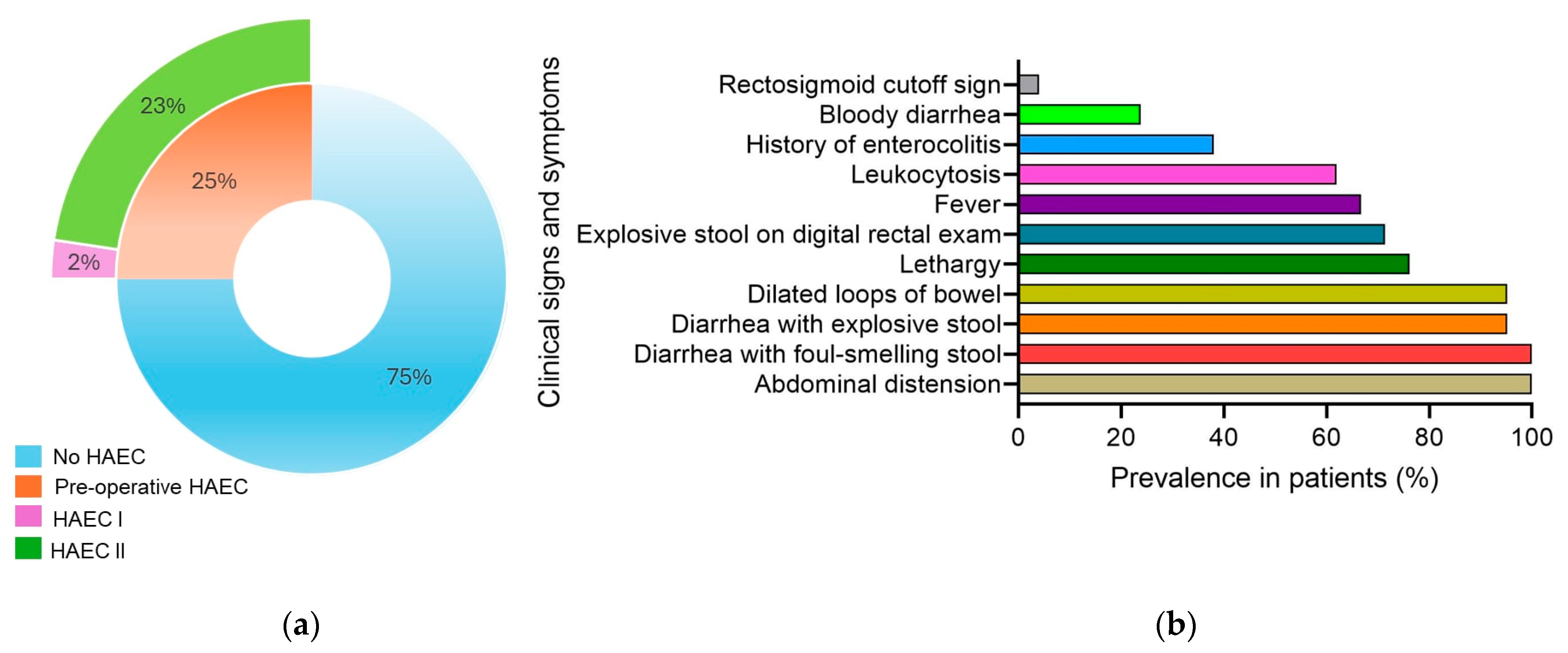
Figure 1. Preoperative enterocolitis in pediatric patients. (a) Pie chart illustrating percentages of patients with preoperative HAEC amongst total 84 HSCR patients; (b) bar chart indicating prevalence of clinical signs and symptoms amongst 21 HSCR patients with HAEC.
Gastrointestdisord 07 00017 g001
Figure 2. Postoperative enterocolitis in pediatric patients. (a) Pie chart illustrating percentages of patients with postoperative HAEC amongst total 83 HSCR patients; (b) bar chart indicating prevalence of clinical signs and symptoms amongst 14 HSCR patients with HAEC.
Gastrointestdisord 07 00017 g002
Figure 3. Kaplan–Meier plot illustrates the risk score performance comparing relative survival between patients with and those without a history of pre-op HAEC. To evaluate the performance of the risk score during the study period after surgery, we used intervals of 30 days, starting from 0 to 360 days, with Cox proportional hazards regression. The hazard ratio is 18.4, 95% CI 2.40–141 with p-value = 0.005.
Gastrointestdisord 07 00017 g003
Table 1. General characteristics of pediatric patients with HAEC.
VariablesSamples (n = 84)
Age at diagnosis (months)Mean ± S.D.7.2 ± 10.7
Interquartile range1.5–8
Range1–68
GenderMale71 (84.5%)
Female13 (15.5%)
GestationFull term81 (96.4%)
Pre-term3 (3.6%)
Congenital abnormalitiesDown syndrome1 (1.2%)
Genitourinary defects4 (4.8%)
Cardiac defects7 (8.4%)
None82 (85.6%)
Clinical presentationAbdominal distension78 (92.9%)
Delayed meconium passage73 (86.9%)
High-pressure rectal decompression38 (45.2%)
Palpable fecal mass6 (7.1%)
Table 2. Risk factors for preoperative enterocolitis.
Patients with HAEC (n = 21)Patients Without HAEC (n = 63)OR [95% CI]p-Value a
Percentile of weight for age b 0.001 **
  Normal7 (13%)47 (87%)1
  Underweight12 (48%)13 (52%)0.17 [0.05; 0.50]
  Malnutrition2 (50%)2 (50%)0.16 [0.01; 1.71]
  Overweight0 (0%)1 (100%)-
History of HAEC 3.53 [1.26; 10.3]0.024 *
  Yes12 (41.4%)17 (58.6%)
  No9 (16.4%)46 (83.6%)
Preoperative medical care 0.18 [0.04; 0.61]0.010 *
  Yes3 (8.82%)31 (91.2%)
  No18 (36.0%)32 (64.0%)
Enemas radiology aganglionosis 1.23 [0.16; 35.1]1.000
  Short20 (25.3%)59 (74.7%)
  Long1 (20%)4 (80%)
Congenital anomalies 0.40 [0.11; 1.57]0.164
  Yes7 (58.3%)7 (58.3%)
  No56 (77.8%)16 (22.2%)
a The p values were calculated using Fisher’s exact tests, ** p-value < 0.01, * p-value < 0.05. b Percentile of weight for age was determined using WHO Anthro software [10].
Table 3. Relationship between postoperative enterocolitis and risk factors.
Patients with HAEC (n = 14)Patients Without HAEC (n = 69)p-Value a
History of preoperative HAEC <0.001 **
Yes11 (37.9%)18 (62.1%)
No3 (5.56%)51 (94.4%)
Procedure 0.581
Swenson-like10 (18.2%)45 (81.8%)
SWlike—open2 (28.6%)5 (7.4%)
SWlike—lap0 (0.0%)7 (100%)
TEPT2 (14.3%)12 (85.7%)
Complication 0.002 **
Stricture4 (80.0%)1 (20.0%)
None10 (12.8%)68 (87.2%)
Hb (g/dL)11.7 (2.94%)11.5 (2.23%)0.823
Blood loss (mL)12.1 (9.73%)8.91 (7.58%)0.224
Aganglionic length (cm)9.676.70.031 *
a The p values were calculated using Fisher’s exact tests, ** p-value < 0.01, * p-value < 0.05.
Disclaimer/Publisher’s Note: The statements, opinions and data contained in all publications are solely those of the individual author(s) and contributor(s) and not of MDPI and/or the editor(s). MDPI and/or the editor(s) disclaim responsibility for any injury to people or property resulting from any ideas, methods, instructions or products referred to in the content.