Article

Advancements in Laparoscopic Techniques for Perineal Hernias—Technical Success and Complications Data

by
Sarah Kalmbach
1,
Hannah Laura Welskopf
1,
Christoph Steidle
1,
Philipp Horvath
1,2 and
Robert Bachmann
1,*
1
Department of General, Visceral and Transplant Surgery, Eberhard-Karls University Tübingen, Hoppe-Seyler Straße 3, 72076 Tübingen, Germany
2
Department of General, Visceral and Thoracic Surgery, Academic Teaching Hospital Feldkirch, Carinagasse 47, 6807 Feldkirch, Austria
*
Author to whom correspondence should be addressed.
Gastrointest. Disord. 2024, 6(4), 976-983; https://doi.org/10.3390/gidisord6040068
Submission received: 12 November 2024 / Revised: 30 November 2024 / Accepted: 11 December 2024 / Published: 17 December 2024

Abstract

:
Purpose: Multimodal approaches, including radiochemotherapy and surgical resection, are commonly used to treat rectal malignancies. Postoperative perineal hernias, though not uncommon, have shown an increasing incidence in recent years. Managing symptomatic perineal hernias presents a challenge, with a high recurrence rate after repair. This retrospective study investigates the laparoscopic placement of a non-absorbable mesh for treating pelvic floor hernias involving bowel displacement. Methods: This study included all consecutive patients requiring hernia repair due to symptomatic perineal hernias following minimally invasive abdominoperineal resection. All patients had a history of local radiation therapy. Data were collected on surgery duration, intra- and postoperative complications, and hernia recurrence rates. Results: Laparoscopic displacement of the small bowel from the pelvis was performed on eight patients, and closure of the pelvic floor hernia area was achieved using non-absorbable mesh placement. All procedures were successful, and no perioperative complications were recorded. Only one early complication occurred, classified as Clavien-Dindo II. Conclusions: The laparoscopic procedure, serving as a safe method for repositioning the small intestine and closing a perineal hernia using a mesh, has shown promising outcomes. The use of a non-absorbable mesh as an alternative to native tissue appears to be effective, with no observed recurrent hernias. However, further evaluation through long-term results or a larger cohort study is still pending.

1. Introduction

The therapy of malignant diseases of the rectum requires, depending on the stage, in addition to radiochemotherapy, a surgical resection, sometimes involving the pelvic floor muscles [1]. Multimodal therapeutic approaches have increased operability in a larger number of patients. The surgical treatment of rectal cancer has evolved in recent years, with the increasing acceptance of the principle of total mesorectal excision (TME). At the same time, the importance of clear surgical resection and the avoidance of tumor perforations or positive circumferential margins during an abdominoperineal excision (APE) are increasingly recognized [2]. This evolution has led to the development of extra-levator abdominoperineal excision (ELAPE), where potential surgical resection margins can be identified through MRI staging [2,3,4].
In the further postoperative course, both after APE and ELAPE, there can be a risk of perineal hernia [5]. In perineal hernias, intra-abdominal viscera protrudes through weakened tissues or surgical defects in the perineal area. The main complications of these hernias, in addition to local discomfort such as pain while sitting, include local skin erosion, bowel obstruction, and urinary dysfunction [6]. The incidence of perineal hernia after APR or ELAPE varies significantly, ranging from 1% to 26% [7]. A higher incidence of postoperative perineal hernias is associated with the more radical ELAPE procedure. The incidence of perineal hernias has increased in recent years [7]. Possible reasons for this may include, in addition to operative aspects, the rise in neoadjuvant radiochemotherapy [1,5,6].
Managing symptomatic perineal hernias post-surgery presents a formidable challenge. The recurrence rate following surgical repair is high, primarily due to difficulties in securely fixing prosthetic materials, and there is no widely accepted standard for the best repair method. The repair of these hernias can be carried out through various approaches, including abdominal, perineal, or combined methods, whereas open methods require a highly invasive approach. Closure of the hernia may involve options such as primary suture closure, mesh placement, or myocutaneous flaps [2,7]. Another challenge is the lack of universally accepted radiological definitions of a perineal hernia. It would be desirable to develop a general definition of a perineal hernia in terms of clinical findings and radiological results, as well as a staging system in the future. This would also increase the comparability of individual studies [8]. The latest studies also explore prophylactic techniques to prevent the development of postoperative perineal hernias [9,10].
Herein, we report a retrospective study of laparoscopic placement of a non-absorbable mesh in patients requiring pelvic floor hernia treatment. This study addresses the pressing need for standardized surgical techniques by presenting a reproducible laparoscopic approach. By doing so, it contributes to the limited body of evidence on minimally invasive solutions for this complex condition. The main objective is to present and describe our surgical method for repairing perineal hernias. Additionally, we will discuss the patient cohort and the postoperative course for further context.

2. Results

In total, eight patients who underwent planned and performed pelvic mesh placement were included. The mesh placement was the sole purpose of the operation. The mean age at the time of hernia operation was 69 ± 7.9 years. All included patients reported local symptoms (pressure/pulling/pain), exacerbated under stress. Additionally, three patients noticed swelling caused by a seroma. One patient presented urgently with ileus symptoms due to incarcerated small bowel loops in the perineal hernia (Figure 1). One patient had a recurrence of the perineal hernia, previously treated openly in another clinic 18 months earlier. The mean duration from APR to the occurrence of the perineal hernia was 30.75 months (10–83 months).
As part of the follow-up after malignant disease, computed tomography of the abdomen and pelvis: Perineal incisional hernia with protrusion of small bowel loops and accompanying interenteric free fluid in the hernia sac (11.4 × 5.7 cm hernia gate dimensions).

2.1. Technical Success

All patients underwent laparoscopic small bowel displacement from the pelvis and closure of the pelvic floor hernia area. The consecutive operation steps are shown in Figure 2. No bleeding complications or injuries to adjacent vessels or organs were observed. There was no need for conversion to an open exploration. In all procedures, the non-absorbable mesh could be adequately placed and secured, resulting in a success rate of 100%.
On average, the surgical procedure lasted 1:44 h ± 0:54 h. The choice of mesh depended on the hernia size. A single 15 × 15 cm non-absorbable intraperitoneal onlay mesh with anti-adhesive coating was sufficient for six patients. For one patient, two 15 × 15 cm meshes were required, while a larger 20 × 15 cm non-absorbable intraperitoneal onlay mesh with anti-adhesive coating was used for another.

2.2. Follow-Up

The average postoperative hospital stay was 3 ± 1.4 days (1–5 days). Upon discharge, all patients required minimal analgesia, corresponding to a pain level of 0–3/10 on the numeric rating scale. One patient experienced an early complication with clinical and radiological obstruction on the fifth postoperative day. This complication was managed conservatively with bowel stimulation, intravenous fluid therapy, and close clinical monitoring. No surgical revision was required, and the patient’s condition resolved without further intervention, consistent with Clavien–Dindo grade II classification. No other early or late complications occurred. The longest follow-up duration reached 599 days (138.8 ± 217.7 days), with no recurrence of perineal hernia observed in any of the patients (Table 1).

3. Materials and Methods

The cohort study received approval from the Tuebingen University Ethics Committee (059/2021BO2).
We retrospectively screened all patients at our clinic who were treated with operation and procedure code OPS 5-536.49 (laparoscopic transperitoneal, with intraperitoneal onlay mesh [IPOM]) or diagnosed with K 46.9 (unspecified abdominal hernia without obstruction and without gangrene). From 2020 to 2023, this resulted in 52 cases, of which 8 met the inclusion criteria for a perineal hernia following APE/ELAPE. The diagnosis of a perineal hernia was established based on clinical examination findings and supported by radiological imaging. Patients with comorbidities that constituted a contraindication for laparoscopic surgery were excluded from the study. Preoperatively, all patients received a CT scan of the abdomen and pelvis, not least to exclude recurrence of the malignant disease. Follow-up continued until 31 December 2023.
Data were collected using the hospital information database (SAP for Healthcare; SAP AG Walldorf). Collected data for each patient include demographic characteristics such as age, gender, underlying disease, neoadjuvant therapy, timing of initial hernia diagnosis, and follow-up time. Additionally, surgical data obtained include the date of primary resection, postoperative TNM stage, date of hernia repair, operation time, type of mesh used, and placement of trocars. Follow-up data consist of the occurrence of recurrent hernia and other complications (date, category, and treatment).
Data collection and analysis were conducted using Microsoft Excel 2023, Version 16.80, Microsoft Corporation, Redmond, DC, USA. Results in the manuscript and figures are reported as mean ± standard deviation (SD) with the complete range.

3.1. Patients

Three patients were female, and five were male. All patients had an underlying malignant disease. Six patients were initially operated on for a low rectal carcinoma, one patient due to the recurrence of an anal carcinoma, and one patient had a local recurrence of a rectum-infiltrating leiomyosarcoma. The oncologic surgeries of the patients were performed at the same center, which is a certified visceral oncology center for colorectal cancer. Seven patients received neoadjuvant radiochemotherapy, and one patient received local radiation therapy. The pelvic floor closure technique was performed during the primary resection, with three patients using the V-Y advancement flap in collaboration with plastic surgeons, and five patients through layered primary closure.

3.2. Definitions

Operative success was defined as complete hernia repair. Complications were classified using the Clavien–Dindo classification [11]. Complications were differentiated as early complications (<30 days after surgery) and late complications (>30 days after surgery). Clinical and imaging follow-up was conducted, which, in addition to postoperative clinical examinations, followed the intervals of surveillance appropriate for the malignant condition (clinical examinations and cross-sectional imaging procedures).

3.3. Surgical Procedure

We performed the hernia repair using a laparoscopic abdominal approach. A urethral catheter was placed to prevent bladder injury and to gain better visibility of the perineal hernia and surrounding structures. Perioperative antibiotic prophylaxis with a cephalosporin antibiotic (Cefotaxime, Fresenius Kabi Deutschland GmbH, Bad Homburg, Germany) was administered. Specific preparation in terms of bowel preparation was not necessary.
An 11 mm supra-umbilical incision was made, and pneumoperitoneum was achieved using a Veress needle, with the placement of an 11 mm port at the umbilicus. Subsequently, a 5 mm port was placed in the right lower abdomen along the midclavicular line, and a 5 mm port in the right mid-abdomen under camera control. Placement of the patient in lithotomy and steep Trendelenburg position facilitated the division of adhesions and extrusion of the small bowel from the pelvic field.
For hernia closure, a non-absorbable intraperitoneal onlay mesh (15 × 15 cm, lightweight material with anti-adhesive coating) was used, which, after size adjustment, was introduced into the abdominal cavity through the 11 mm trocar. Subsequently, it is placed in the lesser pelvic region over the symphysis pubis and pelvic brim. It is important to ensure adequate overlap and proper placement with the anti-adhesive side facing the abdominal cavity.
The fixation of the mesh is achieved through absorbable tackers. These are anchored to the muscular pelvic floor and tangentially to the pelvic peritoneum, under visual control of the internal iliac artery and vein, as well as the ureters. The abdomen was then deflated of gas, and the fascia of the umbilical port closed. Skin incisions were closed in the usual manner.

4. Discussion

Since the initial description of a perineal hernia in 1939, these have become a more frequently occurring problem encountered by colorectal surgeons in recent years [12,13]. To date, the reconstruction is under debate, and there is no consensus.
In our study, we present a laparoscopic surgical approach utilizing a non-absorbable mesh. The procedures were conducted by experienced laparoscopic surgeons specializing in colorectal surgery. Based on the collected data, it appears that this method shows promising safety and efficiency. The technical success rate was 100%, with no need for conversion to an open procedure.
The laparoscopic approach offers several advantages, particularly compared to an open transperineal approach [10,14]. Firstly, it provides an excellent view of the anatomical structures of the pelvis and the hernia defect. Furthermore, the approach allows for diagnostic laparoscopy to exclude tumor recurrence [6]. Postoperatively, patients quickly regain mobility and everyday functionality; discharge in our study was possible on average after three days. No restrictions in the perineal region, as observed after a perineal approach, need to be considered. In our cohort, there were no postoperative wound infections, as reported in studies utilizing a perineal approach [10]. Subsequently, postoperatively, there was only a minor manifestation of pain, as in our method, the tackers are placed in the irradiated areas of the pelvis, thereby reducing pain sensation in these regions. Furthermore, patients demonstrated rapid functional recovery, resuming normal activities within a few days. These results emphasize the effectiveness of our laparoscopic approach in minimizing postoperative discomfort and optimizing recovery time. The technique we presented can be complemented by an omental flap [10,15]. This approach is more elaborate and results in a longer surgical duration. The advantage of an omental flap, however, is a lower incidence of adhesions in the mesh area and potentially greater stability in hernia repair. It is essential to bear in mind that all patients have undergone previous surgery and have a stoma, which may complicate and increase the risk of omentum preparation. A drawback of the laparoscopic method is the remaining perineal skin excess, resulting from the protrusion of abdominal structures. However, this is predominantly a cosmetic aspect and could be addressed in a subsequent procedure.
A perineal hernia typically occurs on average after 13 months (1–51 months) [7]. In our cohort, hernias most commonly occurred between 12 and 18 months. In one case, the hernia was diagnosed after 83 months, although it was presumed to have existed for a longer duration based on the patient’s history. The average age at the time of hernia surgery in our cohort was 69 ± 7.9 years, which is similar to other studies, such as 69.5 years in the study by Sayers et al. and 71 years in the study by Samar et al. [7,16]. Following the completion of our laparoscopic procedure, there were no instances of hernia recurrence observed during the follow-up period. The reported incidences in the literature range from 14.3% to 25% [7,17]. When applying the laparoscopic method, particular attention must be paid to the correct mesh placement with sufficient overlap and thorough fixation. Otherwise, recurrences can also occur with a laparoscopic repair procedure, as reported by Goedhart-de Haan et al. However, a different mesh was used in this study compared to ours, which could also be an important factor [18]. In the literature, there have already been indications that using a mesh for hernia repair results in a lower recurrence rate compared to primary closure [19]. In their pooled analysis, Mjoli et al. reported a total of 43 repairs for perineal hernias. These included 22 perineal repairs, 11 open abdominal operations, 3 combined abdominoperineal approaches, 5 laparoscopic repairs, and 2 laparoscopic-perineal procedures. The highest recurrence rate was observed with the primary closure method at 50% (6/12). Only one patient was found to have undergone repair with an absorbable mesh, and this hernia recurred within 16 months [19]. We employed a non-absorbable mesh made of polyvinylidene fluoride (PVDF). This mesh, due to its properties as a composite mesh, offers not only good mechanical strength but also allows for adequate mesothelialization at the peritoneal level, thus preventing adhesions or fistulas [20]. The choice of mesh should always be individualized, considering patient-specific factors. One study demonstrated that the use of lightweight polypropylene (LW-PP) meshes was associated with fewer postoperative restrictions [21]. In our opinion, a mesh made of poliglecaprone is not suitable for this region due to its self-absorbable properties. The use of biological mesh for repairing perineal hernias is another option. The acellular collagen matrix is believed to promote fibroblast migration, neovascularization, and integration into natural tissue, potentially reducing the risk of wound infections and adhesions. A recently published case series of 15 patients undergoing perineal hernia repair with porcine acellular dermal meshes reported a recurrence rate of 47% after a median follow-up of 17 months (IQR 12–24) [22]. However, it is worth noting that in this study, all patients experiencing a recurrence had a perineal open approach, limiting comparability with the present study. Further research is needed to evaluate the benefits of biological meshes in repairing perineal hernias.
It is noteworthy that our analysis comes with relevant limitations due to the retrospective study design and the small and heterogeneous set of patients. However, other cohort sizes in the literature match ours [17,18,23], and all complications are coded and retained in the electronic notes of our institution. Furthermore, the follow-up time is not uniform, and a reliable statement regarding recurrent hernias is ultimately uncertain. It is noteworthy that designing an appropriate prospective clinical trial to provide robust evidence should be considered.

5. Conclusions

In summary, the management of perineal hernias through a laparoscopic approach presents a technical challenge; however, with the expertise of experienced surgeons, this method appears to be safely and effectively accomplished. In addition to providing an optimal view of the hernia defect, the laparoscopic approach allows for excluding a recurrence of the malignant disease. The effective fixation of the non-absorbable mesh appears achievable, and while long-term results are still awaited in our study, there were no occurrences of hernia recurrence observed. These findings suggest that laparoscopic repair could become a preferred method for managing perineal hernias, offering potential benefits in terms of reduced postoperative pain, quicker recovery times, and improved long-term outcomes. Further research with larger cohorts and extended follow-up is needed to fully establish its role in clinical practice.

Author Contributions

Study conception and design, R.B., S.K. and P.H.; acquisition of data, S.K., H.L.W. and C.S.; analysis and interpretation of the data, S.K.; drafting of manuscript: S.K. and R.B.; revision and critique of the manuscript, all authors. All authors have read and agreed to the published version of the manuscript.

Funding

This study was conducted without funding.

Institutional Review Board Statement

This study was conducted in accordance with the Declaration of Helsinki, and approved by the Ethics Committee of Tuebingen, University Ethics Committee (protocol code 059/2021BO2 and date of approval 23 February 2021).

Informed Consent Statement

Informed consent was obtained from all subjects involved in the study.

Data Availability Statement

The original contributions presented in this study are included in the article. Further inquiries can be directed to the corresponding author.

Conflicts of Interest

The authors have no conflicts of interest or financial ties to disclose.

References

  1. Hawkins, A.T.; Albutt, K.; Wise, P.E.; Alavi, K.; Sudan, R.; Kaiser, A.M.; Bordeianou, L.; Continuing Education Committee of the SSAT. Abdominoperineal Resection for Rectal Cancer in the Twenty-First Century: Indications, Techniques, and Outcomes. J. Gastrointest. Surg. 2018, 22, 1477–1487. [Google Scholar] [CrossRef]
  2. Shihab, O.C.; Brown, G.; Daniels, I.R.; Heald, R.J.; Quirke, P.; Moran, B.J. Patients with low rectal cancer treated by abdominoperineal excision have worse tumors and higher involved margin rates compared with patients treated by anterior resection. Dis. Colon Rectum 2010, 53, 53–56. [Google Scholar] [CrossRef] [PubMed]
  3. Shihab, O.C.; Taylor, F.; Salerno, G.; Heald, R.J.; Quirke, P.; Moran, B.J.; Brown, G. MRI Predictive Factors for Long-Term Outcomes of Low Rectal Tumours. Ann. Surg. Oncol. 2011, 18, 3278–3284. [Google Scholar] [CrossRef] [PubMed]
  4. Grass, J.K.; Perez, D.R.; Izbicki, J.R.; Reeh, M. Systematic review analysis of robotic and transanal approaches in TME surgery-A systematic review of the current literature in regard to challenges in rectal cancer surgery. Eur. J. Surg. Oncol. (EJSO) 2019, 45, 498–509. [Google Scholar] [CrossRef] [PubMed]
  5. Li, J.; Zhang, W. How we do it: Repair of large perineal hernia after abdominoperineal resection. Hernia 2017, 21, 957–961. [Google Scholar] [CrossRef] [PubMed]
  6. Stamatiou, D.; Skandalakis, J.E.; Skandalakis, L.J.; Mirilas, P. Perineal hernia: Surgical anatomy, embryology, and technique of repair. Am. Surg. 2010, 76, 474–479. [Google Scholar] [CrossRef]
  7. Balla, A.; Rodríguez, G.B.; Buonomo, N.; Martinez, C.; Hernández, P.; Bollo, J.; Targarona, E.M. Perineal hernia repair after abdominoperineal excision or extralevator abdominoperineal excision: A systematic review of the literature. Tech. Coloproctol. 2017, 21, 329–336. [Google Scholar] [CrossRef] [PubMed]
  8. Narang, S.K.; Alam, N.N.; Köckerling, F.; Daniels, I.R.; Smart, N.J. Repair of Perineal Hernia Following Abdominoperineal Excision with Biological Mesh: A Systematic Review. Front. Surg. 2016, 3, 49. [Google Scholar] [CrossRef] [PubMed]
  9. Liu, S.; Jiang, T.; Xiao, L.; Yang, S.; Liu, Q.; Gao, Y.; Chen, G.; Xiao, W. Total Neoadjuvant Therapy (TNT) versus Standard Neoadjuvant Chemoradiotherapy for Locally Advanced Rectal Cancer: A Systematic Review and Meta-Analysis. Oncologist 2021, 26, e1555. [Google Scholar] [CrossRef]
  10. McKenna, N.P.; Habermann, E.B.; Larson, D.W.; Kelley, S.R.; Mathis, K.L. A 25 year experience of perineal hernia repair. Hernia 2020, 24, 273–278. [Google Scholar] [CrossRef]
  11. Dindo, D.; Demartines, N.; Clavien, P. Classification of Surgical Complications. Ann. Surg. 2004, 240, 205–213. [Google Scholar] [CrossRef] [PubMed]
  12. Shen, Y.; Yang, T.; Deng, X.; Wu, Q.; Wei, M.; Meng, W.; Wang, Z. Closure of pelvic peritoneum with bladder peritoneum flap reconstruction after laparoscopic extralevator abdominoperineal excision: A prospective stage II study. J. Surg. Oncol. 2023, 128, 851–859. [Google Scholar] [CrossRef] [PubMed]
  13. Thiel, J.T.; Welskopf, H.L.; Yurttas, C.; Farzaliyev, F.; Daigeler, A.; Bachmann, R. Feasibility of Perineal Defect Reconstruction with Simplified Fasciocutaneous Inferior Gluteal Artery Perforator (IGAP) Flaps after Tumor Resection of the Lower Rectum: Incidence and Outcome in an Interdisciplinary Approach. Cancers 2023, 15, 3345. [Google Scholar] [CrossRef] [PubMed]
  14. Moiș, E.; Graur, F.; Horvath, L.; Furcea, L.; Zaharie, F.; Vălean, D.; Moldovan, S.; Al Hajjar, N. Perineal Hernia Mesh Repair Using Only the Perineal Approach: How We Do It. J. Pers. Med. 2023, 13, 1456. [Google Scholar] [CrossRef] [PubMed]
  15. Hultman, C.S.; Sherrill, M.A.; Halvorson, E.G.; Lee, C.N.; Boggess, J.F.; Meyers, M.O.; Calvo, B.A.; Kim, H.J. Utility of the omentum in pelvic floor reconstruction following resection of anorectal malignancy: Patient selection, technical caveats, and clinical outcomes. Ann. Plast. Surg. 2010, 64, 559–562. [Google Scholar] [CrossRef] [PubMed]
  16. Yeomans, F.C. Levator hernia, perineal and pudendal. Am. J. Surg. 1939, 43, 695–697. [Google Scholar] [CrossRef]
  17. A Sayers, E.; Patel, R.K.; Hunter, I.A. Perineal hernia formation following extralevator abdominoperineal excision. Color. Dis. 2015, 17, 351–355. [Google Scholar] [CrossRef] [PubMed]
  18. Haan, A.M.S.G.-D.; Langenhoff, B.S.; Petersen, D.; Verheijen, P.M. Laparoscopic repair of perineal hernia after abdominoperineal excision. Hernia 2016, 20, 741–746. [Google Scholar] [CrossRef]
  19. Mjoli, M.; Sloothaak, D.A.M.; Buskens, C.J.; Bemelman, W.A.; Tanis, P.J. Perineal hernia repair after abdominoperineal resection: A pooled analysis. Color. Dis. 2012, 14, e400–e406. [Google Scholar] [CrossRef]
  20. Rodríguez, M.; Gómez-Gil, V.; Pérez-Köhler, B.; Pascual, G.; Bellón, J.M. Polymer Hernia Repair Materials: Adapting to Patient Needs and Surgical Techniques. Materials 2021, 14, 2790. [Google Scholar] [CrossRef] [PubMed]
  21. Musters, G.D.; Lapid, O.; Stoker, J.; Musters, B.F.; Bemelman, W.A.; Tanis, P.J. Is there a place for a biological mesh in perineal hernia repair? Hernia 2016, 20, 747–754. [Google Scholar] [CrossRef]
  22. Calomino, N.; Poto, G.E.; Carbone, L.; Micheletti, G.; Gjoka, M.; Giovine, G.; Sepe, B.; Bagnacci, G.; Piccioni, S.A.; Cuomo, R.; et al. Weighing the benefits: Exploring the differential effects of light-weight and heavy-weight polypropylene meshes in inguinal hernia repair in a retrospective cohort study. Am. J. Surg. 2024, 238, 115950. [Google Scholar] [CrossRef] [PubMed]
  23. Samar, A.M.; Branagan, G. Perineal hernia repair after extralevator abdominoperineal excision, how we do it (PERineal Laparoscopic Sling: PERLS Technique). Langenbeck’s Arch. Surg. 2022, 407, 2187–2191. [Google Scholar] [CrossRef] [PubMed]
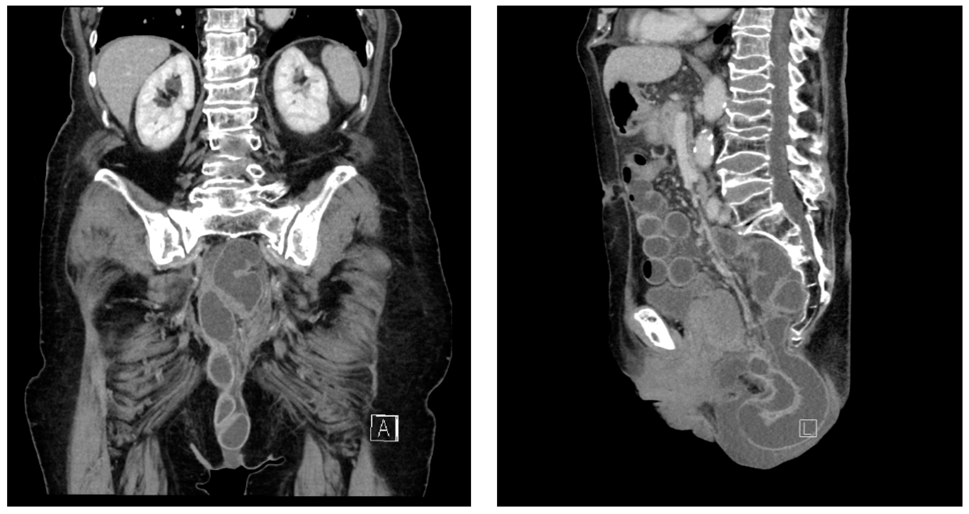
Figure 1. CT scan of a perineal hernia.
Gastrointestdisord 06 00068 g001
Figure 2. Step-by-step guide for laparoscopic repair of a perineal hernia. (a) Initial view of the pelvis, the perineal hernia is obscured by loops of small bowel within; (b) after mobilization and possibly adhesiolysis of the small bowel loops, the entrance into the pelvic cavity and the hernia gate of the perineal hernia are revealed; (c) View of the hernia gate and the hernia sac; (d): after trimming and placement of the non-absorbable intraperitoneal onlay mesh (15 × 15 cm, lightweight material with anti-adhesive coating), it is positioned in the area of the perineal hernia gate; (e) placement of the mesh; initially, the mesh is fixed in the area of the sacrum using absorbable secure strap fixation tackers and then spread out over the two lateral sides ventrally; (f) view of the repaired perineal hernia, the mesh is secured on all sides with Tackers with sufficient overlap of the hernia gate.
Gastrointestdisord 06 00068 g002
Table 1. Baseline patient characteristics.
Patient Characteristics (n = 8)
Age69 years
youngest 58 years
oldest 81 years
Gender5 male
3 female
Underlying diseaseLow rectal cancer (n = 6)
Recurrence of Anal Carcinoma (n = 1)
Local Recurrence of a Rectum-Infiltrating Leiomyosarcoma (n = 1)
Neoadjuvant TherapyRadiochemotherapy (n = 7)
Localized Radiotherapy (n = 1)
Closure Technique following Resection (APE/ELAPE)Layered Primary Suture (n = 5)
V-Y advancement flap (n = 3)
Tumor Stage after ResectionypT2 ypN0 L0 V0 Pn0 R0 (n = 6)
ypT3 ypN1b pM1 (hep) V0 Pn0 R0 (n = 1)
ypT3 ypN1a L0 V1 Pn1 R0 (n = 1)
Disclaimer/Publisher’s Note: The statements, opinions and data contained in all publications are solely those of the individual author(s) and contributor(s) and not of MDPI and/or the editor(s). MDPI and/or the editor(s) disclaim responsibility for any injury to people or property resulting from any ideas, methods, instructions or products referred to in the content.