Article

Body Mass Index: An Unreliable Adiposity Indicator for Predicting Outcomes of Liver Transplantation Due to Hepatocellular Carcinoma

Department of Transplant Medicine, Immunology, Nephrology and Internal Diseases, Medical University of Warsaw, 59 Nowogrodzka Street, 02-006 Warsaw, Poland
*
Author to whom correspondence should be addressed.
Gastrointest. Disord. 2024, 6(3), 607-621; https://doi.org/10.3390/gidisord6030040
Submission received: 6 May 2024 / Revised: 17 June 2024 / Accepted: 19 June 2024 / Published: 25 June 2024

Abstract

:
Obesity is a well-documented risk factor for the development of hepatocellular carcinoma (HCC) in the general population. The applicability of these findings to liver recipients is uncertain, and the results of available data have not been unanimous. The objective of the current study was to investigate the impact of the pre-operative body mass index (BMI) on oncological outcomes of liver transplantation due to HCC. Methods: This observational retrospective study enrolled all patients with histologically confirmed HCC who underwent liver transplantation from a deceased donor in our centre between 2008 and 2018. Results: Overall, 83 patients were enrolled and were subsequently stratified according to their pre-operative BMI into three groups: patients with normal body weight (n = 53), patients with overweight (n = 23), patients with obesity (n = 7). Overall tumour recurrence was 12%. BMI failed to predict the 5-year recurrence-free survival (p = 0.55), risk of tumour recurrence (p = 0.314) and overall 5-year survival (p = 0.19) in liver recipients. Conclusions: BMI was proven to be an unreliable surrogate measure of obesity for predicting oncological outcomes among liver recipients. Other obesity indices should be referenced to assess cancer-related prognosis more accurately in these groups of patients.

1. Introduction

Hepatocellular carcinoma (HCC) constitutes the most common primary liver cancer, the sixth most frequently diagnosed malignancy and is ranked as the third leading cause of cancer-related deaths worldwide [1]. Liver transplantation constitutes a potentially curative treatment modality for selected patients with early-stage HCC that is unsuitable for surgical resection [2,3,4]. A fundamental condition of suitability for the procedure is a low risk of tumour recurrence after the transplant [3]. This is most universally achieved by the utilisation of the Milan criteria (solitary tumour ≤5 cm or two to three tumours, each one ≤3 cm, no evidence of angioinvasion or extrahepatic spread) [5]. Over the years, numerous other classifications have been proposed to address the increasing demand for liver transplantation due to HCC and to mitigate the burden of disease recurrence regarding post-transplantation outcomes [6,7,8,9,10,11,12,13,14].
Despite these efforts, a sizable proportion of patients at an increased risk of HCC re-lapse remain eligible according to the current proposals [15,16]. Consequently, disease recurrence affects between 10 and 25% of liver recipients and constitutes the most important negative predictor of post-transplantation survival [17,18,19,20,21,22,23].
HCC recurrence after liver transplantation is multifactorial. As of now, several well-established risk factors have been identified to increase the likelihood of HCC recurrence in the allograft and associated mortality. Among these, most are related to tumour characteristics (tumour size, its differentiation, number of nodules or presence of vascular invasion) the resultant levels of serum biomarkers of tumour biology (e.g., alpha-fetoprotein (AFP) and neutrophil-to-lymphocyte ratio (NLR)) or are related to the host (underlying liver disease) [5,23,24,25]. As a consequence, most factors are not modifiable or accurately assessable until a pathological examination of the explanted liver is performed. Therefore, it has been a challenge to establish more effective selection protocols and identify pre-operatively available modifiable predictors of HCC recurrence to enhance post-transplantation survival. Obesity is a well-documented and modifiable risk factor for many cancers, including HCC, in the general population [26,27,28,29,30,31,32,33]. The same holds true for the risk of HCC recurrence following hepatectomy [34,35]. Importantly, most of the studies investigating the subject have utilised the body mass index (BMI) as a simple, easily attainable marker of obesity, and its effectiveness for determining oncological outcomes in the general population has been proven. However, BMI utilisation holds some limitations for patients with liver cirrhosis due to the associated ascites and fluid overload. Of note, some transplant-associated factors, namely the removal of underlying liver disease along with potential intra-hepatic micrometastases and ischaemia–reperfusion injury are also known to modify the risk of tumour recurrence in organ recipients [36]. Therefore, it remains a subject of debate to what extent previous findings may be extrapolated to transplant recipients. There is a scarcity of scientific reports conducted in the field, and results have not been unanimous [22,37,38]. Importantly, the existing studies failed to estimate the dry body weight of liver transplant candidates prior to BMI calculation, which may significantly impact their results. To our knowledge, this is the first study investigating the impact of pre-operative BMI on oncological outcomes in liver recipients in which BMI values were corrected for concomitant fluid retention.
That is why the primary endpoint of the current study was to investigate the impact of the pre-operative BMI of liver transplant candidates, adjusted for fluid overload, on the oncological outcomes of liver transplantation due to HCC, defined as 5-year recurrence-free survival (RFS) and 5-year overall survival of graft recipients. We also examined pre- and post-transplant variables to detect predictors of HCC recurrence and all-cause mortality after liver transplantation.

2. Materials and Methods

2.1. Study Population

This observational retrospective study was conducted in the Department of Transplant Medicine, Immunology, Nephrology and Internal Disease, at the Medical University of Warsaw, Poland, an academic primary care centre. All patients with histologically confirmed HCC in an explanted liver who underwent liver transplantation from a deceased donor in our centre between 2008 and 2018 were recruited into this retrospective analysis.
Dry-weight-based BMI was calculated for all study participants. The dry weight of candidates for liver transplantation was evaluated by subtracting a certain percentage of the initial body weight depending on the severity of intercurrent ascites (5% for mild, 10% for moderate, 15% for severe). An additional 5% was subtracted if peripheral oedema was present [39].
The study participants were divided into three groups depending on the pre-operative BMI: patients with BMI < 25 kg/m2 were categorised as Group 1 (patients with normal body weight (n = 53)). Patients with BMI between 25 and 29.9 kg/m2 were categorised as Group 2 (patients with overweight (n = 23)). Patients with BMI ≥30 kg/m2 were categorised as Group 3 (patients with obesity (n = 7)).
For the determination of 5-year RFS, patients were censored at the time of HCC recurrence, last available follow-up, time of non-recurrence-related death or at the fifth year of post-transplant observation—whichever occurred first. For the purpose of the 5-year overall survival determination, the patients were censored at the last available follow-up, at the date of death or once they reached the 5-year observation.
For each of the study participants, predicted 5-year survival after the transplant was also estimated using the Metroticket calculator (available online at www.hcc-olt-metroticket.org, accessed on 13 June 2024). Liver recipients were also assessed based on a simplified version of the AFP model [13].
All patients with HCC were subjected to a regular oncological screening protocol, which consisted of thoracic–abdominal–pelvic CT scans performed twice a year for the first 2 years, and annually thereafter, and abdominal ultrasound along with AFP level testing biannually. All the patients were initially prescribed a triple immunosuppressive regimen with a steroid, mycophenolate mofetil and a calcineurin inhibitor, which was subsequently individually modified in accordance with the effective recommendations for the immunosuppressive treatment of patients undergoing solid organ transplantation from the Polish Transplantation Society [40]. As a general principle, the liver recipients were targeted to be weaned off steroids within 3 to 6 months and were maintained on calcineurin inhibitor in monotherapy or in combined therapy with mycophenolate mofetil or everolimus.

2.2. Data Collection

The patients’ medical files were reviewed, and the following information was retrieved: sociodemographics (age, gender, excessive alcohol consumption, tobacco use); model of end-stage liver disease (MELD) score; BMI at baseline, at 1-, 3- and 5-years post-transplantation; presence of diabetes mellitus at baseline; intercurrent liver disease if applicable; haematological and biochemical parameters at baseline (creatinine, AFP level, neutrophil and lymphocyte count); fulfilment of the Milan criteria and histopathological tumour characteristics (number of tumours, size of the largest tumour, presence of microvascular invasion, tumour differentiation). Information on previous tumour management, if any, was also reported. Peri- and post-operative variables (surgical technique, cold and warm ischaemia time, baseline immunosuppression, duration of steroid administration) and age of the donor were obtained from the National Transplant Registry.
The Metroticket-predicted 5-year survival was calculated using the Metroticket calculator (available online at www.hcc-olt-metroticket.org, accessed on 13 June 2024) based on the following explant data (size of the largest tumour, the number of HCC nodules and the presence of microvascular invasion) [14].

2.3. Statistical Analysis

The qualitative variables are expressed as the median, interquartile range (Q1–Q3) and ranges, while the quantitative variables are expressed as frequencies. The data were tested for normal distributions with the Shapiro–Wilk test and for homogeneity of variances with the Bartlett test. Depending on the results, differences in baseline variables between the groups were compared using either a one-way ANOVA (parametric test) or Kruskal–Wallis (non-parametric test) test. Both tests were followed by Dunn’s post hoc test for pairwise comparisons.
The Kaplan–Meier method was used to compare the overall survival and RFS between the analysed subgroups. Differences in survival curves were evaluated using the Gehan–Breslow test. Additionally, we conducted a Cox regression analysis to estimate the hazard ratios and 95% confidence intervals for the predictors of overall survival. The Wald test was used to assess the statistical significance of the models. Moreover, in cases where the Schoenfeld Individual test showed that the Cox model did not satisfy the proportional hazards assumption, a parametric model based on the Weibull distribution was used.
Both univariate and multivariate logistic models were used to identify risk factors associated with the overall risk of HCC recurrence. Based on the stepwise forward regression, along with the Akaike Information Criterion, the best-fit model was selected.
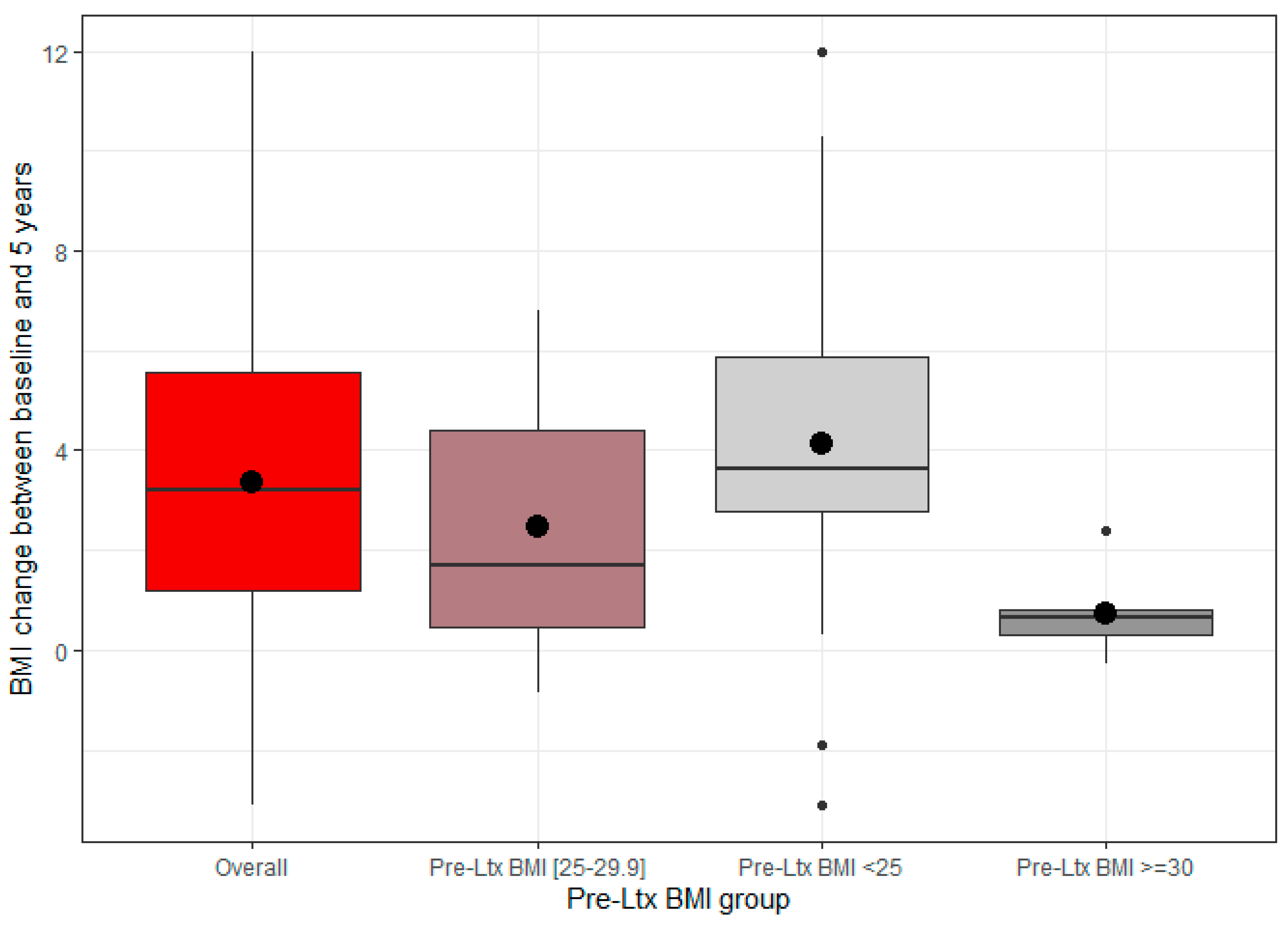
Box plots were constructed to visually depict the distribution of BMI changes over the 5-year observation and its skewness by displaying the data quartiles and averages. The upper and lower whiskers represented scores outside the middle 50%. The median was marked by the line dividing the box into two parts. Big dots represented the mean value while smaller dots represented the outliers in the dataset.
The level of statistical significance was set to p = 0.05. The p-values indicating a statistically significant result are shown in bold. All calculations were carried out in the R statistical software package version 4.3.2 (R Core Team, Vienna, Austria).
Due to the retrospective study design, the requirement to obtain approval from the local Ethics Committee was waived. The study protocol was submitted to the Ethics Committee of the Medical University of Warsaw for acknowledgement only.

3. Results

A total of 83 patients underwent liver transplantation within the pre-defined timeframes and had histologically confirmed HCC in the explanted liver. The median age of the study participants was 57 years, and the majority were males (84.3%). Overall, 83.1% of the participants were transplanted within the Milan criteria. The median number of nodules at transplantation was 1 (range: 1–4), with the median size of the largest tumour being 2 cm (range 0.5–8 cm). Most of the tumours showed moderate differentiation. Microvascular invasion was confirmed in approximately one-third of the tumours. The median AFP level at transplantation was 10.2 ng/mL. Most of the liver recipients did not receive any form of bridging therapy prior to transplantation. All patients, except for one, had accompanying chronic liver disease, of which hepatitis C (HCV) infection was the most frequently reported. All patients underwent orthotopic liver transplantation with a median MELD score of 11 at the time of transplantation.
The baseline features of the study participants, stratified by BMI class, are presented in Table 1. The sociodemographics and most of the pre- and post-operative features of liver recipients were comparable between the groups. However, the subgroups were not well-balanced in terms of the baseline AFP level, presence of diabetes mellitus, and diameter of the largest tumour: patients with diabetes mellitus were underrepresented among the patients with obesity; pre-operative AFP levels were unevenly distributed among the groups, as they were more varied in Groups 2 and 3; and the median diameter of the largest tumour was greater in patients in BMI Group 3—3 cm, compared to the median values of 2.5 cm and 2 cm observed in the other two groups. However, the statistical significance of these differences was not confirmed.
The overall median expected 5-year survival estimated using the Metroticket model was 73.3% (73.8%, 71.3%, 70.9% 5-year survival predicted for Groups 1, 2 and 3, respectively).

3.1. Five-Year Overall Survival and Determinants

Overall, 19.3% (n = 16) of the patients died during a median follow-up of 60 months. HCC recurrence was responsible for half of the fatal events. The remaining eight were attributed to a major cardiovascular event (one case), infectious complications (one case), post-operative complications (two cases), post-transplant lymphoproliferative disorder (one case), and allograft failure (three cases). Of those, 13 and 3 occurred in patients in BMI Groups 1 and 2, respectively. All patients from Group 3 completed the 5-year observation (Figure 1). Kaplan–Meier curves constructed to assess the overall survival of patients who underwent liver transplantation for HCC revealed no significant differences between the three groups (p = 0.19) (Figure 1). Based on the univariate Cox regression models, liver recipients were more likely to survive as the calculated Metroticket-predicted survival increased (HR: 0.953, 95 CI 0.920–0.988, p = 0.0243). Furthermore, histologically confirmed microvascular invasion and HCC recurrence increased the risk of death of the liver recipients by approximately 3.15 times (p = 0.0349) and 12.5 times (p < 0.001), respectively. In the multivariate analysis, only HCC recurrence remained associated with the all-cause mortality of the liver recipients (HR: 13.961; 95 CI 3.442–56.6; p < 0.001). Neither baseline BMI (p = 0.2667) nor early post-transplantation BMI alterations (p = 0.3621) impacted the survival probability of the liver recipients.

3.2. Five-Year Recurrence-Free Survival and Determinants of HCC Recurrence

Overall, the tumour recurrence rate was 12% (n = 10), with a median of 12.8 months (range 2.1–59 months) from liver transplantation to disease recurrence. Of note, 80% of the events occurred within the first 2 years after transplantation. Cases of HCC relapse presented predominantly as extrahepatic manifestations (90%) affecting mainly the lungs (five cases) and bones (three cases). A single instance of HCC recurrence showed a disseminated presentation involving multiple organs. Only one patient experienced an intra-hepatic relapse. All recurrence events had a fatal outcome: eight within the 5-year observation, and the remaining two within one and two months after the 5-year observation was completed. HCC recurred in five patients (9.4%), four patients (17.4%) and one patient (14.3%) from BMI Groups 1, 2 and 3, respectively (p = 0.4828). No significant differences were identified in terms of the 5-year RFS between patients from the three analysed groups (p = 0.55) (Figure 2).
Considering the univariate logistic regression, the presence of microvascular invasion (p = 0.003), number of HCC nodules in the native liver (p = 0.017), AFP level at transplantation (p = 0.004) and increasing value of AFP model (p = 0.007) increased the risk of HCC recurrence. Fulfilment of the Milan criteria was the only factor that mitigated this risk (p = 0.0007). Of the above variables, only three were confirmed in the multivariate models, with microvascular invasion identified as the most prominent determinant of HCC recurrence, and with fulfilment of the Milan criteria as an important variable which decreased the risk of relapse by approximately seven times (Table 2). Neither BMI at 1-year follow-up (p = 0.314) nor BMI change between baseline and 1-year post-transplantation (p = 0.721), when BMI increases tended to be the most pronounced, showed an association with an increased risk of HCC recurrence.

3.3. BMI Alterations over the Course of the Study

Over the course of the study, significant differences were noted in post-transplant gains in weight (expressed as BMI values) between patients with an initially normal body weight, those who were overweight and those who were initially obese (p = 0.0027). However, post hoc Dunn’s multiple comparisons tests indicated that statistically significant differences only existed between patients from BMI Groups 1 and 3 (p = 00028). Patients with a normal body weight at baseline gained a median of 3.65 kg over the course of the 5 years, which considerably exceeded the values reported in the remaining two groups (0.66 kg for BMI Group 3; 1.69 kg for BMI Group 2). Consequently, the patients from Group 1 upgraded their BMI class by one rank at the end of the 5-year observation, with a median BMI value of 26.57 kg/m2 (Figure 3). As expected, the liver recipients gained the most post-transplant weight within the first year after the procedure, with a median of 2.31 kg (range from −0.33 to 9.49 kg), with no significant differences noted between the analysed subgroups (p = 0.0582).

4. Discussion

This retrospective study investigated the impact of pre-operative BMI on the post-transplantation prognosis of liver recipients with histologically confirmed HCC in the explanted liver. Our results demonstrated that BMI, as a surrogate measure of obesity, was not predictive of inferior oncological outcomes among the studied population. BMI failed to predict the 5-year RFS, risk of tumour recurrence and overall 5-year survival of the liver recipients.
Considering that HCC is a highly angiogenic malignancy that arises from chronic inflammation, it has been widely determined that obesity has a significant influence on the risk of HCC development and recurrence after hepatectomy [38,41]. Siegel et al. demonstrated that obesity at the time of transplantation is associated with a greater risk of tumour recurrence, likely as a result of the excessive expression of vascular endothelial growth factor and the resultant tumour angiogenesis [37]. In agreement with Siegel et al., Mathur et al. evidenced that obesity-related alterations of the adipocytokine levels (increased production of oncogenic leptin in parallel with decreased levels of cancer-protective adiponectin) promote the proliferation, migration and invasiveness of HCC [38]. Importantly, the vast majority of previous studies reached the above-mentioned conclusions by utilising BMI as a marker of obesity. On the other hand, a longitudinal retrospective study by El-Domiaty et al. examined 427 liver recipients and showed a comparable incidence of tumour recurrence and post-transplant survival regardless of the tumour characteristics. Therefore, the authors concluded that BMI/obesity should not be factored in during the selection of liver transplant candidates for HCC-related liver transplant procedures [22]. Some other studies agreed with this conclusion [42]. Our study replicated these findings, showing no association between pre-operative BMI values and the risk of HCC recurrence or all-cause mortality. The fact remains that BMI is an unreliable adiposity indicator in patients with liver cirrhosis, which likely remains imprecise even after adequate correction for fluid retention. Furthermore, BMI, as an indicator of overall adiposity, does not account for structural and functional differences between adipose tissue deposits and resultant health consequences. Considering the above, we concluded that BMI is not a reliable indicator of post-transplant oncological outcomes in liver recipients, and other obesity indices should be referenced in order to assess cancer-related prognosis more accurately in liver recipients. Most current scientific data point towards distinct differences in the arrangements of adipose tissue depositions as having a prominent influence. Most reports emphasise the central role of abdominal obesity, specifically excessive accumulation of visceral adipose tissue [30,43,44]. However, emerging scientific reports also suggest the potential involvement of its subcutaneous counterpart as a main leptin secretor [45,46,47,48]. Given this, it appears reasonable to shift our attention to augmentation of our current understanding of abdominal adiposity’s contribution to HCC recurrence. It is also crucial to consciously differentiate between BMI and obesity when formulating conclusions, avoiding the interchangeable use of both terms. Interestingly, many previous reports investigating the impact of BMI on the risk of HCC recurrence did not report a significant weight increase during post-transplantation observations [22]. However, weight gain is prevalent among liver recipients, who typically gain weight as a result of recuperation from chronic liver disease. The process is additionally fuelled by additional transplant-specific risk factors, such as immunosuppressive treatment, and frequently results in excessive accumulation of adipose tissue. As opposed to previous research, our results demonstrated a significant weight increase (expressed as BMI values) among all subgroups both in the early post-transplantation phase and cumulatively over the 5-year observation. The overall BMI increase was the most pronounced in BMI Group 1, and the least in BMI Group 3, which may be explained by their pre-operative status. Nevertheless, neither overall BMI increases nor early post-transplant BMI alterations, which are recognised as the most pronounced, showed an association with the risk of HCC recurrence.
We demonstrated a pattern of HCC recurrence consistent with those previously reported. The overall recurrence rate was 12%, with most of the cases occurring early (up to 2 years) after the transplant procedure and predominantly as an extrahepatic spread [20,21].
In our study, HCC recurrence was confirmed as the only independent determinant of 5-year all-cause mortality, and it was associated with an approximately 14-fold increase in the risk of fatal outcomes after liver transplantation. Interestingly, the Metroticket-predicted survival and observed 5-year survival of liver recipients did not correspond. The matter can be at least partially explained by the fact that Metroticket was designed to predict recurrence-related survival, while in our study population, only half of fatal outcomes were HCC-related, and none of them were reported in Group 3 during the study period.
The fulfilment of the Milan criteria, a lack of microvascular invasion and a low AFP level at baseline predicted the 5-year RFS. These are the most reproducible predictors for determining post-transplant prognosis [23]. Of note, the Milan criteria largely rely on the accurate interpretation of radiological images before transplantation. Furthermore, the presence of microvascular invasion can be accurately assessed no sooner than during the post-transplantation pathology exam. Another limitation is the presence of non-AFP-secreting HCC lesions, which reportedly account for up to 50% of all HCC tumours [49]. Consequently, the most impactful determinants of disease recurrence remain unknown at the time of eligibility determination or are reliant on the expertise and experience of the attending radiologist. Our findings reinforce the need for more precise prognostic models that could translate into improved post-transplantation survival, mainly via the more accurate qualification of potential liver transplant candidates, thus curtailing the probability of HCC recurrence after the procedure. With the development of new evaluation scales, enhanced long-term survival is becoming more likely. Accordingly, the risk-stratified MORAL score has been proven to be superior to the Milan criteria in terms of the accurate evaluation of the risk of disease relapse [6]. The pre-MORAL score adds a significant advantage to the selection algorithms, introducing additional insight into HCC biology that the Milan criteria lack. This scoring system has the additional benefit that its post-transplantation assessment, based solely on the evaluation of histological findings, appears to be able to successfully aid in the identification of patients with a very high/high risk of HCC recurrence, and it can assist with the planning of tailored immunosuppressive therapies and surveillance plans. With the dynamic development in the field of article intelligence (AI), a growing body of evidence indicates that AI-driven solutions can become a great facilitator in improving HCC management. Currently available data suggest that AI could successfully complement clinical decision-making in organ allocation and donor-recipient matching in order to reduce waitlist mortality and improve post-transplant outcomes [50]. AI also demonstrated its advantage in tumour characterisation, especially for lesions of indeterminate nature in radiological examination [51]. Additionally, preliminary results suggest that AI could also guide treatment decision-making and subsequent management of liver recipients [50,51]. Despite these promising results, there is still much work to be done before AI-driven solutions are safely deployed in clinical practice. Most of the current experience derives from small sample sizes, which constitutes a significant limitation for deep learning algorithms. Therefore, there is a need for large datasets and multicentre efforts to confirm the generalisability and interpretability of the AI models across populations. One should not forget about the lack of external validation of AI-driven methods as of now.
The present study has several drawbacks, including its retrospective and monocentric study design. Moreover, the study is also limited by its small sample size with an uneven distribution of variables among the three groups. The strengths of the study were its inclusion criteria of only histologically confirmed cases of HCC and clearly stated method of the dry-weight-based BMI calculation. Importantly, the utilised method of BMI calculation has not been validated. Nevertheless, it presented excellent inter-observer accuracy. Owing to an underrepresentation of patients with obesity pre-operatively, we could not formulate clear conclusions and reliably compare tumour characteristics between the three BMI groups.

5. Conclusions

BMI was proven to be an unreliable surrogate measure of obesity for predicting oncological outcomes among liver transplant recipients. Tumour recurrence constituted the sole determinant of overall 5-year survival, whereas 5-year RFS was independently associated with the fulfilment of the Milan criteria, a lack of microvascular invasion and a low AFP level at baseline. Our findings reinforce the still unmet need for more precise prognostic models that can translate into improved post-transplantation survival, mainly via more accurate qualification of potential liver transplant candidates, thereby reducing the probability of HCC recurrence after the procedure. It is of equal importance to enhance our knowledge and pursue efforts in identifying modifiable determinants of disease recurrence in parallel. With the development of new evaluation scales, the chances for enhanced long-term survival are increasing.

Author Contributions

Conceptualisation, K.C.; formal analysis, K.C. and P.C.; methodology, K.C. and P.C.; visualisation, K.C.; writing—original draft, K.C.; writing—review and editing, O.T., T.B. and M.D. All authors have read and agreed to the published version of the manuscript.

Funding

No funding was received to assist with the preparation of this manuscript.

Institutional Review Board Statement

In view of the retrospective study design, the Ethics Committee approval was not required. The study protocol was submitted to the Ethics Committee of Medical University of Warsaw for acknowledgement only (AKBE/21/2024).

Informed Consent Statement

Not applicable.

Data Availability Statement

Data supporting the results reported in the article can be found under the following https://data.mendeley.com/datasets/mkbv3z8pm4/1, accessed on 13 June 2024.

Conflicts of Interest

The authors declare no conflicts of interest.

References

  1. Bray, F.; Laversanne, M.; Sung, H.; Ferlay, J.; Siegel, R.L.; Soerjomataram, I.; Jemal, A. Global cancer statistics 2022: GLOBOCAN estimates of incidence and mortality worldwide for 36 cancers in 185 countries. CA Cancer J. Clin. 2024, 74, 229–263. [Google Scholar] [CrossRef]
  2. Clavien, P.-A.; Lesurtel, M.; Bossuyt, P.M.; Gores, G.J.; Langer, B.; Perrier, A. Recommendations for liver transplantation for hepatocellular carcinoma: An international consensus conference report. Lancet Oncol. 2012, 13, e11–e22. [Google Scholar] [CrossRef]
  3. Marrero, J.A.; Kulik, L.M.; Sirlin, C.B.; Zhu, A.X.; Finn, R.S.; Abecassis, M.M.; Roberts, L.R.; Heimbach, J.K. Diagnosis, Staging, and Management of Hepatocellular Carcinoma: 2018 Practice Guidance by the American Association for the Study of Liver Diseases. Hepatology 2018, 68, 723–750. [Google Scholar] [CrossRef]
  4. Puigvehí, M.; Hashim, D.; Haber, P.K.; Dinani, A.; Schiano, T.D.; Asgharpour, A.; Kushner, T.; Kakked, G.; Tabrizian, P.; Schwartz, M.; et al. Liver transplant for hepatocellular carcinoma in the United States: Evolving trends over the last three decades. Am. J. Transplant. 2020, 20, 220–230. [Google Scholar] [CrossRef]
  5. Mazzaferro, V.; Regalia, E.; Doci, R.; Andreola, S.; Pulvirenti, A.; Bozzetti, F.; Montalto, F.; Ammatuna, M.; Morabito, A.; Gennari, L. Liver Transplantation for the Treatment of Small Hepatocellular Carcinomas in Patients with Cirrhosis. N. Engl. J. Med. 1996, 334, 693–700. [Google Scholar] [CrossRef]
  6. Halazun, K.J.; Najjar, M.; Abdelmessih, R.M.; Samstein, B.; Griesemer, A.D.; Guarrera, J.V.; Kato, T.; Verna, E.C.; Emond, J.C.; Brown, R.S. Recurrence After Liver Transplantation for Hepatocellular Carcinoma. Ann. Surg. 2017, 265, 557–564. [Google Scholar] [CrossRef]
  7. Mehta, N.; Heimbach, J.; Harnois, D.M.; Sapisochin, G.; Dodge, J.L.; Lee, D.; Burns, J.M.; Sanchez, W.; Greig, P.D.; Grant, D.R.; et al. Validation of a Risk Estimation of Tumor Recurrence After Transplant (RETREAT) Score for Hepatocellular Carcinoma Recurrence After Liver Transplant. JAMA Oncol. 2017, 3, 493. [Google Scholar] [CrossRef]
  8. Kim, J.H.; Kim, Y.D.; Lee, M.; Jun, B.G.; Kim, T.S.; Suk, K.T.; Kang, S.H.; Kim, M.Y.; Cheon, G.J.; Kim, D.J.; et al. Modified PAGE-B score predicts the risk of hepatocellular carcinoma in Asians with chronic hepatitis B on antiviral therapy. J. Hepatol. 2018, 69, 1066–1073. [Google Scholar] [CrossRef]
  9. Yao, F.Y.; Ferrell, L.; Bass, N.M.; Watson, J.J.; Bacchetti, P.; Venook, A.; Ascher, N.L.; Roberts, J.P. Liver transplantation for hepatocellular carcinoma: Expansion of the tumor size limits does not adversely impact survival. Hepatology 2001, 33, 1394–1403. [Google Scholar] [CrossRef]
  10. Grąt, M.; Wronka, K.M.; Stypułkowski, J.; Bik, E.; Krasnodębski, M.; Masior, Ł.; Lewandowski, Z.; Grąt, K.; Patkowski, W.; Krawczyk, M. The Warsaw Proposal for the Use of Extended Selection Criteria in Liver Transplantation for Hepatocellular Cancer. Ann. Surg. Oncol. 2017, 24, 526–534. [Google Scholar] [CrossRef]
  11. Mazzaferro, V.; Sposito, C.; Zhou, J.; Pinna, A.D.; De Carlis, L.; Fan, J.; Cescon, M.; Di Sandro, S.; Yi-Feng, H.; Lauterio, A.; et al. Metroticket 2.0 Model for Analysis of Competing Risks of Death After Liver Transplantation for Hepatocellular Carcinoma. Gastroenterology 2018, 154, 128–139. [Google Scholar] [CrossRef]
  12. Halazun, K.J.; Tabrizian, P.; Najjar, M.; Florman, S.; Schwartz, M.; Michelassi, F.; Samstein, B.; Brown Jr, R.S.; Emond, J.C.; Busuttil, R.W.; et al. Is it Time to Abandon the Milan Criteria? Ann. Surg. 2018, 268, 690–699. [Google Scholar] [CrossRef]
  13. Duvoux, C.; Roudot–Thoraval, F.; Decaens, T.; Pessione, F.; Badran, H.; Piardi, T.; Francoz, C.; Compagnon, P.; Vanlemmens, C.; Dumortier, J.; et al. Liver Transplantation for Hepatocellular Carcinoma: A Model Including α-Fetoprotein Improves the Performance of Milan Criteria. Gastroenterology 2012, 143, 986–994.e3. [Google Scholar] [CrossRef]
  14. Mazzaferro, V.; Llovet, J.M.; Miceli, R.; Bhoori, S.; Schiavo, M.; Mariani, L.; Camerini, T.; Roayaie, S.; Schwartz, M.E.; Grazi, G.L.; et al. Predicting survival after liver transplantation in patients with hepatocellular carcinoma beyond the Milan criteria: A retrospective, exploratory analysis. Lancet Oncol. 2009, 10, 35–43. [Google Scholar] [CrossRef]
  15. Grąt, M.; Krasnodębski, M.; Krawczyk, M.; Stypułkowski, J.; Morawski, M.; Wasilewicz, M.; Lewandowski, Z.; Grąt, K.; Patkowski, W.; Zieniewicz, K. Extremes of Liver Transplantation for Hepatocellular Carcinoma. J. Clin. Med. 2019, 8, 787. [Google Scholar] [CrossRef]
  16. Grat, M.; Stypulkowski, J.; Morawski, M.; Wronka, K.M.; Wasilewicz, M.; Lewandowski, Z.; Grat, K.; Wójcik, Z.; Patkowski, W.; Zieniewicz, K. Shadows Behind Using Simple Risk Models in Selection of Hepatocellular Carcinoma Patients for Liver Transplantation. Ann. Surg. 2020, 271, 1124–1131. [Google Scholar] [CrossRef]
  17. Pomfret, E.A.; Washburn, K.; Wald, C.; Nalesnik, M.A.; Douglas, D.; Russo, M.; Roberts, J.; Reich, D.J.; Schwartz, M.E.; Mieles, L.; et al. Report of a national conference on liver allocation in patients with hepatocellular carcinoma in the United States. Liver Transplant. 2010, 16, 262–278. [Google Scholar] [CrossRef]
  18. Yao, F.Y.; Mehta, N.; Flemming, J.; Dodge, J.; Hameed, B.; Fix, O.; Hirose, R.; Fidelman, N.; Kerlan, R.K., Jr.; Roberts, J.P. Downstaging of hepatocellular cancer before liver transplant: Long-term outcome compared to tumors within Milan criteria. Hepatology 2015, 61, 1968–1977. [Google Scholar] [CrossRef]
  19. Levi, D.M.; Tzakis, A.G.; Martin, P.; Nishida, S.; Island, E.; Moon, J.; Selvaggi, G.; Tekin, A.; Madrazo, B.L.; Narayanan, G.; et al. Liver Transplantation for Hepatocellular Carcinoma in the Model for End-Stage Liver Disease Era. J. Am. Coll. Surg. 2010, 210, 727–734. [Google Scholar] [CrossRef]
  20. Bodzin, A.S.; Lunsford, K.E.; Markovic, D.; Harlander-Locke, M.P.; Busuttil, R.W.; Agopian, V.G. Predicting Mortality in Patients Developing Recurrent Hepatocellular Carcinoma After Liver Transplantation. Ann. Surg. 2017, 266, 118–125. [Google Scholar] [CrossRef]
  21. de’Angelis, N.; Filippo, L.; Carra, M.; Azoulay, D. Managements of recurrent hepatocellular carcinoma after liver transplantation: A systematic review. World J. Gastroenterol. 2015, 21, 11185. [Google Scholar] [CrossRef] [PubMed]
  22. El-Domiaty, N.; Saliba, F.; Karam, V.; Sobesky, R.; Ibrahim, W.; Vibert, E.; Pittau, G.; Amer, K.; Saeed, M.A.; Shawky, J.A.; et al. Impact of body mass index on hepatocellular carcinoma recurrence after liver transplantation through long-term follow-up. Hepatobiliary Surg. Nutr. 2021, 10, 598–609. [Google Scholar] [CrossRef] [PubMed]
  23. Tan, D.J.H.; Wong, C.; Ng, C.H.; Poh, C.W.; Jain, S.R.; Huang, D.Q.; Muthiah, M.D. A Meta-Analysis on the Rate of Hepatocellular Carcinoma Recurrence after Liver Transplant and Associations to Etiology, Alpha-Fetoprotein, Income and Ethnicity. J. Clin. Med. 2021, 10, 238. [Google Scholar] [CrossRef] [PubMed]
  24. Amado, V.; Rodríguez-Perálvarez, M.; Ferrín, G.; De la Mata, M. Selecting patients with hepatocellular carcinoma for liver transplantation: Incorporating tumor biology criteria. J. Hepatocell. Carcinoma 2018, 6, 1–10. [Google Scholar] [CrossRef] [PubMed]
  25. Filgueira, N.A. Hepatocellular carcinoma recurrence after liver transplantation: Risk factors, screening and clinical presentation. World J. Hepatol. 2019, 11, 261–272. [Google Scholar] [CrossRef]
  26. Shiota, M.; Takeuchi, A.; Sugimoto, M.; Kashiwagi, E.; Dejima, T.; Kiyoshima, K.; Inokuchi, J.; Tatsugami, K.; Yokomizo, A.; Eto, M. The Differential Impact of Body Mass Index and the Feature of Metabolic Syndrome on Oncological Outcomes Following Different Surgical Procedures in Japanese Men with Prostate Cancer. Ann. Surg. Oncol. 2017, 24, 1443–1450. [Google Scholar] [CrossRef] [PubMed]
  27. Kambara, Y.; Yuasa, N.; Takeuchi, E.; Miyake, H.; Nagai, H.; Yoshioka, Y.; Okuno, M.; Miyata, K. Overweight or Obesity is an Unfavorable Long-Term Prognostic Factor for Patients who Underwent Gastrectomy for Stage II/III Gastric Cancer. World J. Surg. 2019, 43, 1766–1776. [Google Scholar] [CrossRef] [PubMed]
  28. Okumura, S.; Kaido, T.; Hamaguchi, Y.; Kobayashi, A.; Shirai, H.; Yao, S.; Yagi, S.; Kamo, N.; Hatano, E.; Okajima, H.; et al. Visceral Adiposity and Sarcopenic Visceral Obesity are Associated with Poor Prognosis After Resection of Pancreatic Cancer. Ann. Surg. Oncol. 2017, 24, 3732–3740. [Google Scholar] [CrossRef]
  29. Karczewski, J.; Begier-Krasińska, B.; Staszewski, R.; Popławska, E.; Gulczynska-Elhadi, K.; Dobrowolska, A. Obesity and the Risk of Gastrointestinal Cancers. Dig. Dis. Sci. 2019, 64, 2740–2749. [Google Scholar] [CrossRef]
  30. Hamaguchi, Y.; Kaido, T.; Okumura, S.; Kobayashi, A.; Shirai, H.; Yao, S.; Yagi, S.; Kamo, N.; Seo, S.; Taura, K.; et al. Preoperative Visceral Adiposity and Muscularity Predict Poor Outcomes after Hepatectomy for Hepatocellular Carcinoma. Liver Cancer 2019, 8, 92–109. [Google Scholar] [CrossRef]
  31. Fujihara, S.; Mori, H.; Kobara, H.; Nishiyama, N.; Kobayashi, M.; Oryu, M.; Masaki, T. Metabolic Syndrome, Obesity, and Gastrointestinal Cancer. Gastroenterol. Res. Pract. 2012, 2012, 483623. [Google Scholar] [CrossRef]
  32. Bhaskaran, K.; Douglas, I.; Forbes, H.; dos-Santos-Silva, I.; Leon, D.A.; Smeeth, L. Body-mass index and risk of 22 specific cancers: A population-based cohort study of 5·24 million UK adults. Lancet 2014, 384, 755–765. [Google Scholar] [CrossRef]
  33. Gupta, A.; Das, A.; Majumder, K.; Arora, N.; Mayo, H.G.; Singh, P.P.; Beg, M.S.; Singh, S. Obesity is Independently Associated With Increased Risk of Hepatocellular Cancer–related Mortality. Am. J. Clin. Oncol. 2018, 41, 874–881. [Google Scholar] [CrossRef] [PubMed]
  34. Itoh, S.; Shirabe, K.; Matsumoto, Y.; Yoshiya, S.; Muto, J.; Harimoto, N.; Yamashita, Y.-I.; Ikegami, T.; Yoshizumi, T.; Nishie, A.; et al. Effect of Body Composition on Outcomes after Hepatic Resection for Hepatocellular Carcinoma. Ann. Surg. Oncol. 2014, 21, 3063–3068. [Google Scholar] [PubMed]
  35. Utsunomiya, T.; Okamoto, M.; Kameyama, T.; Matsuyama, A.; Yamamoto, M.; Fujiwara, M.; Mori, M.; Aimitsu, S.; Ishida, T. Impact of obesity on the surgical outcome following repeat hepatic resection in Japanese patients with recurrent hepatocellular carcinoma. World J. Gastroenterol. 2008, 14, 1553. [Google Scholar]
  36. Grąt, M.; Krawczyk, M.; Wronka, K.M.; Stypułkowski, J.; Lewandowski, Z.; Wasilewicz, M.; Krawczyk, P.; Grąt, K.; Patkowski, W.; Zieniewicz, K. Ischemia-reperfusion injury and the risk of hepatocellular carcinoma recurrence after deceased donor liver transplantation. Sci. Rep. 2018, 8, 8935. [Google Scholar]
  37. Siegel, A.B.; Lim, E.A.; Wang, S.; Brubaker, W.; Rodriguez, R.D.; Goyal, A.; Jacobson, J.S.; Hershman, D.L.; Verna, E.C.; Zaretsky, J.; et al. Diabetes, Body Mass Index, and Outcomes in Hepatocellular Carcinoma Patients Undergoing Liver Transplantation. Transplantation 2012, 94, 539–543. [Google Scholar] [PubMed]
  38. Mathur, A.; Franco, E.S.; Leone, J.P.; Osman-Mohamed, H.; Rojas, H.; Kemmer, N.; Neff, G.W.; Rosemurgy, A.S.; Alsina, A.E. Obesity portends increased morbidity and earlier recurrence following liver transplantation for hepatocellular carcinoma. HPB 2013, 15, 504–510. [Google Scholar]
  39. Merli, M.; Berzigotti, A.; Zelber-Sagi, S.; Dasarathy, S.; Montagnese, S.; Genton, L.; Plauth, M. and Parés, A. EASL Clinical Practice Guidelines on nutrition in chronic liver disease. J. Hepatol. 2019, 70, 172–193. [Google Scholar]
  40. The Polish Transplant Society. Available online: https://p-t-t.org/artykul/zalecenia-dotyczace-leczenia-immunosupresyjnego-po-przeszczepieniu-narzadow-unaczynionych (accessed on 15 March 2024).
  41. Parida, S.; Siddharth, S.; Sharma, D. Adiponectin, Obesity, and Cancer: Clash of the Bigwigs in Health and Disease. Int. J. Mol. Sci. 2019, 20, 2519. [Google Scholar] [CrossRef]
  42. Angin, Y.S.; Usta, S.; Ceylan, C.; Ince, V.; Isik, B.; Carr, B.I.; Yilmaz, S. Is Obesity a Risk Factor. for Recurrence in HCC Patients Who Undergo Liver Transplantation? J. Inonu Liver Transplant. Inst. 2024, 94–99. [Google Scholar] [CrossRef]
  43. Montano-Loza, A.J.; Mazurak, V.C.; Ebadi, M.; Meza-Junco, J.; Sawyer, M.B.; Baracos, V.E.; Kneteman, N. Visceral adiposity increases risk for hepatocellular carcinoma in male patients with cirrhosis and recurrence after liver transplant. Hepatology 2018, 67, 914–923. [Google Scholar] [CrossRef] [PubMed]
  44. Kobayashi, A.; Kaido, T.; Hamaguchi, Y.; Okumura, S.; Shirai, H.; Yao, S.; Kamo, N.; Yagi, S.; Taura, K.; Okajima, H.; et al. Impact of Sarcopenic Obesity on Outcomes in Patients Undergoing Hepatectomy for Hepatocellular Carcinoma. Ann. Surg. 2019, 269, 924–931. [Google Scholar] [CrossRef] [PubMed]
  45. Grąt, K.; Pacho, R.; Grąt, M.; Krawczyk, M.; Zieniewicz, K.; Rowiński, O. Impact of Body Composition on the Risk of Hepatocellular Carcinoma Recurrence After Liver Transplantation. J. Clin. Med. 2019, 8, 1672. [Google Scholar] [CrossRef]
  46. Ribatti, D.; Belloni, A.S.; Nico, B.; Di Comite, M.; Crivellato, E.; Vacca, A. Leptin–leptin receptor are involved in angiogenesis in human hepatocellular carcinoma. Peptides 2008, 29, 1596–1602. [Google Scholar] [CrossRef]
  47. Saxena, N.K.; Sharma, D.; Ding, X.; Lin, S.; Marra, F.; Merlin, D.; Anania, F.A. Concomitant Activation of the JAK/STAT, PI3K/AKT, and ERK Signaling Is Involved in Leptin-Mediated Promotion of Invasion and Migration of Hepatocellular Carcinoma Cells. Cancer Res. 2007, 67, 2497–2507. [Google Scholar] [CrossRef] [PubMed]
  48. Duan, X.-F.; Tang, P.; Li, Q.; Yu, Z.-T. Obesity, adipokines and hepatocellular carcinoma. Int. J. Cancer 2013, 133, 1776–1783. [Google Scholar] [CrossRef] [PubMed]
  49. Bai, D.-S.; Zhang, C.; Chen, P.; Jin, S.-J.; Jiang, G.-Q. The prognostic correlation of AFP level at diagnosis with pathological grade, progression, and survival of patients with hepatocellular carcinoma. Sci. Rep. 2017, 7, 12870. [Google Scholar] [CrossRef] [PubMed]
  50. Bhat, M.; Rabindranath, M.; Chara, B.S.; Simonetto, D.A. Artificial intelligence, machine learning, and deep learning in liver transplantation. J. Hepatol. 2023, 78, 1216–1233. [Google Scholar] [PubMed]
  51. Calderaro, J.; Seraphin, T.P.; Luedde, T.; Simon, T.G. Artificial intelligence for the prevention and clinical management of hepatocellular carcinoma. J. Hepatol. 2022, 76, 1348–1361. [Google Scholar] [CrossRef]
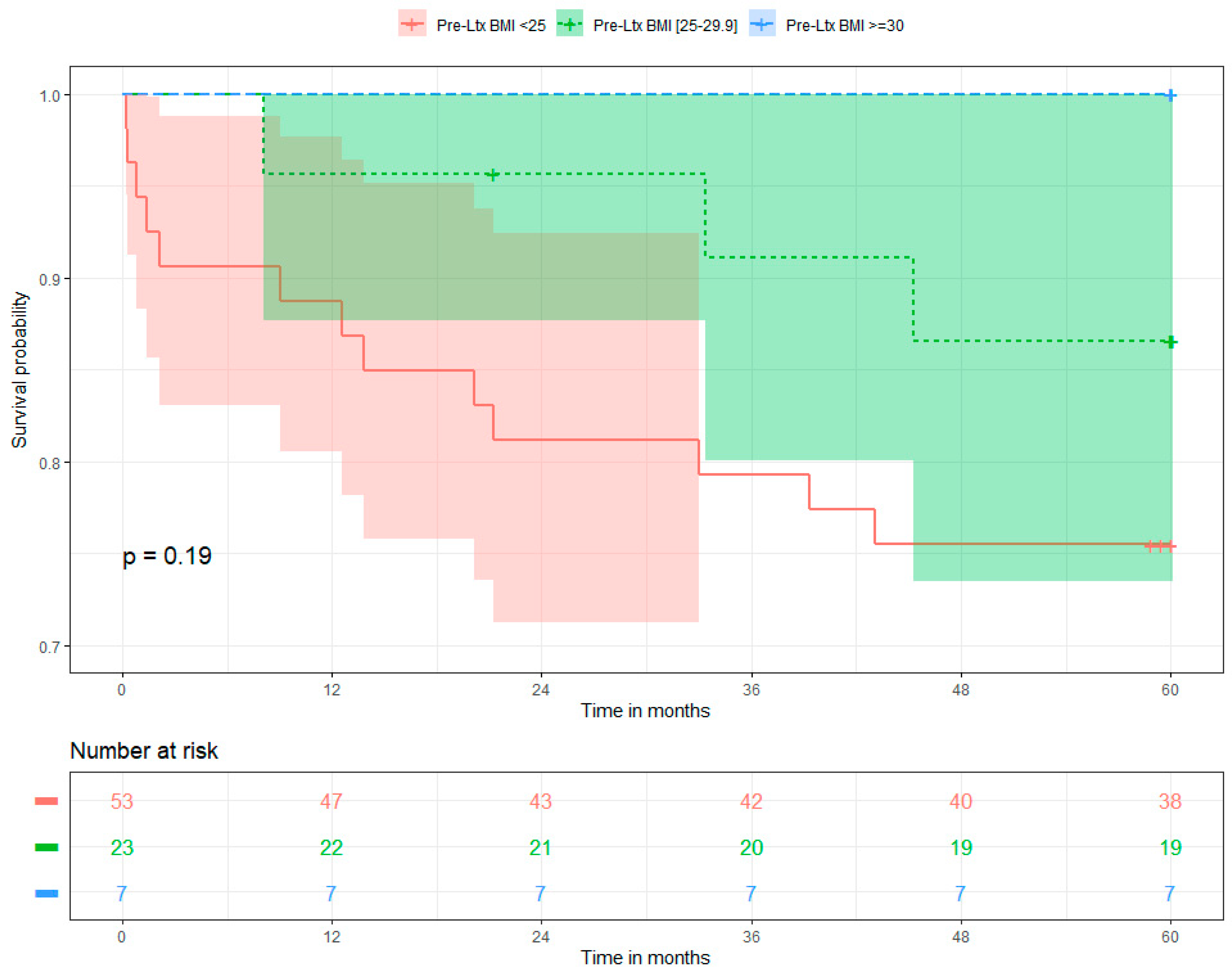
Figure 1. Overall survival of the liver recipients stratified by pre-operative BMI group.
Gastrointestdisord 06 00040 g001
Figure 2. Recurrence-free survival of the liver recipients stratified by pre-operative BMI group.
Gastrointestdisord 06 00040 g002
Figure 3. BMI changes over the course of the 5-year observation stratified by baseline BMI group.
Gastrointestdisord 06 00040 g003
Table 1. Baseline characteristics of the study population.
VariableOverall (N = 83)Pre-Ltx BMI < 25 kg/m2 (n = 53)Pre-Ltx BMI [25–29.9 kg/m2] (n = 23)Pre-Ltx BMI ≥ 30 kg/m2 (n = 7)Statistical Testp-Value
Sociodemographics
Age at transplantation (years)
Median (Q1–Q3)
Range

57.01 (51.27–59.5)
32.85–69.75

56.58 (49.26–59.52)
32.85–69.75

56.42 (52.98–59.22)
45.15–64.5

57.62 (49.66–62.14)
42.1–69.14
Kruskal–Wallis0.9195
Male 84.3% (n = 70)84.9% (n = 45)82.6% (n = 19)85.7% (n = 6)Fisher1
Excessive alcohol consumption12% (n = 10)9.4% (n = 5)17.4% (n = 4)14.3% (n = 1)Fisher0.4828
Indication for liver transplantation
HCV + HCC72.3% (n = 60)75.5% (n = 40)56.5% (n = 13)100% (n = 7)Fisher0.062
HBV + HCC9.6% (n = 8)9.4% (n = 5)13% (n = 3)0% (n = 0)Fisher0.8527
ALD + HCC6% (n = 5)5.7% (n = 3)8.7% (n = 2)0% (n = 0)Fisher0.768
PBC, AIH, PSC + HCC3.6% (n = 3)5.7% (n = 3)0% (n = 0)0% (n = 0)Fisher0.6551
MASH + HCC6% (n = 5)3.8% (n = 2)13% (n = 3)0% (n = 0)Fisher0.3352
Cryptogenic + HCC1.2% (n = 1)0% (n = 0)4.3% (n = 1)0% (n = 0)Fisher0.3614
HCC1.2% (n = 1)0% (n = 0)4.3% (n = 1)0% (n = 0)Fisher0.3614
Baseline features of the host
MELD score at transplantation
Median (Q1–Q3)
Range

11 (9–14.5)
7–35

11 (10–15)
7–35

11 (8–13)
7–20

10 (10–15)
9–23
Kruskal–Wallis0.6258
AFP at transplant [ng/mL]
Median (Q1–Q3)
Range

10.2 (4.6–31.75)
1.4–334

10.2 (4.6–20)
1.9–147.5

9.4 (5.65–59.5)
1.4–334

28.6 (8.85–34.55)
1.5–271
Kruskal–Wallis0.461
Neutrophil/lymphocyte ratio at transplant
Median (Q1–Q3)
Range

2.28 (1.64–3.35)
0.89–11.5

2.29 (1.63–3.2)
0.94–11.5

2.78 (1.86–3.8)
1.04–10.42

1.87 (1.3–2.97)
0.89–5.57
Kruskal–Wallis0.33
AFP model
Median (Q1–Q3)
Range

0 (0–0)
0–6

0 (0–1)
0–6

0 (0–1)
0–3

1 (0–1)
0–6
Kruskal–Wallis0.6306
Creatinine at baseline (mg/dL)
Median (Q1–Q3)
Range

0.9 (0.75–1.1)
0.45–1.8

0.9 (0.8–1.1)
0.52–1.8

0.81 (0.7–1.1)
0.45–1.7

0.84 (0.72–1)
0.5–1
Kruskal–Wallis0.651
Diabetes mellitus at baseline30.1% (n = 25)22.6% (n = 12)47.8% (n = 11)28.6% (n = 2)Fisher0.0922
Pre-operative BMI
Median (Q1–Q3)
Range

23.59 (21.49–26.24)
17.3–32.61

22.08 (20.98–23.37)
17.3–24.98

26.78 (25.88–27.49)
25.06–29.17

30.8 (30.46–31.04)
30.11–32.61
Kruskal–Wallis<0.001
Bridging therapy before Ltx
No treatment
Radiofrequency ablation
Resection
Percutaneous ethanol injection

84.3% (n = 70)
10.8% (n = 9)
3.6% (n = 3)
1.2% (n = 1)

83% (n = 44)
11.3% (n = 6)
5.7% (n = 3)
0% (n = 0)

87% (n = 20)
8.7% (n = 2)
0% (n = 0)
4.3% (n = 1)

85.7% (n = 6)
14.3% (n = 1)
0% (n = 0)
0% (n = 0)
Fisher0.6317
Donor and transplant variables
Donor age (years)
Median (Q1–Q3)
Range

42 (32.5–50.5)
10–65

44 (32–53)
17–65

41 (33–45)
10–59

46 (35–47)
20–51
ANOVA0.7662
Cold ischaemia time (min)
Median (Q1–Q3)
Range

385 (312.5–457.5)
200–730

385 (305–455)
200–730

360 (320–420)
255–720

430 (322.5–493.5)
245–560
Kruskal–Wallis0.8395
Warm ischaemia time (min)
Median (Q1–Q3)
Range

42 (35–47)
28–175

41 (35–46)
28–175

45 (40–49)
30–70

40 (32.5–45)
30–54
Kruskal–Wallis0.2758
Immunosuppression at baseline
Steroids100% (n = 83)100% (n = 53)100% (n = 23)100% (n = 7)Fisher1
Tacrolimus95.2% (n = 79)98.1% (n = 52)87% (n = 20)100% (n = 7)Fisher0.1478
MMF94% (n = 78)96.2% (n = 51)91.3% (n = 21)85.7% (n = 6)Fisher0.3352
Cyclosporine4.8% (n = 4)1.9% (n = 1)13% (n = 3)0% (n = 0)Fisher0.1478
Everolimus2.4% (n = 2)0% (n = 0)4.3% (n = 1)14.3% (n = 1)Fisher0.0535
Tumour characteristics
Diameter of the largest tumour (cm)
Median (Q1–Q3)
Range

2 (1.5–3)
0.5–8

2 (1.5–3)
0.5–7

2.5 (1.75–2.75)
0.7–6

3 (1.9–5.25)
1.5–8
Kruskal–Wallis0.3232
Number of nodules
Median (Q1–Q3)
Range

1 (1–2)
1–4

1 (1–2)
1–4

1 (1–2)
1–3

1 (1–1)
1–2
Kruskal–Wallis0.4546
Fulfilment of Milan criteria83.1% (n = 69)83% (n = 44)87% (n = 20)71.4% (n = 5)Fisher0.6605
Microvascular invasion33.7% (n = 28)30.2% (n = 16)43.5% (n = 10)28.6% (n = 2)Fisher0.5101
Tumour differentiation
G120.5% (n = 17)20.8% (n = 11)26.1% (n = 6)0% (n = 0)Fisher0.7669
G273.5% (n = 61)71.7% (n = 38)69.6% (n = 16)100% (n = 7)
G34.8% (n = 4)5.7% (n = 3)4.3% (n = 1)0% (n = 0)
G41.2% (n = 1)1.9% (n = 1)0% (n = 0)0% (n = 0)
Time of follow-up (months)
Median (Q1–Q3)
Range

60.03 (59.74–60.03)
0.2–60.16

60.03 (58.85–60.03)
0.2–60.07

60.03 (60.03–60.07)
5.72–60.16

60.03 (60.03–60.03)
30.44–60.07
Kruskal–Wallis0.1144
Abbreviations: AIH, autoimmune hepatitis; AFP, alfa-fetoprotein; ALD, alcoholic liver disease; BMI, body mass index; HBV, hepatitis B virus; HCC, hepatocellular carcinoma; HCV, hepatitis C virus; MASH, metabolic dysfunction-associated steatohepatitis; MELD, model for end-stage liver disease; PBC, primary biliary cirrhosis; pre-Ltx, pre-transplant; PSC, primary sclerosing cholangitis.
Table 2. Multivariate logistic regression models for the risk of HCC recurrence in liver recipients.
VariableEstimateORLCIUCIp-Value
Intercept−2.7260.0660.0030.4380.018
Presence of microvascular invasion3.00820.2392.891426.5490.010
AFP level at transplant0.0131.0131.0021.0270.030
Fulfilment of Milan criteria−1.9370.1440.0180.9210.046
Disclaimer/Publisher’s Note: The statements, opinions and data contained in all publications are solely those of the individual author(s) and contributor(s) and not of MDPI and/or the editor(s). MDPI and/or the editor(s) disclaim responsibility for any injury to people or property resulting from any ideas, methods, instructions or products referred to in the content.