1. Introduction
Median arcuate ligament syndrome (MALS), otherwise called celiac artery compression syndrome (CACS), is an uncommon disorder first described in 1965 by Dunbar et al. [
1]. The median arcuate ligament is a muscular arch that connects the diaphragmatic crura to form the anterior margin of the aortic hiatus. In patients with MALS, it causes compression of the celiac artery on expiration and entrapment of the celiac plexus (a dense network of ganglia that lies over the anterolateral aorta at the T12 vertebral level) [
2]. The pathophysiology of MALS is related to both ischemic and neuropathic mechanisms due to the anatomic relationship of the MAL to the origin of the arterial celiac plexus and ganglion [
3] (
Figure 1).
Theories have been proposed to explain the altered anatomy of the median arcuate ligament, including a congenital anomaly [
2]. A traumatic origin has also been hypothesized, causing a change of the diaphragm anatomy, resulting in an alteration of the diaphragm’s aortic hiatus [
5,
6,
7]. The manifestations of symptoms in adults could be explained by a combination of a congenitally early take-off origin of the celiac artery and/or an abnormally thickened MAL with or without arteriosclerosis that may predispose to the “arterial narrowing” on expiration.
The typical symptom in MALS is chronic epigastric pain aggravated by meals. Other symptoms include nausea, vomiting, and accompanying weight loss. In about 35% of the patients, an abdominal bruit can be auscultated [
8]. MALS is a diagnosis of exclusion that should be considered in patients with severe upper abdominal pain that is out of proportion and does not correlate with the objective findings. The cardinal feature used in the diagnosis of MALS, which is demonstrated by imaging studies, is the compression of the celiac artery during expiration and reduction in this compression during inspiration, demonstrated on ultrasound or conventional angiographic imaging. The detection of an abnormal gastric electric rhythm in patients with MALS is suggestive of neurogenic pathogenesis [
9]. The definitive treatment is the surgical release of the median arcuate ligament to achieve decompression of the celiac plexus. This article describes our experience with this entity, focusing on symptom presentation, diagnostic challenges, and management, including long-term follow-up.
2. Results
Four patients with MALS were identified from the experience of a gastroenterologist (RMC) at an academic referral center directing a center for GI motility and functional bowel disorders from January 2017–June 2020. The demographics of all patients included in the study are summarized in
Table 1. All four patients were females with a median age of 34.5 (IQR 28.5, 42.3) years. The initial presenting complaint in all four patients was chronic abdominal pain of unknown etiology despite an extensive evaluation from the previous providers. All four patients were found to have delayed gastric emptying that did not improve with the medical treatment they had received, and hence they were referred to our center. The median duration of symptoms before presentation was 3 (IQR 2, 4.8) years. All the patients had increased peak systolic velocity (PSV) on the celiac artery on Doppler ultrasound during expiration and more than 50% narrowing of the celiac artery on the aortic aortogram during expiration. Most of the patients (75%) underwent a celiac plexus block using xylocaine with a temporary improvement of symptoms. Eventually, they were referred to surgical evaluation, and all the patients underwent release of the median arcuate ligament (MAL). All patients recovered well from the surgery with the resolution of the abdominal pain during long-term follow-up along with documentation of improvement in the abdominal aortogram and duplex ultrasound (
Table 2).
3. Discussion
In these four cases, the surgical intervention led to the resolution of chronic abdominal pain. All our patients were females, mostly in the 20–40 years age group, and had characteristic chronic abdominal pain exacerbated by eating [
1].
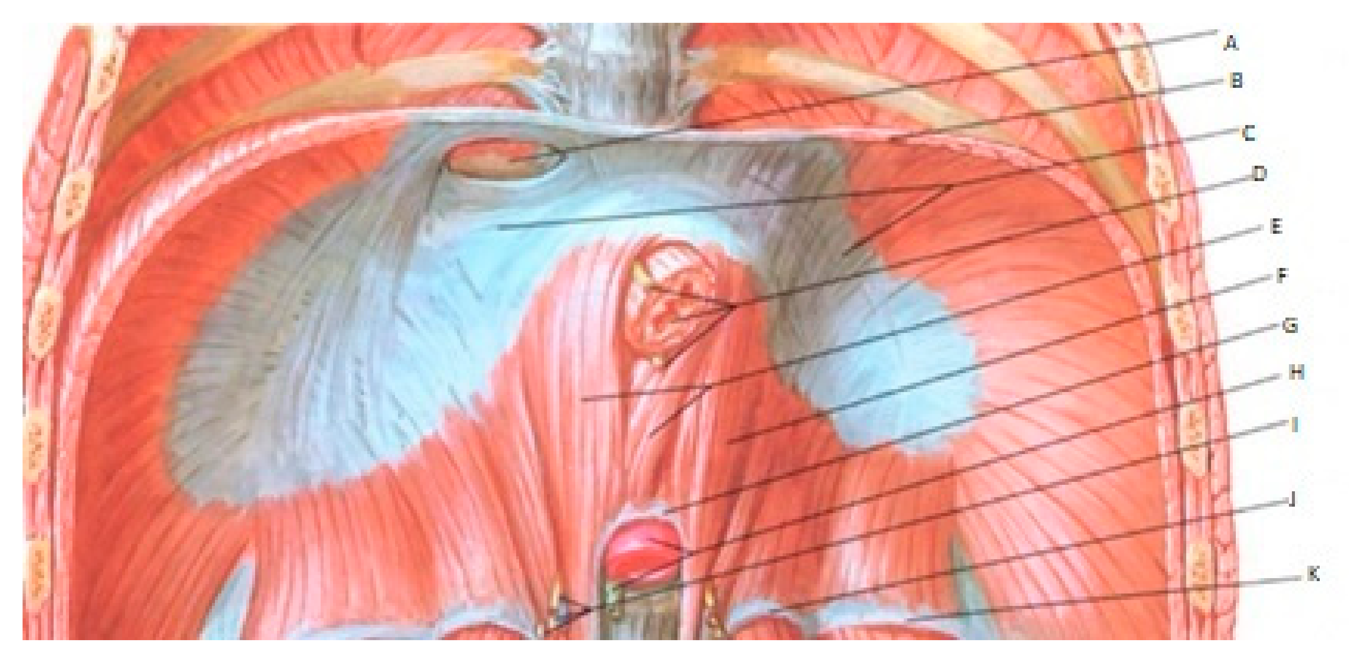
3.1. Clinical Manifestation
Most patients report epigastric and right upper quadrant abdominal pain, aggravated after meals. The fear of provoking pain and delayed gastric emptying can result in weight loss. The abdominal pain can lead to intermittent use of narcotics for pain control plus emergency room visits. In our study, all patients presented with abdominal pain, and 75% had nausea/vomiting. Only 50% of the patients had significant weight loss. Weight loss is typically considered as a classic “red flag” in the Rome criteria for suspecting an organic cause [
10]. Hence, it is essential to consider organic causes, where weight loss often, but not invariably, occurs in addition to post-prandial abdominal pain [
10]. Previous studies have reported abdominal bruit in the upper abdomen in up to 35% of patients [
3]. However, we did not identify this finding in our series.
3.2. Delayed Gastric Emptying
Nausea and vomiting can be prominent to the degree that gastric emptying studies indicate a diagnosis of “idiopathic gastroparesis”. In our third patient, the gastric emptying study was abnormal at presentation with 31% meal retention at 4 h, normal being less than 10% retention by scintigraphic solid meal methodology. Gastric emptying normalized after the surgical intervention. The gastroparesis component is attributed to the abnormal gastric electrical rhythm (e.g., tachygastria) from celiac ganglion compression [
9]. These findings have suggested the role of neurogenic pathogenesis resulting from the inhibition of the gastric neuromuscular function [
9].
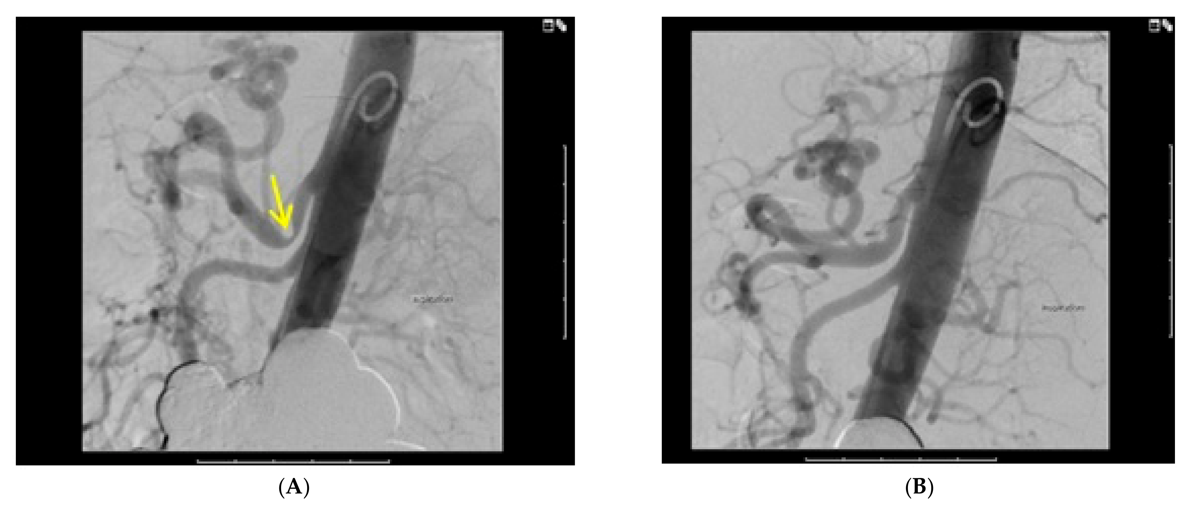
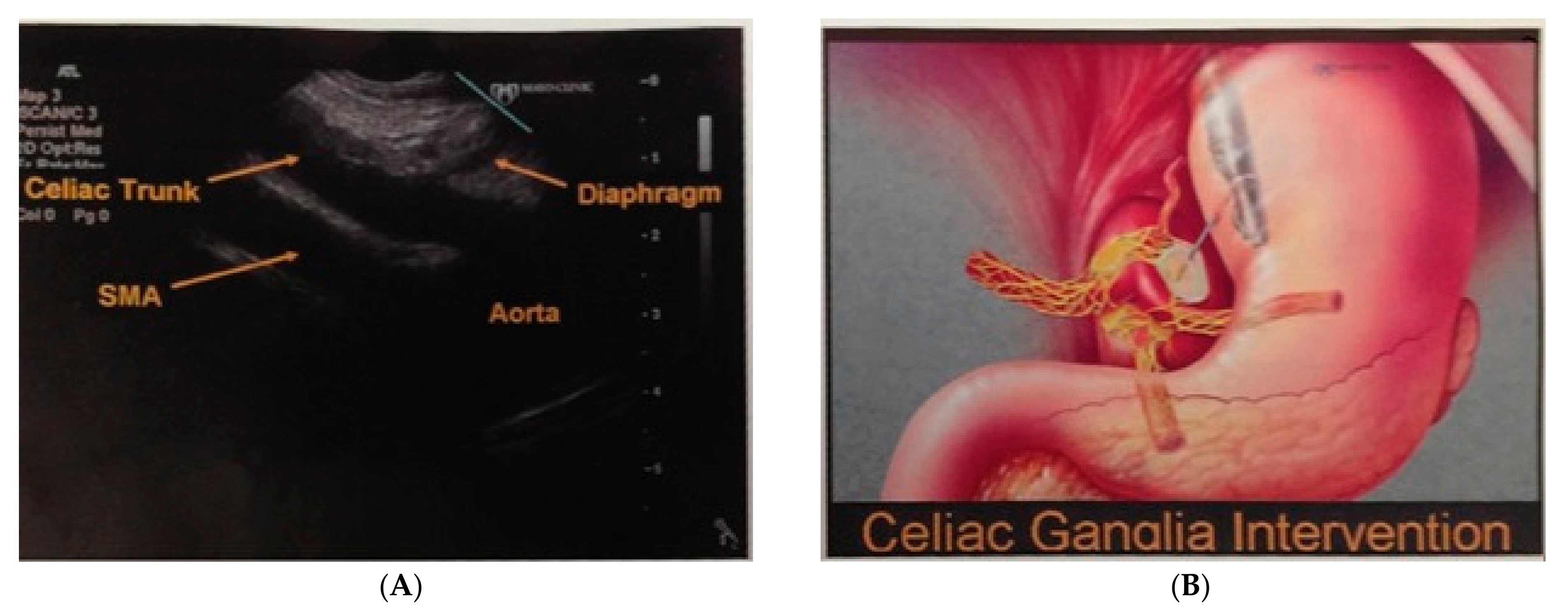
3.3. Diagnostic Studies
Most of the patients diagnosed with MALS have previously undergone extensive workups, including abdominal computed tomography (CT) scans, abdominal magnetic resonance imaging (MRI), aortic angiography, and some even undergo surgical procedures including cholecystectomies, appendectomies, or laparoscopic or gynecological procedures in an attempt to make the diagnosis or at least try to relieve the symptoms [
11]. These studies are usually repeated, and the average length of time prior to making a diagnosis can be a number of years. In our study, the median length of time that the patients were symptomatic before the diagnosis was 3 (Interquartile range 2–4.8) years.
MALS is a diagnosis that should be considered in patients with chronic post-prandial abdominal pain in the absence of any objective findings, as well as lack of response to all standard empiric therapies for acid-related conditions, irritable bowel syndrome, and gastroparesis. The imaging modalities that are utilized to diagnose MALS include duplex ultrasound, CT angiography, magnetic resonance angiography, and conventional angiography. The clinicians should be aware that these patients may be wrongly diagnosed with functional dyspepsia or irritable bowel syndrome because of the negative workup from a “standard” testing. This could lead to delayed diagnosis due to a clinical belief that the patient has dyspepsia.
The major findings specific for the diagnosis are compression of the celiac artery with respiratory variations, post-compression dilation, and elevated velocities of blood flow [
3,
12]. The compressive effect of the MAL on the celiac artery is relieved during inspiration since the aorta moves anteriorly and inferiorly. Downward diaphragm movement during inspiration relaxes the crura and the compression on the celiac artery. The gold standard for diagnosing MALS is lateral view aortic angiography, which shows more than 50% narrowing in the origin of the celiac artery, with or without distal dilation, with improvement during inspiration (
Figure 2). These findings were well demonstrated in the second patient, in whom the duplex ultrasound showed a PSV of 213 cm/s on inspiration and 323 cm/s on expiration. Furthermore, the abdominal aortogram was diagnostic for MALS with >60% narrowing of the celiac artery on expiration (
Figure 2).
3.4. Management
Surgery is the definitive treatment to divide the median arcuate ligament to decompress the celiac artery and celiac plexus [
3,
13]. Laparoscopic and robotic-assisted laparoscopic approaches have been successfully utilized [
14,
15,
16]. Intraoperative pre- and post-decompression flow velocity studies are performed routinely to assess success of the procedure. Post-operative angiography may be normal or could show some continuing minimal compression of the celiac artery (<30%) during expiration due to the chronicity of the condition inducing some arterial “stiffness” from intimal hyperplasia. The first patient who underwent surgical release of the median arcuate ligament (MAL) demonstrated PSV on expiration reduced from 362 cm/s before surgery to 130 cm/s after surgery. The patient had a post-operative abdominal aortogram that was normal, and over a 6-year follow-up, she had 100% relief of abdominal pain, with no more narcotic requirement, stable weight, and improved quality of life.
Endoscopic ultrasound (EUS)-guided celiac block is used to predict response to surgical decompression. The EUS allows injecting the celiac ganglion with xylocaine and monitor for symptom improvement (
Figure 3). Improvement of symptoms increases confidence in the diagnosis and hence the decision for surgery. In our study, the fourth patient underwent EUS-guided celiac plexus block, which resulted in amelioration of the pain for 3 to 4 weeks. Another role for EUS-guided celiac block is to enhance the results of post-surgical decompression further in patients who have some residual abdominal pain or some recurrence during long-term follow-up.
Debate in the radiology literature occurs regarding the roles of celiac artery angioplasty and even vascular reconstruction. These vascular approaches do not address the role of the celiac plexus [
17]. Blood flow to the stomach can occur through extensive collateral networks and multiple branches from the superior mesenteric and inferior mesenteric arteries that supplement the contribution of the celiac artery. Therefore, the therapeutic focus should be on the decompression of celiac ganglion.
4. Conclusions
MALS is a rare disorder that is often misdiagnosed and not considered in the differential diagnosis, resulting in the exhaustive negative workup, after which suspicion may arise for this entity. Diagnosis relies on the utilization of imaging modalities like angiogram and color Doppler ultrasound with the respiratory variation of celiac blood flow. In addition, there must be a realization that the symptoms are not explained by ischemia. The surgical approach of minimally invasive laparoscopic and robotic techniques focuses on ligating the ligament, and often times results in partial surgical disruption of the celiac plexus, thus overcoming the compression of the celiac ganglion from which the symptoms are emanating. Finally, it is important for the long-term follow-up of these patients to make sure that they are symptom-free, thus confirming the sustainability of the surgery and the accuracy of the diagnosis.
5. Materials and Methods
We performed an observational retrospective study with chart review identifying four patients who were referred to the academic GI motility center directed by one of the authors (Dr. Richard McCallum), who saw these patients and provided ongoing care. All the patients presented with chronic abdominal pain with unknown etiology and were later diagnosed with MALS. All these patients already underwent extensive evaluation of their symptoms with no definitive diagnosis and persistence of symptoms. They were comprehensively evaluated to exclude other explanations, and the diagnosis of MALS was confirmed initially with Doppler ultrasound, and definitively with abdominal aortogram. Most of the patients were treated with celiac plexus block and eventually referred to surgical evaluation. All the patients underwent surgical release of the median arcuate ligament (MAL) with an improvement of symptoms, along with improvement in Doppler ultrasound PSV and abdominal aortograms. Baseline clinical characteristics, diagnostic studies, management, and outcomes were reviewed and recorded.
Author Contributions
Conceptualization, I.A.B., B.R.D., J.R.D., M.G., and R.W.M.; methodology, M.G.; resources, I.A.B., M.G.; data curation, I.A.B., B.R.D., J.R.D.; writing—original draft preparation, I.A.B.; writing—review and editing, I.A.B., M.G., B.R.D., J.R.D. and R.W.M.; supervision, R.W.M. All authors have read and agreed to the published version of the manuscript.
Funding
This research received no external funding.
Institutional Review Board Statement
Ethical review and approval were waived for this study by the Texas Tech University Health Sciences Center El Paso Institutional Review Board (IRB), since this is a case series.
Informed Consent Statement
Informed consent was obtained from all subjects involved in the study.
Data Availability Statement
The data that support the findings of this study are available from the corresponding author upon reasonable request.
Conflicts of Interest
The authors declare no conflict of interest.
References
- Dunbar, J.D.; Molnar, W.; Beman, F.F.; Marable, S.A. Compression of the celiac trunk and abdominal angina: Preliminary report of 15 cases. Am. J. Roentgenol. 1965, 95, 731–744. [Google Scholar] [CrossRef] [PubMed]
- Rathmell, J.P.; Gallant, J.M.; Brown, D.L. Computed tomography and the anatomy of celiac plexus block. Reg. Anesth. Pain Med. 2000, 25, 411–416. [Google Scholar] [CrossRef] [PubMed]
- Jimenez, J.C.; Harlander-Locke, M.; Dutson, E.P. Open and laparoscopic treatment of median arcuate ligament syndrome. J. Vasc. Surg. 2012, 56, 869–873. [Google Scholar] [CrossRef] [PubMed] [Green Version]
- McCallum, R.W.; Al-Bayati, I. Median Arcuate Ligament Syndrome. Pract. Gastroenterol. 2015, 21. [Google Scholar] [CrossRef]
- Bech, F.; Loesberg, A.; Rosenblum, J.; Glagov, S.; Gewertz, B.L. Median arcuate ligament compression syndrome in monozygotic twins. J. Vasc. Surg. 1994, 19, 934–938. [Google Scholar] [CrossRef] [Green Version]
- Okten, R.S.; Kucukay, F.; Tola, M.; Bostanci, B.; Cumhur, T. Is celiac artery compression syndrome genetically inherited?: A case series from a family and review of the literature. Eur. J. Radiol. 2012, 81, 1089–1093. [Google Scholar] [CrossRef] [PubMed]
- Schreiber, J.P.; Angle, J.F.; Matsumoto, A.H.; Young, J.S.; Hagspiel, K.D.; Spinosa, D.J. Acute visceral ischemia occurring subsequent to blunt abdominal trauma: Potential culpability of median arcuate ligament compression. J. Trauma Acute Care Surg. 1998, 45, 404–406. [Google Scholar] [CrossRef] [PubMed]
- Kim, E.N.; Lamb, K.; Relles, D.; Moudgill, N.; DiMuzio, P.J.; Eisenberg, J.A. Median Arcuate Ligament Syndrome—Review of This Rare Disease. JAMA Surg. 2016, 151, 471–477. [Google Scholar] [CrossRef] [PubMed]
- Balaban, D.H.; Chen, J.; Lin, Z.; Tribble, C.G.; McCallum, R.W. Median arcuate ligament syndrome: A possible cause of idiopathic gastroparesis. Am. J. Gastroenterol. 1997, 92, 519–523. [Google Scholar] [PubMed]
- Tack, J.; Talley, N.J.; Camilleri, M.; Holtmann, G.; Hu, P.; Malagelada, J.-R.; Stanghellini, V. Functional Gastroduodenal Disorders. Gastroenterology 2006, 130, 1466–1479. [Google Scholar] [CrossRef] [PubMed] [Green Version]
- Watson, W.; Sadikali, F. Celiac axis compression: Experience with 20 patients and a critical appraisal of the syndrome. Ann. Intern. Med. 1977, 86, 278–284. [Google Scholar] [CrossRef] [PubMed]
- Aschenbach, R.; Basche, S.; Vogl, T.J. Compression of the celiac trunk caused by median arcuate ligament in children and adolescent subjects: Evaluation with contrast-enhanced MR angiography and comparison with Doppler US evaluation. J. Vasc. Interv. Radiol. 2011, 22, 556–561. [Google Scholar] [CrossRef] [PubMed]
- Delis, K.T.; Gloviczki, P.; Altuwaijri, M.; McKusick, M.A. Median arcuate ligament syndrome: Open celiac artery reconstruction and ligament division after endovascular failure. J. Vasc. Surg. 2007, 46, 799–802. [Google Scholar] [CrossRef] [PubMed] [Green Version]
- Carbonell, A.; Kercher, K.; Heniford, B.; Matthews, B. Laparoscopic management of median arcuate ligament syndrome. Surg. Endosc. 2005, 19, 729. [Google Scholar] [CrossRef] [PubMed]
- Jaik, N.P.; Stawicki, S.P.; Weger, N.S.; Lukaszczyk, J.J. Celiac artery compression syndrome: Successful utilization of robotic-assisted laparoscopic approach. J. Gastrointest. Liver Dis. 2007, 16, 93. [Google Scholar]
- Roddy, S.P. Laparoscopic versus open celiac ganglionectomy in patients with median arcuate ligament syndrome. J. Vasc. Surg. 2010, 52, 1419. [Google Scholar] [CrossRef] [Green Version]
- Kohn, G.P.; Bitar, R.S.; Farber, M.A.; Marston, W.A.; Overby, D.W.; Farrell, T.M. Treatment options and outcomes for celiac artery compression syndrome. Surg. Innov. 2011, 18, 338–343. [Google Scholar] [CrossRef] [PubMed]
| Publisher’s Note: MDPI stays neutral with regard to jurisdictional claims in published maps and institutional affiliations. |
© 2021 by the authors. Licensee MDPI, Basel, Switzerland. This article is an open access article distributed under the terms and conditions of the Creative Commons Attribution (CC BY) license (http://creativecommons.org/licenses/by/4.0/).