Perspective

Comparison of the Passive Behavior of NiTi and CoNiCrMo in Simulated Physiological Solutions

Materials and Corrosion Engineering, Exponent, Menlo Park, CA 94025, USA
Corros. Mater. Degrad. 2025, 6(1), 4; https://doi.org/10.3390/cmd6010004
Submission received: 24 December 2024 / Revised: 10 January 2025 / Accepted: 24 January 2025 / Published: 30 January 2025

Abstract

:
Biomedical alloys in general, except for the biodegradable type, exhibit passive behavior in neutral chloride solutions. Two commonly used biomedical alloys are nitinol (NiTi) and Co-35Ni-20Cr-10Mo (CoNiCrMo). In this work, the passive behavior of electropolished NiTi and CoNiCrMo in a simulated physiological solution (phosphate-buffered saline) was compared using data largely obtained from our previous studies involving potentiodynamic polarization and electrochemical impedance spectroscopy (EIS). The potentiodynamic results showed a marked difference in passive behavior between the alloys, with NiTi remaining completely passive up to the oxidation of water and CoNiCrMo, in contrast, undergoing solid-state oxidation and then transpassive dissolution. Both alloys exhibited Tafel-type behavior over the initial part of the passive range. A small but distinct difference in the apparent Tafel slopes was found between the two alloys and can be attributed to the difference in their predominant oxide; that is, TiO2 versus Cr2O3. The EIS results also showed marked differences between the alloys in terms of the oxide thickness and resistivity. The thickness was greater for NiTi—consistent with surface analytical results—and differed in potential dependence between the two alloys in the passive region. The oxide resistivity, conversely, was substantially lower for NiTi and showed a similar potential dependence for the two alloys.

1. Introduction

Biomedical alloys used for implantable medical devices are exposed to physiological liquids that all contain chloride. These alloys, except for the biodegradable type, exhibit passive behavior in neutral and near-neutral chloride solutions such as simulated physiological solutions; for instance, Hanks solution and phosphate-buffered saline (PBS). Some type of surface treatment, such as electropolishing, is commonly used to enhance the passive behavior of the alloys [1,2,3].
Most of the biomedical alloys fall into two groups: Ti and its alloys, and CoCr alloys. Two commonly used alloys in these groups are nitinol (NiTi) and Co-35Ni-20Cr-10Mo (CoNiCrMo). Both alloys are used in certain applications where they are exposed to blood. Nitinol is a nearly equiatomic alloy of Ni and Ti but can be viewed as a Ti alloy because its surface oxide is predominantly TiO2 [4]. Of the various CoCr alloys used for medical implants, interest has largely focused on Co-28Cr-6Mo (CoCrMo) and Co-35Ni-20Cr-10Mo (CoNiCrMo). Although their composition differs, the passive film is predominantly Cr2O3 in both cases, and the two alloys exhibit the same essential features in their electrochemical behavior in simulated physiological solutions [5].
In this work, the passive behavior of electropolished (EP) NiTi and CoNiCrMo in PBS was compared using data largely obtained from our previous studies involving potentiodynamic polarization and electrochemical impedance spectroscopy (EIS) [1,6,7]. Each alloy was investigated separately in those studies, and a comparison of their passive behavior has not been made until now. Both alloys have thin passive films, but the composition and other characteristics of these films are quite different. This work was focused on contrasting the passive behavior and characteristics of the passive film on the two alloys, thereby providing further insight into the nature of the film on biomedical alloys under physiological conditions.
Potentials in corrosion studies of biomedical alloys are generally reported with respect to a saturated calomel electrode (SCE) or silver/silver chloride electrode. Unless otherwise stated, the potentials cited in this work are referenced to an SCE.

2. Surface Analysis

2.1. Oxide Film Composition

Studies using X-ray photoelectron spectroscopy (XPS) have indicated that the passive film on EP NiTi is predominantly TiO2 with a small amount of nickel oxide [2,4]. Electropolishing was shown to increase the TiO2 content and decrease the Ni concentration in the oxide, resulting in a large increase in the Ti:Ni ratio for the oxide [3,4]. The composition of the oxide was found to be unaffected by potential during a potentiodynamic scan [2].
The passive film on CoNiCrMo tends to be more complex, depending on the potential. XPS showed that the film on both CoNiCrMo and CoCrMo in simulated physiological solutions (buffered NaCl and Hanks solution) is predominantly Cr2O3 at lower potentials with only small amounts of other metal oxides [5,8]. However, the fraction of oxidized Co and Ni increases at higher potentials. The oxide formed on CoNiCrMo in Hanks solution was shown to contain (i) a higher cationic fraction of Co and Ni at 0.6 V than at 0.1 V, and (ii) Co(III) at 0.6 V but not at 0.1 V (data were not obtained between these potentials). Moreover, Cr(VI) was shown to be present in the oxide on CoCrMo and CoNiCrMo at 0.5 V and higher [5,8]. X-ray near-edge spectroscopy indicated that it is trapped in the outermost part of the Cr2O3 film prior to transpassive dissolution [9].

2.2. Oxide Film Thickness
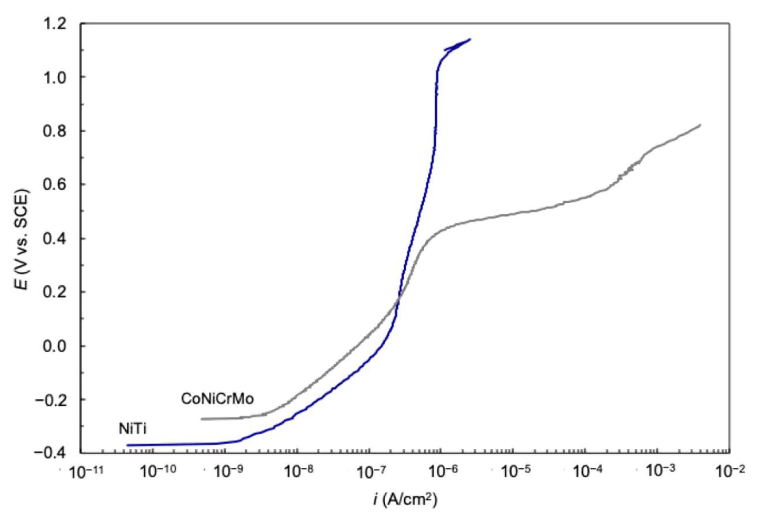
The passive film on NiTi and CoCr alloys is thin. Work involving Auger electron spectroscopy (AES) found the oxide thickness on EP NiTi to be 3–4 nm [2,3,4,10]. In the case of EP CoNiCrMo, the most applicable values from surface analysis appear to be 1.5 ± 0.5 nm for mechanically polished (MP) CoNiCrMo in an acetate buffer solution [11] and 0.8–1.0 nm estimated from AES together with film growth for MP CoCrMo in PBS [12].

3. Passive Behavior

Potentiodynamic polarization curves showing the dependence of current density (i) on potential (E) for EP NiTi and CoNiCrMo in deaerated PBS at 37 °C are compared in Figure 1. The two alloys show a marked difference in passive behavior. NiTi remained completely passive up to the oxidation of water with no evidence of breakdown (localized or general) of the oxide film. Although NiTi can be susceptible to pitting corrosion, depending on various factors such as potential and test solution (Figure 2) [13], electropolishing has been found to greatly enhance its resistance to pitting corrosion [1,2,3]. This enhancement results primarily from the removal of surface inclusions that have been shown to act as pit initiation sites [14]. CoNiCrMo can also contain inclusions, but no evidence of pitting has been found either from cyclic polarization tests up to 1 V or from scanning electron microscopy after the tests [7,15].
CoNiCrMo, in contrast to NiTi, exhibited an increase in current density at about 0.4 V that was identified with two solid-state oxidation reactions: Cr(III) to Cr(VI) and Co(II) to Co(III) [7]. The second increase in current density for CoNiCrMo at about 0.7 V is associated with transpassive dissolution [7]. Metal ion release has been found to increase in this region for CoCrMo and CoNiCrMo [7,8]. Figure 3 shows the concentration of dissolved Ni and Cr at selected potentials for CoNiCrMo in relation to the current density in the polarization curve [7]. The metal ion concentrations were determined by using inductively coupled plasma-mass spectrometry to analyze samples of the solution obtained after 1 h at each potential in separate potentiostatic tests. The Ni and Cr concentrations were low at 0.5 V, even though the current density had already increased by over an order of magnitude. However, they exhibited a progressively sharp increase at 0.7 V and 0.9 V coincident with the second increase in current density, reflecting the occurrence of transpassive dissolution.
Both NiTi and CoNiCrMo exhibited a Tafel-type current-potential dependence over the initial part of the passive range in PBS, as found in general for biomedical alloys in buffered physiological solutions [16]. This dependence is represented by Equation (1) [16]:
E = b a log i log i c o r r + E c o r r
where Ecorr is the corrosion potential, icorr is the corrosion current density, and ba′ is the anodic slope. Equation (1) is similar in form to the Tafel equation for corrosion processes under charge-transfer control, so ba′ can be regarded as an apparent Tafel slope.
Values of ba′ and icorr obtained from the polarization curves are given in Table 1. The ba′ values showed a small difference between the alloys, but they were consistent with those for Ti alloys (0.15–0.16 V) and CoCr alloys (0.17–0.19 V) [16]. For comparison, 316 L stainless steel exhibits somewhat higher values of 0.24–0.27 V.
NiTi and CoNiCrMo also showed a small difference in icorr. In the case of NiTi, the value compares well with values of 9 ± 5 and 10 ± 4 nA/cm2 reported for EP discs and wire, respectively, in deaerated Hanks solution [17,18]. Data do not appear to be available elsewhere for EP CoNiCrMo (or CoCrMo) in a deaerated solution to allow a similar comparison.
The Tafel-type dependence observed for the biomedical alloys in buffered physiological solutions is in sharp contrast to the more limited potential dependence typically observed for the alloys in unbuffered chloride solutions. This dependence may be associated with the buffering capability of these solutions or the presence of phosphate. Phosphate is known to adsorb on the surface of NiTi [3,19] and CoCr alloys [12,20], and it appears to hinder the normal aging of the passive film in the case of TiO2 [21]. Phosphate adsorption probably has a similar effect on other oxides such as Cr2O3, and would be expected to restrict film growth during a potentiodynamic scan. The absence of phosphate in an unbuffered solution could well enable sufficient growth of the oxide film such that the current becomes limited at lower potentials in the passive region.

4. Model for Film Growth and Dissolution

NiTi and CoNiCrMo appear to have the same corrosion mechanism, based on the fact that their ba′ values are close. In both cases, the anodic reaction is considered to be controlled by migration through the oxide film with limited growth, because (1) the passive film is thin (~1–4 nm), (2) migration of charged species through thin films is considered generally to be rate-controlling [22,23], and (3) film growth is likely impeded in buffered solutions.
The Tafel-type dependence and associated ba′ values were analyzed using the Generalized Growth Model (GGM) developed by Marcus and coworkers for passive film growth and dissolution [22,24]. The GGM gave rise to equations for the film growth rate and dissolution rate, based on the assumption that transport of charged species through the film is the rate-controlling step under most conditions. The dissolution current density (idiss) for thin films is expressed by [16]
i d i s s = R e x p n e V α k T
where R′ is a multicomponent parameter containing constants such as Boltzmann’s constant (k) and Faraday as well as variables such as pH and activation energy of the dissolution reaction, T is the absolute temperature, e is the elementary charge, ∆V is the variation in the potential drop (V) across the metal/oxide/solution, n relates to the metal oxide (MOn/2) undergoing dissolution, and α is the fraction of ∆V that is imparted to the film/solution potential drop. The parameter of particular interest in this work is α, since (1 − α) is the fraction of ∆V imparted to the potential drop in the film and so can be expected to differ between alloys with different oxides.
The measured anodic current density (ia) is the sum of idiss and the film growth current density (if). Since film growth appears to be impeded by the adsorption of phosphate, if was assumed to be small enough that ia can be approximated to idiss. As discussed previously, ∆V was taken to be EEcorr, and Equation (2) can then be rearranged to yield in logarithmic form [16]:
E E c o r r = 2.303 k T n e α ( log i d i s s log i c o r r )
Equation (3), as with Equation (1), is similar in form to the Tafel equation for corrosion processes under charge-transfer control. In this case, the apparent Tafel slope is given by ba′ = 2.303kT/neα.
For both NiTi and CoNiCrMo, ia could be equated to idiss as the rate of film growth can be neglected [16]. Dissolution was considered to involve only Ni, Co, and Mo ions, where appropriate. Ti and Cr ions were viewed as making negligible contributions to dissolution and being involved only in oxide formation. Studies have shown that Ni is released without any detectable Ti from NiTi in a buffered (Hanks-type) solution [25], and that Co and Mo are released without any detectable Cr from CoCrMo in PBS [26].
The behavior predicted by the GGM through Equation (3) is consistent with the log iE dependence exhibited by NiTi and CoNiCrMo. The value of α could therefore be obtained from ba′ to determine the fraction (1 − α) of ∆V applied across the film. Based on the values of ba′ in Table 1, α was calculated to be 0.19 for NiTi and 0.16 for CoNiCrMo at 37 °C, indicating that the majority of the potential drop (81-84%) resulting from ∆V occurs across the oxide in both cases. Although the values are similar for the two alloys, some difference is to be expected in α (and therefore ba′) because of the difference in the predominant oxide on each alloy.

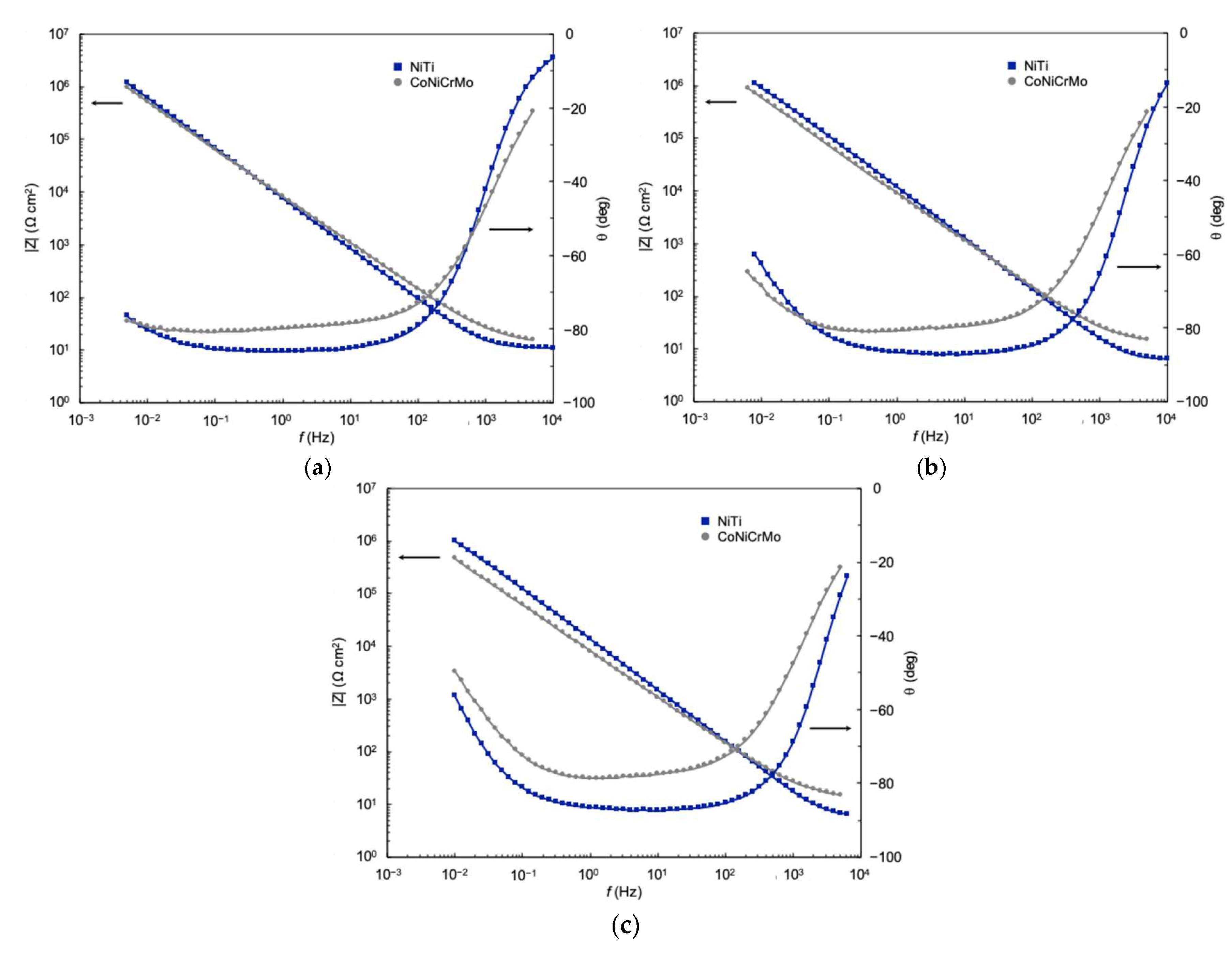
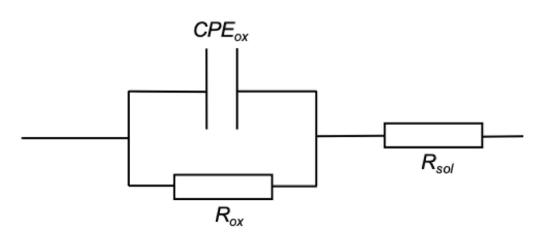
5. EIS Characterization

Impedance spectra for EP NiTi and CoNiCrMo in deaerated PBS at 37 °C are compared at different potentials in Figure 4. The comparison was limited to potentials up to 0.2 V because of the changes in the passive film on CoNiCrMo at higher potentials. Both alloys exhibit near-capacitive behavior over a wide frequency range, irrespective of the potential.
The spectra reflect an equivalent circuit with a single time constant and indicate that the impedance of NiTi and CoNiCrMo under passive conditions can be adequately represented by a simple equivalent circuit involving a parallel combination of the resistance of the oxide film (Rox), and a constant phase element (CPEox) associated with the oxide film (Figure 5) [6,7,27].
A constant phase element was used in place of a capacitance (Cox) to account for the nonideal capacitive behavior of the oxide. The impedance associated with a CPE is given by Equation (4):
Z C P E = 1 Y 0 ( j ω ) a
where Y0 is the constant-phase-element parameter, j = √ − 1, ω is the angular frequency, and a is equal to 1 for an ideal capacitor. Values of Rox, Y0, and a were obtained by using a curve fitting procedure. Figure 6 shows the close fit between the calculated and experimental data that was typical for NiTi and CoNiCrMo in PBS. The value of a was found to differ slightly between the two alloys—0.97 for NiTi [6] and 0.88–0.91 for CoNiCrMo [7]—but was close enough to 1 in both cases for Cox to be taken as the value of Y0.

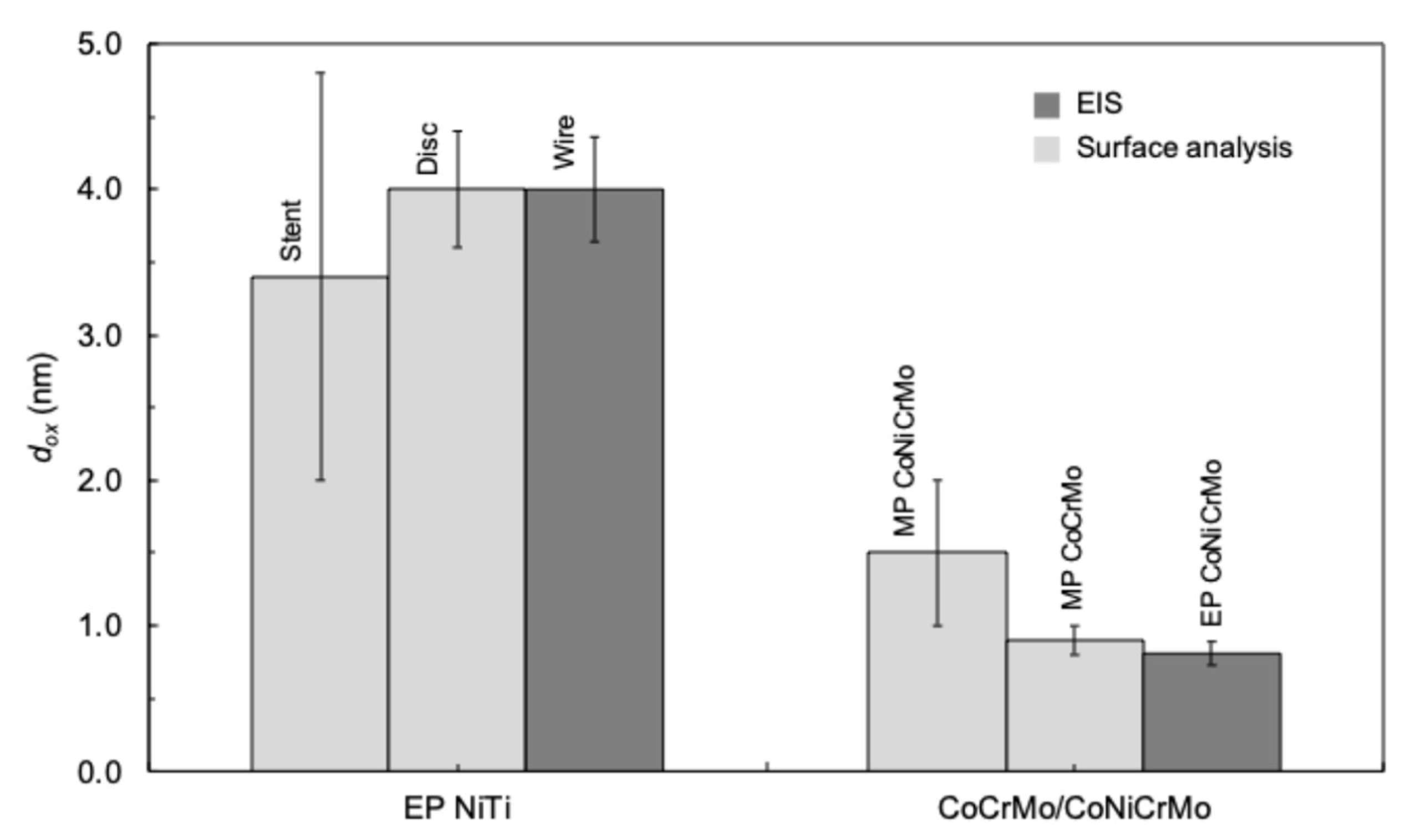
5.1. Oxide Thickness

The oxide thickness (dox) was calculated from Cox (normalized to area) using Equation (5):
d o x = ε ε 0 C o x
where ε is the dielectric constant of the oxide and ε0 is the permittivity of free space. The passive film formed on NiTi and Cr-containing alloys exhibits semiconducting properties [28,29], and the capacitance of a semiconducting oxide is typically associated with the space charge layer in the oxide. Equation (5), however, can be used for semiconducting oxide films if they are thin [7]. Surface analytical studies, as discussed above, have shown that the oxide on NiTi and CoCr alloys is in fact thin (~1–4 nm). Accordingly, dox can be calculated from Cox for these alloys.
Values of dox obtained from the EIS data at Ecorr are given in Table 2. The value for NiTi is substantially greater than that for CoNiCrMo, which is consistent with values obtained through surface analysis, as shown in Figure 7.
The EIS values for both alloys are shown to compare well with the surface analytical results. The NiTi match with wire is notable for both the disc [3,10] and stent [2] form of the alloy. In the case of CoNiCrMo, the most applicable values from surface analyses were for MP CoNiCrMo in an acetate buffer (phosphate-free) solution and MP CoCrMo in PBS, as noted above. Even so, the EIS value for EP CoNiCrMo compares particularly well with the AES-estimated thickness of 0.8–1.0 nm for MP CoCrMo in PBS at Ecorr [12].
The effect of potential on dox for NiTi and CoNiCrMo is compared in Figure 8. In the case of CoNiCrMo, dox showed little change over the potential range of interest [5], whereas it was found to increase linearly for NiTi [6]. An EIS study of Ti in a simulated physiological solution (Ringer’s solution with phosphate) showed a small decrease in Cox over the same potential range, indicating an increase in dox corresponding to that for NiTi [30]. In addition, the linear dependence observed for NiTi has also been reported for Ti in acidified 0.6 M NaCl [31].

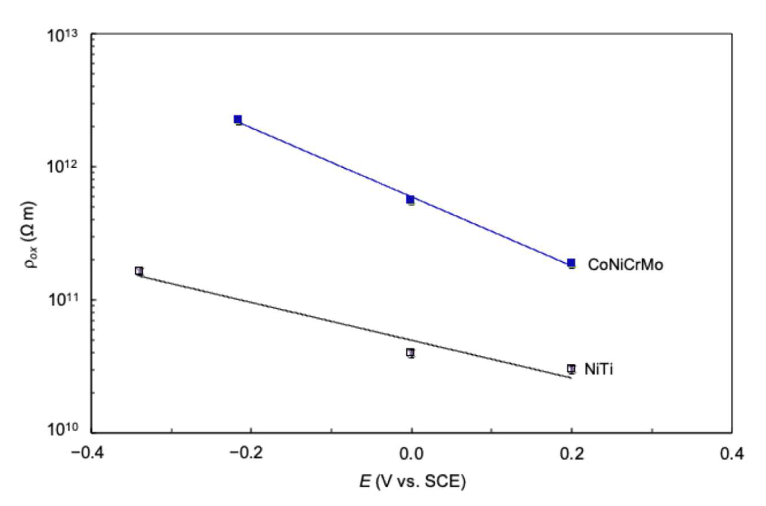
5.2. Oxide Resistivity

The oxide resistivity (ρox) was determined from Rox (normalized to area) using Equation (6), based on the assumption that the oxide resistance was a linear function of thickness:
ρ o x = R o x d o x
The value of ρox at Ecorr was found to be on the order of 1011 Ω m for NiTi versus 1012 Ω m for CoNiCrMo—that is, an order-of-magnitude difference—as shown in Table 2. The value for NiTi was consistent with the range (1011 to 1016 Ω m [32]) given for bulk TiO2. In contrast, the value for CoNiCrMo was higher than that reported for bulk Cr2O3 (~1010 Ω m [33]), and Cr2O3 films can have even lower values; for example, ~5 × 10⁸ Ω m was obtained for a 440 nm film [34]. In the case of CoNiCrMo, the phosphate layer on the oxide film may have had a relatively large influence on the ρox value obtained using EIS because of the film being so thin (<2 nm).
The dependence of ρox on potential for the two alloys is compared in Figure 9. In both cases, ρox decreased as the potential was increased, suggesting that the oxide becomes increasingly defective. Comparable behavior has been found for Ti in acidified 0.6 M NaCl [31]. The similarity in the potential dependence between the alloys is striking since the oxide is a semiconductor in both cases but TiO2 is an n-type [35,36] whereas Cr2O3 is a p-type [37]. Notwithstanding this difference, the respective charge carriers are evidently influenced in a similar manner by the electric field across the oxide film.

6. Summary

EP NiTi and CoNiCrMo are characterized by a thin (~1–4 nm) passive film in PBS and other buffered physiological solutions. Both alloys exhibit Tafel-type behavior consistent with that predicted by the GGM for dissolution in the case of thin films where the rate-controlling step is cation migration through the film. Values of α obtained from the apparent Tafel slopes indicate that the corrosion mechanism is similar for NiTi and CoNiCrMo. A small but distinct difference in α (and therefore the apparent Tafel slopes) was found between the two alloys and can be attributed to the difference in their predominant oxide; that is, TiO2 versus Cr2O3. Moreover, the oxide film on each alloy showed marked differences in thickness and resistivity. The thickness was greater for NiTi—consistent with surface analytical results—and differed in potential dependence between the two alloys in the passive region. The oxide resistivity, conversely, was substantially lower for NiTi and showed a similar potential dependence for the two alloys, even though the oxide is an n-type semiconductor in one case and p-type in the other.

Funding

This research received no external funding.

Data Availability Statement

No new data were created or analyzed in this study.

Conflicts of Interest

The author is employed by Exponent. The research was conducted in the absence of any commercial or financial relationships that could be construed as potential conflicts of interest.

References

  1. Pound, B.G. Susceptibility of nitinol to localized corrosion. J. Biomed. Mater. Res. Part A 2006, 77, 185–191. [Google Scholar] [CrossRef]
  2. Trepanier, C.; Tabrizian, M.; Yahia, L.H.; Bilodeau, L.; Piron, D.L. Effect of modification of oxide layer on NiTi stent corrosion resistance. J. Biomed. Mater. Res. 1998, 43, 433–440. [Google Scholar] [CrossRef]
  3. Thierry, B.; Tabrizian, M.; Trepanier, C.; Savadogo, O.; Yahia, L.H. Effect of surface treatment and sterilization processes on the corrosion behavior of NiTi shape memory alloy. J. Biomed. Mater. Res. 2000, 51, 685–693. [Google Scholar] [CrossRef]
  4. Trigwell, S.; Hayden, R.D.; Nelson, K.F.; Selvaduray, G. Effects of surface treatment on the surface chemistry of NiTi alloy for biomedical applications. Surf. Interface Anal. 1998, 26, 483–489. [Google Scholar] [CrossRef]
  5. Kocijan, A.; Milosev, I.; Pihlar, B. Cobalt-based alloys for orthopaedic applications studied by electrochemical and XPS analysis. J. Mater. Sci. Mater. Med. 2004, 15, 643–650. [Google Scholar] [CrossRef]
  6. Pound, B.G. The electrochemical behavior of nitinol in simulated physiological solutions. J. Biomed. Mater. Res. Part A 2008, 85, 1103–1113. [Google Scholar] [CrossRef]
  7. Pound, B.G. Electrochemical behavior of cobalt-chromium alloys in a simulated physiological solution. J. Biomed. Mater. Res. Part A 2010, 94, 93–102. [Google Scholar] [CrossRef]
  8. Hodgson, A.W.E.; Kurz, S.; Virtanen, S.; Fervel, V.; Olsson, C.-O.A.; Mischler, S. Passive and transpassive behavior of CoCrMo in simulated biological solutions. Electrochim. Acta 2004, 49, 2167–2178. [Google Scholar] [CrossRef]
  9. Schmuki, P.; Virtanen, S.; Davenport, A.J.; Vitus, C.M. Transpassive dissolution of Cr and sputter-deposited Cr oxide studied by in situ x-ray near-edge spectroscopy. J. Electrochem. Soc. 1996, 143, 3997–4005. [Google Scholar] [CrossRef]
  10. Tabrizian, M.; Thierry, B.; Savadago, O.; Yahia, L.H. Surface characterization of sterilized electropolished NiTi shape memory alloy as biomaterials. In Proceedings of the Sensory Phenomena and Measurement Instrumentation for Smart Structures and Materials, Newport Beach, CA, USA, 1 March 1999; SPIE: Bellingham, WA, USA, 1999; Volume 3670, pp. 106–114. [Google Scholar]
  11. Pound, B.G.; Becker, C.H. Composition of surface films on nickel based superalloys. J. Electrochem. Soc. 1991, 138, 696–700. [Google Scholar] [CrossRef]
  12. Igual Muñoz, A.; Mischler, S. Interactive effects of albumin and phosphate ions on the corrosion of CoCrMo implant alloy. J. Electrochem. Soc. 2007, 154, C562–C570. [Google Scholar] [CrossRef]
  13. Pound, B.G. Pit initiation on nitinol in simulated physiological solutions. J. Biomed. Mater. Res. Part B 2018, 106, 1605–1610. [Google Scholar] [CrossRef]
  14. Pound, B.G. Pit initiation on biomedical alloys—A review. J. Biomed. Mater. Res. Part B 2024, 112, e35367. [Google Scholar] [CrossRef]
  15. Hu, R.; Ornberg, A.; Pan, J. Investigation of influence of small particles in MP35N on the corrosion resistance in synthetic biological environment. J. Electrochem. Soc. 2009, 156, C341–C344. [Google Scholar] [CrossRef]
  16. Pound, B.G. The passive behavior of biomedical alloys in simulated physiological solutions. J. Biomed. Mater. Res. Part B 2022, 110, 768–775. [Google Scholar] [CrossRef]
  17. Venugopalan, R.; Trepanier, C. Assessing the corrosion behavior of Nitinol for minimally-invasive device design. Minim. Invasive Ther. Allied Technol. 2000, 9, 67–74. [Google Scholar] [CrossRef]
  18. Trepanier, C.; Pelton, A.R. Effect of strain on the corrosion resistance of nitinol and stainless steel in simulated physiological environment. In Proceedings of the International Conference on Shape Memory and Superelastic Technologies, SMST-2003, Pacific Grove, CA, USA, 5–8 May 2003; pp. 393–398. [Google Scholar]
  19. Wever, D.J.; Veldhuizen, A.G.; de Vries, J.; Busscher, H.J.; Uges, D.R.A.; van Horn, J.R. Electrochemical and surface characterization of a nickel-titanium alloy. Biomaterials 1998, 19, 761–769. [Google Scholar] [CrossRef]
  20. Ouerd, A.; Alemany-Dumont, C.; Normand, B.; Szunerits, S. Reactivity of CoCrMo alloy in physiological medium: Electrochemical characterization of the metal/protein interface. Electrochim. Acta 2008, 53, 4461–4469. [Google Scholar] [CrossRef]
  21. Alkhateeb, E.; Virtanen, S. Influence of surface self-modification in Ringer’s solution on the passive behavior of titanium. J. Biomed. Mater. Res. Part A 2005, 75, 934–940. [Google Scholar] [CrossRef]
  22. Seyeux, A.; Maurice, V.; Marcus, P. Oxide film growth kinetics on metals and alloys. I. Physical model. J. Electrochem. Soc. 2013, 160, C189–C196. [Google Scholar] [CrossRef]
  23. Lutton, K.; Scully, J.R. Kinetics of oxide growth of passive films on transition metals. In Encyclopedia of Interfacial Chemistry; Wandelt, K., Ed.; Elsevier: New York, NY, USA, 2018; pp. 284–290. [Google Scholar]
  24. Leistner, K.; Toulemonde, C.; Diawara, B.; Seyeux, A.; Marcus, P. Oxide film growth kinetics on metals and alloys. II. Numerical simulation of transient behavior. J. Electrochem. Soc. 2013, 160, C197–C205. [Google Scholar] [CrossRef]
  25. Yeung, K.W.K.; Poon, R.W.Y.; Chu, P.K.; Chung, C.Y.; Liu, X.Y.; Lu, W.W.; Chan, D.; Chan, S.C.W.; Luk, K.D.K.; Cheung, K.M.C. Surface mechanical properties, corrosion resistance, and cytocompatibility of nitrogen plasma-implanted nickel–titanium alloys: A comparative study with commonly used medical grade materials. J. Biomed. Mater. Res. Part A 2007, 82, 403–414. [Google Scholar] [CrossRef] [PubMed]
  26. Okazaki, Y.; Gotoh, E. Comparison of metal release from various metallic biomaterials in vitro. Biomaterials 2005, 26, 11–21. [Google Scholar] [CrossRef] [PubMed]
  27. Ornberg, A.; Pan, J.; Herstedt, M.; Leygraf, C. Corrosion resistance, chemical passivation, and metal release of 35N LT and MP35N for biomedical material application. J. Electrochem. Soc. 2007, 154, C546–C551. [Google Scholar] [CrossRef]
  28. Schroeder, V. Evolution of the passive film on mechanically damaged nitinol. J. Biomed. Mater. Res. Part A 2009, 90, 1–17. [Google Scholar] [CrossRef]
  29. Ferreira, M.G.S.; Da Cunha Belo, M.; Hakiki, N.E.; Goodlet, G.; Montemor, M.F.; Simões, A.M.P. Semiconducting properties of oxide and passive films formed on AISI 304 stainless steel and Alloy 600. J. Braz. Chem. Soc. 2002, 13, 433–440. [Google Scholar] [CrossRef]
  30. Popa, M.V.; Demetrescu, I.; Vasilescu, E.; Drob, P.; Lopez, A.S.; Mirza-Rosca, J.; Vasilescu, C.; Ionita, D. Corrosion susceptibility of implant materials Ti-5Al-4V and Ti-6Al-4Fe in artificial extra-cellular fluids. Electrochim. Acta 2004, 49, 2113–2121. [Google Scholar] [CrossRef]
  31. Kolman, D.G.; Scully, J.R. Electrochemistry and passivity of Ti-15V-3Cr-3Al-3Sn β-titanium alloy in ambient temperature aqueous chloride solutions. J. Electrochem. Soc. 1994, 141, 2622–2641. [Google Scholar] [CrossRef]
  32. Weast, R.C. (Ed.) CRC Handbook of Chemistry and Physics, 64th ed.; CRC Press: New York, NY, USA, 1983–1984; p. E-55. [Google Scholar]
  33. Kwan, C.-P.; Chen, R.; Singisetti, U.; Bird, J.P. Electric-field dependent conduction mechanisms in crystalline chromia. Appl. Phys. Lett. 2015, 106, 112901. [Google Scholar] [CrossRef]
  34. Kaspar, T.C.; Sushko, P.V.; Bowden, M.E.; Heald, S.M.; Papadogianni, A.; Tschammer, C.; Bierwagen, O.; Chambers, S.A. Defect compensation by Cr vacancies and oxygen interstitials in Ti4+-doped Cr2O3 epitaxial thin films. Phys. Rev. B 2016, 94, 155409. [Google Scholar] [CrossRef]
  35. McAleer, J.F.; Peter, L.M. Photocurrent spectroscopy of anodic oxide films on titanium. Faraday Disc. Chem. Soc. 1980, 70, 67–80. [Google Scholar] [CrossRef]
  36. Torresi, R.M.; Camara, O.R.; De Pauli, C.P. Influence of the hydrogen evolution reaction on the anodic titanium oxide film properties. Electrochim. Acta 1987, 32, 1357–1363. [Google Scholar] [CrossRef]
  37. Sekizawa, K.; Oh-ishi, K.; Morikawa, T. Photoelectrochemical water-splitting over a surface modified p-type Cr2O3 photocathode. Dalton Trans. 2020, 49, 659–666. [Google Scholar] [CrossRef] [PubMed]
Figure 1. Potentiodynamic polarization curves for EP NiTi and CoNiCrMo in deaerated PBS [1,7].
Cmd 06 00004 g001
Figure 2. Pitting on NiTi after potentiodynamic polarization in 0.9% NaCl; (a) 45X; (b) 500X [13].
Cmd 06 00004 g002aCmd 06 00004 g002b
Figure 3. Concentration of total dissolved Ni and total dissolved Cr in PBS shown in relation to the polarization curve for EP CoNiCrMo for the potential range of interest. The metal ion concentrations were determined after 1 h at 0.5 V, 0.7 V, and 0.9 V [7]. The data shown are the mean values for three tests, with the error bars representing the standard deviation.
Cmd 06 00004 g003
Figure 4. Impedance spectra for NiTi and CoNiCrMo in deaerated PBS at 37 °C: (a) Ecorr; (b) 0.0 V; (c) 0.2 V [6,7]. |Z| is the impedance magnitude, θ is the phase angle, and f is the frequency.
Cmd 06 00004 g004
Figure 5. Equivalent circuit for EP NiTi and CoNiCrMo in deaerated PBS. Rox and CPEox are the resistance and constant phase element associated with the oxide film, and Rsol is the solution resistance.
Cmd 06 00004 g005
Figure 6. Comparison of fitted and experimental data for EP NiTi and CoNiCrMo in PBS at 0.0 V [6,7].
Cmd 06 00004 g006
Figure 7. Comparison of dox values obtained for EP NiTi and CoNiCrMo from EIS and surface analysis. Surface analytical data: EP NiTi stent [2] and disc [3,10]; MP CoNiCrMo [11] and CoCrMo [12]. Error bars show the standard deviation for NiTi and CoNiCrMo and the range of values for CoCrMo.
Cmd 06 00004 g007
Figure 8. Dependence of dox on E for EP NiTi and CoNiCrMo from Ecorr to 0.2 V [6,7].
Cmd 06 00004 g008
Figure 9. Dependence of ρox on E for EP NiTi and CoNiCrMo from Ecorr to 0.2 V [6,7].
Cmd 06 00004 g009
Table 1. Values of ba′ and icorr for NiTi and CoNiCrMo in deaerated PBS 1.
Alloyba′ (V)icorr (nA/cm2)
NiTi0.16 ± 0.14.7 ± 0.2
CoNiCrMo0.19 ± 0.13.6 ± 0.1
1 Mean ± standard deviation for three tests with each alloy.
Table 2. Values of dox and ρox for NiTi and CoNiCrMo in deaerated PBS at Ecorr [6,7] 1.
Alloydox (nm)ρox (1012 Ω m)
NiTi4.0 ± 0.40.16 ± 0.02
CoNiCrMo0.8 ± 0.12.22 ± 0.11
1 Mean ± standard deviation for three tests with each alloy.
Disclaimer/Publisher’s Note: The statements, opinions and data contained in all publications are solely those of the individual author(s) and contributor(s) and not of MDPI and/or the editor(s). MDPI and/or the editor(s) disclaim responsibility for any injury to people or property resulting from any ideas, methods, instructions or products referred to in the content.

