Review

A Review of Evidence for the Involvement of the Circadian Clock Genes into Malignant Transformation of Thyroid Tissue

by
Arcady A. Putilov
1,2,*,
Elena V. Budkevich
1 and
Roman O. Budkevich
1
1
Laboratory of Nanobiotechnology and Biophysics, North-Caucasus Federal University, 355029 Stavropol, Russia
2
Laboratory of Sleep/Wake Neurobiology, Institute of Higher Nervous Activity and Neurophysiology of the Russian Academy of Sciences, 117865 Moscow, Russia
*
Author to whom correspondence should be addressed.
Clocks & Sleep 2023, 5(3), 384-398; https://doi.org/10.3390/clockssleep5030029
Submission received: 29 March 2023 / Revised: 10 July 2023 / Accepted: 11 July 2023 / Published: 13 July 2023
(This article belongs to the Section Disorders)

Abstract

:
(1) Background: In 2013, the results of a pioneer study on abnormalities in the levels and circadian rhythmicity of expression of circadian clock genes in cancerous thyroid nodules was published. In the following years, new findings suggesting the involvement of circadian clockwork dysfunction into malignant transformation of thyroid tissue were gradually accumulating. This systematic review provides an update on existing evidence regarding the association of these genes with thyroid tumorigenesis. (2) Methods: Two bibliographic databases (Scopus and PubMed) were searched for articles from inception to 20 March 2023. The reference lists of previously published (nonsystematic) reviews were also hand-searched for additional relevant studies. (3) Results: Nine studies published between 2013 and 2022 were selected. In total, 9 of 12 tested genes were found to be either up- or downregulated. The list of such genes includes all families of core circadian clock genes that are the key components of three transcriptional–translational feedback loops of the circadian clock mechanism (BMAL1, CLOCK, NPAS2, RORα, REV-ERBα, PERs, CRYs, and DECs). (4) Conclusions: Examination of abnormalities in the levels and circadian rhythmicity of expression of circadian clock genes in thyroid tissue can help to reduce the rate of inadequate differential preoperative diagnosis for thyroid carcinoma.

1. Introduction

The first results pointing to the alternation of expression of circadian clock genes in human malignant thyrocytes were published 10 years ago [1]. In the following years, further evidence for the association of these genes with thyroid cell oncogenic transformation was gradually accumulating [2,3,4,5,6,7,8,9].
As stressed in several (nonsystematic) reviews of such findings [10,11,12,13,14,15,16,17,18], the examination of the pattern of expression of circadian clock genes might be of practical importance for the establishment of an adequate presurgical diagnosis of thyroid tumors [19]. Although the frequency of thyroid nodules (i.e., discrete masses in the gland) is very high (they are detected in up to 65% of general populations), most of them are benign. A relatively low percentage of cancerous nodules (about 5%) increases the risk of unnecessary surgeries in the case of asymptomatic benign nodules and increases the risk of delays with diagnosis and treatment in the case of asymptomatic cancerous nodules [20,21].
Thyroid cancers (TCs) are usually divided into five main histological types: “papillary,” “follicular,” “poorly differentiated,” “undifferentiated/anaplastic,” and “medullary” (PC, FC, PDTC, ATC, and MTC, respectively) [22,23]. The origin from neuroendocrine C cells distinguishes the last type (MTC) from other four types. Two of the remaining four types (PC and FC) are classified as well differentiated. An incomplete tumor capsule with expansive growth is the most typical feature of PDTC, and among five histological types, ATC is regarded as the most aggressive form. The vast majority of TCs are classified as PC and FC types (85%), while only a small fraction of thyroid tumors (5–7%) lose features of cell origin and are classified as either PDTC or ATC [24]. The prognosis of patients with PC and FC is rather optimistic (e.g., they are usually curable with radioactive iodine therapy or surgery [25,26,27]), while the risk of mortality for ATC is high due to its rapid progression [28].
Until recently, fine-needle aspiration biopsy aimed at identification of thyroid malignancies has served as a safe and accurate tool for clinical evaluation of nonsecreting thyroid nodules [20]. Although surgery is not recommended without evaluating the results of such biopsies, the rate of unnecessary surgery remains high (e.g., after surgical interventions, 70–90% of thyroid cases were found to be benign [29]). The difficulty of distinguishing between benign and malignant thyroid nodules has been recognized as one of the most challenging issues of diagnostic approaches in oncology of the thyroid gland [30]. The rate of unnecessary surgeries and delays with diagnosis and treatment of thyroid cancer can be lowered by the development of preoperative markers for thyroid malignancies [31]. In order to improve preoperative diagnosis, the molecular testing platforms have been introduced. In efforts to reduce unnecessary surgical interventions, these platforms can be used as an integral part of the cytological evaluation in conjunction with fine-needle aspiration biopsy [32,33,34,35,36]. Therefore, it is critical to determine whether presurgical molecular biomarkers for thyroid carcinoma might include indices of abnormal functioning of circadian clocks [10,11,12,13,14].
The importance of the circadian clockwork for multicell organisms has become evident from findings indicating that almost each cell in almost each tissue type of almost each organism contains a set of genes involved in the construction and functioning of its own circadian clocks with a near 24 h (circadian) period [37,38].
A transcriptional–translational feedback loop is a key feature of this regulating mechanism [37,38,39]. In a mammalian cell, the circadian cycle is initiated by BMAL1 (brain and muscle aryl hydrocarbon receptor nuclear translocator-like), CLOCK (circadian locomotor output cycles kaput), and NPAS2 (neuronal PAS domain protein 2). Their proteins are members of the basic helix–loop–helix/Per-Arnt-Sim (bHLH-PAS) family of transcription factors. In the cytoplasm, they interact with each other to pro-duce heterodimers (BMAL1/CLOCK and BMAL1/NPAS2). At the next phase, these heterodimers translocate to the nucleus to activate the transcription of several other clock genes, including CRYs (cryptochrome circadian regulator 1 and cryptochrome circadian regulator 2), PERs (period circadian regulator 1, period circadian regulator 2, and period circadian regulator 3), and DECs (differentially expressed in chondrocyte 1 and differentially expressed in chondrocyte 2). BMAL1/CLOCK and BMAL1/NPAS2 heterodimers act as transcription factors and bind the E-box regions on the promoters of these target genes that in turn encode for the repressor components of the circadian clocks. At a later phase of the cycle, the protein products of these genes dimerize and form complexes between themselves (PERs with CRYs and DEC1 with DEC2). At the final phase, the feedback loop is completed by transporting these cytoplasmic dimers (PERs/CRYs and DEC1/DEC2) back into the nucleus to suppress activity of BMAL1/CLOCK and BMAL1/NPAS2 heterodimers. When they are suppressing activity of these heterodimers, they are also repressing their own expression, thus giving rise to a new cycle (i.e., the next cycle is started with allowing the transcription of BMAL1, CLOCK, and NPAS2) [38,39,40]. The circadian expression of BMAL1 and NPAS2 is also influenced by two nuclear receptors, REV-ERBα (or NR1D1, nuclear receptor subfamily 1 group D member 1) and RORα (RAR related orphan receptor alpha). Both receptors are activated by BMAL1/CLOCK, and they in turn regulate expression of BMAL1 and NPAS2 genes by acting on their promoters [41] (Figure 1).
In addition to controlling each other’s expression, these molecular regulators drive the rhythms of expression of many thousands of target genes. Circadian transcription factors interact with various corepressors, coactivators, and chromatin-associated factors, which in turn read or write or erase chromatin histone modification marks for activating or repressing transcription [42,43,44,45].
Overall, the cellular circadian clocks govern the expression of approximately half of the protein coding genes, thus providing coordination of circadian rhythms originating from different biochemical, physiological, and behavioral systems in the organism [46,47].
At any higher (multicell) hierarchical levels, the same set of circadian clock genes constitutes a basis of more complex circadian clocks with a similar molecular makeup. The list of such multicell clocks includes the peripheral clocks of various organs and tissue, the clocks of endocrine and physiological systems, and finally the central clocks in the supra-chiasmatic nuclei, which entrain the circadian rhythms within an organism and correct their phases by responding to environmental time cues [48,49].
Overall, the numerous modulating effects of the circadian clocks on gene expression, DNA repair, proliferation, cellular metabolism, inflammation, apoptosis, etc. [50] gave rise to hypotheses that the abnormal functioning of circadian clock genes may have various pathological consequences, including tumorigenesis [51,52,53,54,55,56,57,58,59]. In particular, it was assumed that the characteristics of clock genes signaling their abnormal functioning can serve as specific molecular markers of malignant transformation of thyroid tissue [10,11,12,13,14,15,16,17,18].
In 2018, Angelousi et al. [15] presented results of a systematic search for studies testing the association of circadian clock genes with different types of endocrine cancer. Results on thyroid cancer were reported in only one of 23 eligible publications [1]. A majority of new studies confirming this association were published more recently [5,6,7,8,9].
Therefore, the aim of the present systematic review was to summarize evidence supporting the assumption of abnormal expression of circadian clock genes in thyroid carcinoma.

2. Results

The main results of the literature search are summarized in Table 1. These indicated that each of the circadian clock genes mentioned in the Introduction was tested at least once in thyroid carcinoma between 2013 and 2022 (Table 1) and the results of such testing were reported in nine selected publications, in the pioneer publication of Mannic et al. (2013) [1] and eight following articles authored by Mond et al. (2014) [2], Chitikova et al. (2015) [3], Makhlouf et al. (2016) [4], Gallo et al. (2018) [5], Sadowski et al. (2019) [6], Lou et al. (2021) [7], Xu et al. (2022) [8], and Mou et al. (2022) [9] (article in Chinese with abstract in English). The authors of the nine selected publications interpreted the detected increase and decrease in expression of a gene compared to normal samples as its up- and downregulation, respectively. Most frequently, the transcripts of BMAL1 (brain and muscle aryl hydrocarbon receptor nuclear translocator-like) were studied under this and another name (ARNTL). In five published studies, the results suggested its upregulation. BMAL1 forms heterodimers with CLOCK (circadian locomotor output cycles kaput) and NPAS2 (neuronal PAS domain protein 2), whose transcripts were less frequently tested (in three studies and one study, respectively). The results reported in two studies and one study, respectively, suggested that they were (like BMAL) upregulated (Table 1). Upregulation was also reported for REV-ERBα (or NR1D1, nuclear receptor subfamily 1 group D member 1) and RORα (RAR-related orphan receptor alpha) in one of three and one of two studies, respectively (Table 1).
Downregulation was consistently (in three of four articles) revealed for CRY2 (cryptochrome circadian regulator 2). DECs (differentially expressed in chondrocyte 1 and differentially expressed in chondrocyte 2) were tested in one study the results of which indicated that, similarly to the results on CRY2, these two genes were downregulated (Table 1). For only one gene, PER2 (period circadian regulator 2), the results on its transcripts were contradictory. This gene was reported to be upregulated in unspecified thyroid cancer (TC [7]) and downregulated in follicular and poorly differentiated cancer (FTC and PDTC [4]). The results on the transcripts of this gene were reported to be nonsignificant (Table 1) in two other studies of well-differentiated papillary cancer (PTC [1,3]) and follicular cancer (FTC [1]).
Nonsignificant results were obtained for the transcripts of three genes: CRY1 (cryptochrome circadian regulator 1) and PER1 (period circadian regulator 1) in three studies and for PER 3 (period circadian regulator 3) in two studies (Table 1).
The pioneer publication [1] contains results of additional comparisons of diurnal profiles of expression of seven circadian clock genes in thyrocytes cultured in vitro for 7 days and harvested every 6 h during 36 h (Table 2).
The results showed that thyrocytes from healthy and benign (HB) thyroid nodules kept their circadian properties (i.e., the clock gene expressions in these nodules exhibited circadian oscillatory patterns in synchronized thyrocytes). In contrast, only alternated circadian profiles were detected in thyrocytes from poorly differentiated thyroid carcinomas (PDTCs) [1]. As shown in Table 2, Mannic et al. [1] revealed abnormal circadian phase positions for six of seven tested transcripts. Moreover, they failed to determine this position for the seventh transcript due to an abnormally low circadian amplitude [1]. Sadly, there have been no attempts so far to provide support to these very promising findings. Therefore, replicability and reliability of the abnormalities in circadian oscillatory patterns remain to be elaborated.
In sum, upregulation was reported for all three circadian clock genes involved in the circadian cycle in its earlier phases: BMAL1, CLOCK and NPAS2. These genes form heterodimers with the function of transcription activators. It was found that all three genes were upregulated in thyroid carcinoma [1,3,6,7,8,9] (Table 1). Upregulation was also shown for the genes producing receptors REV-ERBα and RORα, which are activated by BMAL1/CLOCK (Table 1). Downregulation was reported for CRYs, DECs, and PERs. These genes play the role of suppressors of activity of BMAL1, CLOCK and NPAS2 at later phases of the cycle. Downregulation of genes that form dimers with the function of transcription inhibitors was revealed for CRY2 [1,4,7], DEC1 and DEC2 [5], and PER2 ([4], but see contradicting results reported in [7]; Table 1).
Overall, either up- or downregulation was reported for 9 of 12 tested circadian clock genes. This result suggested that if two or more such genes were tested, there was a good chance of finding evidence of dysregulation of expression of at least one of them (Table 1).

3. Discussion

In 2013, Mannic et al. [1] published results of their pioneer study providing evidence for the involvement of circadian clockwork dysfunctions into malignant transformation of thyroid tissue [1]. Further evidence for the abnormal expression of circadian clock genes in cancerous thyroid nodules has gradually accumulated in the following 10 years [2,3,4,5,6,7,8,9]. The main aim of the present systematic review was to identify articles reporting results of testing the association between thyroid carcinoma and expression of circadian clock genes. The findings of nine relevant studies [1,2,3,4,5,6,7,8,9] were summarized and allowed the conclusion of plausibility of the assumption of abnormal expression of circadian clock genes in thyroid carcinoma. The reviewed findings provided evidence that if just two of these genes were tested, there was a 100% chance of revealing that expression of at least one of them was abnormal.
Additional results of the pioneer study of Mannic et al. (2013) [1] on the circadian patterns of gene expression indicated that these patterns were alternated in each of seven circadian clock genes tested in poorly differentiated thyroid nodules (PDTCs). Therefore, the integration of such studies on circadian biology of the thyroid gland into management of thyroid cancer might lead to the development of new methodology for diagnosis of malignant transformation of thyroid tissue. It is plausible to expect that an examination of abnormalities in the levels and circadian rhythmicity of expression of circadian clock genes in this tissue can help to decrease the rate of inadequate differential preoperative diagnosis for thyroid carcinoma.
In more detail, upregulation of BMAL1 was reported for the first time in the pioneer study [1] and confirmed by results of four subsequent studies: Chitikova et al. (2015) [3], Sadowski et al. (2019) [6], Lou et al. (2021) [7] and Mou et al. (2022) [9]. Expression of this gene was not tested in the remaining four studies of Mond et al. (2014) [2], Makhlouf et al. (2016) [4], Gallo et al. (2018) [5], or Xu et al. (2022) [8], but the results of these studies allowed the extension of the list of circadian clock genes demonstrating abnormal expression in thyroid carcinoma. These results provided evidence for dysfunction of practically all core circadian clock genes in malignant thyroid tissue.
The only contradictory results were obtained for the transcription of one of 12 tested genes (PER2). Downregulation of PER2 was reported for follicular and poorly differentiated cancer (FTC and PDTC) [4], while upregulation was found in a study of unspecified thyroid cancer (TC) [7]. Results on other genes were not contradictory. Overall, upregulation was found for BMAL1, CLOCK, NPAS2, REV-ERBα, and RORα, and downregulation was revealed for CRY2, DEC1, and DEC2.
Such results regarding the directions of changes in gene expression in thyroid carcinoma (increased or decreased transcription) were in good agreement with the directions expected from the roles of the transcripts of the circadian clock genes in the transcriptional–translational feedback loop regulating the body clocks (see Introduction). The general rule was that if the dimers of genes acted as promoters of transcription of other circadian clock genes, they were found to be upregulated, while if the dimers of genes suppressed transcription of other circadian clock genes, they were found to be downregulated.
In more detail, dimers with opposite functions of transcription activator and transcription inhibitor are formed by BMAL1, CLOCK and NPAS2 and by CRYs, DECs, and PERs, respectively. Therefore, if it were consistently found in the reviewed studies that BMAL1, CLOCK and NPAS2 were upregulated in thyroid carcinoma [1,3,6,7,8,9], the suppressors of activity of BMAL1, CLOCK and NPAS2 (CRYs, DECs, and PERs) would be expected to be downregulated in this carcinoma. Indeed, this expectation was confirmed by the reports indicating downregulation of the majority of genes controlling expression of BMAL1, CLOCK and NPAS2. Upregulation was revealed for CRY2 [1,4,7], DEC1 and DEC2 [5], and PER2 ([4] but not [7]; see above).
Moreover, upregulation was obtained for the genes coding REV-ERBα and RORα monomers [2], which are activated by BMAL1 and CLOCK heterodimer.
In oncological diseases, patient prognosis might be potentially improved by normalization of circadian rhythms [18]. Therefore, chronobiotic therapeutic factors were recommended to be used for prevention and/or treatment of these diseases (e.g., [17]). Two major treatment approaches were suggested for avoiding the pathological consequences of circadian clock dysfunction. The first is a direct pharmacological activation of some circadian clock genes [60]. Another is a restoration of normal circadian rhythms with the help of small molecules that are able to modulate activity of the core components of circadian clocks [61]. However, such studies remain at a stage of basic experimental exploration.
It is well established that thyroid-stimulating hormone (TSH) produced by the pituitary gland plays a pivotal role in regulating the hypothalamic–pituitary–thyroid axis. Therefore, this hormone serves as the major marker of hormonal and physiological activity of the pituitary and thyroid gland [13]. It was proposed that elevated TSH levels in serum can have diagnostic value in the presurgical management of thyroid carcinoma [62,63,64,65,66,67,68], and that the measurement of these levels can be recommended as an easily performed additional tool for decision-making in patients with indeterminate cytological findings [69,70,71]. Moreover, TSH exhibits a robust 24 h rhythm of secretion, and the diurnal profile of this hormone in serum is regarded as one of three most reliable markers of the circadian rhythmicity of the human organism [10,72,73,74,75]. TSH is rhythmically secreted in response to the neuronal and humoral signals from the central clocks, but thyroid tissue that contains the same set of circadian clock proteins as the set expressed in the central clocks also exerts its influence on regulation of this hormone [76,77]. Therefore, it is plausible to suggest that thyroid cancer is linked to both an abnormal circadian rhythm of TSH and an abnormal expression of the circadian clock genes. Empirical support of this suggestion might lead to the recommendation of evaluation of both this hormone rhythmicity and these genes’ expression. However, with the exception of one publication [78], the literature is lacking studies examining the 24 h rhythm of TSH in patients with different types of thyroid carcinoma [12]. Therefore, it remains unknown whether the levels and daily pattern of TSH in serum or thyroid issue might be challenged in parallel with the levels and daily pattern of expression of circadian clock genes.
TSH directly bound to its receptor (TSH-R) [79] and levels of TSH circulating in thyroid tissue regulate the receptor signaling in the thyrocyte’s membrane [80]. Studies suggested the involvement of TSH-R in thyroid cancer (e.g., [81,82,83,84]). In particular, a reduced number of binding sites was found in carcinoma tissue [85], and levels of TSH-R gene expression were shown to be lower in such tissue [82,86]. Expression was decreased and even lost in poorly differentiated and undifferentiated thyroid cancer, respectively [87,88,89,90,91]. Therefore, it was proposed that TSH-R might have important diagnostic value for thyroid cancer, e.g., as a marker of thyroid differentiation [84,85,86,87,88,89,90,91,92,93]. However, almost nothing has been published about the diurnal profile of TSH-R expression, and this expression has not been examined in relation to expression of any circadian clock genes [12].
Therefore, future research might be aimed at testing whether there are associations between pathological changes in levels and diurnal profiles of thyroid hormones, their receptors, and transcripts of circadian clock genes. For instance, further in vitro experiments on cultured malignant thyrocytes might examine whether thyroid tumorigenesis is linked to parallel pathological changes in regulation of circadian clock genes and circadian rhythmicity of TSH and its receptor (TSH-R) [12].
Environmental factors can contribute to the risks of both thyroid cancer and circadian dysregulation. Several types of endocrine cancers were found to be associated with chronic circadian disruption caused by travel across time zones, shift and night work, sleep insufficiency, and irregular sleep–wake cycles [94]. Patients with more severely disturbed sleep and/or circadian disruption have worse prognosis than those having good sleep and normal circadian rhythmicity [15]. In respect to thyroid cancer, night and shift work was reported to be associated with an increased risk of thyroid nodules [95]. Moreover, disturbances of the sleep–wake cycle were shown to be linked to a higher risk of thyroid cancer in postmenopausal nonobese women [96]. Such a higher risk was also reported for women who used a sedative–hypnotic drug [97].
Light at night was found to be associated with an increased risk of thyroid cancer [98]. This association may be partially driven by melatonin’s deficiency leading to a decrease in its tumor suppression function and to the disruptions of sleep and circadian rhythms [98]. Future studies may provide deeper insights into the potential roles of melatonin in thyroid cancer etiology and into the biological pathways underlying the relationship between light at night and thyroid cancer.
Several traits of the sleep–wake cycle, such as sleep duration, insomnia, and chronotype, may be risk factors for cancer [99]. Lou et al. [7] found that patients with thyroid cancer reported sleep disturbances more often when they also expressed elevated levels of CLOCK, BMAL1, and PER2 and reduced levels of CRY2 compared to age-matched cancer-free controls. Evidence for the extensive cross-talk between sleep, circadian rhythms, and metabolic pathways involved in malignancy suggests a possibility for recommending screening cancer patients for sleep and circadian disruptions [100,101].
Given that several variants of circadian clock genes were found to be associated with incidents of thyroid carcinoma (e.g., [102,103,104]), polymorphism of human circadian clock genes might also contribute to the risks of development of thyroid cancer. These associations and phenotypic traits underpinned by these variants require further exploration.
The mechanisms underlying the links between abnormalities of the circadian clock machinery and malignant transformation of thyroid tissue remain to be elucidated. Some proposed causes of the associations between circadian disturbances and cancer (including thyroid cancer) were discussed in several previously published reviews (e.g., [10,11,16,18,51,52,53,54,55,56,57,58]). Analysis of existing evidence [105] indicated that the circadian clocks may be dysregulated in many forms of cancer, but such dysregulation is not caused solely by inactivation of core circadian clock genes. Rather, it is accompanied by large-scale changes in circadian gene expression and coexpression because the cellular circadian clocks play an important role in tumorigenesis, tumor immune escape, tumor growth, metastasis, and the processes of regulation of proliferation, apoptosis, intracellular metabolism, etc.
As mentioned in the Introduction, the risks of unnecessary surgeries and delays to diagnosis and treatment of thyroid carcinoma remain high due to a relatively low percentage of cancerous nodules [20,21]. This problem was addressed in recent years by introducing two new approaches to the diagnosis of cancerous nodules: liquid biopsy and molecular testing platforms. Liquid biopsy provided a possibility to eliminate the invasive procedures needed to obtain tissue samples. Because such biopsy detects and analyzes biological samples released from the tumor into the bloodstream, it can be repeatedly performed in a noninvasive way, at lower cost and without the risks associated with classic tissue biopsy [30,106]. Using molecular platforms for stratification of patients with neoplasm provides more definitive guidance for decision-making in the clinic, including the decision to avoid unnecessary surgical interventions [32,33,34,35,36]. As already emphasized in previously published reviews [10,11,12,13,14,15,16,17,18], the examination of expression of circadian clock genes in thyroid carcinoma might be of practical importance for adequate presurgical diagnosis of thyroid cancer. The reviewed findings here allow a recommendation to be made for trying to improve the preoperational management of thyroid carcer by examining whether the levels and circadian rhythmicity of expression of these genes are altered in thyroid nodules.
This review had several limitations. The number of studies selected for this review was small. Therefore, we cannot evaluate replicability of these studies. Although all five main histological types of thyroid carcer (PC, FC, PDTC, ATC, and MTC) were tested in nine reviewed studies, their results remain insufficient for comparisons of these types to the extent of abnormal changes in expression of circadian clock genes. Some of these types (e.g., PDTC and ATC) might be more vulnerable than other types to the development of circadian clockwork disfunction. Therefore, the results obtained on one type should be generalized to other types with caution. We reviewed here only studies on expression of core circadian genes in thyroid cancerous nodules. Consequently, we cannot compare the results on down- and upregulation of these genes in these nodules with the findings on down- and upregulation of the same genes in other endocrine cancers. Moreover, this review was limited to studies on expression of core circadian clock genes. We did not search for studies exploring the relationships of these genes with genes playing an important role in the processes of regulation of proliferation, apoptosis, and intracellular metabolism of thyroid tissue. These relationships might be addressed in future reviews.

4. Materials and Methods

The main aim of this systematic review was to identify publications reporting results of testing significance of associations between thyroid carcinoma and the levels and diurnal pattern of expression of core circadian clock genes. PRISMA (Preferred Reporting Items for Systematic Reviews and Meta-Analyses) guidelines for systematic review were followed [107]. Two bibliographic databases (Scopus and PubMed) were searched for articles from inception to 20 March 2023. The reference lists of previously published nonsystematic reviews [10,11,12,13,14,16,17,18] were also hand-searched for additional relevant articles.
Syntax for conducting the search within Scopus was the following:
TITLE-ABS-KEY (((circadian AND transcription) OR (clock AND expression) OR (clock AND genes) OR (circadian AND genes)) AND ((thyroid AND cancer) OR (thyroid AND tumor) OR (thyroid AND nodule) OR (thyroid AND neoplasm)))
For PubMed, the same search syntax was applied:
((circadian transcription) OR (clock expression) OR (clock genes) OR (circadian clock)) AND ((thyroid cancer) OR (thyroid nodule) OR (thyroid tumor) OR (thyroid neoplasm))
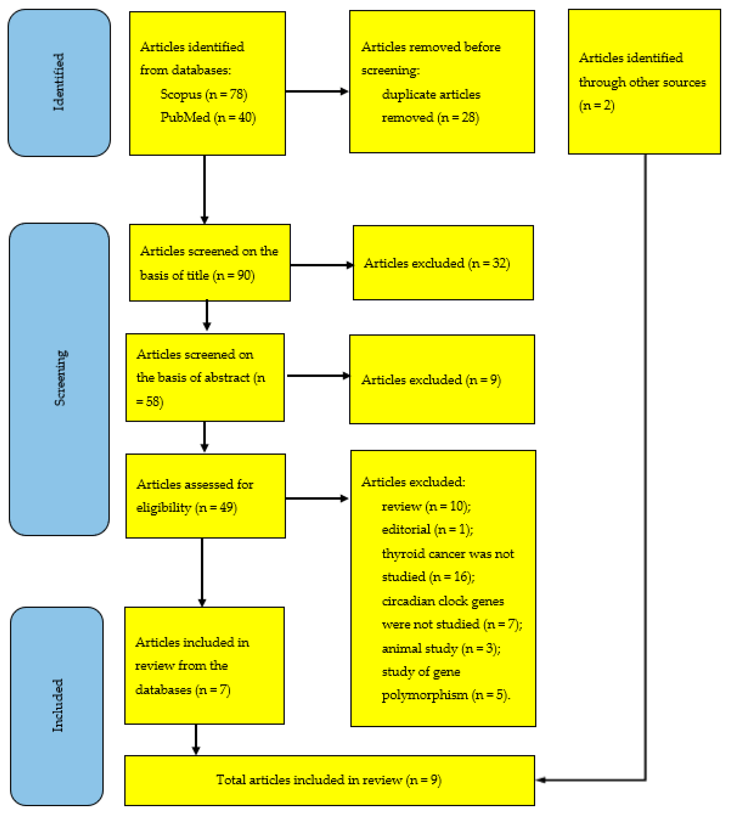
In total, 118 articles were identified (the study selection process is reported in Figure 2 in the PRISMA format). The hand search in the reference lists of previously published reviews [10,11,12,13,14,15,16,17,18] gave two additional relevant studies [2,5] that were previously mentioned in some such reviews [10,11,12].
After exclusion of 28 duplicates, 23 and 9 articles were additionally excluded on the basis of their title and abstract, respectively. The assessment for eligibility was performed by applying the following exclusion criteria: (1) review (n = 10), (2) editorial (n = 1), (3) thyroid cancer was not studied (n = 16), (4) circadian clock genes were not studied (n = 7), (5) animal study (n = 3), and 6) study of gene polymorphism (n = 5).
Seven and two potentially eligible full-text articles [1,2,3,4,5,6,7,8,9] were finally selected by searching the databases and reference lists, respectively (Figure 2).

5. Conclusions

The first result suggesting the involvement of circadian clockwork dysfunction in thyroid tumorigenesis was published in 2013. In the following 10 years, further evidence for abnormalities in expression of circadian clock genes in thyroid carcinoma has been gradually accumulating. The present systematic review of existing evidence for such abnormalities found nine studies published from 2013 to 2022. In each of the studies, an abnormal expression in malignant thyroid tissue was documented for one or more circadian clock genes. In total, 9 of 12 tested genes were found to be either up- or downregulated. The list of up- or downregulated genes includes all families of core circadian clock genes, BMAL1, CLOCK, NPAS2, CRYs, PERs, DECs, REV-ERBα, and RORα, constituting all three transcriptional–translational feedback loops of the circadian clock mechanism. These findings allowed the conclusion that the assumption of association of thyroid carcinoma with circadian clockwork dysfunctions was supported. Therefore, the integration of circadian biology into management of thyroid cancer can improve the methods of preoperative diagnosis of thyroid cancer. In particular, such diagnosis might account for the results of evaluation of circadian rhythmicity and mean levels of expression of circadian clock genes.

Author Contributions

A.A.P.: conceptualization, formal analysis, investigation, methodology, project administration, supervision, validation, visualization, writing—original draft, review and editing. E.V.B.: investigation, writing—review and editing. R.O.B.: investigation, funding acquisition, writing—review and editing. All authors have read and agreed to the published version of the manuscript.

Funding

This research received no external funding.

Institutional Review Board Statement

Not applicable.

Informed Consent Statement

Not applicable.

Data Availability Statement

Not applicable.

Acknowledgments

The North-Caucasus Federal University also provided technical and other similar support to R.O.B., E.V.B. and A.A.P.

Conflicts of Interest

The authors declare no conflict of interest.

References

  1. Mannic, T.; Meyer, P.; Triponez, F.; Pusztaszeri, M.; Le Martelot, G.; Mariani, O.; Schmitter, D.; Sage, D.; Philippe, J.; Dibner, C. Circadian clock characteristics are altered in human thyroid malignant nodules. J. Clin. Endocrinol. Metab. 2013, 98, 4446–4456. [Google Scholar] [CrossRef] [PubMed] [Green Version]
  2. Mond, M.; Alexiadis, M.; Eriksson, N.; Davis, M.J.; Muscat, G.E.; Fuller, P.J.; Gilfillan, C. Nuclear receptor expression in human differentiated thyroid tumors. Thyroid 2014, 24, 1000–1011. [Google Scholar] [CrossRef] [PubMed]
  3. Chitikova, Z.; Pusztaszeri, M.; Makhlouf, A.M.; Berczy, M.; Delucinge-Vivier, C.; Triponez, F.; Meyer, P.; Philippe, J.; Dibner, C. Identification of new biomarkers for human papillary thyroid carcinoma employing NanoString analysis. Oncotarget 2015, 6, 10978–10993. [Google Scholar] [CrossRef] [PubMed] [Green Version]
  4. Makhlouf, A.M.; Chitikova, Z.; Pusztaszeri, M.; Berczy, M.; Delucinge-Vivier, C.; Triponez, F.; Meyer, P.; Philippe, J.; Dibner, C. Identification of CHEK1, SLC26A4, c-KIT, TPO and TG as new biomarkers for human follicular thyroid carcinoma. Oncotarget 2016, 7, 45776–45788. [Google Scholar] [CrossRef] [Green Version]
  5. Gallo, C.; Fragliasso, V.; Donati, B.; Torricelli, F.; Tameni, A.; Piana, S.; Ciarrocchi, A. The bHLH transcription factor DEC1 promotes thyroid cancer aggressiveness by the interplay with NOTCH1. Cell Death Dis. 2018, 9, 871. [Google Scholar] [CrossRef]
  6. Sadowski, S.M.; Petrenko, V.; Meyer, P.; Pusztaszeri, M.; Brulhart-Meynet, M.C.; Heddad Masson, M.; Triponez, F.; Philippe, J.; Dibner, C. Validation of molecular biomarkers for preoperative diagnostics of human papillary thyroid carcinoma in fine needle aspirates. Gland. Surg. 2019, 8 (Suppl. S2), S62–S76. [Google Scholar] [CrossRef]
  7. Lou, X.; Wang, H.; Tu, Y.; Tan, W.; Jiang, C.; Sun, J.; Bao, Z. Alterations of sleep quality and circadian rhythm genes expression in elderly thyroid nodule patients and risks associated with thyroid malignancy. Sci. Rep. 2021, 11, 13682. [Google Scholar] [CrossRef]
  8. Xu, T.; Jin, T.; Lu, X.; Pan, Z.; Tan, Z.; Zheng, C.; Liu, Y.; Hu, X.; Ba, L.; Ren, H.; et al. A signature of circadian rhythm genes in driving anaplastic thyroid carcinoma malignant progression. Cell. Signal. 2022, 95, 110332. [Google Scholar] [CrossRef]
  9. Mou, Y.K.; Ren, C.; Li, Y.M.; Yu, G.H.; Zheng, G.B.; Song, H.; Lu, C.X.; Tian, R.X.; Song, X.C. Correlation analysis of clock genes and MEN2 medullary thyroid carcinoma. Chin. J. Otorhinolaryngol. Head Neck Surg. 2022, 7, 1079–1086. (In Chinese) [Google Scholar] [CrossRef]
  10. Ikegami, K.; Refetoff, S.; Van Cauter, E.; Yoshimura, T. Interconnection between circadian clocks and thyroid function. Nat. Rev. Endocrinol. 2019, 15, 590–600. [Google Scholar] [CrossRef]
  11. Malaguarnera, R.; Ledda, C.; Filippello, A.; Frasca, F.; Francavilla, V.C.; Ramaci, T.; Parisi, M.C.; Rapisarda, V.; Piro, S. Thyroid cancer and circadian clock disruption. Cancers 2020, 12, 3109. [Google Scholar] [CrossRef]
  12. Putilov, A. Prospects of testing diurnal profiles of expressions of TSH-R and circadian clock genes in thyrocytes for identification of preoperative biomarkers for thyroid carcinoma. Int. J. Mol. Sci. 2022, 23, 12208. [Google Scholar] [CrossRef]
  13. Philippe, J.; Dibner, C. Thyroid circadian timing: Roles in physiology and thyroid malignancies. J. Biol. Rhythm. 2015, 30, 76–83. [Google Scholar] [CrossRef]
  14. Dibner, C.; Sadowski, S.M.; Triponez, F.; Philippe, J. The search for preoperative biomarkers for thyroid carcinoma: Application of the thyroid circadian clock properties. Biomark. Med. 2017, 11, 285–293. [Google Scholar] [CrossRef]
  15. Angelousi, A.; Kassi, E.; Nasiri-Ansari, N.; Weickert, M.O.; Randeva, H.; Kaltsas, G. Clock genes alterations and endocrine disorders. Eur. J. Clin. Investig. 2018, 48, e12927. [Google Scholar] [CrossRef] [Green Version]
  16. Angelousi, A.; Kassi, E.; Ansari-Nasiri, N.; Randeva, H.; Kaltsas, G.; Chrousos, G. Clock genes and cancer development in particular in endocrine tissues. Endocr.-Relat. Cancer 2019, 26, R305–R317. [Google Scholar] [CrossRef] [Green Version]
  17. Bellastella, G.; Maiorino, M.I.; Scappaticcio, L.; De Bellis, A.; Mercadante, S.; Esposito, K.; Bellastella, A. Chronothyroidology: Chronobiological aspects in thyroid function and diseases. Life 2021, 11, 426. [Google Scholar] [CrossRef]
  18. Miro, C.; Docimo, A.; Barrea, L.; Verde, L.; Cernea, S.; Sojat, A.S.; Marina, L.V.; Docimo, G.; Colao, A.; Dentice, M.; et al. “Time” for obesity-related cancer: The role of the circadian rhythm in cancer pathogenesis and treatment. Semin. Cancer Biol. 2023, 91, 99–109. [Google Scholar] [CrossRef]
  19. Valderrabano, P.; McIver, B. Evaluation and management of indeterminate thyroid nodules: The revolution of risk stratification beyond cytological diagnosis. Cancer Control. 2017, 24, 1073274817729231. [Google Scholar] [CrossRef]
  20. Durante, C.; Grani, G.; Lamartina, L.; Filetti, S.; Mandel, S.J.; Cooper, D.S. The diagnosis and management of thyroid nodules: A review. JAMA 2018, 319, 914–924. [Google Scholar] [CrossRef]
  21. Gharib, H. Fine-needle aspiration biopsy of thyroid nodules: Advantages, limitations, and effect. Mayo Clin. Proc. 1994, 69, 44–49. [Google Scholar] [CrossRef] [PubMed]
  22. Chmielik, E.; Rusinek, D.; Oczko-Wojciechowska, M.; Jarzab, M.; Krajewska, J.; Czarniecka, A.; Jarzab, B. Heterogeneity of Thyroid Cancer. Pathobiology 2018, 85, 117–129. [Google Scholar] [CrossRef] [PubMed]
  23. Baloch, Z.W.; Asa, S.L.; Barletta, J.A.; Ghossein, R.A.; Juhlin, C.C.; Jung, C.K.; LiVolsi, V.A.; Papotti, M.G.; Sobrinho-Simões, M.; Tallini, G.; et al. Overview of the 2022 WHO Classification of Thyroid Neoplasms. Endocr. Pathol. 2022, 33, 27–63. [Google Scholar] [CrossRef] [PubMed]
  24. Ibrahimpasic, T.; Ghossein, R.; Carlson, D.L.; Chernichenko, N.; Nixon, I.; Palmer, F.L.; Lee, N.Y.; Shaha, A.R.; Patel, S.G.; Tuttle, R.M.; et al. Poorly differentiated thyroid carcinoma presenting with gross extrathyroidal extension: 1986–2009 Memorial Sloan-Kettering Cancer Center experience. Thyroid 2013, 23, 997–1002. [Google Scholar] [CrossRef]
  25. Elisei, R.; Molinaro, E.; Agate, L.; Bottici, V.; Masserini, L.; Ceccarelli, C.; Lippi, F.; Grasso, L.; Basolo, F.; Bevilacqua, G.; et al. Are the clinical and pathological features of differentiated thyroid carcinoma really changed over the last 35 years? Study on 4187 patients from a single Italian institution to answer this question. J. Clin. Endocrinol. Metab. 2010, 95, 1516–1527. [Google Scholar] [CrossRef] [Green Version]
  26. Luster, M.; Hänscheid, H.; Freudenberg, L.S.; Verburg, F.A. Radioiodine therapy of metastatic lesions of differentiated thyroid cancer. J. Endocrinol. Investig. 2012, 35 (Suppl. S6), 21–29. [Google Scholar]
  27. Haugen, B.R. 2015 American Thyroid Association Management Guidelines for adult patients with thyroid nodules and differentiated thyroid cancer: What is new and what has changed? Cancer 2017, 123, 372–381. [Google Scholar] [CrossRef]
  28. Chiacchio, S.; Lorenzoni, A.; Boni, G.; Rubello, D.; Elisei, R.; Mariani, G. Anaplastic thyroid cancer: Prevalence, diagnosis and treatment. Minerva Endocrinol. 2008, 33, 341–357. [Google Scholar]
  29. Walsh, P.S.; Wilde, J.I.; Tom, E.Y.; Reynolds, J.D.; Chen, D.C.; Chudova, D.I.; Pagan, M.; Pankratz, D.G.; Wong, M.; Veitch, J.; et al. Analytical performance verification of a molecular diagnostic for cytology-indeterminate thyroid nodules. J. Clin. Endocrinol. Metab. 2012, 97, E2297–E2306. [Google Scholar] [CrossRef]
  30. Rappa, G.; Puglisi, C.; Santos, M.F.; Forte, S.; Memeo, L.; Lorico, A. Extracellular vesicles from thyroid carcinoma: The new frontier of liquid biopsy. Int. J. Mol. Sci. 2019, 20, 1114. [Google Scholar] [CrossRef] [Green Version]
  31. Alexander, E.K.; Kennedy, G.C.; Baloch, Z.W.; Cibas, E.S.; Chudova, D.; Deggans, J.; Friedman, L.; Kloos, R.T.; LiVolsi, V.A.; Mandel, S.J.; et al. Preoperative diagnosis of benign thyroid nodules with indeterminate cytology. N. Engl. J. Med. 2012, 367, 705–715. [Google Scholar] [CrossRef] [Green Version]
  32. Rossi, E.D.; Pantanowitz, L.; Faquin, W.C. The role of molecular testing for the indeterminate thyroid FNA. Genes 2019, 10, 736. [Google Scholar] [CrossRef] [Green Version]
  33. Muzza, M.; Colombo, C.; Pogliaghi, G.; Karapanou, O.; Fugazzola, L. Molecular markers for the classification of cytologically indeterminate thyroid nodules. J. Endocrinol. Investig. 2020, 43, 703–716. [Google Scholar] [CrossRef]
  34. Rao, S.N.; Bernet, V. Indeterminate thyroid nodules in the era of molecular genomics. Mol. Genet. Genom. Med. 2020, 8, e1288. [Google Scholar] [CrossRef]
  35. Karapanou, O. The role of molecular genetics in the presurgical management of thyroid nodules. Minerva Endocrinol. 2021, 46, 21–34. [Google Scholar] [CrossRef]
  36. Patel, S.G.; Carty, S.E.; Lee, A.J. Molecular testing for thyroid nodules including its interpretation and use in clinical practice. Ann. Surg. Oncol. 2021, 28, 8884–8891. [Google Scholar] [CrossRef]
  37. Rosbash, M. Circadian rhythms and the transcriptional feedback loop (Nobel lecture). Angew. Chem. Int. Ed. Engl. 2021, 60, 8650–8666. [Google Scholar] [CrossRef]
  38. Ye, R.; Selby, C.P.; Ozturk, N.; Annayev, Y.; Sancar, A. Biochemical analysis of the canonical model for the mammalian circadian clock. J. Biol. Chem. 2011, 286, 25891–25902. [Google Scholar] [CrossRef] [Green Version]
  39. Ko, C.H.; Takahashi, J.S. Molecular components of the mammalian circadian clock. Hum. Mol. Genet. 2006, 15, R271–R277. [Google Scholar] [CrossRef]
  40. Sato, F.; Kawamoto, T.; Fujimoto, K.; Noshiro, M.; Honda, K.K.; Honma, S.; Honma, K.; Kato, Y. Functional analysis of the basic helix-loop-helix transcription factor DEC1 in circadian regulation. Interaction with BMAL1. Eur. J. Biochem. 2004, 271, 4409–4419. [Google Scholar] [CrossRef]
  41. Crumbley, C.; Wang, Y.; Kojetin, D.J.; Burris, T.P. Characterization of the core mammalian clock component, NPAS2, as a REV-ERBalpha/RORalpha target gene. J. Biol. Chem. 2010, 285, 35386–35392. [Google Scholar] [CrossRef] [PubMed] [Green Version]
  42. Brown, S.A.; Azzi, A. Peripheral circadian oscillators in mammals. Handb. Exp. Pharmacol. 2013, 217, 45–66. [Google Scholar] [CrossRef]
  43. Albrecht, U. Timing to perfection: The biology of central and peripheral circadian clocks. Neuron 2012, 74, 246–260. [Google Scholar] [CrossRef] [PubMed] [Green Version]
  44. Panda, S. Circadian physiology of metabolism. Science 2016, 354, 1008–1015. [Google Scholar] [CrossRef] [PubMed]
  45. Honma, S. The mammalian circadian system: A hierarchical multi-oscillator structure for generating circadian rhythm. J. Physiol. Sci. 2018, 68, 207–219. [Google Scholar] [CrossRef]
  46. Gamble, K.L.; Berry, R.; Frank, S.J.; Young, M.E. Circadian clock control of endocrine factors. Nat. Rev. Endocrinol. 2014, 10, 466–475. [Google Scholar] [CrossRef] [PubMed] [Green Version]
  47. Zhang, R.; Lahens, N.F.; Ballance, H.I.; Hughes, M.E.; Hogenesch, J.B. A circadian gene expression atlas in mammals: Implications for biology and medicine. Proc. Natl. Acad. Sci. USA 2014, 111, 16219–16224. [Google Scholar] [CrossRef]
  48. Dibner, C.; Schibler, U.; Albrecht, U. The mammalian circadian timing system: Organization and coordination of central and peripheral clocks. Annu. Rev. Physiol. 2010, 72, 517–549. [Google Scholar] [CrossRef] [PubMed] [Green Version]
  49. Koronowski, K.B.; Sassone-Corsi, P. Communicating clocks shape circadian homeostasis. Science 2021, 371, eabd0951. [Google Scholar] [CrossRef]
  50. Patke, A.; Young, M.W.; Axelrod, S. Molecular mechanisms and physiological importance of circadian rhythms. Nat. Rev. Mol. Cell Biol. 2020, 21, 67–84. [Google Scholar] [CrossRef]
  51. Fu, L.; Kettner, N.M. The circadian clock in cancer development and therapy. Prog. Mol. Biol. Transl. Sci. 2013, 119, 221–282. [Google Scholar] [CrossRef] [Green Version]
  52. Li, H.X. The role of circadian clock genes in tumors. OncoTargets Ther. 2019, 12, 3645–3660. [Google Scholar] [CrossRef] [Green Version]
  53. Morgan, M.N.; Dvuchbabny, S.; Martinez, C.A.; Kerr, B.; Cistulli, P.A.; Cook, K.M. The cancer clock is (not) ticking: Links between circadian rhythms and cancer. Clocks Sleep 2019, 1, 435–458. [Google Scholar] [CrossRef] [Green Version]
  54. Sulli, G.; Lam, M.T.Y.; Panda, S. Interplay between circadian clock and cancer: New frontiers for cancer treatment. Trends Cancer 2019, 5, 475–494. [Google Scholar] [CrossRef]
  55. Hernández-Rosas, F.; López-Rosas, C.A.; Saavedra-Vélez, M.V. Disruption of the molecular circadian clock and cancer: An epigenetic link. Biochem. Genet. 2020, 58, 189–209. [Google Scholar] [CrossRef]
  56. Tsuchiya, Y.; Umemura, Y.; Yagita, K. Circadian clock and cancer: From a viewpoint of cellular differentiation. Int. J. Urol. 2020, 27, 518–524. [Google Scholar] [CrossRef]
  57. Sancar, A.; Van Gelder, R.N. Clocks, cancer, and chronochemotherapy. Science 2021, 371, eabb0738. [Google Scholar] [CrossRef]
  58. Yang, Y.; Lindsey-Boltz, L.A.; Vaughn, C.M.; Selby, C.P.; Cao, X.; Liu, Z.; Hsu, D.S.; Sancar, A. Circadian clock, carcinogenesis, chronochemotherapy connections. J. Biol. Chem. 2021, 297, 101068. [Google Scholar] [CrossRef]
  59. Huang, C.; Zhang, C.; Cao, Y.; Li, J.; Bi, F. Major roles of the circadian clock in cancer. Cancer Biol. Med. 2023, 20, 1–24. [Google Scholar] [CrossRef]
  60. Sulli, G.; Rommel, A.; Wang, X.; Kolar, M.J.; Puca, F.; Saghatelian, A.; Plikus, M.V.; Verma, I.M.; Panda, S. Pharmacological activation of REV-ERBs is lethal in cancer and oncogene-induced senescence. Nature 2018, 553, 351–355. [Google Scholar] [CrossRef]
  61. Cha, H.K.; Chung, S.; Lim, H.Y.; Jung, J.W.; Son, G.H. Small molecule modulators of the circadian molecular clock with implications for neuropsychiatric diseases. Front. Mol. Neurosci. 2019, 11, 496. [Google Scholar] [CrossRef] [PubMed] [Green Version]
  62. Jin, J.; Machekano, R.; McHenry, C.R. The utility of preoperative serum thyroid-stimulating hormone level for predicting malignant nodular thyroid disease. Am. J. Surg. 2010, 199, 294–297, discussion 298. [Google Scholar] [CrossRef] [PubMed]
  63. Fiore, E.; Rago, T.; Provenzale, M.A.; Scutari, M.; Ugolini, C.; Basolo, F.; Di Coscio, G.; Berti, P.; Grasso, L.; Elisei, R.; et al. Lower levels of TSH are associated with a lower risk of papillary thyroid cancer in patients with thyroid nodular disease: Thyroid autonomy may play a protective role. Endocr. Relat. Cancer 2009, 16, 1251–1260. [Google Scholar] [CrossRef] [Green Version]
  64. Kim, S.S.; Lee, B.J.; Lee, J.C.; Song, S.H.; Kim, B.H.; Son, S.M.; Kim, I.J.; Kim, Y.K.; Kang, Y.H. Preoperative serum thyroid stimulating hormone levels in well-differentiated thyroid carcinoma is a predictive factor for lateral lymph node metastasis as well as extrathyroidal extension in Korean patients: A single-center experience. Endocrine 2011, 39, 259–265. [Google Scholar] [CrossRef] [PubMed]
  65. McLeod, D.S.; Cooper, D.S.; Ladenson, P.W.; Ain, K.B.; Brierley, J.D.; Fein, H.G.; Haugen, B.R.; Jonklaas, J.; Magner, J.; Ross, D.S.; et al. Prognosis of differentiated thyroid cancer in relation to serum thyrotropin and thyroglobulin antibody status at time of diagnosis. Thyroid 2014, 24, 35–42. [Google Scholar] [CrossRef] [Green Version]
  66. Shi, R.L.; Liao, T.; Qu, N.; Liang, F.; Chen, J.Y.; Ji, Q.H. The usefulness of preoperative thyroid-stimulating hormone for predicting differentiated thyroid microcarcinoma. Otolaryngol. Head Neck Surg. 2016, 154, 256–262. [Google Scholar] [CrossRef]
  67. Su, A.; Zhao, W.; Wu, W.; Wei, T.; Ruan, M.; Li, Z.; Zhu, J. The association of preoperative thyroid-stimulating hormone level and the risk of differentiated thyroid cancer in patients with thyroid nodules: A systematic review and meta-analysis. Am. J. Surg. 2020, 220, 634–641. [Google Scholar] [CrossRef]
  68. Kim, H.; Jung, J.; Cho, Y.S.; Choi, J.Y.; Park, H.; Lee, Y.B.; Kim, S.W.; Chung, J.H.; Kim, T.H. Pattern analysis for prognosis of differentiated thyroid cancer according to preoperative serum thyrotropin levels. Sci. Rep. 2021, 11, 22322. [Google Scholar] [CrossRef]
  69. Baser, H.; Topaloglu, O.; Tam, A.A.; Evranos, B.; Alkan, A.; Sungu, N.; Dumlu, E.G.; Ersoy, R.; Cakir, B. Higher TSH can be used as an additional risk factor in prediction of malignancy in euthyroid thyroid nodules evaluated by cytology based on Bethesda system. Endocrine 2016, 53, 520–529. [Google Scholar] [CrossRef]
  70. Kaliszewski, K.; Diakowska, D.; Rzeszutko, M.; Nowak, Ł.; Wojtczak, B.; Sutkowski, K.; Ludwig, M.; Ludwig, B.; Mikuła, A.; Greniuk, M.; et al. Assessment of preoperative TSH serum level and thyroid cancer occurrence in patients with AUS/FLUS thyroid nodule diagnosis. Biomedicines 2022, 10, 1916. [Google Scholar] [CrossRef]
  71. Golbert, L.; de Cristo, A.P.; Faccin, C.S.; Farenzena, M.; Folgierini, H.; Graudenz, M.S.; Maia, A.L. Serum TSH levels as a predictor of malignancy in thyroid nodules: A prospective study. PLoS ONE 2017, 12, e0188123. [Google Scholar] [CrossRef] [Green Version]
  72. Lucke, C.; Hehrmann, R.; von Mayersbach, K.; von zur Mühlen, A. Studies on circadian variations of plasma TSH, thyroxine and triiodothyronine in man. Acta Endocrinol. 1977, 86, 81–88. [Google Scholar] [CrossRef]
  73. Brabant, G.; Prank, K.; Ranft, U.; Schuermeyer, T.; Wagner, T.O.; Hauser, H.; Kummer, B.; Feistner, H.; Hesch, R.D.; von zur Mühlen, A. Physiological regulation of circadian and pulsatile thyrotropin secretion in normal man and woman. J. Clin. Endocrinol. Metab. 1990, 70, 403–409. [Google Scholar] [CrossRef]
  74. Roelfsema, F.; Veldhuis, J.D. Thyrotropin secretion patterns in health and disease. Endocr. Rev. 2013, 34, 619–657. [Google Scholar] [CrossRef] [Green Version]
  75. Danilenko, K.V.; Putilov, A.A. Diurnal and seasonal variations in cortisol, prolactin, TSH and thyroid hormones in women with and without seasonal affective disorder. J. Interdisc. Cycle Res. 1993, 24, 185–196. [Google Scholar] [CrossRef]
  76. Mohawk, J.A.; Green, C.B.; Takahashi, J.S. Central and peripheral circadian clocks in mammals. Annu. Rev. Neurosci. 2012, 35, 445–462. [Google Scholar] [CrossRef] [Green Version]
  77. Morris, C.J.; Aeschbach, D.; Scheer, F.A. Circadian system, sleep and endocrinology. Mol. Cell. Endocrinol. 2012, 349, 91–104. [Google Scholar] [CrossRef] [Green Version]
  78. Custro, N.; Scafidi, V.; Notarbartolo, A. Alterations in circadian rhythm of serum thyrotropin in critically ill patients. Acta Endocrinol. 1992, 127, 18–22. [Google Scholar] [CrossRef]
  79. Chiamolera, M.I.; Wondisford, F.E. Minireview: Thyrotropin-releasing hormone and the thyroid hormone feedback mechanism. Endocrinology 2009, 150, 1091–1096. [Google Scholar] [CrossRef] [Green Version]
  80. García-Jiménez, C.; Santisteban, P. TSH signalling and cancer. Arq. Bras. Endocrinol. Metabol. 2007, 51, 654–671. [Google Scholar] [CrossRef] [Green Version]
  81. Spitzweg, C.; Joba, W.; Heufelder, A.E. Expression of thyroid-related genes in human thymus. Thyroid 1999, 9, 133–141. [Google Scholar] [CrossRef] [PubMed]
  82. Lazar, V.; Bidart, J.M.; Caillou, B.; Mahé, C.; Lacroix, L.; Filetti, S.; Schlumberger, M. Expression of the Na+/I symporter gene in human thyroid tumors: A comparison study with other thyroid-specific genes. J. Clin. Endocrinol. Metab. 1999, 84, 3228–3234. [Google Scholar] [CrossRef]
  83. Tanaka, K.; Sonoo, H.; Yamamoto, Y.; Udagawa, K.; Kunisue, H.; Arime, I.; Yamamoto, S.; Kurebayashi, J.; Shimozuma, K. Changes of expression level of the differentiation markers in papillary thyroid carcinoma under thyrotropin suppression therapy in vivo immunohistochemical detection of thyroglobulin, thyroid peroxidase, and thyrotropin receptor. J. Surg. Oncol. 2000, 75, 108–116. [Google Scholar] [CrossRef] [PubMed]
  84. D’Agostino, M.; Sponziello, M.; Puppin, C.; Celano, M.; Maggisano, V.; Baldan, F.; Biffoni, M.; Bulotta, S.; Durante, C.; Filetti, S.; et al. Different expression of TSH receptor and NIS genes in thyroid cancer: Role of epigenetics. J. Mol. Endocrinol. 2014, 52, 121–131. [Google Scholar] [CrossRef] [PubMed] [Green Version]
  85. Takahashi, H.; Jiang, N.S.; Gorman, C.A.; Lee, C.Y. Thyrotropin receptors in normal and pathological human thyroid tissues. J. Clin. Endocrinol. Metab. 1978, 47, 870–876. [Google Scholar] [CrossRef]
  86. Ohta, K.; Endo, T.; Onaya, T. The mRNA levels of thyrotropin receptor, thyroglobulin and thyroid peroxidase in neoplastic human thyroid tissues. Biochem. Biophys. Res. Commun. 1991, 174, 1148–1153. [Google Scholar] [CrossRef]
  87. Elisei, R.; Pinchera, A.; Romei, C.; Gryczynska, M.; Pohl, V.; Maenhaut, C.; Fugazzola, L.; Pacini, F. Expression of thyrotropin receptor (TSH-R), thyroglobulin, thyroperoxidase, and calcitonin messenger ribonucleic acids in thyroid carcinomas: Evidence of TSH-R gene transcript in medullary histotype. J. Clin. Endocrinol. Metab. 1994, 78, 867–871. [Google Scholar] [CrossRef]
  88. Brabant, G.; Maenhaut, C.; Köhrle, J.; Scheumann, G.; Dralle, H.; Hoang-Vu, C.; Hesch, R.D.; von zur Mühlen, A.; Vassart, G.; Dumont, J.E. Human thyrotropin receptor gene: Expression in thyroid tumors and correlation to markers of thyroid differentiation and dedifferentiation. Mol. Cell. Endocrinol. 1991, 82, R7–R12. [Google Scholar] [CrossRef]
  89. Hoang-Vu, C.; Dralle, H.; Scheumann, G.; Maenhaut, C.; Horn, R.; von zur Mühlen, A.; Brabant, G. Gene expression of differentiation- and dedifferentiation markers in normal and malignant human thyroid tissues. Exp. Clin. Endocrinol. 1992, 100, 51–56. [Google Scholar] [CrossRef]
  90. Tanaka, K.; Inoue, H.; Miki, H.; Masuda, E.; Kitaichi, M.; Komaki, K.; Uyama, T.; Monden, Y. Relationship between prognostic score and thyrotropin receptor (TSH-R) in papillary thyroid carcinoma: Immunohistochemical detection of TSH-R. Br. J. Cancer 1997, 76, 594–599. [Google Scholar] [CrossRef] [Green Version]
  91. Zhou, C.; Li, J.; Wang, Y.; Xue, S.; Zhang, Y. Association of BRAF gene and TSHR with cervical lymph node metastasis of papillary thyroid microcarcinoma. Oncol. Lett. 2019, 17, 183–194. [Google Scholar] [CrossRef] [Green Version]
  92. Chinnappa, P.; Taguba, L.; Arciaga, R.; Faiman, C.; Siperstein, A.; Mehta, A.E.; Reddy, S.K.; Nasr, C.; Gupta, M.K. Detection of thyrotropin-receptor messenger ribonucleic acid (mRNA) and thyroglobulin mRNA transcripts in peripheral blood of patients with thyroid disease: Sensitive and specific markers for thyroid cancer. J. Clin. Endocrinol. Metab. 2004, 89, 3705–3709. [Google Scholar] [CrossRef] [Green Version]
  93. Chia, S.Y.; Milas, M.; Reddy, S.K.; Siperstein, A.; Skugor, M.; Brainard, J.; Gupta, M.K. Thyroid-stimulating hormone receptor messenger ribonucleic acid measurement in blood as a marker for circulating thyroid cancer cells and its role in the preoperative diagnosis of thyroid cancer. J. Clin. Endocrinol. Metab. 2007, 92, 468–475. [Google Scholar] [CrossRef] [Green Version]
  94. Haus, E.L.; Smolensky, M.H. Shift work and cancer risk: Potential mechanistic roles of circadian disruption, light at night, and sleep deprivation. Sleep Med. Rev. 2013, 17, 273–284. [Google Scholar] [CrossRef]
  95. Rizza, S.; Neri, A.; Capanna, A.; Grecuccio, C.; Pietroiusti, A.; Magrini, A.; Federici, M.; Coppeta, L. Night shift working is associated with an increased risk of thyroid nodules. J. Occup. Environ. Med. 2020, 62, 1–3. [Google Scholar] [CrossRef]
  96. Luo, J.; Sands, M.; Wactawski-Wende, J.; Song, Y.; Margolis, K.L. Sleep disturbance and incidence of thyroid cancer in postmenopausal women the Women’s Health Initiative. Am. J. Epidemiol. 2013, 177, 42–49. [Google Scholar] [CrossRef] [Green Version]
  97. Jung, S.J.; Lee, J.; Choi, J.W.; Kim, S.; Shin, A.; Lee, Y.J. Association between sedative-hypnotic medication use and incidence of cancer in Korean Nation Health Insurance Service data. Sleep Med. 2019, 60, 159–164. [Google Scholar] [CrossRef]
  98. Zhang, D.; Jones, R.R.; James, P.; Kitahara, C.M.; Xiao, Q. Associations between artificial light at night and risk for thyroid cancer: A large US cohort study. Cancer 2021, 127, 1448–1458. [Google Scholar] [CrossRef]
  99. Peeri, N.C.; Tao, M.H.; Demissie, S.; Nguyen, U.D.T. Sleep duration, chronotype, and insomnia and the risk of lung cancer: United Kingdom Biobank Cohort. Cancer Epidemiol. Biomark. Prev. 2022, 31, 766–774. [Google Scholar] [CrossRef]
  100. Jensen, L.D.; Oliva, D.; Andersson, B.Å.; Lewin, F. A multidisciplinary perspective on the complex interactions between sleep, circadian, and metabolic disruption in cancer patients. Cancer Metastasis Rev. 2021, 40, 1055–1071. [Google Scholar] [CrossRef]
  101. Almaida-Pagan, P.F.; Torrente, M.; Campos, M.; Provencio, M.; Madrid, J.A.; Franco, F.; Morilla, B.R.; Cantos, B.; Sousa, P.A.; Madrid, M.J.M.; et al. Chronodisruption and Ambulatory Circadian Monitoring in Cancer Patients: Beyond the Body Clock. Curr. Oncol. Rep. 2022, 24, 135–149. [Google Scholar] [CrossRef] [PubMed]
  102. Kettner, N.M.; Katchy, C.A.; Fu, L. Circadian gene variants in cancer. Ann. Med. 2014, 46, 208–220. [Google Scholar] [CrossRef] [Green Version]
  103. Morales-Santana, S.; Morell, S.; Leon, J.; Carazo-Gallego, A.; Jimenez-Lopez, J.C.; Morell, M. Overview of the polymorphisms of circadian genes associated with endocrine cancer. Front. Endocrinol. 2019, 10, 104. [Google Scholar] [CrossRef] [PubMed] [Green Version]
  104. Zhang, Q.; Jiang, C.; Li, H.; Zhang, C.; Wu, H.; Huang, F. Effect of the interaction between cadmium exposure and CLOCK gene polymorphisms on thyroid cancer: A case-control study in China. Biol. Trace Elem. Res. 2020, 196, 86–95. [Google Scholar] [CrossRef] [PubMed]
  105. Shilts, J.; Chen, G.; Hughey, J.J. Evidence for widespread dysregulation of circadian clock progression in human cancer. PeerJ 2018, 6, e4327. [Google Scholar] [CrossRef]
  106. Pogliaghi, G. Liquid biopsy in thyroid cancer: From circulating biomarkers to a new prospective of tumor monitoring and therapy. Minerva Endocrinol. 2021, 46, 45–61. [Google Scholar] [CrossRef]
  107. Page, M.J.; McKenzie, J.E.; Bossuyt, P.M.; Boutron, I.; Hoffmann, T.C.; Mulrow, C.D.; Shamseer, L.; Tetzlaff, M.J.; Akl, E.A.; Brennan, S.E.; et al. The PRISMA 2020 statement: An updated guideline for reporting systematic reviews. BMJ 2021, 372, n71. [Google Scholar] [CrossRef]
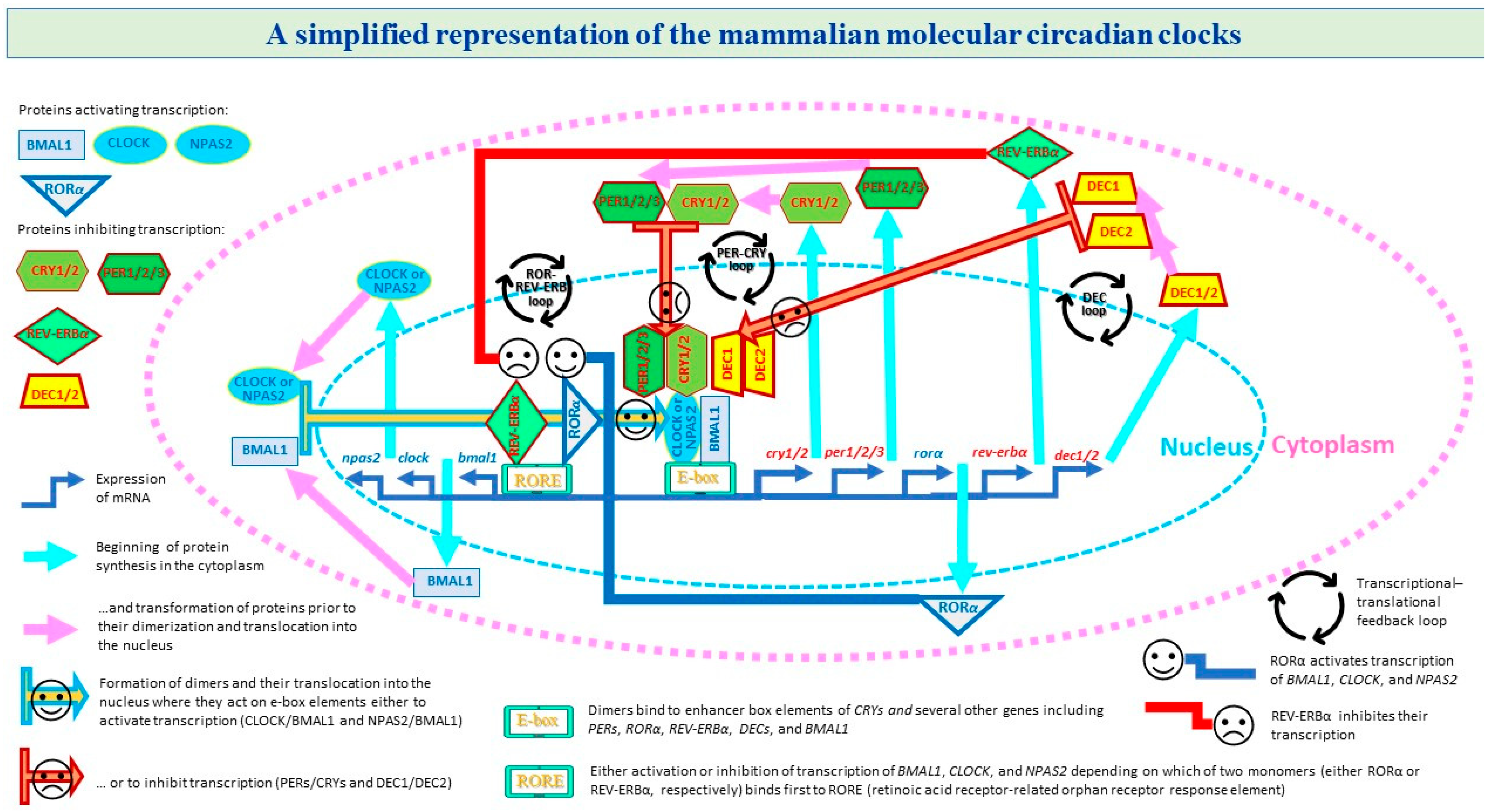
Figure 1. A simplified representation of three transcriptional–translational feedback loops. A transcriptional–translational feedback loop is a key feature of the circadian clock mechanism. In a mammalian cell, this mechanism includes several transcriptional–translational feedback loops involving clock genes. The circadian cycle is initiated by three circadian clock genes producing proteins BMAL1, CLOCK, and NPAS2 (brain and muscle aryl hydrocarbon receptor nuclear translocator-like 1, circadian locomotor output cycles kaput, and neuronal PAS domain protein 2, respectively) that are the members of the basic helix–loop–helix/Per-Arnt-Sim (bHLH-PAS) family of transcription factors. In the cytoplasm, they start to interact with each other to produce heterodimers BMAL1/CLOCK and BMAL1/NPAS2. At the next phase, these heterodimers translocate into the nucleus to activate the transcription of several other clock genes, including CRYs, PERs, (cryptochrome circadian regulator 1 and cryptochrome circadian regulator 2, period circadian regulator 1, period circadian regulator 2, and period circadian regulator 3), DECs (differentially expressed in chondrocyte 1 and differentially expressed in chondrocyte 2), and BMAL1 itself. Namely, BMAL1/CLOCK and BMAL1/NPAS2 heterodimers act as transcription factors by binding the E-box regions on the promoters of the target genes. In turn, CRYs, PERs, DECs encode for the repressor components of the circadian clocks. At a later phase of the cycle, the protein products of these genes dimerize and form complexes between themselves (PERs with CRYs and DEC1 with DEC2). At the final phase, the core feedback loop (PER-CRY) is completed by transporting the cytoplasmic dimers PERs/CRYs back into the nucleus to suppress activity of BMAL1/CLOCK and BMAL1/NPAS2 heterodimers. When they are suppressing activity of these heterodimers, they are also repressing their own expression, thus giving rise to a new cycle (i.e., this next cycle is started with the initiation of transcription of BMAL1, CLOCK, and NPAS2). The cytoplasmic dimers DEC1 and DEC2 are included in another feedback loop (DEC). As with PER and CRY dimers, they translocate into the nucleus to suppress activity of BMAL1/CLOCK and BMAL1/NPAS2 heterodimers. One more transcriptional–translational feedback loop (ROR-REV-ERB) includes two nuclear receptors, REV-ERBα (nuclear receptor subfamily 1 group D member 1) and RORα (RAR-related orphan receptor alpha), that additionally contribute to the circadian expression of BMAL1 and NPAS2. Both receptors are activated by BMAL1/CLOCK, and in turn regulate expression of BMAL1 and NPAS2 genes by acting on their promoter RORE (retinoic acid receptor-related orphan receptor response element) in the opposite direction, i.e., to either inhibit or activate transcription depending on which of two proteins binds first to the promoter (either REV-ERBα or RORα, respectively).
Clockssleep 05 00029 g001
Figure 2. PRISMA flow diagram for study selection.
Clockssleep 05 00029 g002
Table 1. Involvement of 12 circadian clock genes into thyroid carcinoma.
Feedback
Loop
NameCircadian Clock Transcript
GeneProteinUpregulatedDownregulatedNonsignificantNo Data
Monomers or dimers are promoters
All threeBMAL1 or ARNTLBMAL1 or ARNTLFTC and PTC [1] PTC [3] PTC [6] TC [7] MTC [9] [2,4,5,8]
All threeCLOCKCLOCKTC [7] MTC [9] PTC [3][1,2,4,5,6,8]
All threeNPAS2NPAS2ATC [8] [1,2,3,4,6,7,9]
ROR-REV-ERBRORαNuclear receptor ROR-alphaPTC [2] PTC [3][1,4,5,6,7,8,9]
Monomers or dimers are suppressors
ROR-REV-ERBREV-ERBα or NR1D1REV-ERBαPTC [2] FTC, PTC [1] PTC [3] [4,5,6,7,8,9]
PER-CRYCRY1CRY1 FTC, PTC [1] PTC [3] TC [7][2,4,5,6,8,9]
PER-CRYCRY2CRY2 FTC and PTC [1] PDTC [4] TC [7]PTC [3][2,5,6,8,9]
PER-CRYPER1PER1 FTC, PTC [1] PTC [3] TC [7] [2,4,5,6,8,9]
PER-CRYPER2PER2TC [7]FTC and PDTC [4]FTC, PTC [1] PTC [3] [2,5,6,8,9]
PER-CRYPER3PER3 FTC, PTC [1] PTC [3][2,4,5,6,7,8,9]
DECDEC1DEC1 PTC [5] [1,2,3,4,6,7,8,9]
DECDEC2DEC2 PTC [5] [1,2,3,4,6,7,8,9]
Notes. TC: thyroid carcinoma. Malignant neoplasms: not further specified (TC), well-differentiated papillary (PTC), follicular (FTC), poorly differentiated (PDTC), undifferentiated/anaplastic (ATC). Thyroid C-cell–derived carcinoma: medullary (MTC). References (publication year): [1] Mannic et al. (2013); [2] Mond et al. (2014); [3] Chitikova et al. (2015); [4] Makhlouf et al. (2016); [5] Gallo et al. (2018); [6] Sadowski et al. (2019); [7] Lou et al. (2021); [8] Xu et al. (2022); [9] Mou et al. (2022). No data: this transcript of circadian clock genes was not tested/mentioned in this article. An increase and a decrease in expression compared to normal samples are interpreted as up- and downregulation, respectively. See Figure 1 for more details on the involvement of monomers (RORα and REV-ERBα) and dimers formed by BMAL1, CLOCK, NPAS2, PERs, CRYs, and DECs in the circadian clock mechanism.
Table 2. Abnormal circadian rhythmicity of transcription of 7 clock genes in thyroid carcinoma.
NameCircadian Rhythmicity of TranscriptCompared to
GeneProteinAltered PhaseAltered AmplitudeOther TC Type
BMAL1 or ARNTLBMAL1 or ARNTLPDTC PTC or HB
REV-ERBα or NR1D1REV-ERBαPDTC PTC or HB
CRY1CRY1PDTC PTC or HB
CRY2CRY2PDTC PTC or HB
PER1PER1 PDTCPTC or HB
PER2PER2PDTC PTC or HB
PER3PER3PDTC PTC or HB
Notes. Summary of the findings on diurnal profiles of expression of 7 circadian clock genes reported in Mannic et al. (2013) [1]. HB: healthy tissue or benign nodule. See other notes in Table 1.
Disclaimer/Publisher’s Note: The statements, opinions and data contained in all publications are solely those of the individual author(s) and contributor(s) and not of MDPI and/or the editor(s). MDPI and/or the editor(s) disclaim responsibility for any injury to people or property resulting from any ideas, methods, instructions or products referred to in the content.