Case Report

Twice the Leak: Managing CSF Fistulas in a Recurrent Thoracic Arachnoid Cyst—A Case Report

Neurosurgery Unit, Department of Neuroscience “Rita Levi Montalcini”, A.O.U. Città della Salute e della Scienza, University of Turin, 10127 Turin, Italy
*
Author to whom correspondence should be addressed.
Reports 2025, 8(3), 152; https://doi.org/10.3390/reports8030152
Submission received: 30 June 2025 / Revised: 3 August 2025 / Accepted: 19 August 2025 / Published: 21 August 2025
(This article belongs to the Section Surgery)

Abstract

Background and Clinical Significance: Spinal arachnoid cysts are rare lesions that may become symptomatic through progressive spinal cord compression. We present a complex case of a thoracic extradural SAC in a 17-year-old male, managed through a stepwise, multidisciplinary approach. Case Presentation: The patient presented with progressive lower limb weakness, right knee paresthesia, and urinary hesitancy following physical exertion. MRI revealed a large posterior extradural SAC extending from T2–T3 to T8, with associated spinal cord compression. Initial management involved T8 laminectomy and cyst fenestration under intraoperative neurophysiological monitoring, with partial clinical improvement. However, early recurrence with pseudomeningocele formation prompted a second surgery, including external CSF drainage. Persistent cerebrospinal fluid (CSF) leakage led to targeted epidural blood patching, followed by temporary stabilization. Due to continued cyst enlargement and spinal cord compression, definitive surgical repair was undertaken: fistula clipping at T3 and embolization with platinum coils inside the cystic cavity, combined with a new blood patch. This novel technique resulted in radiological improvement and clinical stabilization. Conclusions: This case highlights the diagnostic and therapeutic challenges of managing symptomatic extradural SACs, particularly in young patients. Our experience underscores the utility of a staged approach involving surgical decompression, neuroimaging-guided interventions, and definitive dural repair. The combination of fistula clipping and coil embolization may offer a promising strategy for refractory cases, potentially reducing recurrence and preserving neurological function.

1. Introduction and Clinical Significance

Spinal arachnoid cysts (SACs) are rare but clinically relevant lesions, accounting for approximately 1–3% of all intraspinal masses. These cerebrospinal fluid (CSF)-filled sacs arise from the arachnoid membrane and may be located intradurally, extradurally, or in both compartments. Although often asymptomatic, SACs can cause a range of neurological symptoms when they compress the spinal cord or nerve roots. The thoracic spine is the most commonly affected region, with cysts typically positioned dorsally or posterolaterally to the spinal cord.
While asymptomatic cysts may be managed conservatively, surgical intervention is generally indicated for symptomatic cases. Complete resection is preferred due to its lower recurrence rate, although cyst fenestration or CSF shunting may be considered when excision is not feasible.
Herein, we present a rare case of a recurrent, symptomatic thoracic SAC with both intradural and extradural components, complicated by dual CSF fistulas in an adolescent patient. We discuss the clinical presentation, radiological features, surgical approach, and postoperative course.

2. Case Presentation

2.1. First Admission

A previously healthy 17-year-old male began experiencing right knee paresthesia and gradually worsening right foot weakness in September 2023, shortly after engaging in intense physical exercise during gym activities. Over the subsequent months, he developed gait instability and urinary hesitancy, without frank incontinence. Due to worsening symptoms, he presented to the emergency department of our institution in February 2024.
Neurological examination revealed marked right foot dorsiflexion weakness (Medical Research Council [MRC] grade 1/5), moderate plantarflexion weakness (MRC 3/5), and hyperactive bilateral patellar reflexes with right-sided reflexogenic spread. Bilateral Achilles clonus and a right Babinski sign were also noted. Sensory examination, including perineal sensation, was preserved.
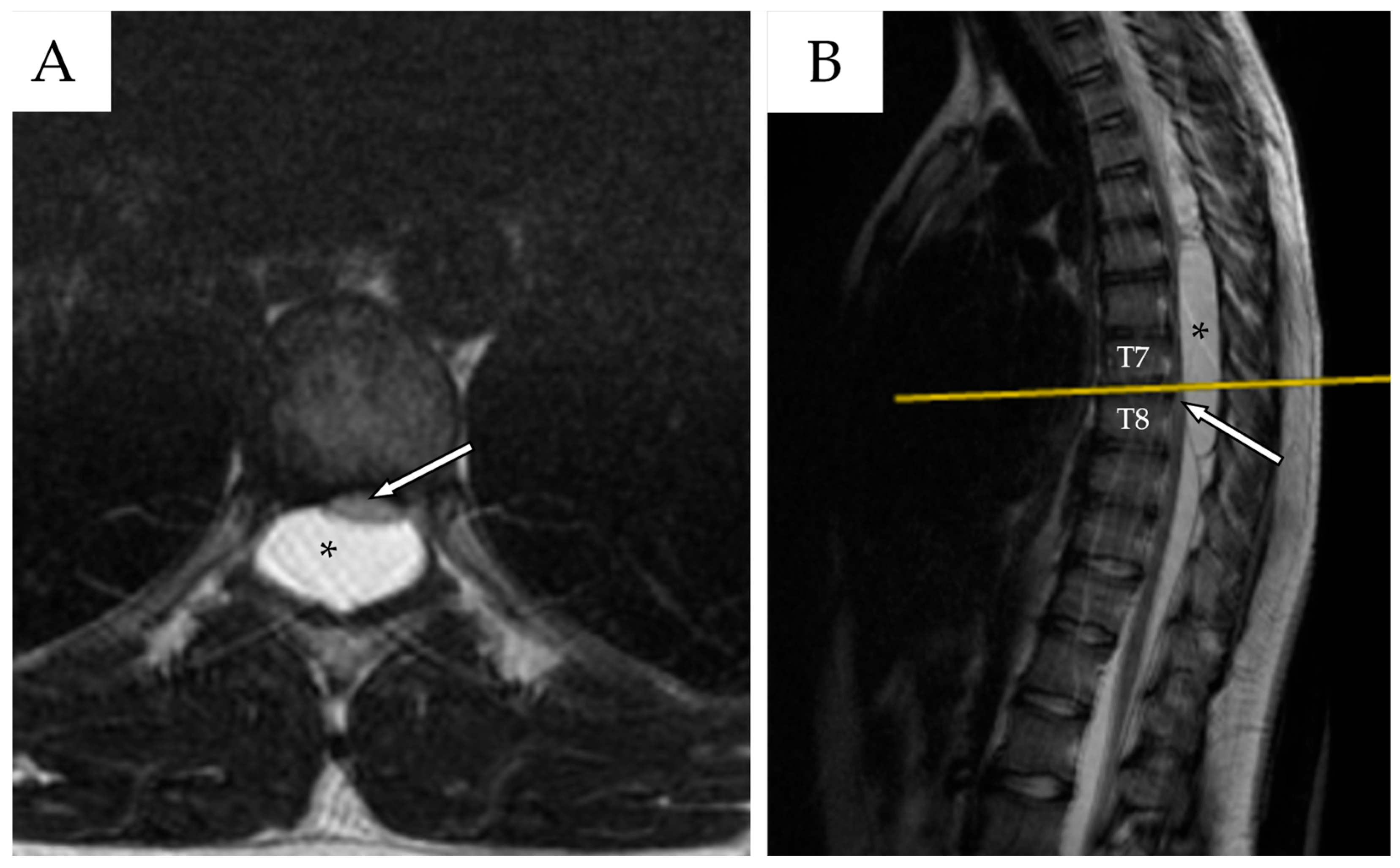
Whole-spine contrast-enhanced Magnetic Resonance Imaging (ceMRI) demonstrated a posterior, extradural intraspinal lesion extending from T2 to T8, causing significant compression of the dural sac and spinal cord (Figure 1). The lesion was formed by two components: a cranial one (T2–T4) and a larger caudal one (T5–T8). The area of maximal spinal cord compression was identified at T7–T8, where subtle intramedullary T2 hyperintensity was observed, suggestive of early spinal cord changes consistent with possible myelopathy (Figure 2). The lesion appeared hyperintense on T2-weighted images, isointense with CSF, and showed no contrast enhancement—findings consistent with a spinal arachnoid cyst.
No features typically associated with arachnoiditis—such as nerve root clumping, leptomeningeal enhancement, or intrathecal adhesions—were present on MRI. Moreover, there was no prior history of trauma, infection, or spinal instrumentation, further supporting a congenital origin of the cyst.
Given the longitudinal extent of the arachnoid cyst along the spinal canal, complete resection would have required a multilevel laminectomy from T2 to T8. This approach carried the potential need for subsequent spinal stabilization and would have necessitated a large surgical exposure. Considering the patient’s young age (17 years), we opted instead for cyst fenestration as a more conservative yet effective strategy.
The patient therefore underwent emergent thoracic decompression with a T8 laminectomy and cyst fenestration, performed under continuous intraoperative neurophysiological monitoring (IONM). Upon completion of the laminectomy, a thin, transparent membrane was observed. The cyst was punctured dorsally and found to contain a clear, cerebrospinal fluid-like liquid. After exposing the dural plane and the resection of a small portion of the cyst wall, the cyst was released toward the ventral side. Ultimately, communication was established between the cyst and the subarachnoid space on both the dorsal and ventral sides. Duroplasty was performed using a dural substitute and 6-0 vascular Prolene, reinforced with fibrin glue. Hermetic closure was confirmed by performing a Valsalva maneuver. The diagnosis of an arachnoid cyst was confirmed based on intraoperative findings, although a pathological diagnosis could not be established due to the limited amount of tissue available. No intraoperative complications occurred, and neurophysiological monitoring remained stable throughout the procedure.
Postoperatively, the patient showed improvement in right lower limb strength. He remained on bed rest for the first 24 h, with the surgical wound remaining clean and dry. Active mobilization was initiated on postoperative day 2.
Initial attempts at urinary catheter removal were unsuccessful. However, with intermittent catheterization, spontaneous voiding progressively resumed, and the patient achieved complete urinary independence by postoperative day 4.
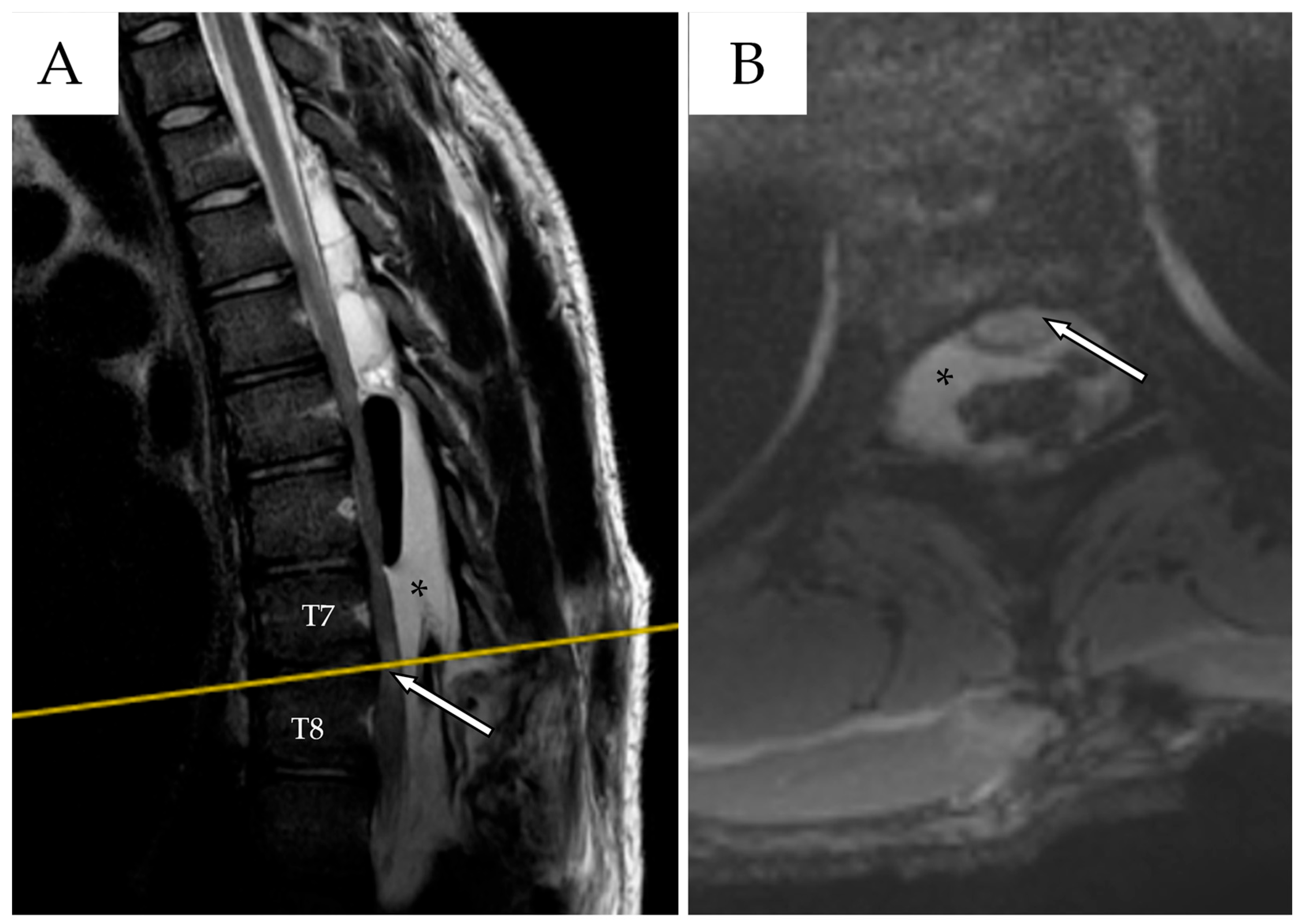
A postoperative MRI demonstrated a mild reduction in cyst volume and clear spinal cord decompression (Figure 3). The patient was enrolled in outpatient rehabilitation and was discharged on postoperative day 10. Upon discharge, the patient demonstrated independent ambulation with minimal aid. Right knee paresthesia had completely resolved. A motor deficit of the right foot persisted, predominantly affecting dorsiflexion (MRC 3/5), with mild residual weakness in plantarflexion.

2.2. First Follow-Up

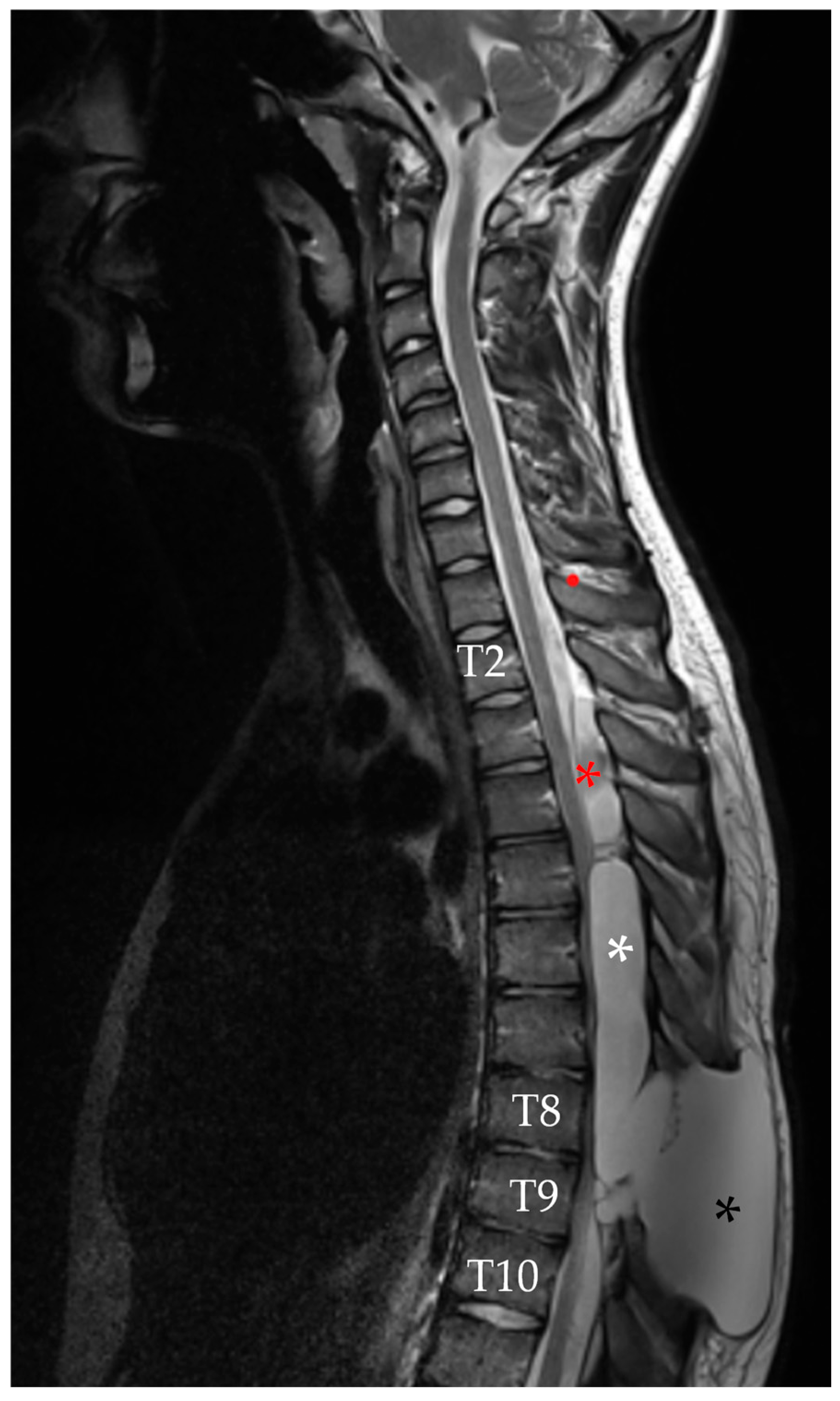
At the routine outpatient follow-up visit two weeks after discharge, urinary function had fully recovered. However, the control thoracic spine MRI revealed enlargement of the residual cystic components, both cranially at T2–T4 and caudally at T8–T9, adjacent to the previous fenestration site. Additionally, an 8.5 cm pseudomeningocele was identified, along with spinal cord signal changes between T5 and T8 (Figure 4). Given the clinical stability despite the MRI findings, a decision was made to continue close follow-up, with repeat imaging scheduled in two weeks.
At four weeks post-discharge right leg strength had improved to MRC 4/5, but a mild weakness developed on the left side (MRC 4/5). The patient also experienced new onset bladder tenesmus. The follow-up MRI with CSF flowmetry demonstrated no communication between the intradural space and the extradural cystic lesion. Given the MRI findings, outpatient myelography was scheduled, followed by clinical and radiological reassessment.

2.3. Second Admission

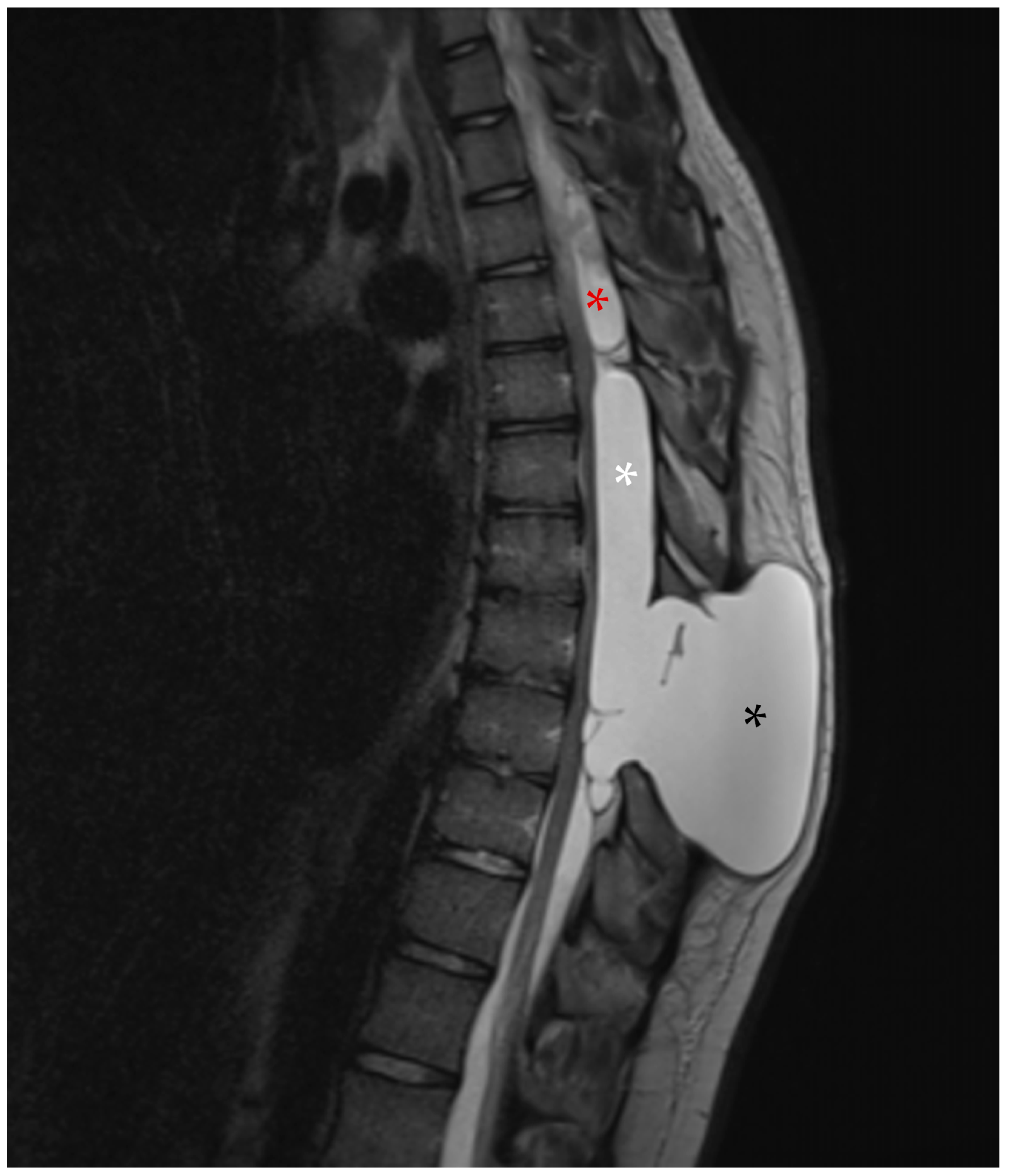
However, in early April, the patient returned to our emergency department with worsening of the bilateral lower limb weakness, gait disturbance, and urge incontinence. Neurological examination revealed bilateral leg weakness (M2 on the right, M3 on the left, globally), bilateral Babinski signs, and preserved perineal sensation. An emergent MRI was consistent with a further enlargement of the cyst and of the pseudomeningocele (Figure 5).
A second surgery was performed, again with continuous intraoperative neurophysiological monitoring. The procedure included repeat cyst fenestration, pseudomeningocele drainage, and placement of an external spinal CSF shunt. The latter was positioned to help control the volume of the pseudomeningocele and to promote wound healing, as the surgical site had already been reopened twice. CSF cultures obtained intraoperatively were negative for infection.
The immediate postoperative MRI showed minimal reduction in cyst volume (Figure 6). The shunt had a steady output of about 10 mL/h, and was removed on postoperative day 5. A new MRI performed at two weeks after surgery showed cranial enlargement of the fluid collection with persistent spinal cord compression. Myelography identified two CSF fistulas: one at the T3–T4 level communicating with the subarachnoid space, and another at T8–T9 connecting to the pseudomeningocele.
Following multidisciplinary consultation with the interventional neuroradiology team, a targeted epidural blood patch under CT guidance was performed, leading to the apparent exclusion of the cyst and radiographic closure of both fistulous tracts.
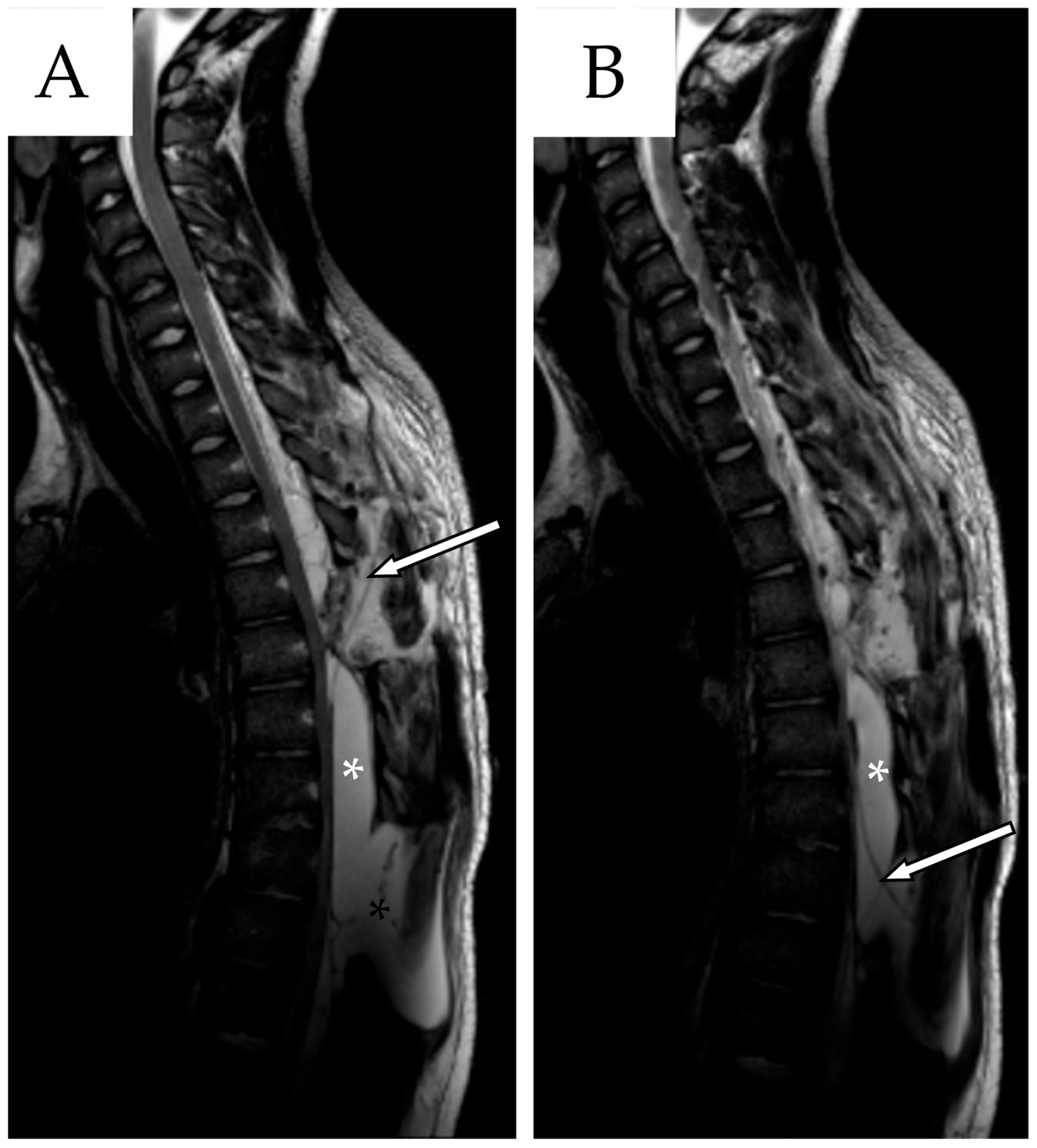
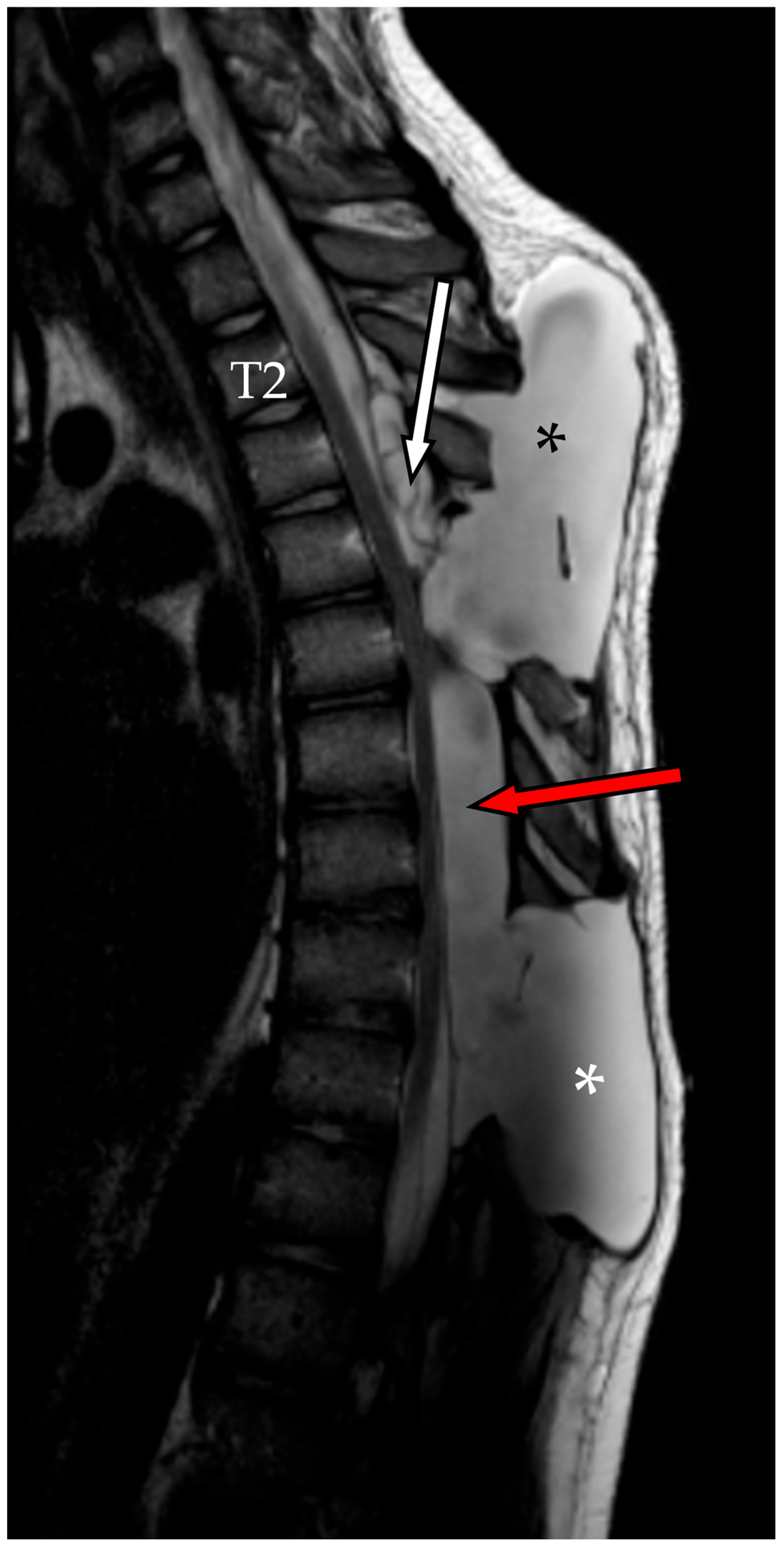
Although the blood patch was successfully performed, follow-up imaging at two weeks revealed the onset of a new cystic formation at the T4–T5 level extending in the soft tissues from T2 to T5 compatible with another pseudomeningocele; it probably developed at the point of entrance of the previously placed external drainage. The cranial component of the SAC was stable, whereas the caudal component together with the caudal pseudomeningocele appeared refilled and slightly enlarged (Figure 7). The patient’s neurological status remained stable though and a conservative management of “watch and see” was adopted.
During this period lasting three weeks, the pseudomeningocele collection continued to increase in size, and was appreciable at the inspection evaluation. MRI confirmed ongoing spinal cord compression at the T7–T8 level, and myelography demonstrated persistent cerebrospinal fluid leakage from a dorsal fistula at T3–T4.
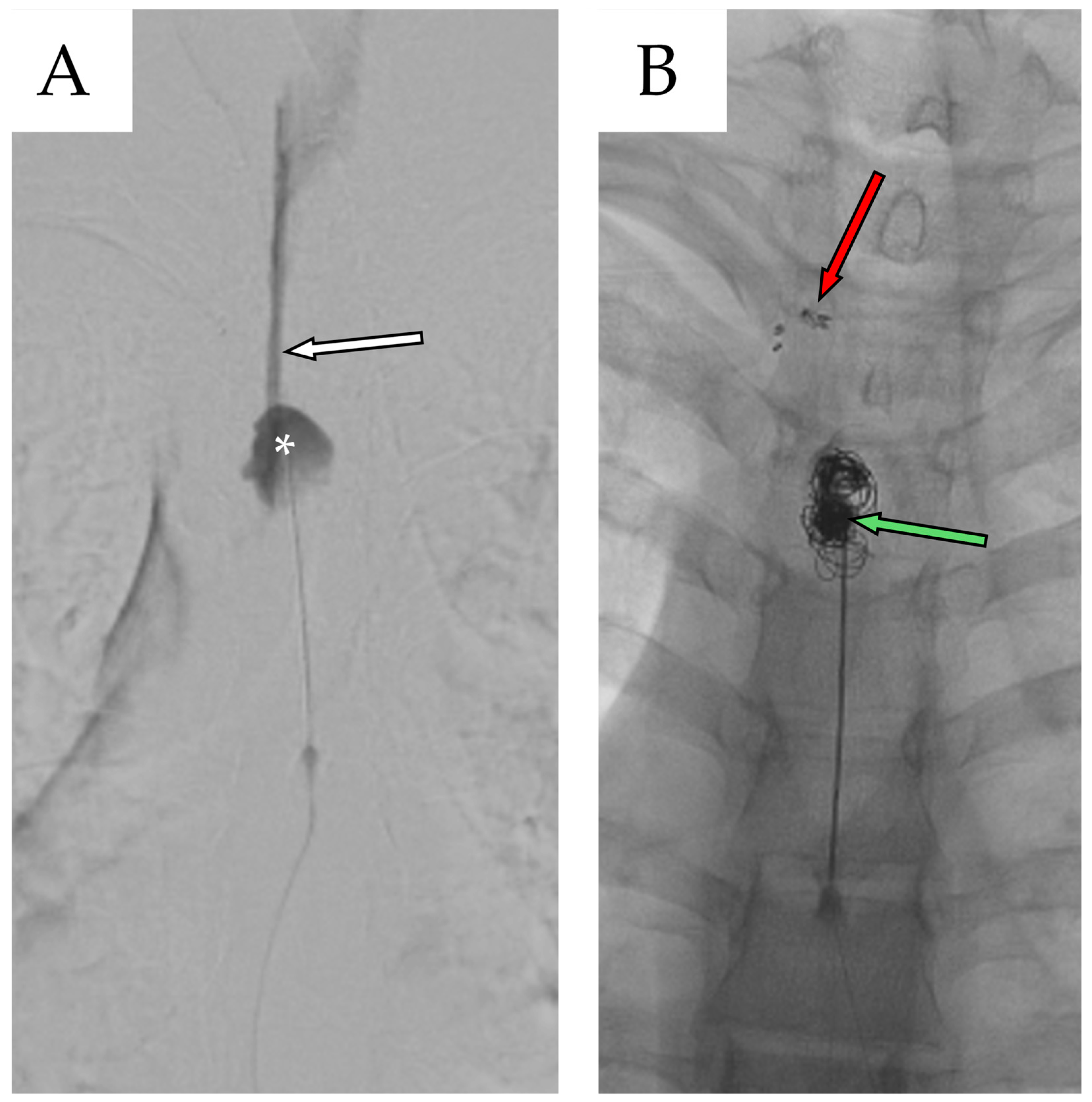
A new multidisciplinary consultation was convened to address the need for definitive surgical repair. Two months after second admission, the patient underwent a third and final surgical procedure, consisting of bilateral T4 laminectomy with clipping of the T3 fistulous tract combined with percutaneous repair of the dural leak using metallic coils placed within the cystic cavity followed by a new blood patch (Figure 8).
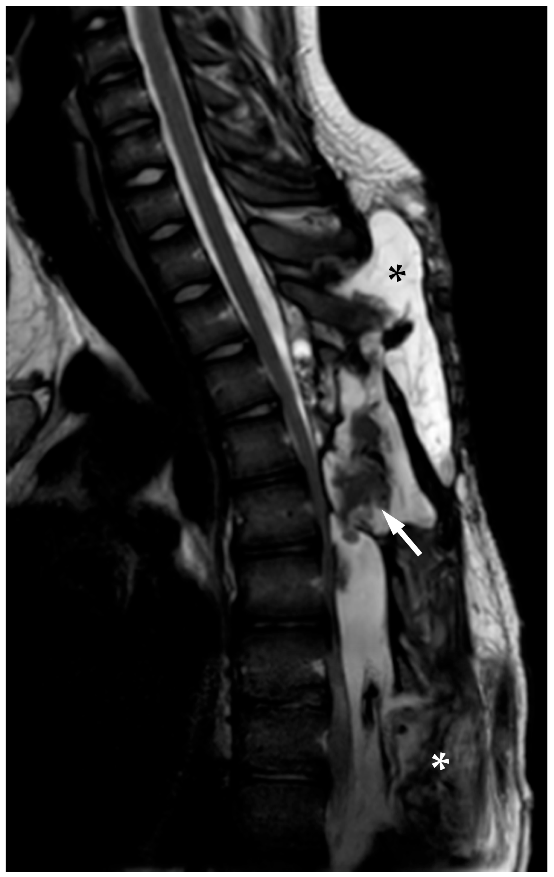
Finally, MRI at 1 week after surgery showed a reduction in size of the pseudomeningocele at the T2–T4 level, along with decreased overall spinal cord compression and almost complete resolution of the caudal pseudomeningocele (Figure 9). The patient remained neurologically stable with mild improvement and no new deficits were observed.
The patient was subsequently discharged to their home. At discharge, the patient presented with residual motor deficits in the lower extremities, with muscle strength graded as MRC 4+/5 on the left and MRC 4−/5 proximally and 4/5 distally on the right. No sensory deficits were noted across thermal-nociceptive, light touch, or discriminatory modalities (both epicritic and protopathic). Bladder function was preserved, and bowel transit remained normal. Patellar reflexes were hyperelicitable, with an expanded reflexogenic zone. Independent ambulation was achieved, with partial weight-bearing allowed on the more affected limb, although gait remained slightly circumductory.

2.4. Second Follow-Up

The patient remained in good clinical condition, with no further neurological deterioration. He underwent serial follow-up evaluations every three months during the first year, including both clinical assessments and control MRI scans. These demonstrated a gradual regression of radiological signs of myelopathy and a reduction in the size of the pseudomeningocele. He continued occupational and physical therapy and was educated on strict return precautions and warning signs (i.e., “red flag” symptoms). The patient is currently undergoing regular outpatient follow-up for ongoing monitoring, now scheduled annually.

3. Discussion

According to the classification proposed by Nabors et al. [1], spinal arachnoid cysts (SACs) are categorized into three types: Type I, II, and III. Type I cysts are extradural without involvement of nerve root fibers; Type II cysts are extradural with involvement of nerve root fibers; and Type III cysts are intradural. In the present case report, we describe a Type I cyst. The origin of the lesion remains unclear: its size and radiological appearance suggest a congenital SAC, whereas the onset of symptoms following physical exertion could indicate a secondary SAC. Nevertheless, the congenital origin appears more likely.
Congenital SACs are thought to originate either from diverticula of the septum posticum (anatomic theory) [2] or from hypertrophic and dilated arachnoid granulations [3,4]. These congenital cysts are sometimes associated with central nervous system malformations, such as syringomyelia [5], not presented in our case. Secondary SACs may result from subarachnoid hemorrhage [6], trauma, infection, neoplasms, arachnoiditis, or iatrogenic causes, including diagnostic procedures such as myelography and, in rare cases, post-laminectomy changes [7,8].
Type I spinal arachnoid cysts, which are extradural and do not involve nerve root fibers, are more common than intradural cysts (Type III) and Type II extradural cysts. They are most often located in the thoracic spine, typically posterior or posterolateral to the spinal cord, as seen in the present case, but they may also occur in the cervical and lumbar regions [3,9]. While primary extradural SACs (including Type I) are thought to be congenital and dorsally located, secondary cysts may present ventrally and can involve a valve-like mechanism that disrupts CSF dynamics [10].
The pathogenesis of spinal arachnoid cysts (SACs) remains incompletely understood, in part due to the still unclear mechanisms regulating cerebrospinal fluid (CSF) dynamics and pressure within the central nervous system [11]. Several theories have been proposed to explain both the development and progressive enlargement of these cysts. According to the ‘ball-valve’ mechanism described by Rohrer [12], once an arachnoid protrusion forms, CSF becomes trapped within the cyst without adequate drainage. The ‘osmotic enlargement’ theory suggests that hyperosmolar cyst fluid may draw water into the cyst via osmotic gradients [13]. A third hypothesis posits that the cyst walls themselves may secrete fluid [14].
Extradural SACs may originate from arachnoid herniation through dural defects, whereas intradural cysts are thought to result from alterations of the arachnoid trabeculae [15]. In general, any mechanism that increases CSF pressure may contribute to cyst growth.
There appears to be no clear gender or age predilection reported in the literature [4,5]. However, when symptomatic, SACs most commonly present in early adulthood, typically between the ages of 30 and 50 [2]. Earlier onset has been reported in some cases—for example, in the case series by Abdel-Hameed & Morsy [7], which included a patient as young as 3 years old—and our case again shows its unicity for the young age of onset.
Magnetic resonance imaging with contrast is the modality of choice for diagnosing spinal arachnoid cysts. On T2-weighted sequences, SACs appear as lesions isointense with CSF and are best visualized on sagittal views [7]. MRI allows for precise localization of the lesion and can reveal internal features such as septations or loculations [4].
MRI also plays a critical role in postoperative evaluation, particularly in assessing residual spinal cord compression. Its high spatial resolution enables detailed characterization of the cyst and aids in the differential diagnosis from other lesions, such as neurenteric cysts (commonly located in the cervicothoracic region), perineural cysts, synovial cysts, epidermoid cysts, and dermoid cysts [7].
Currently, there are no definitive guidelines for the treatment of SACs. In general, surgical intervention is indicated in symptomatic cases, particularly in the presence of myelopathy; all criteria for operation were presented by our patient [3].
For patients with mild or minimal symptoms, a conservative approach with clinical monitoring may be appropriate, with surgery reserved for those who experience significant neurological deterioration [16]. According to the literature, delaying surgical treatment in mildly symptomatic patients—even in the event of later clinical progression—does not appear to adversely affect long-term neurological outcomes [17].
Treatment options include surgical excision, fenestration, and cysto-subarachnoid shunting. The choice of procedure depends on the cyst’s location, type, size, and the degree of adhesion to surrounding structures [18]. Complete surgical excision is considered the gold standard, as it is associated with lower recurrence rates and improved long-term clinical outcomes, while most reported recurrences have occurred in patients who underwent fenestration alone [9,18]. As previously said, the choice of undergoing a fenestration in the myelopathic level instead of an excision was based on the young age of the patient and the cranial to caudal extension of the SAC itself, which would have needed a long incision and a wide decompression with the need of thoracic spine stabilization with fixation from T1 to T9.
Identifying the precise communication point between the cyst and the subarachnoid space is essential to minimize surgical exposure, reduce the risk of postoperative CSF leakage, and prevent pseudomeningocele formation [19].
Intraoperatively, distinguishing the cyst wall from the surrounding arachnoid membrane can be challenging. This often precludes en bloc resection and increases the risk of leaving residual tissue, thereby contributing to recurrence. The use of intraoperative dyes has been proposed to facilitate complete and safe excision by delineating the cyst boundaries from adjacent arachnoid tissue [20]. Additionally, staining the SAC may assist in defining its margins, potentially avoiding unnecessary laminectomies or dural incisions that could increase the risk of postoperative complications such as infection, hemorrhage, or spinal instability.
To minimize surgical trauma, endoscopic techniques may be utilized to localize the site of CSF leakage, thereby enabling more targeted laminectomies and reducing the risk of postoperative complications [3].
In all surgical procedures performed on our patient—including in the emergency setting—intraoperative neurophysiological monitoring (IONM) was employed to minimize the risk of spinal cord injury. This was made possible by the availability of dedicated neurophysiologists and IONM technicians at our institution.
As expected—and consistent with the higher recurrence rate associated with fenestration alone—the patient experienced a recurrence of symptoms after initial postoperative improvement, necessitating revision surgery. The clinical course was further complicated by the development of a significant pseudomeningocele, a known complication following arachnoid cyst surgery, particularly when the fistulous tract cannot be clearly identified. Fortunately, to date, no CSF infection has been observed in the present case.
In cases of recurrence, cystoperitoneal, cystopleural, or cystoatrial shunting may be considered as alternative treatment options [21,22]. In our case, a cystoperitoneal shunt was proposed during the second multidisciplinary discussion as a definitive solution. However, the decision was ultimately made to forgo this approach, due to concerns about placing a permanent silicone device in a 17-year-old patient and the associated risk of shunt-related infections—particularly in the context of an existing pseudomeningocele, which may further increase infection risk.
In the present case, a novel technique was implemented, combining closure of the fistulous tract using titanium aneurysmal clips with the placement of metallic coils inside the SAC. To the best of our knowledge, no similar attempts have been previously reported in the available literature. The rationale behind the combination of a blood patch and coiling was that a blood patch alone would probably not have been sufficient to seal the SAC, as the presence of the pseudomeningocele and the high volume of CSF would have diluted the blood, preventing the coagulation mechanism from taking place. By inserting the coil together with the blood patch, a pro-thrombotic surface is provided to help trigger the coagulation cascade.
Neo M. et al. [23] described in 2004 the closure of a fistulous tract in an extradural spinal arachnoid cyst using metal clips, resulting in an almost complete disappearance of the cyst just 12 days after the operation. The underlying principle is the same: to close the communication between the subarachnoid space and the cyst. However, in our case, platinum coiling was added to promote coagulation of the cyst’s contents, in combination with a new percutaneous blood patch.
The combined approach proved effective, though the results were not as striking as those reported by Neo M. et al., likely due to the presence of the pseudomeningocele. Nevertheless, the patient achieved a satisfactory neurological outcome and favorable imaging findings at the last outpatient follow-up, one year after discharge.
To further characterize the SACs and contextualize the present case within the existing literature, we conducted a comprehensive review of case reports and case series published between 2005 and 2024. A total of 37 articles describing the clinical history of 157 patients with spinal arachnoid cysts were identified.
Based on the collected data, this pathology predominantly affects a younger population, with most patients ranging between 20 and 50 years of age. However, pediatric cases (as young as 4 years) and elderly patients (up to 80 years) were also reported. The mean age across cohorts ranged between 8 and 60 years in larger studies. A slight female predominance was observed (approximately 85 females vs. 72 males).
Risk factors were identified in a subset of patients, mainly including spinal trauma, prior spinal surgery, central nervous system anomalies, and, less frequently, epidural steroid injections, schwannoma, or post-surgical meningitis.
The thoracic spine was the most commonly affected region, followed by the Thoracic–lumbar and lumbar regions. Less frequently, cysts were located in the cervical or Cervical–thoracic spine. Multisegmental involvement was observed in rare cases.
Regarding cyst classification, Type III (intradural or intramedullary cysts) was the most frequent, accounting for the majority of cases. Some articles reported mixed-type cysts or unspecified classification.
Symptoms varied depending on cyst location and size. The most common clinical presentations were motor weakness, particularly in the lower limbs, urinary and/or bowel dysfunction, and radicular pain. Sensory deficits, gait ataxia, myelopathy, and in some pediatric cases, tetraparesis or quadriplegia, were also noted. A few cases were asymptomatic and discovered incidentally.
Surgical treatment was the mainstay in nearly all symptomatic cases. Complete excision was considered the gold standard and was performed in most patients, often with good outcomes and remission of symptoms. In other cases, fenestration alone or followed by a new intervention of excision was used. Less frequently, shunt placement or conservative management was adopted in selected asymptomatic or high-risk patients.
Postoperative outcomes were generally favorable. The majority of patients showed full or significant neurological recovery, even in those requiring reoperation for complications such as recurrence, CSF fistulas, or pseudomeningoceles. Only a small subset experienced persistent or progressive symptoms. All data are summarized in Table 1 and Table 2.

4. Conclusions

This case highlights both the diagnostic challenges and therapeutic complexities associated with spinal arachnoid cysts, particularly in young patients. Despite initial surgical management through fenestration, recurrence and complications such as pseudomeningocele necessitated innovative treatment strategies. The implementation of a novel technique combining aneurysmal clipping and platinum coiling, along with a percutaneous blood patch, represents a promising approach in selected cases where standard surgical options are limited or pose significant risks. While the outcome was ultimately satisfactory, the experience underscores the importance of individualized treatment planning, multidisciplinary collaboration, and the need for continued research into less invasive and more effective management options for SACs. Further studies and long-term follow-up will be essential to validate the efficacy and safety of such combined interventions.

Author Contributions

Conceptualization, L.B. and F.B.; methodology, F.B.; investigation, L.B., F.B.; resources, F.B.; data curation, L.B.; writing—original draft preparation, F.B.; writing—review and editing, L.B.; visualization, M.A. and D.G.; supervision, M.A.; project administration, D.G. All authors have read and agreed to the published version of the manuscript.

Funding

This research received no external funding.

Institutional Review Board Statement

The study was conducted in accordance with the Declaration of Helsinki. According to current Italian regulations as well as the policies of our institution, ethical approval from an Institutional Review Board is not required for a case report, provided that the patient has given informed consent for publication. This is in line with the national guidelines and the position of the Italian Data Protection Authority, which does not mandate ethics committee approval for single-patient case reports that do not include sensitive personal data beyond what is necessary and where appropriate consent is obtained.

Informed Consent Statement

Informed consent was obtained from all subjects involved in the study. Written informed consent has been obtained from the patient to publish this paper.

Data Availability Statement

Due to patient privacy concerns, the data presented in this study are available from the corresponding author upon reasonable request.

Acknowledgments

We would like to thank Mauro Bergui for his support during the multidisciplinary meeting and Carola Juenemann for her assistance and expertise with the IONM during surgery.

Conflicts of Interest

The authors declare no conflicts of interest.

Abbreviations

The following abbreviations are used in this manuscript:
SACsSpinal Arachnoid Cysts
CSFCerebrospinal Fluid
MRIMagnetic Resonance Imaging
MRCMedical Research Council
ceMRIcontrast-enhanced Magnetic Resonance Imaging
IONMIntraoperative Neurophysiological Monitoring
TSETurbo Spin Echo
WIWeighted Imaging
NANot Available
LBPLow Back Pain
TLIFTransforaminal Lumbar Interbody Fusion

References

  1. Nabors, M.W.; Pait, T.G.; Byrd, E.B.; Karim, N.O.; Davis, D.O.; Kobrine, A.I.; Rizzoli, H.V. Updated assessment and current classification of spinal meningeal cysts. J. Neurosurg. 1988, 68, 366–377. [Google Scholar] [CrossRef] [PubMed]
  2. Kriss, T.C.; Kriss, V.M. Symptomatic spinal intradural arachnoid cyst development after lumbar myelography: Case report and review of the literature. Spine 1997, 22, 568–572. [Google Scholar] [CrossRef] [PubMed]
  3. Papadimitriou, K.; Cossu, G.; Maduri, R.; Valerio, M.; Vamadevan, S.; Daniel, R.; Messerer, M. Endoscopic treatment of spinal arachnoid cysts. Heliyon 2021, 7, e06736. [Google Scholar] [CrossRef] [PubMed] [PubMed Central]
  4. Levin, T.L.; Zimmerman, R.D.; Lieberman, H. Intradural spinal arachnoid cyst: Case report. Clin. Imaging 1990, 14, 245–247. [Google Scholar] [CrossRef] [PubMed]
  5. Mirza, A.B.; Bartram, J.; Vastani, A.; Gebreyohanes, A.; Al Banna, Q.; Lavrador, J.P.; Vasan, A.K.; Grahovac, G. Systematic review of surgical management of spinal intradural arachnoid cysts. World Neurosurg. 2022, 158, e298–e309. [Google Scholar] [CrossRef] [PubMed]
  6. Hussain, O.; Treffy, R.; Reecher, H.M.; DeGroot, A.L.; Palmer, P.; Bakhaidar, M.; Shabani, S. Management of a recurrent spinal arachnoid cyst presenting as arachnoiditis in the setting of spontaneous spinal subarachnoid hemorrhage: Illustrative case. J. Neurosurg. Case Lessons 2024, 7, CASE23660. [Google Scholar] [CrossRef] [PubMed] [PubMed Central]
  7. Abdelhameed, E.; Morsy, A.A. Surgical outcome of primary intradural spinal arachnoid cysts: A series of 10 cases. Egypt J. Neurol. Psychiatry Neurosurg. 2021, 57, 42. [Google Scholar] [CrossRef]
  8. Nath, P.C.; Mishra, S.S.; Deo, R.C.; Satapathy, M.C. Intradural spinal arachnoid cyst: A long-term postlaminectomy complication: Case report and review of the literature. World Neurosurg. 2016, 85, e1–e367. [Google Scholar] [CrossRef] [PubMed]
  9. Ramazanoglu, A.F.; Sarikaya, C.; Varol, E.; Aydin, S.O.; Etli, M.U.; Avci, F.; Naderi, S. Surgical treatment of spinal arachnoid cysts: Cyst excision or fenestration? Turk Neurosurg. 2022, 32, 1002–1006. [Google Scholar] [CrossRef]
  10. Wang, Y.B.; Wang, D.H.; Deng, S.L. Symptomatic secondary spinal arachnoid cysts: A systematic review. Spine J. 2023, 23, 1199–1211. [Google Scholar] [CrossRef] [PubMed]
  11. Theologou, M.; Natsis, K.; Kouskouras, K.; Chatzinikolaou, F.; Varoutis, P.; Skoulios, N.; Tsitouras, V.; Tsonidis, C. Cerebrospinal fluid homeostasis and hydrodynamics: A review of facts and theories. Eur. Neurol. 2022, 85, 313–325. [Google Scholar] [CrossRef] [PubMed]
  12. Rohrer, D.C.; Burchiel, K.J.; Gruber, D.P. Intraspinal extradural meningeal cyst demonstrating ball-valve mechanism of formation: Case report. J. Neurosurg. 1993, 78, 122–125. [Google Scholar] [CrossRef] [PubMed]
  13. Bhaisora, K.S.; Singh, S.; Sardhara, J.; Das, K.K.; Attri, G.; Mehrotra, A.; Srivastava, A.K.; Jasiwal, A.K.; Behari, S. Symptomatic extradural spinal arachnoid cyst: More than a simple herniated sac. J. Craniovertebr. Junction Spine 2019, 10, 64–71. [Google Scholar] [CrossRef] [PubMed] [PubMed Central]
  14. Kulkarni, A.G.; Goel, A.; Thiruppathy, S.P.; Desai, K. Extradural arachnoid cysts: A study of seven cases. Br. J. Neurosurg. 2004, 18, 484–488. [Google Scholar] [CrossRef] [PubMed]
  15. Marbacher, S.; Barth, A.; Arnold, M.; Seiler, R.W. Multiple spinal extradural meningeal cysts presenting as acute paraplegia: Case report and review of the literature. J. Neurosurg. Spine 2007, 6, 465–472. [Google Scholar] [CrossRef] [PubMed]
  16. Kodali, S.; Panigrahi, M. Strategies to improve surgical outcome of spinal arachnoid cyst—A single center, single surgeon experience from a tertiary care center. J. Spinal Surg. 2025, 12, 3–7. [Google Scholar]
  17. Chatain, G.P.; Shrestha, K.; Kortz, M.W.; Serva, S.; Hosokawa, P.; Ward, R.C.; Sethi, A.; Finn, M. Impact of surgical timing on neurological outcomes for spinal arachnoid cyst: A single institution series. Neurospine 2022, 19, 453–462. [Google Scholar] [CrossRef] [PubMed] [PubMed Central]
  18. El-Hajj, V.G.; Singh, A.; Pham, K.; Edström, E.; Elmi-Terander, A.; Fletcher-Sandersjöö, A. Long-term outcomes following surgical treatment of spinal arachnoid cysts: A population-based consecutive cohort study. Spine J. 2023, 23, 1869–1876. [Google Scholar] [CrossRef] [PubMed]
  19. Zhang, P.; Liu, H.; Sun, Z.; Guo, Y.; Wang, G.; Wang, J.J. Ultrafine flexible endoscope visualization to assist in the removal of a huge spinal extradural arachnoid cyst: Case report and literature review. World Neurosurg. 2022, 159, 130–133. [Google Scholar] [CrossRef] [PubMed]
  20. Takamiya, S.; Seki, T.; Yamazaki, K.; Sasamori, T.; Houkin, K. Intraoperative visualization of a spinal arachnoid cyst using pyoktanin blue. World Neurosurg. 2018, 109, 18–23. [Google Scholar] [CrossRef] [PubMed]
  21. Nakahashi, M.; Uei, H.; Tokuhashi, Y. Recurrence of a symptomatic spinal intradural arachnoid cyst 29 years after fenestration. J. Int. Med. Res. 2019, 47, 4530–4536. [Google Scholar] [CrossRef] [PubMed] [PubMed Central]
  22. DeGroot, A.L.; Treffy, R.W.; Bakhaidar, M.; Palmer, P.; Rahman, M.; Shabani, S. Minimally invasive management of a spinal arachnoid cyst with ultrasound-assisted catheter placement: Illustrative case. J. Neurosurg. Case Lessons 2024, 8, CASE24461. [Google Scholar] [CrossRef] [PubMed] [PubMed Central]
  23. Neo, M.; Koyama, T.; Sakamoto, T.; Fujibayashi, S.; Nakamura, T. Detection of a dural defect by cinematic magnetic resonance imaging and its selective closure as a treatment for a spinal extradural arachnoid cyst. Spine 2004, 29, E426–E430. [Google Scholar] [CrossRef] [PubMed]
  24. Ichinose, T.; Miyashita, K.; Tanaka, S.; Oikawa, N.; Oishi, M.; Nambu, I.; Kinoshita, M.; Nakada, M. Recurrent spinal intramedullary arachnoid cyst: Case report and literature review. World Neurosurg. 2020, 138, 68–72. [Google Scholar] [CrossRef]
  25. Novak, L.; Dobai, J.; Nemeth, T.; Fekete, M.; Prinzinger, A.; Csecsei, G.I. Spinal extradural arachnoid cyst causing cord compression in a 15-year-old girl: A case report. Zentralbl. Neurochir. 2005, 66, 43–46. [Google Scholar] [CrossRef]
  26. Lee, H.G.; Kang, M.S.; Na, Y.C.; Jin, B.H. Spinal intradural arachnoid cyst as a complication of insertion of an interspinous device. Br. J. Neurosurg. 2023, 37, 811–815. [Google Scholar] [CrossRef]
  27. Krstačić, A.; Krstačić, G.; Butković Soldo, S. Atypical cause of radiculopathy—Intradural spinal arachnoid cyst. Acta Clin. Belg. 2016, 71, 267–268. [Google Scholar] [CrossRef]
  28. Lee, H.J.; Cho, D.Y. Symptomatic spinal intradural arachnoid cysts in the pediatric age group: Description of three new cases and review of the literature. Pediatr. Neurosurg. 2001, 35, 181–187. [Google Scholar] [CrossRef]
  29. Bond, A.E.; Zada, G.; Bowen, I.; McComb, J.G.; Krieger, M.D. Spinal arachnoid cysts in the pediatric population: Report of 31 cases and a review of the literature. J. Neurosurg. Pediatr. 2012, 9, 432–441. [Google Scholar] [CrossRef]
  30. Liu, J.K.; Cole, C.D.; Sherr, G.T.; Kestle, J.R.W.; Walker, M.L. Noncommunicating spinal extradural arachnoid cyst causing spinal cord compression in a child. J. Neurosurg. 2005, 103 (Suppl. 3), 266–269. [Google Scholar] [CrossRef] [PubMed]
  31. El-Hajj, V.G.; Edström, E.; Elmi-Terander, A.; Fletcher-Sandersjöö, A. An unusual cause of chronic neuropathic pain: Report of a case of multiple intradural spinal arachnoid cysts and review of the literature. Acta Neurochir. 2023, 165, 2699–2705. [Google Scholar] [CrossRef] [PubMed]
  32. Özdemir, M.; Kavak, R.P.; Gülgönül, N. Spinal extradural arachnoid cyst in cervicothoracic junction. Spinal Cord. Ser. Cases 2019, 5, 45. [Google Scholar] [CrossRef]
  33. Ebot, J.; Domingo, R.; Ruiz Garcia, H.; Chen, S. Intradural thoracic arachnoid cyst fenestration for spinal cord compression: A case illustration and video demonstration. Cureus 2020, 12, e6572. [Google Scholar] [CrossRef]
  34. Alugolu, R.; Arradi, V.; Sahu, B.P. Intramedullary arachnoid cyst in an adult: Case report and review. Asian J. Neurosurg. 2016, 11, 70. [Google Scholar] [CrossRef] [PubMed]
  35. Nayak, R.; Chaudhuri, A.; Sadique, S.; Attry, S. Multiple spinal extradural arachnoidal cysts: An uncommon cause of thoracic cord compression. Asian J. Neurosurg. 2017, 12, 321–323. [Google Scholar] [CrossRef]
  36. Sharif, S.; Afsar, A.; Qadeer, M. Conus medullaris arachnoid cyst presenting as cauda equina syndrome. Asian J. Neurosurg. 2017, 12, 707–709. [Google Scholar] [CrossRef]
  37. Lin, L.C.; Jason, R. A rare case of spinal extradural arachnoid cyst with cord compression. Asian J. Neurosurg. 2018, 13, 468–470. [Google Scholar] [CrossRef]
  38. Shaaban, A.; Abdelrahman, A.; Jarir, R.; Al-Bozom, I.; Raza, A. Thoracic spinal intramedullary arachnoid cyst presented with myelopathy with marked postoperative improvement: A case report and review of literature. Asian J. Neurosurg. 2019, 14, 981–984. [Google Scholar] [CrossRef]
  39. Raes, K.; Oostra, K.M. Correlation of spinal cord injury with development of spinal arachnoid cysts: Two case reports. J. Rehabil. Med. Clin. Commun. 2021, 4, 1000066. [Google Scholar] [CrossRef]
  40. Taccone, M.S.; Theriault, P.G.; Roffey, D.M.; AlShumrani, M.; Alkherayf, F.; Wai, E.K. Intradural hematoma and arachnoid cyst following lumbar spinal surgery: A case report. Can. J. Neurol. Sci. 2018, 45, 114–116. [Google Scholar] [CrossRef] [PubMed]
  41. Turel, M.K.; Kerolus, M.G.; Deutsch, H. Intradural spinal arachnoid cyst—A complication of lumbar epidural steroid injection. Neurol. India 2017, 65, 863–864. [Google Scholar] [CrossRef]
  42. Kong, W.K.; Cho, K.T.; Hong, S.K. Spinal extradural arachnoid cyst: A case report. Korean J. Spine 2013, 10, 32–34. [Google Scholar] [CrossRef]
  43. Fischer, K.M.; Madsen, P.J.; Tucker, A.M.; Long, C.J. Thoracic spinal arachnoid cyst in a pediatric patient presenting with isolated bladder and bowel incontinence. Urol. Case Rep. 2022, 44, 102129. [Google Scholar] [CrossRef]
  44. Sharma, R.; Kumarasamy, S.; Tiwary, S.K.; Kedia, S.; Sawarkar, D.; Doddamani, R.; Laythalling, R.K. Multiple spinal extradural arachnoid cysts presenting as compressive myelopathy in a teenager. Childs Nerv. Syst. 2024, 40, 729–747. [Google Scholar] [CrossRef]
  45. Engelhardt, J.; Vignes, J.R. Anterior cervical intradural arachnoid cyst, a rare cause of spinal cord compression: A case report with video systematic literature review. Eur. Spine J. 2016, 25 (Suppl. 1), 19–26. [Google Scholar] [CrossRef]
  46. Zekaj, E.; Saleh, C.; Servello, D. Intramedullary cyst formation after removal of multiple intradural spinal arachnoid cysts: A case report. Surg. Neurol. Int. 2016, 7 (Suppl. 17), S473–S474. [Google Scholar] [CrossRef]
  47. Watanabe, A.; Nakanishi, K.; Kataoka, K. Intradural spinal arachnoid cyst contributing to sudden paraparesis. Surg. Neurol. Int. 2019, 10, 102. [Google Scholar] [CrossRef] [PubMed]
  48. Cavalcante-Neto, J.F.; Silva-Neto, L.S.; Leal, P.R.L.; Moreira, C.H.S.; Ribeiro, E.M.L.; Cristino-Filho, G.; da Ponte, K.F. Multiple extradural spinal arachnoid cysts: A case report. Surg. Neurol. Int. 2021, 12, 101. [Google Scholar] [CrossRef] [PubMed]
  49. Marrone, S.; Kharbat, A.F.; Palmisciano, P.; Umana, G.E.; Haider, A.S.; Iacopino, D.G.; Nicoletti, G.F.; Scalia, G. Thoracic spinal extradural arachnoid cyst: A case report and literature review. Surg. Neurol. Int. 2022, 13, 55. [Google Scholar] [CrossRef] [PubMed]
  50. Alanazi, R.F.; Namer, T.S.; Almalki, A.; AlSufiani, F.; Arias, D.P. Idiopathic thoracolumbar spinal epidural arachnoid cysts: A case report and systematic review. Surg. Neurol. Int. 2022, 13, 599. [Google Scholar] [CrossRef]
  51. Sawaya, J.; Savla, P.; Minasian, T. Extradural spinal cyst in a pediatric patient: A case report. Surg. Neurol. Int. 2024, 15, 123. [Google Scholar] [CrossRef]
  52. Ayantayo, T.O.; Owagbemi, O.F.; Rasskazoff, S.; Sulaiman, O.A.R. Thoracic spinal intradural arachnoid cyst with a fulminant course. Ochsner. J. 2023, 23, 332–342. [Google Scholar] [CrossRef]
  53. Werner, C.; Mathkour, M.; Scullen, T.; Dallapiazza, R.F.; Dumont, A.S.; Maulucci, C.M. Recurrent arachnoid cysts secondary to spinal adhesive arachnoiditis successfully treated with a ventriculoperitoneal shunt. Clin. Neurol. Neurosurg. 2020, 194, 105835. [Google Scholar] [CrossRef]
  54. Pillai, M.K. Dorsal cervical spinal arachnoid cyst (Type III) presenting with dorsal column dysfunction: A case report. J. Spinal Cord. Med. 2017, 40, 250–252. [Google Scholar] [CrossRef]
  55. French, H.; Somasundaram, A.; Biggs, M.; Parkinson, J.; Allan, R.; Ball, J.; Little, N. Idiopathic intradural dorsal thoracic arachnoid cysts: A case series and review of the literature. J. Clin. Neurosci. 2017, 40, 147–152. [Google Scholar] [CrossRef]
  56. Shi, L.; Su, Y.; Yan, T.; Wang, H.; Wang, K.; Liu, L. Early microsurgery on thoracolumbar spinal extradural arachnoid cysts: Analysis of a series of 41 patients. J. Clin. Neurosci. 2021, 94, 257–265. [Google Scholar] [CrossRef]
  57. Yılmaz, E.; Gezer, B.; Şen, H.E.; Gündüz, B.; Etuş, V.; Karabagli, H.; Karabagli, P. Intradural spinal arachnoid cysts in children: A collective experience of 2 centers. World Neurosurg. 2023, 177, e637–e643. [Google Scholar] [CrossRef]
  58. Tokmak, M.; Ozek, E.; Iplikcioglu, A.C. Spinal extradural arachnoid cysts: A series of 10 cases. J. Neurol. Surg. A Cent. Eur. Neurosurg. 2015, 76, 348–352. [Google Scholar] [CrossRef] [PubMed]
  59. Menezes, A.H.; Hitchon, P.W.; Dlouhy, B.J. Symptomatic spinal extradural arachnoid cyst with cord compression in a family: Case report. J. Neurosurg. Spine 2017, 27, 341–345. [Google Scholar] [CrossRef] [PubMed]
  60. Kalsi, P.; Hejrati, N.; Charalampidis, A.; Wu, P.H.; Schneider, M.; Wilson, J.R.; Gao, A.F.; Massicotte, E.M.; Fehlings, M.G. Spinal arachnoid cysts: A case series & systematic review of the literature. Brain Spine 2022, 2, 100904. [Google Scholar] [CrossRef] [PubMed]
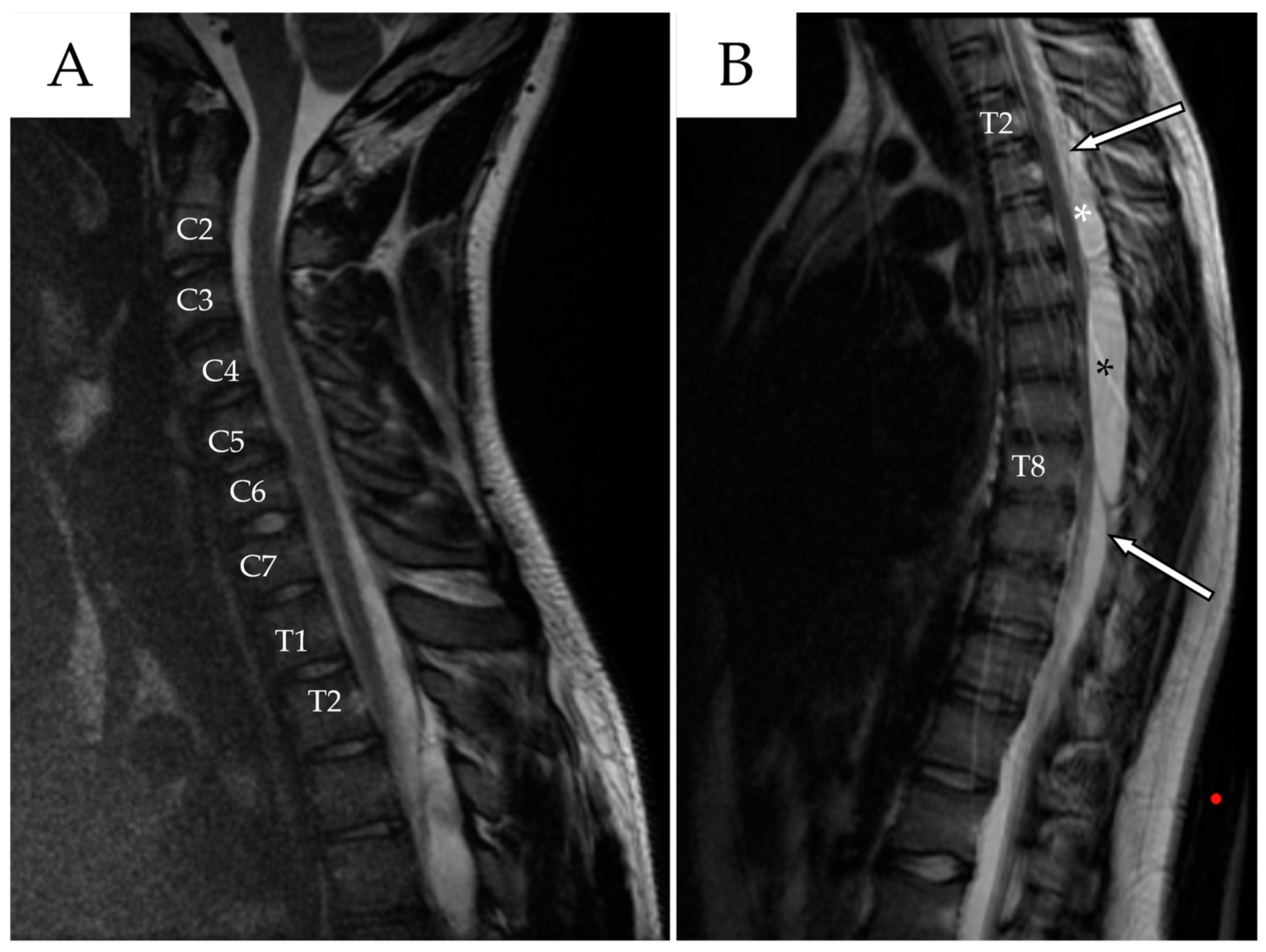
Figure 1. (A) Cervical spine ceMRI, sagittal section, TSE T2-WI. The cranial portion of the SAC is visible at the T2 level. To facilitate counting, the cervical vertebrae and the first two thoracic vertebrae are marked with their corresponding names. (B) Thoracic spine ceMRI, sagittal section, TSE T2-WI. The SAC is visible in its entirety, composed of a smaller cranial portion (T2–T4, white asterisk) and a larger caudal portion (T5–T8, black asterisk). The dural sac is easily identifiable (white arrows with black outlines), confirming the extradural location.
Reports 08 00152 g001
Figure 2. (A) Thoracic spine ceMRI, axial section, TSE T2-WI. The section is taken at the highest point of spinal cord compression (T7–T8), with early signs of radiological myelopathy (white arrow with black outlines). The spinal cord is dislocated anteriorly against the vertebral posterior wall. The SAC is marked with a black asterisk. (B) The yellow line indicates the level at which the axial section in (A) was obtained.
Reports 08 00152 g002
Figure 3. (A) Postoperative MRI, sagittal section, TSE T2-WI. After T8 laminectomy with cyst fenestration, the spinal cord appeared less compressed at the T7–T8 level (white arrows with black outlines). Fenestration was confirmed by the presence of an air sac within the caudal portion of the SAC (black asterisk). The yellow line indicates the level at which the axial section in (B) was obtained. (B) The spinal cord, although not yet re-expanded, appeared less compressed.
Reports 08 00152 g003
Figure 4. First follow-up MRI, sagittal section, TSE T2-WI. Both the cranial (red asterisk) and caudal (white asterisk) components of the SAC appear enlarged compared to the MRI in Figure 3, especially the latter. Additionally, a new pseudomeningocele (black asterisk) is visible at the level of the previous fenestration.
Reports 08 00152 g004
Figure 5. Emergent MRI, sagittal section, TSE T2-WI. Although the cranial component (red asterisk) remained stable, the caudal component (white asterisk), along with the pseudomeningocele (black asterisk), had enlarged and appeared under tension, which could explain the onset of the patient’s symptoms.
Reports 08 00152 g005
Figure 6. (A,B) Immediate post-second surgery MRI. The external drainage (white arrows with black outlines) is visible along the length of the SAC (white asterisk); it was inserted at the T4–T5 level and exits at the level of the pseudomeningocele (black asterisk), designed to drain as much CSF collection as possible.
Reports 08 00152 g006
Figure 7. Control MRI after percutaneous blood patch procedure. The cranial component of the SAC remained stable (white arrow with black outlines), while the caudal component (red arrow with black outlines), along with the pseudomeningocele (white asterisk), was refilled and slightly enlarged. At the T4–T5 level, a new pseudomeningocele (black asterisk) has appeared, extending into the soft tissues from T2 to T5.
Reports 08 00152 g007
Figure 8. (A) Preoperative fluoroscopic myelography after contrast agent infusion shows the caudal portion of the cyst (white asterisk), along with the fistulous stream originating from the overlying fistulous tract (white arrow with black outline). (B) Postoperative fluoroscopic myelography confirms closure of the fistula at the T3–T4 level using metal clips (red arrow with black outline), while the remaining subarachnoid cavity (SAC) is filled with metallic coils and blood (green arrow with black outline).
Reports 08 00152 g008
Figure 9. Sagittal T2-weighted MRI performed 7 days after the last surgery, which included percutaneous repair of the dural leak. The result of the blood patch is visible in the upper portion of the spinal arachnoid cavity (white arrow). A marked reduction in the cranial pseudomeningocele (black asterisk) is noted, along with near-complete resolution of the inferior pseudomeningocele (white asterisk).
Reports 08 00152 g009
Table 1. Summary of the literature review data (first part). * Age refers to the age at the time of surgery (in years); for case series, the median age is reported. ** When referring to Type I cysts, we considered only Type Ia, as Type Ib cysts are outside the scope of this study. NA: Not Available. LBP: Low Back Pain. TLIF: Transforaminal Lumbar Interbody Fusion.
AuthorCountry/YearAge *GenderRisk FactorsLocation of the CystType of Cyst **SymptomsManagement
Ichinose T et al. [24]Japan, 202014MNoCervicalType IIITetraparesis, bladder/bowel dysfunctionFenestration
Novak L et al. [25]Hungary, 2005115FSpine traumaThoracicType ISpastic paraparesis inferior limbs, sensory lossExcision
Lee HG et al. [26]Korea, 2019150MSpine surgeryLumbarType IIILBP, right S1 radiculopathyFenestration, excision
Krstačić A et al. [27]Croatia, 2016150FNoLumbarType IIILBP, right S1 radiculopathy, right leg hypostheniaNA
Lee & Cho [28]China, 200138, 9, 9M, M, FNo, No, NoThoracic–lumbar, cervical–thoracic, thoracicType III1: Cauda equina
1: Spastic gait
1: Quadriplegia
Fenestration, excision
Bond AE et al. [29]USA, 2012316, 9 (median)17 F, 14 M16 CNS anomalies11 thoracic, 2 lumbar, 3 sacral, 4 cervical–thoracic, 4 thoracic–lumbar, 4 lumbar–sacral, 2 cervical19: Type III
11: Type I,
1: Type III/I
21: Radiculopathy/myelopathy
3: Asymptomatic
29: Excision
2: Fenestration, excision
Liu JK et al. [30]USA, 2005111FNoThoracicType IBladder dysfunction, gait ataxia, lower limbs hypostheniaExcision
El-Hajj VG et al. [31]Sweden, 2023135FSurgery for cranial arachnoid cystCervical–thoracicType IIINeck and back pain, upper limbs weaknessExcision
Özdemir M et al. [32]Turkey, 2019122MNoCervical–thoracicType INeck pain, numbness and weakness of the left upper armExcision
Ebot J et al. [33]USA, 2020180FNoThoracicType IIILower extremity weakness, decreased sensation and hyperreflexiaFenestration
Alugolu R et al. [34]India, 2016154FNoThoracicType IIISpastic weakness of lower limbs, decreased sensations below T12Excision
Nayak R et al. [35]India, 201517FNoThoracicType INumbness and weakness of lower limbsExcision
Sharif S et al. [36]Pakistan, 2017125MNoLumbarType IIILBP, numbness of lower limbs, cauda equinaExcision
Lin & Jason [37]Malaysia, 2018135MNoThoracicType INumbness and weakness of lower limbsExcision
Shaaban A et al. [38]Quatar, 2019131MNoThoracicType IIIT6 sensory level, LBP, bladder/bowel disfunctionExcision
Raes & Oostra [39]Belgium, 2021238, 45F, MSpine trauma with T12 and L1 fractureThoracicType IIILoss of strength of lower limbs, sensory disturbances in the right leg, paraplegia and T11 neurological levelFenestration
Taccone MS et al. [40]Canada, 2018172MTLIF L4-L5LumbarType IIILower limbs weakness in first day after surgeryFenestration
Turel MK et al. [41]USA, 2017136FEpidural steroid injectionsLumbarType IIIAsymptomaticConservative managed
Kong WK et al. [42]Korea, 2013165MSpine traumaThoracic–lumbarType IWeakness of lower limbs, bladder dysfunctionFenestration
Fischer KM et al. [43]USA, 202217FNoThoracicType IIIUrinary and fecal incontinenceExcision
Sharma R et al. [44]India, 2023116MNoCervical–thoracic–lumbarType IWeakness of lower limbs, numbness of the right legExcision
Engelhardt J et al. [45]France, 2015118MNoCervicalType IIICervical pain, paresthesia and weakness of both armsExcision
Zekaj E et al. [46]Italy, 2016147FNoThoracicType IIIRight dorsal radicular pain, weakness of the left lower limbFenestration
Watanabe A et al. [47]Japan, 2019137FNoThoracicType IIIAbdominal pain, numbness and weakness of both lower extremitiesExcision
Cavalcante-Neto JF et al. [48]Brazil, 2021138FNoThoracic–lumbarType ITransient weakness of lower limbs, bilateral patellar hyperreflexiaExcision
Marrone S et al. [49]Italy, 2022170MNoThoracicType IBilateral lower limb weakness and paresthesiaFenestration, excision
Alanazi RF et al. [50]Saudi Arabia, 2022147FNoThoracic–lumbarType IProgressive lower extremity paraparesisExcision
Sawaya J et al. [51]USA, 2024115FNoThoracic–lumbarType ILBP radiating bilateral in posterior thighs and kneesFenestration, excision
Ayantayo TO et al. [52]Nigeria, 2023149MNoThoracicType IIIGait instability, urinary retention and paraplegiaExcision
Werner C et al. [53]USA, 2020158MSpinal schwannoma
Post-surgical meningitis
ThoracicType IIIParaplegia and loss of sensation below T10, urinary retentionFenestration
Pillai MK [54]Oman, 2016129FNoCervical–thoracicType IIIPosterior column dysfunctionFenestration
French H et al. [55]Australia, 20171060 (mean)F:M 2:1NAThoracicType IIIThoracic myelopathy, gait ataxia
3: thoracic radicular pain
1: sphincter dysfunction
6: Fenestration
4: Excision
Shi L et al. [56]China, 20214141 (mean)24F, 17M NAThoracic–lumbarType IRadicular pain, gait ataxia, myelopathy
1: cauda equina syndrome
Excision
Yılmaz E et al. [57]Turkey, 202388 (mean)4F, 4MNAThoracic–lumbarType IIIMotor weakness, urinary incontinence, sensory disturbance7: Excision
1: Shunt placement
Tokmak M et al. [58]Turkey, 20241043 (mean)6F, 4MNAThoracic–lumbarType INumbness and progressive weakness of the lower extremities, back pain9: Excision
1: Conservative managed
Menezes AH et al. [59]USA, 2017214, 34M, FBack traumaThoracic–lumbar, lumbarType IGait instability, urinary retention, pain in the left legExcision
Kalsi P et al. [60]Canada, 20221147 (mean)3F, 8M 3 presented with associated syrinx10 cervical–thoracic, 1 cervicalType IIINeuropathic pain, back pain, weakness, gait and balance problems, sensory issues, sphincter disturbance, radicular pain, long tract signs, gait ataxia10: Fenestration
1: Excision
1: Shunt placement
Table 2. Summary of the literature review data (second part).
ImprovementRelapse and Complications
NoNo
YesNo
YesNA
NANo, no, no
2: Yes
1: Partial
1: Fistula
1: Pseudomeningocele
1: Relapse and shunt
27: Yes
4: No
No
YesYes
YesNo
YesNo
YesPseudomeningocele
YesNo
YesNo
YesNo
YesNo
YesYes, yes
Yes, yesNo
YesNA
NANo
YesNo
YesNo
YesNo
YesYes
YesNo
YesNo
YesNo
YesNo
YesNo
YesNo
YesYes
YesNo
Yes1: Pseudomeningocele
1: Syrinx
Yes2: Yes
YesNo
Yes1: Fistula
YesNo
YesNo
4: No
7: Yes
Yes
Disclaimer/Publisher’s Note: The statements, opinions and data contained in all publications are solely those of the individual author(s) and contributor(s) and not of MDPI and/or the editor(s). MDPI and/or the editor(s) disclaim responsibility for any injury to people or property resulting from any ideas, methods, instructions or products referred to in the content.