Case Report

A Novel Case of Recurrent Mucinous Borderline Ovarian Tumor: Early Relapse and Fatal Outcome

1
Department of Obstetrics and Gynecology, Shimane University Faculty of Medicine, Izumo 693-8501, Japan
2
Department of Obstetrics and Gynecology, Uji-Tokushukai Medical Center, Uji, Kyoto 611-0041, Japan
*
Author to whom correspondence should be addressed.
Reports 2022, 5(2), 15; https://doi.org/10.3390/reports5020015
Submission received: 25 January 2022 / Revised: 18 April 2022 / Accepted: 26 April 2022 / Published: 12 May 2022

Abstract

:
Unlike ovarian carcinomas, borderline ovarian tumors (BOTs) are associated with a favorable prognosis: their recurrence rate is around 5–7%, and the survival rate is more than 97% when diagnosed early. There are only a few reports of recurrence and fatal outcomes. Herein, we report a novel case of recurrent mucinous BOT, with a literature review. A 63-year-old woman presented to a local doctor with abdominal fullness. She was diagnosed as having a polycystic tumor. The lesion was suspected to be a mucinous BOT (M-BOT) on magnetic resonance imaging. Upper and lower gastrointestinal endoscopy revealed no digestive cancerous lesions, and surgery was performed. Intra-operative diagnosis confirmed the diagnosis, and total abdominal hysterectomy, bilateral salpingo-oophorectomy, and omentectomy were performed. The final pathological diagnosis was non-invasive M-BOT (stage I c1 (T1c1N0M0)). The result of immunohistochemical staining supported the diagnosis of primary ovarian mucinous tumor.Four months after surgery, relapse occurred. Blood tests revealed an elevated carbohydrate antigen 19-9 level, and computed tomography revealed multiple liver metastases, peritoneal dissemination, left ureter infiltration, and carcinomatous peritonitis. Although the patient underwent chemotherapy, she died. This case of a very short progression-free and overall survival in stage I M-BOT indicates that some M-BOTs could result in fatal clinical outcomes despite diagnosis at an early stage. Frequent follow-up appointments after surgery could help detect relapse and increase survival in such cases.

1. Introduction

Borderline ovarian tumors (BOTs) are histologically characterized by cellular proliferation and nuclear atypia without destructive stromal invasion. Taylor, in 1929, described BOTs as ovarian neoplasms with a favorable prognosis despite peritoneal involvement, and called these tumors “semi-malignant” ovarian tumors [1]. The term ‘”borderline tumors” was used for the first time by the World Health Organization (WHO) in 2003 [2]. In 2016, the Japan Society of Obstetrics and Gynecology (JSOG) and the Japanese Society of Pathology defined epithelial ovarian tumors of borderline malignancy as tumors that have “histologic characteristics intermediate between benign and malignant tumors. Patients with these tumors have a better prognosis because of low malignancy. Although recurrences occur occasionally, fatality from BOTs is rare” [3].
On the basis of the epithelial features, epithelial ovarian tumors of borderline malignancy are categorized into six subtypes: serous, mucinous, endometrioid, clear cell, seromucinous, and Brenner tumors. One of the histological features of borderline ovarian epithelial neoplasms is microinvasion. In 2014, the WHO defined an invasive lesion as a lesion of less than 5 mm. This definition has been adapted to serous, mucinous, and endometrioid tumors [4].
Mucinous BOTs (M-BOTs) and serous BOTs (S-BOTs) account for more than 90% of epithelial ovarian tumors of borderline malignancy [5]. In Japan, histologically, M-BOTs contribute to most cases (57.7%), followed by S-BOTs (20.4%) [6]. M-BOTs were formerly known as intestinal-type, and seromucinous borderline tumors as endocervical-type mucinous BOTs. Most BOTs are diagnosed at International Federation of Gynecology and Obstetrics (FIGO) stage I (≥90%). The overall recurrence rate for BOTs is 5–7%, and the five-year overall survival rate is more than 97% for early-stage disease [7].
A key characteristic of the M-BOT is recurrence that occurs a long time after treatment. On the basis of the clinical features of BOTs, the Japan Society of Gynecologic Oncology (JSGO) recommends long-term follow-up (over 5 years) after treatment [4].
Although M-BOTs are usually associated with a good prognosis, there are reports of recurrence and fatal outcomes in a few cases. Herein, we report a novel case of a recurrent M-BOT in a 63-year-old woman, along with a literature review.

2. Case Presentation Section

The patient was a 63-year-old woman who had had three normal vaginal deliveries. She presented to a local physician with abdominal fullness, and was diagnosed as having a polycystic tumor that had reached over the navel level. Magnetic resonance imaging revealed a 24 cm polycystic tumor (Figure 1). The lesion was suspected to be an M-BOT, and a laparotomy was planned.
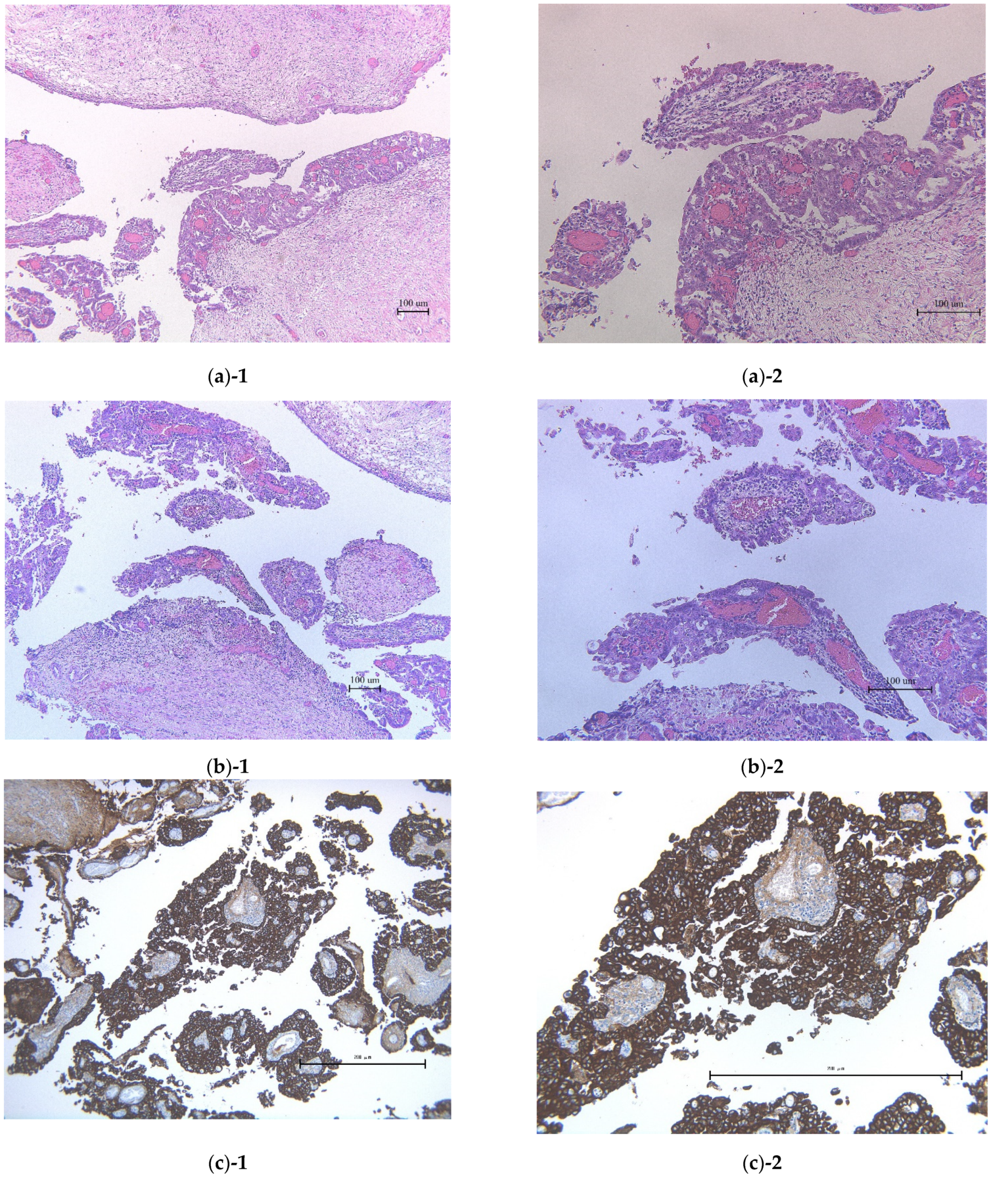
Before surgery, the patient’s serum carbohydrate antigen 125 (CA-125) and carbohydrate antigen 19-9 (CA19-9) levels were measured. Only the CA19-9 level was elevated (176 U/mL), and the CA-125 level was within the normal range (24.8 U/mL). No cancerous lesions were detected at upper and lower gastrointestinal endoscopy. Subsequently, surgery was performed. The tumor was limited to the left ovary and the size of the left ovarian tumor was 210 × 165 mm. The diagnosis of M-BOT was confirmed intra-operatively. In view of the diagnosis, a total abdominal hysterectomy, bilateral salpingo-oophorectomy, and omentectomy were performed. A key observation during surgery was that the left ureter had adhered to the tumor. The tumor capsule was ruptured by operative handling. The final pathological diagnosis was a stage Ⅰ c1 (T1cN0M0) M-BOT, with no stromal invasion and no mural nodules (Figure 2). To distinguish from ovarian metastases of digestive organ cancers, the immunohistochemical staining was performed (CK7, CK20, and PAX-8). CK7 was strongly positive, CK20 was weakly positive, and PAX-8 was moderately positive in this tumor (Figure 2). These results were consistent with the suspicion that this tumor was a primary ovarian mucinous tumor.
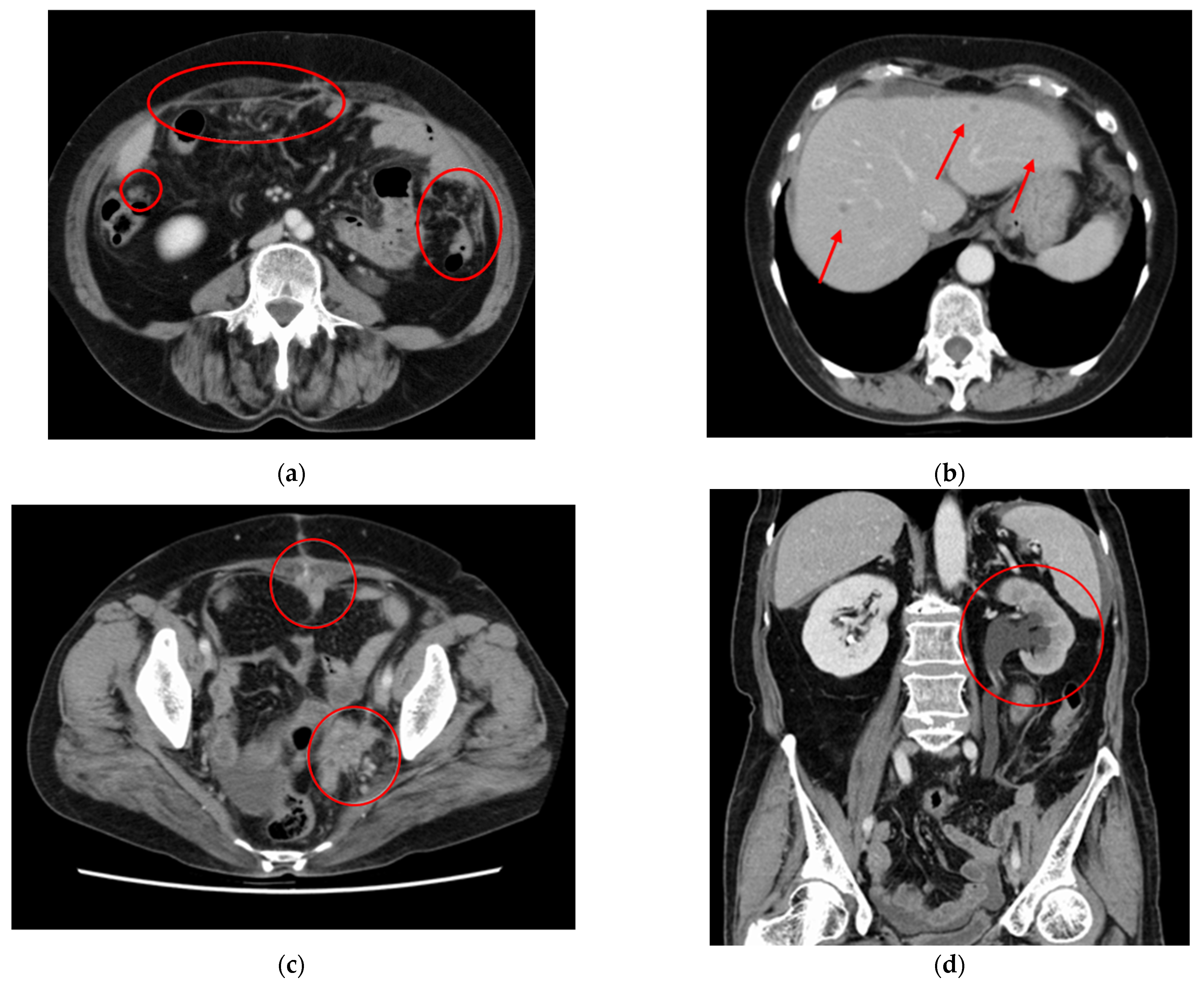
The patient was followed up as an outpatient at our gynecologic department after discharge. Although she had no symptoms, blood tests continued to reveal high CA19-9 levels (179.5 U/mL) 4 months after surgery (Figure 3). CA-125 levels were not measured. Ultrasonography revealed ascites, and computed tomography (CT) indicated multiple liver metastases, peritoneal metastasis, left ureter infiltration, and carcinomatous peritonitis (Figure 4). Pathological analysis revealed no stromal invasions. The diagnosis was the same as that reached previously. Therefore, we considered that we had detected a recurrence of BOT.
Owing to multiple liver metastases, secondary debulking surgery was not performed. The microsatellite instability test yielded negative results. The patient received five courses of paclitaxel and carboplatin therapy (TC) (concomitant with bevacizumab (BEV) after the first course). The serum CA-125 and CA19-9 levels were measured three times after the first chemotherapy (Figure 3). At the start of the first course of TC, both CA-125 and CA19-9 levels were elevated at 721.3 and 253 U/mL, respectively. At the end of the second course of TC + BEV, CT revealed that the disseminated intrapelvic lesions had partially shrunk. Although CA-125 levels decreased (443 U/mL), CA19-9 levels were still elevated at 599 U/mL. We concurred that the tumor showed stable disease at this point. Because the clinical course was nonspecific for BOT (FIGO stage I), it was necessary to check whether the recurrence had been a BOT or invasive carcinoma. After the fourth course of TC + BEV, a liver biopsy was performed to make a decision on recurrence. However, no malignant tissue was detected. Therefore, we concluded that the tumor recurrence had occurred, also taking the clinical course into account. Although the patient continued the therapy, she became unresponsive to it. Cancerous peritonitis and increasing ascites resulted in a poor performance status in this patient. Chemotherapy was withheld after the fifth course of TC + BEV. The patient died 5 months after the relapse, that is, 9 months after the primary surgery.

3. Discussion

In contrast to the present case, BOTs usually have a favorable prognosis. According to reports of the Committee on Gynecologic Oncology, by the JSOG, >90% of patients with BOT were diagnosed as FIGO stage Ⅰ between 2003 and 2018 [8]. Another report suggests that the overall recurrence rates in these patients are 5–7%, and the overall survival is >97% for early-stage disease [7].
Most cases of epithelial ovarian tumors of borderline malignancy are S-BOTs or M-BOTs. In Japan, M-BOTs contribute to a larger share of cases (57.7%) [6]. The onset of the disease is usually between the ages of 40 and 50 (average 48 [ ±16.7]) [9].
Compared to that in ovarian carcinoma, the time between standard of care and recurrence is usually longer in cases of epithelial ovarian tumors of borderline malignancy. A systematic review [10] showed that 37% of recurrences occurred within 2 years of successful treatment, 31% within 2–5 years, and 32% > 5 years later (10% of these patients developed a relapse ≥ 10 years later). A Swedish follow-up study between 1993 and 2004 reported that 8 of 399 BOT cases showed recurrence. Three of these eight cases relapsed ≥ 5 years after the primary treatment [11]. In response to these characteristics, the JSGO has recommended long-term follow-up (>5 years) after treatment. However, in our unique case of M-BOT (FIGO stage Ⅰ), relapse occurred 4 months after optimal surgery, which had a fatal outcome. To the best of our knowledge, this case showed the shortest reported prognosis in both recurrence and overall survival.
Upon a review of the literature on recurrent BOTs, we found that there were more reports of advanced-stage cases or those with a conservative surgical approach and limited reports of FIGO stage Ⅰ wherein basic surgery had been performed [12,13,14]. One such previous case of recurrence of stage Ⅰ M-BOT was described by Simons et al. [15]. The patient with M-BOT FIGO stage Ⅰ A after staging laparotomy (total abdominal hysterectomy, bilateral salpingo-oophorectomy, lymph node sampling, and appendectomy) was followed up every 6 months after surgery. Four years after primary surgery, the patient reported severe dyspnea and right-sided thoracic pain. A final diagnosis of right pleural metastases was made. Subsequently, palliative chemotherapeutic treatment was initiated. According to the findings from this case, recurrences usually occur several years after surgery. Our review of previous cases did not show any other case with an early relapse such as in our case. The earliest report of relapse in M-BOT is relapse occurring 11 months after surgery [13].
Some studies have investigated the recurrence risk for ovarian tumors in borderline malignancy and reported risk factors for relapse [16,17,18]. On the basis of histological characteristics, S-BOTs have a higher risk of recurrence compared with M-BOTs [16]. In addition, cystectomy (compared to unilateral salpingo-oophorectomy), intraoperative rupture, and residual tumor have been reported as risk factors for recurrence [17,18].
In a retrospective review [19] of patients with BOTs treated between 1998 and 2008 at 24 German centers, 950 BOT patients were analyzed. Of the 950 patients, there were only 74 cases (7.8%) of recurrent disease, and in 48 cases (64.9%) there was a recurrence of BOT; 22 (29.7%) cases had relapses resulting in invasive carcinoma, and data were unavailable in 4 cases (5.4%). When patients younger and older than 40 years were analyzed, recurrence was significantly more frequent in patients younger than 40 years, whereas patients ≥40 years had a higher rate of malignant transformation and often experienced recurrent disease in peritoneal implants or distant localizations. Another retrospective review [20] reports that age >40 years, a late stage at diagnosis, and incomplete surgery are significantly associated with invasive recurrence.
In our patient, the left ureter, which had adhered to the tumor, showed infiltration upon relapse. A possible explanation for this is that there was residual disease despite optimal surgery. Furthermore, only CA19-9 levels were elevated before the surgery, whereas both CA19-9 and CA-125 levels were elevated after recurrence. The fluctuation in tumor markers, advanced age, and possible residual disease are indicators of malignant transformation.
An analysis report [21] on stage Ⅰ M-BOTs with fatal outcome indicated that most of these fatal tumors are related to inadequate peritoneal sampling, and in some reports, carcinomas in the other sections were overlooked. M-BOTs usually showed huge-sized tumors, which are known to have a highly heterogeneous histology. A single tumor can often contain coexisting elements of benign cystadenoma, borderline tumor, and carcinoma [22]. In addition, mucinous tumors are generally larger than serous tumors (average: 18 cm). All of these characteristics suggest a discordance between frozen section and final pathology that results in overlooking invasive carcinoma [23]. In our patient, the tumor size was 210 × 165 mm, and the total number of sections of the tumor was 22 (1cm interval). It was more difficult to make a correct pathological diagnosis. It is also possible that invasive carcinoma was present in sites other than those sampled for histological sections.
The guidelines published by JSGO [4] recommend the resection of the recurrent lesion as it contributes to long-term prognosis, if the tumor is borderline. The pathological characteristics of the lesion also influence the treatment plan. According to the JSGO guidelines [4], if a recurrent lesion is not an invasive carcinoma, follow-up is essential. If the recurrent lesion is an invasive implant or low-grade or high-grade invasive carcinoma, chemotherapy should be considered. In our patient, based on liver biopsy results, no malignant tissue was detected, and a conclusive diagnosis could not be made. It is possible that our patient indeed developed an invasive carcinoma and not an M-BOT after relapse. If she had been monitored regularly and frequently at short intervals, the recurrent lesion might have been revealed earlier and could have been resected under favorable conditions. Since M-BOT with fatal outcome is a rare occurrence, it is necessary to collect similar cases at other facilities to clarify using real-world data.

4. Conclusions

We report a case of stage Ⅰ M-BOT that recurred 4 months after optimal surgery. The patient died 5 months after the recurrence. As far as we know, this case showed the shortest reported interval in both progression-free and overall survival. It should be noted that very rare cases of early-stage M-BOT are associated with inferior clinical outcomes, and close follow-up may be essential in these cases to improve prognosis.

Author Contributions

Conceptualization, K.N. (Kyoko Nakagawa) and K.N. (Kentaro Nakayama); writing—original draft preparation, S.R (Sultana Razia); carried out critical revision of manuscript, S.K. (Satoru Kyo), K.N. (Kyoko Nakagawa) and K.N. (Kentaro Nakayama); data curation, A.N., N.H., S.K. (Sonomi Kurose) and S.A.; co-ordination, S.K. (Satoru Kyo). All authors have read and agreed to the published version of the manuscript.

Funding

This research received no external funding.

Institutional Review Board Statement

The case report protocol was approved by the institutional ethics and research review boards at Shimane University.

Informed Consent Statement

Written informed consent was obtained from the patient.

Data Availability Statement

The data presented in this study are available on request from the corresponding author. The data are not publicly available due to personally identifiable information.

Acknowledgments

We thank Fumihiko Kono for pathological support (Department of Diagnostic Pathology, Uji-Tokushukai Medical Center, Uji, Kyoto, Japan).

Conflicts of Interest

The authors declare no conflict of interest.

References

  1. Hauptmann, S.; Friedrich, K.; Redline, R.; Avril, S. Ovarian borderline tumors in the 2014 WHO classification: Evolving concepts and diagnostic criteria. Virchows Arch. 2017, 470, 125–142. [Google Scholar] [CrossRef] [PubMed] [Green Version]
  2. Tavassolo, F.A.; Devilee, P. (Eds.) WHO Classification of Tumors: Pathology and Genetics of the Tumors of the Breast and Female Genital Organs. IARC Press: Lyon, France, 2003; pp. 113–202. [Google Scholar]
  3. Japan Society of Obstetrics and Gynecology, The Japanese Society of Pathology. The General Rules for Clinical and Pathological Management of Ovarian Tumor, Fallopian Tube Cancer, and Primary Peritoneal Cancer Pathological Edition, 1 ed; Kanehara Shuppan: Kyoto, Japan, 2016; pp. 15–65. [Google Scholar]
  4. Japan Society of Gynecologic Oncology. Guidelines for Treatment of Ovarian Cancer, Fallopian Tube Cancer and Primary Peritoneal Cancer; Japan Society of Gynecologic Oncology (JSGO): Kyoto, Japan, 2020; pp. 159–173. [Google Scholar]
  5. Song, T.; Lee, Y.-Y.; Choi, C.H.; Kim, T.-J.; Lee, J.-W.; Bae, D.-S.; Kim, B.-G. Histologic distribution of borderline ovarian tumors worldwide: A systematic review. J Gynecol. Oncol. 2013, 24, 44–51. [Google Scholar] [CrossRef] [PubMed] [Green Version]
  6. Nagase, S.; Ohta, T.; Takahashi, F.; Yaegashi, N.; Board members of the 2020 Committee on Gynecologic Oncology of the Japan Society of Obstetrics and Gynecology. Annual report of the committee on gynecologic oncology, the Japan Society of Obstetrics and Gynecology: Annual patient report for 2017 and annual treatment report for 2012. J. Obstet. Gynaecol. Res. 2021, 47, 1631–1642. [Google Scholar] [CrossRef] [PubMed]
  7. Obermair, A.; Hiebl, S. Laparoscopy in the treatment of ovarian tumours of low malignant potential. Aust. N. Z. J. Obs. Gynaecol. 2007, 47, 438–444. [Google Scholar] [CrossRef] [PubMed]
  8. Nagase, S.; Ohta, T.; Takahashi, F.; Enomoto, T. The 2017 Committee on Gynecologic Oncology of the Japan Society of Obstetrics and Gynecology Annual report of the committee on gynecologic oncology, the Japan Society of Obstetrics and Gynecology: Annual patients report for 2015 and annual treatment report for 2010. J. Obstet. Gynaecol. Res. 2018, 45, 289–298. [Google Scholar] [CrossRef] [PubMed] [Green Version]
  9. Matsuo, K.; Machida, H.; Mandelbaum, R.S.; Grubbs, B.H.; Roman, L.D.; Sood, A.K.; Gershenson, D.M. Mucinous borderline ovarian tumor versus invasive well-differentiated mucinous ovarian cancer: Difference in characteristics and outcomes. Gynecol. Oncol. 2019, 153, 230–237. [Google Scholar] [CrossRef] [PubMed]
  10. du Bois, A.; Ewald-Riegler, N.; Du Bois, O.; Harter, P. Borderline tumors of the ovary-a systematic review. Geburtshilfe Und Frauenheilkd. 2009, 69, 807–833. [Google Scholar] [CrossRef]
  11. Åkeson, M.; Zetterqvist, B.-M.; Dahllöf, K.; Jakobsen, A.-M.; Brännström, M.; Horvath, G. Population-based cohort follow-up study of all patients operated for borderline ovarian tumor in western Sweden during an 11-year period. Int. J. Gynecol. Cancer 2008, 18, 453–459. [Google Scholar] [CrossRef] [PubMed]
  12. Parker, R.L.; Clement, P.B.; Chercover, D.J.; Sornarajah, T.; Gilks, C.B. Early recurrence of ovarian serous borderline tumor as high-grade carcinoma: A report of two cases. Int. J. Gynecol. Pathol. 2004, 23, 265–272. [Google Scholar] [CrossRef] [PubMed]
  13. Nakata, S.; Kasai, M.; Deguchi, M. Ovarian carcinoma development after laparoscopically-assisted ovarian cystectomy for a mucinous cystic tumor of borderline malignancy. Jpn. J. Gynecol. Obstet. Endosc. 2008, 24, 322–325. [Google Scholar] [CrossRef]
  14. Ma, J.-W.; Miao, Y.; Liang, C.-N.; Wang, N.; Jiang, B.; Wang, Q.-Y.; Kang, J.; Hou, G.; Yin, Y. Malignant transformation of a borderline ovarian tumor with pulmonary and pleural metastases after years of latency: A case report and literature review. Front Med. 2020, 7, 571348. [Google Scholar] [CrossRef] [PubMed]
  15. Simons, M.; Nagtegaal, I.D.; Overbeek, L.I.; Flucke, U.; Massuger, L.F.; Bulten, J. A patient with a noninvasive mucinous ovarian borderline tumor presenting with late pleural metastases. Int. J. Gynecol. Pathol. 2015, 34, 143–150. [Google Scholar] [CrossRef] [PubMed]
  16. Ferrero, A.; Strada, I.; Di Marcoberardino, B.; Maccarini, L.R.; Pozzati, F.; Rossi, M.; Biglia, N.; De Iaco, P. Clinical significance of microinvasion in borderline ovarian tumors and its impact on surgical management. Int. J. Gynecol. Cancer 2012, 22, 1158–1162. [Google Scholar] [CrossRef] [PubMed]
  17. Ushijima, K.; Kawano, K.; Tsuda, N.; Nishio, S.; Terada, A.; Kato, H.; Tasaki, K.; Matsukuma, K. Epithelial borderline ovarian tumor: Diagnosis and treatment strategy. Obstet. Gynecol. Sci. 2015, 58, 183–187. [Google Scholar] [CrossRef] [PubMed] [Green Version]
  18. Morice, P.; Uzan, C.; Fauvet, R.; Gouy, S.; Duvillard, P.; Darai, E. Borderline ovarian tumour: Pathological diagnostic dilemma and risk factors for invasive or lethal recurrence. Lancet Oncol. 2012, 13, e103–e115. [Google Scholar] [CrossRef]
  19. Trillsch, F.; Mahner, S.; Woelber, L.; Vettorazzi, E.; Reuss, A.; Ewald-Riegler, N.; de Gregorio, N.; Fotopoulou, C.; Schmalfeldt, B.; Burges, A.; et al. Age-dependent differences in borderline ovarian tumours (BOT) regarding clinical characteristics and outcome: Results from a sub-analysis of the Arbeitsgemeinschaft Gynaekologische Onkologie (AGO) ROBOT study. Ann. Oncol. 2014, 25, 1320–1327. [Google Scholar] [CrossRef] [PubMed]
  20. Yasmeen, S.; Hannan, A.; Sheikh, F.; Syed, A.A.; Siddiqui, N. Borderline tumors of the ovary: A clinicopathological study. Pak. J. Med. Sci. 2017, 33, 369–373. [Google Scholar] [CrossRef] [PubMed]
  21. Ronnett, B.M.; Kajdacsy-Balla, A.; Gilks, C.B.; Merino, M.J.; Silva, E.; Werness, B.A.; Young, R.H. Mucinous borderline ovarian tumors: Points of general agreement and persistent controversies regarding nomenclature, diagnostic criteria, and behavior. Hum. Pathol. 2004, 35, 949–960. [Google Scholar] [CrossRef] [PubMed]
  22. Moroney, M.R.; Post, M.D.; Berning, A.A.; Sheeder, J.; Corr, B.R. An evaluation of frozen section and lymph node dissection results for mucinous ovarian tumors. Int. J. Gynecol. Cancer 2018, 28, 92–98. [Google Scholar] [CrossRef] [PubMed]
  23. Salomon, L.J.; Lhommé, C.; Pautier, P.; Duvillard, P.; Morice, P. Safety of simple cystectomy in patients with unilateral mucinous borderline tumors. Fertil. Steril. 2006, 85, 1510.e1–1510.e4. [Google Scholar] [CrossRef] [PubMed]
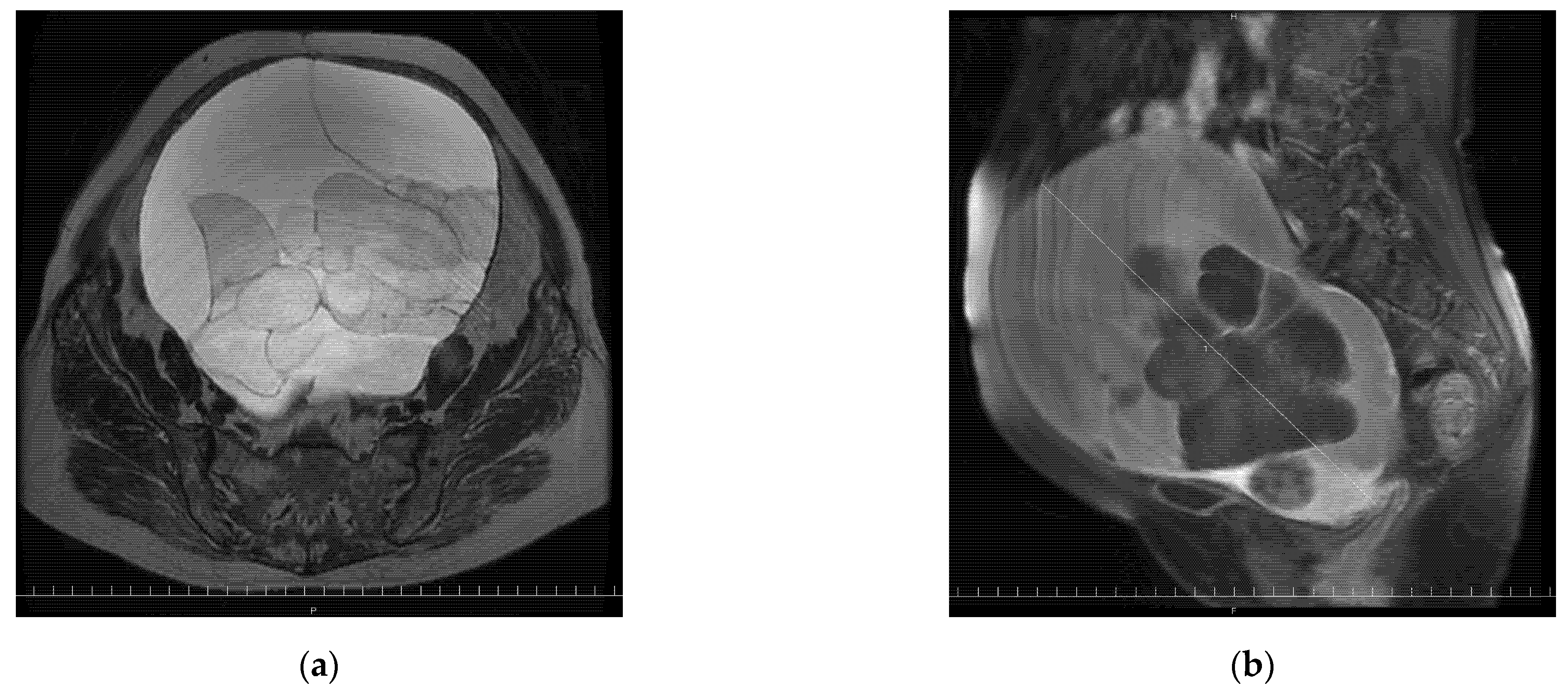
Figure 1. The pelvic magnetic resonance image (MRI) taken before staging a laparotomy. (a) T2-weighted pelvic MRI coronary showing giant ovarian polycystic tumor. (b) T2-weighted MRI sagittal showing a partially solid polycystic tumor 24 cm in diameter.
Reports 05 00015 g001
Figure 2. Histopathological examination of mucinous borderline ovarian tumor of the patient of this current case. (a) Histology of a case of mucinous borderline ovarian tumor showing the absence of stromal invasions (slides H and E). (b) Further sections of slides H and E, also indicating a lack of stromal invasions. (c) CK7: strongly positive. (d) CK20: weakly positive. (e) PAX-8: moderately positive. (a)-1, (b)-1, (c)-1, (d)-1, (e)-1, low-magnification; (a)-2, (b)-2, (c)-2, (d)-2, (e)-2, high magnification.
Reports 05 00015 g002aReports 05 00015 g002b
Figure 3. Tumor markers (CA-125 and CA19-9) and clinical course.
Reports 05 00015 g003
Figure 4. Abdominal and pelvic computed tomography scans after the recurrence was revealed. (a) The circled areas show peritoneal metastasis. (b) Arrows indicate multiple liver metastases. (c) The circles show peritoneal metastasis and left ureter infiltration. (d) The circles show left hydronephrosis.
Reports 05 00015 g004
Publisher’s Note: MDPI stays neutral with regard to jurisdictional claims in published maps and institutional affiliations.