Case Report

Tear Liquid for Predictive Diagnosis of Alzheimer’s Disease

1
Service Biotech s.r.l., 80132 Napoli, Italy
2
Dipartimento di Salute Mentale e Fisica e Medicina Preventiva, Università degli Studi della Campania Luigi Vanvitelli, 80100 Naples, Italy
3
Department of Neurosciences and Reproductive and Dentistry Sciences, School of Medicine and Surgery, University of Naples Federico II, 80100 Naples, Italy
4
Ophthalmologic Unit, Salerno Hospital University, 84100 Salerno, Italy
*
Author to whom correspondence should be addressed.
Reports 2021, 4(3), 26; https://doi.org/10.3390/reports4030026
Submission received: 3 August 2021 / Revised: 16 August 2021 / Accepted: 20 August 2021 / Published: 25 August 2021
(This article belongs to the Special Issue Cyto-Histopathogical Correlations in Pathology Diagnostics)

Abstract

:
The common approach of the diagnosis of Alzheimer’s Disease (AD) is made with an analysis of the cerebrospinal fluid or the study of retinal fundus and the plaques formation through optical corneal tomography (OCT), or more simply with a fundus camera. Tears analysis is widely discussed in literature as an essential method to describe molecular and biochemical alterations in different diseases. The aim of our study was the identification with immunocytochemistry of Amyloid Beta-42 in tears from patients with or without familiarity for Alzheimer Disease, in order to make the diagnosis earlier and more accessible compared to other invasive methods. Our study was performed on tears from three phenotypically healthy subjects: two of them were Caucasian with Alzheimer familiarity (48 and 55 years old) and the other one was Asian without Alzheimer familiarity (45 years old) and affected by an adenoviral keratoconjunctivitis at the moment of withdrawal. Tear samples were collected from eye fornix and were examinated by immunocytochemistry (ICC) assay using anti-Amyloid Beta X-42 antibody. Two out of three tears samples showed positive Amyloid Beta-42. Considering that our patients were phenotypically healthy, the identification of Amyloid Beta-42 by ICC could be a candidable method to make the diagnosis of the disease earlier and more accessible and available then other current and invasive methods and it could be a candidate for a screening method too.

1. Introduction

Alzheimer’s disease is a neurodegenerative pathology of the central nervous system. The diagnosis is currently performed according to the classical methods through bioptic withdrawal of brain tissue or withdrawal of cerebrospinal liquid, subjecting the patients to significative physical stress and limiting the execution of the practice which cannot be extended to diagnostic screening. Human tears represent an exceptional biomaterial rich in information regarding the health status of eyes and, more generally, of whole-body functionality. This is mainly due to the presence in tears of a large variety of organic components including proteins, lipids, metabolites, nucleic acids, and electrolytes, whose concentrations can be altered in pathologies of the whole body too [1,2]. An increasing attention is presently given to the analysis of this human body fluid. The small amounts of substances considered and the typical low concentration of organic compounds hamper the access to a direct analysis by biochemical methods, so the use of tears in diagnosis is in progress. We focused our attention on tears analysis [3], to detect the presence of Amyloid Beta-42 in order to hypothesize a less invasive method and more rapid diagnosis of Alzheimer disease. Native beta-amyloid is a transmembrane protein is derived by the proteolytic processing of amyloid precursor protein (APP), resulting in a peptide predominantly 40 or 42 amino acids in length, with a short cytoplasmic domain that undergoes proteolytic clevage by secretase on its N-terminal intraluminal domain; clevated protein found in the extracellular fluid and in tears [4]. Recent studies have demonstrated that Amyloid Beta-42 and Amyloid Beta-40 have different conformation and assembly states; the first one is associated with the formation of plaques and parenchymal damage in AD and respect the classical vascular AD that associated with Amyloid Beta-40 [5]. In vivo, small, stable oligomers of A-(1–42) have been isolated from brain, plasma, and cerebrospinal fluid and correlate with the severity of neurodegeneration in AD [6,7]. We focused our study on Amyloid Beta-42. These plaques are formed from an altered composition of the chemical barrier, and the changes in the retinal vasculature and retinal morphology were detected in the eyes of a patient with AD so a relationship was observed between beta-amyloid deposition in the retina, in brain and AD [8,9]. In that way, the eyes are a good indicator for the study of AD and its progression. In this context, the detection of Amyloid Beta-42 in tears could be a useful method for an early and less invasive diagnosis of AD [10,11].

2. Materials and Methods

2.1. Methods

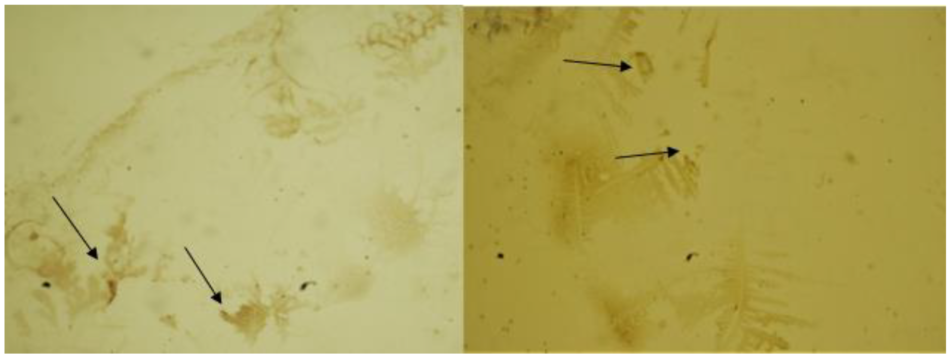
Our study was performed on three phenotypically healthy subjects: two of them were Caucasian with Alzheimer familiarity (48 and 55 years old) and the other one was Asian without Alzheimer familiarity (45 years old) and affected by an adenoviral keratoconjunctivitis at the moment of the withdrawal. Patients were examinated with fundus camera to highlight retinal plaques as described in literature; only the Caucasian patient presented a strong alteration in the posterior chamber with numerous plaques on retina (Figure 1). In this present work, human tear samples were examined with ICC using anti-Amyloid Beta X-42 antibody; samples were stained with DAB if positive, no staining if negative. (Figure 2). NaCl solution was employed as negative control and protein Amyloid Beta-42 as positive control (Figure 3). Immunocytochemistry (ICC) allows the identification by light microscopy of an antigen and its location in cells through specific antigen-antibody reaction. In our study, we employed ICC indirect method: the specific antigen was recognized by an unlabeled primary antibody which binds the secondary antibody (or post-primary), conjugated to the horseradish peroxidase (HRP or polymer) which reacts with the substrate yielding a chromogenic development at the antigen site. Samples were counterstained with hematoxylin, covers lipped and results were interpreted at light microscopy. Our test was not influenced by irritative and infective phenomena that can occur in tissue during the execution of withdrawal.

2.2. Materials

Tear samples were dispensed on Thermo Scientific adhesion slides, dried at 76 °C for 4 h and hydrated with 100% ethanol for 3 min, 95% ethanol for 3 min, 70% ethanol for 3 min and distilled water for 3 min. The area of interest was marked using a PAP pen, which draws a hydrophobic barrier to prevent the waste of reagents by keeping liquid pooled in a small droplet. The endogenous peroxidases were neutralized using peroxidase block (3–4% v/v hydrogen peroxide) for 10 min, followed by protein block for 10 min (0.4% casein in phosphate-buffered saline, with stabilizers, surfactant and 0.2% bronidox L as preservative) to reduce non-specific binding of primary antibody and polymer. Samples were incubated with the primary antibody (Anti beta-Amyloid 1–42 product code: AB5078P, Millipore) (anti-Amyloid Beta X-42, clone 12F4, a purified mouse monoclonal IgG1k in buffer containing 0.1 M Tris-Glycine pH 7.4, 150 mM NaCl with 0.05% sodium azide, Millipore) diluted 1:200 for 60 min. Post-primary (rabbit anti-mouse IgG < 10 µg/mL in 10% v/v animal serum in tris-buffered saline/0.09% Proclin 950) was incubated for 20 min, followed by Novolink Polymer (anti-rabbit Poly-HRP-IgG < 25 µg/mL containing 10% v/v animal serum in tris-buffered saline/0.09% Proclin 950) for 20 min. To avoid the presence of residual reagent from the previous step, starting from peroxidase block, each step was interspersed with a washing with wash buffer (diluted 1:10, <1%-2-Methyl-2H-Isothiazol-3-One) for 5 min. Peroxidase activity was developed with DAB working solution (DAB chromogen 1.74% v/v 3,3′-diaminobenzidine, in a stabilizer solution) diluted 1:20 in DAB substrate buffer and after 5 min the excess of reagent was washed with distilled water for 5 min. Samples were counterstained with hematoxylin for 30 s, washed again with running water for 5 min and distilled water for 3 min, and dehydrated with 70% ethanol for 3 min, 95% ethanol for 3 min and 100% ethanol for 3 min. Finally, they were covers lipped with synthetic mounting medium and results were interpreted using a light microscope. The positive control test is made on the purified protein (Amyloid b Protein Fragment 1–42 Catalog Number A9810 Storage Temperature −20 °C, Sigma-Aldrich), following the same procedure adopted for samples.

3. Results

We observed that patient 1 and 2, with familiarity for Alzheimer, compared with normal patient (Figure 1a) show a significant correlation between the presence of Retinal plaques highlighted in Figure 1b obtained with Fundus Camera Canon CR2 AF plus with normal view and FAF (Filter in Auto Fluorescence) view (black and white figures) and presence Amyloid Beta-42 residues on ICC samples (Figure 2), patient 3 not showed retinal plaques (Figure 1a) and not has Amyloid Beta-42 residues in ICC (Figure 3). We have compared the samples 1, 2 and 3 with a negative control and positive control (1 mg/5 mL of Amyloid Beta-42, Figure 4) and we observe that the expression of Amyloid Beta-42 in sample 1 and 2 is comparable to positive control and the sample 3 is comparable with negative control. We observed that the appearance of retinal plaques were directly linked with the presence of residues of Amyloid Beta-42 in tears [12]. We can also note that the residues of beta 42 is not linked to expression of symptoms in the patients but to the appearance of the retinal plaques. The Amyloid Beta-42 residues in tears could have a predictive value in the diagnosis of AD.

4. Discussion

Recent studies have investigated the concentration of Amyloid Beta-42 and other potential biomarkers in tear fluid and blood of patients with Alzheimer’s disease and other forms of dementia with controversial results [13]. In our study, for the first time, a high concentration of Amyloid Beta-42 was found in the tear fluid of the two healthy subjects tested with familiarity for Alzheimer’s disease. This preliminary data suggests the possibility of being able to identify subjects with a genetic predisposition to the development of the disease early with non-invasive alternatives to cerebrospinal fluid that could serve as front-line diagnostics for Alzheimer’s disease risk before the development of any sign of the pathology. The negative test for Amyloid Beta-42 in the patient with adenoviral keratoconjunctivitis observed also suggests that inflammatory conditions do not cause the production of this substance and therefore are not responsible for false-negative findings. To demonstrate that the inflammatory process does not generate a false positivity, we tested a subject with viral conjunctivitis, who gave a negative response to the test (Figure 3). Our findings suggest that Amyloid Beta-42 expression is exclusively linked to Alzheimer’s disease [10].

5. Conclusions

Our results could suggest that tear analysis may have a predictive role in the diagnosis of AD until 20 years before [1,10]; we detected the presence of the Amyloid Beta-42 protein with DAB staining in the tears of patients with familiarity for AD. The closed relationship between the expression of retinal plaques and the expression of Amyloid Beta- 42. This preliminary study on residues of Amyloid Beta-42 in tears sets the stage for a larger study in order to verify our hypothesis for the real predictive response of the test we propose.

Author Contributions

Conceptualization: S.D.P. and A.D.P.; methodology: S.D.P., R.S., F.Z.M., D.M. and A.D.P.; formal analysis: F.Z.M., R.S.; investigation: D.M. and A.D.P.; resources: S.D.P.; data curation: D.M.; writing—original draft preparation: S.D.P.; writing—review and editing: M.T. and S.T.; visualization: R.S.; supervision: G.C. and R.F.; project administration: A.D.P. and S.D.P. All authors have read and agreed to the published version of the manuscript.

Funding

No funding was received for this study.

Institutional Review Board Statement

The study was conducted according to the guidelines of the Declaration of Helsinki.

Informed Consent Statement

The patient consented to participate and have their clinical data published as a case report.

Data Availability Statement

The data presented in this study are available upon request of the corresponding author. The data are not publicly available as they are elements obtained from direct study on biological samples provided by the patient and treated by the doctor. These patients have freely subscribed to participation in this study for the supply of biological material.

Acknowledgments

All the authors thank Alessandro Gravina for his active participation.

Conflicts of Interest

The authors declare that they have no competing interests.

Ethics Approval

All procedures performed in studies involving human participants were in accordance with the ethical standards of the institutional national research committee and with the 1964 Helsinki declaration and its later amendments or comparable ethical standards.

References

  1. Hagan, S.; Martin, E.; Enríquez-De-Salamanca, A. Tear fluid biomarkers in ocular and systemic disease: Potential use for predictive, preventive and personalised medicine. EPMA J. 2016, 7, 15. [Google Scholar] [CrossRef] [PubMed] [Green Version]
  2. Börger, M.; Funke, S.; Bähr, M.; Grus, F.; Lingor, P. Biomarker sources for Parkinson’s disease: Time to shed tears? Basal Ganglia 2015, 5, 63–69. [Google Scholar] [CrossRef]
  3. Rusciano, G.; Zito, G.; Pesce, G.; Del Prete, S.; Cennamo, G.; Sasso, A. Assessment of conjunctival microvilli abnormality by micro-Raman analysis—By G. Rusciano et al. J. Biophotonics 2016, 9, 551–559. [Google Scholar] [CrossRef] [PubMed] [Green Version]
  4. van Setten, G.; Nilsson, L.; Hahne, S.; Jhonston, A.; Kvanta, A.; Gandy, S.; Naslund, J.; Nordstedt, C. b-amyloid protein precursor expression in lacrimal glands and tears fluid. Investig. Opthalmol. Vis. Sci. 1996, 37, 13. [Google Scholar]
  5. Yu, X.; Hayden, E.Y.; Xia, M.; Liang, O.; Cheah, L.; Teplow, D.B.; Xie, Y.-H. Surface enhanced Raman spectroscopy distinguishes amyloid Β-protein isoforms and conformational states. Protein Sci. 2018, 27, 1427–1438. [Google Scholar] [CrossRef] [PubMed]
  6. Lue, L.-F.; Kuo, Y.-M.; Roher, A.E.; Brachova, L.; Shen, Y.; Sue, L.; Beach, T.; Kurth, J.H.; Rydel, R.E.; Rogers, J. Soluble Amyloid β Peptide Concentration as a Predictor of Synaptic Change in Alzheimer’s Disease. Am. J. Pathol. 1999, 155, 853–862. [Google Scholar] [CrossRef]
  7. McLean, C.A.; Cherny, R.A.; Fraser, F.W.; Fuller, S.J.; Smith, M.J.; Beyreuther, K.; Bush, A.I.; Masters, C.L. Soluble pool of Abeta amyloid as a determinant of severity of neurodegeneration in Alzheimer’s disease. Ann. Neurol. 1999, 46, 860–866. [Google Scholar]
  8. Colligris, P.; De Lara, M.J.P.; Colligris, B.; Pintor, J. Ocular Manifestations of Alzheimer’s and Other Neurodegenerative Diseases: The Prospect of the Eye as a Tool for the Early Diagnosis of Alzheimer’s Disease. J. Ophthalmol. 2018, 2018, 8538573. [Google Scholar] [CrossRef] [PubMed]
  9. Roda, M.; Ciavarella, C.; Giannaccare, G.; Versura, P. Biomarkers in Tears and Ocular Surface: A Window for Neurodegenerative Diseases. Eye Contact Lens 2020, 46, S129–S134. [Google Scholar] [CrossRef] [PubMed]
  10. Camerlingo, C.; Lisitskiy, M.; Lepore, M.; Portaccio, M.; Montorio, D.; Del Prete, S.; Cennamo, G. Characterization of Human Tear Fluid by Means of Surface-Enhanced Raman Spectroscopy. Sensors 2019, 19, 1177. [Google Scholar] [CrossRef] [PubMed] [Green Version]
  11. Hart, N.; Koronyo, Y.; Black, K.L.; Koronyo-Hamaoui, M. Ocular indicators of Alzheimer’s: Exploring disease in the retina. Acta Neuropathol. 2016, 132, 767–787. [Google Scholar] [CrossRef] [PubMed] [Green Version]
  12. Kalló, G.; Emri, M.; Varga, Z.; Ujhelyi, B.; Tőzsér, J.; Csutak, A.; Csősz, É. Changes in the Chemical Barrier Composition of Tears in Alzheimer’s Disease Reveal Potential Tear Diagnostic Biomarkers. PLoS ONE 2016, 11, e0158000. [Google Scholar] [CrossRef] [PubMed] [Green Version]
  13. Sundelöf, J.; Giedraitis, V.; Irizarry, M.C.; Sundström, J.; Ingelsson, E.; Rönnemaa, E.; Ärnlöv, J.; Gunnarsson, M.D.; Hyman, B.T.; Basun, H.; et al. Plasma β Amyloid and the Risk of Alzheimer Disease and Dementia in Elderly Men. Arch. Neurol. 2008, 65, 256–263. [Google Scholar] [CrossRef] [PubMed] [Green Version]
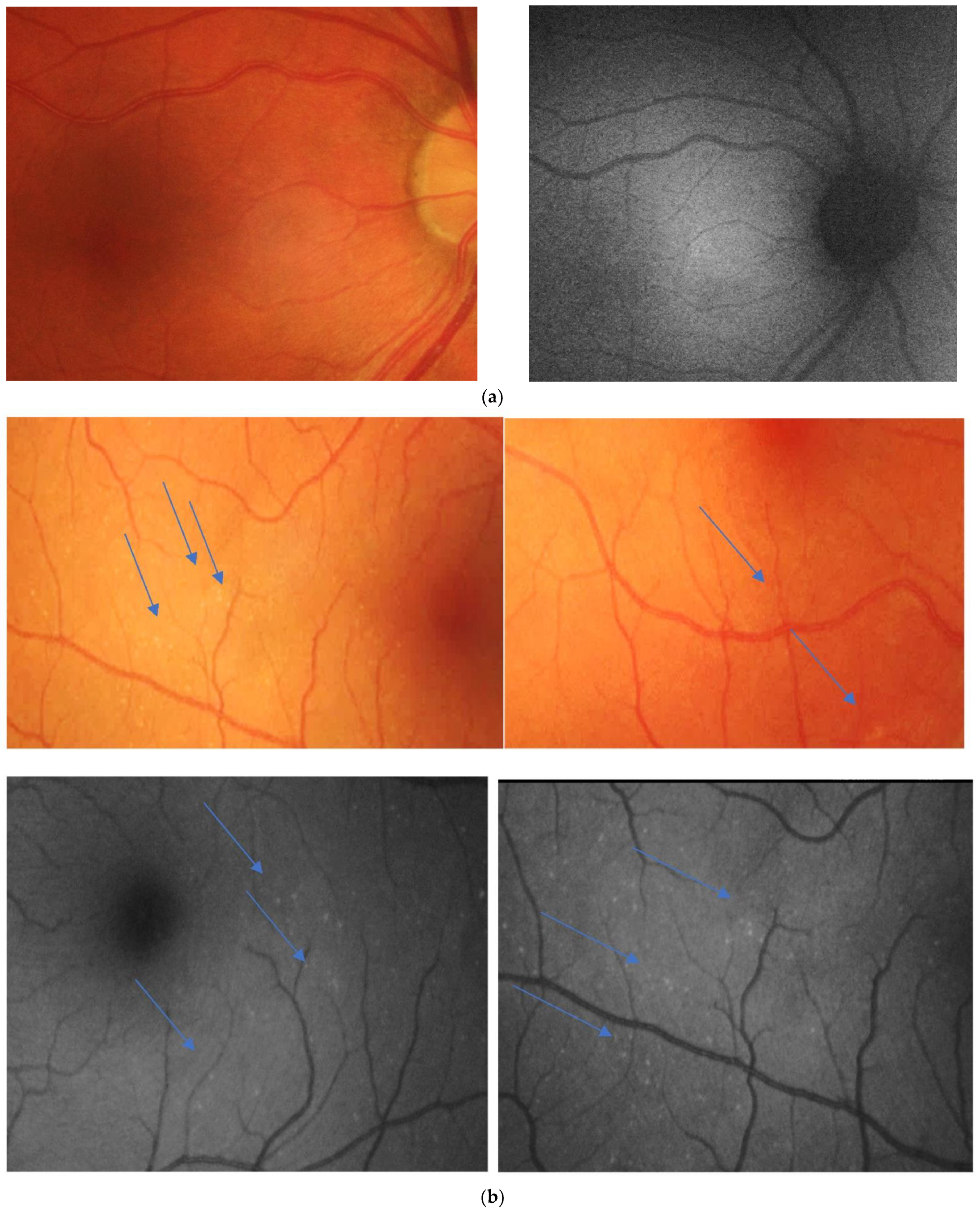
Figure 1. (a) Normal retina in normal view on left side and with autofluorescence filter (FAF) on right side. (b) Retinal plaques of patients 1 and 2 with AD familiarity the arrow shows the plaques on the retinal plane, on upside in normal view and on down side with autofluorescence filter (FAF).
Reports 04 00026 g001
Figure 2. The arrows shows the concentration of Amyloid Beta-42 in patient 1 (left side) and 2 (right side) with familiarity of Alzheimer.
Reports 04 00026 g002
Figure 3. Negative control patient with infective conjunctivitis and no familiarity for AD. Here we can’t find any presence of Beta-42 Amyloid.
Reports 04 00026 g003
Figure 4. The arrow shows the presence of protein Amyloid Beta-42 in a prepared in vitro sample with a concentration of 5mg/mL of purified protein Amyloid Beta 42.
Reports 04 00026 g004
Publisher’s Note: MDPI stays neutral with regard to jurisdictional claims in published maps and institutional affiliations.