Case Report

An Unusual Case of Vomiting Caused by Myeloid Sarcoma

1
Hematology Department, Royal Blackburn Hospital, Haslingden Road, Blackburn, Lancashire BB2 3HH, UK
2
Gastroenterology Department, Royal Blackburn Hospital, Haslingden Road, Blackburn, Lancashire BB2 3HH, UK
3
Radiology Department, Royal Blackburn Hospital, Haslingden Road, Blackburn, Lancashire BB2 3HH, UK
4
Pathology Department, Royal Blackburn Hospital, Haslingden Road, Blackburn, Lancashire BB2 3HH, UK
*
Author to whom correspondence should be addressed.
Submission received: 23 November 2018 / Revised: 25 December 2018 / Accepted: 3 January 2019 / Published: 8 January 2019

Abstract

:
Myeloid sarcoma is an extramedullary mass consisting of myeloblasts that may present simultaneously or precede a bone marrow disorder. It has been reported to occur without a known preexisting diagnosis of acute leukemia, myelodysplastic syndrome or a myeloproliferative neoplasm and this is known as primary myeloid sarcoma. Here, we report a case of an 80-year-old male who presented with intermittent vomiting and significant weight loss for 3 months. The imaging and histological findings were consistent with a mesenteric myeloid sarcoma encasing the coeliac trunk and superior mesenteric artery, abutting and obstructing the proximal small bowel, causing subacute bowel obstruction. Systemic chemotherapy with low dose cytarabine achieved a reduction in the size of myeloid sarcoma and improved patient’s symptomatology but unfortunately our patient succumbed to progression 11 months later.

1. Introduction

According to the World Health Organisation (WHO) classification, acute myeloid leukaemia is classified based on morphology, immunophenotype, cytogenetics and clinical features. There are six main groups of AML recognized in this classification system [1,2]:
  • AML with recurrent genetic abnormalities
  • AML with myelodysplasia-related features
  • Therapy-related AML and MDS
  • AML, not otherwise specified
  • Myeloid sarcoma
  • Myeloid proliferations related to Down syndrome
Myeloid sarcoma (MS) is a rare and aggressive tumour [3]. It consists of extra medullary myeloblasts and immature myeloid cells [4]. MS is usually a sign of relapse of Acute Myeloid Leukaemia (AML) but may also present simultaneously or precede the development of AML. MS may present at various anatomical sites; the most common being soft tissue, bone, skin, lymph nodes and the periosteum [5]. The gastrointestinal tract is only involved in 6.5% of the reported cases of MS [4]. The literature only features case reports of MS affecting the gastrointestinal tract because of the rarity of the condition. In this paper, we present a case of isolated MS involving the mesentery with atypical clinical presentation.

2. Case Report

An 80-year old Asian male presented to hospital with intermittent vomiting and abdominal discomfort for the past 3 months. He had lost about 12 kg in weight during that period. There was no history of haematemesis or melaena. He denied any lower gastrointestinal symptoms.
His past medical history included coronary artery bypass graft (CABG), hypertension, hypercholesterolaemia, transurethral resection of prostate (TURP) and haemorrhoidectomy. He was taking regular amlodipine, atenolol, simvastatin, aspirin and tamsulosin. He was a non-smoker and did not drink any alcohol. There was no significant family history.
Examination revealed an 8 cm tender epigastric mass and a paraumbilical hernia.
Two weeks prior to admission, his General Practitioner (GP) organised an oesophagogastroduodenoscopy (OGD) which showed a hyperplastic polyp in the antrum of the stomach. Full blood count organised by his GP was normal two months prior to admission.
Blood results on admission are shown in Table 1. His blood film revealed approximately 30% circulating myeloid blasts.
Repeat inpatient OGD was challenging as there was extrinsic compression in the area of third part of the duodenum (D3), preventing further passage of the endoscope, Figure 1.
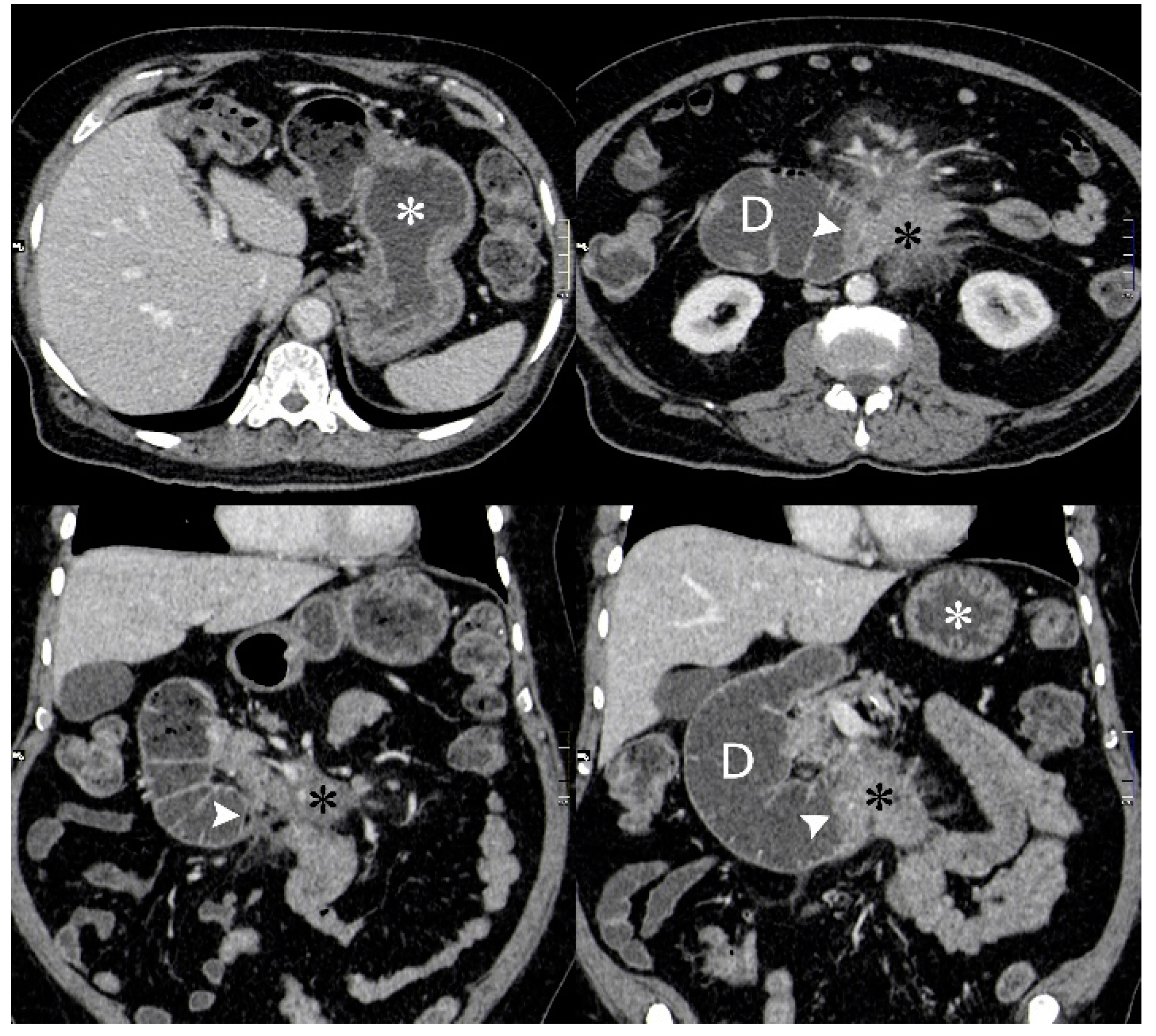
Computed Tomography (CT) of his chest, abdomen and pelvis showed a large (8 cm × 10 cm × 9.5 cm) mesenteric mass encasing the mesenteric vessels with proximal small bowel obstruction (Figure 2). There were multiple enlarged mesenteric and retroperitoneal lymph nodes.
His extremely elevated white cell count and lactate dehydrogenase (LDH) raised the suspicion of AML. A subsequent bone marrow biopsy showed that the normal bone marrow cells and architecture were replaced by myeloblasts. This confirmed the diagnosis of AML. Cytogenetics testing showed a normal karyotype; molecular studies detected a mutation in the nucleophosmin gene (NPM1) and there was a suspicion of a FLT3 ITD mutation. However, this mutant peak represented <5% of the amplified FLT3 sequence from the genomic DNA sample. Flow cytometry identified a composite phenotype of myeloblasts with CD45+, CD117+, CD33++ CD34−, CD15+, HLADR+/−, CD13+/−, CD3++.
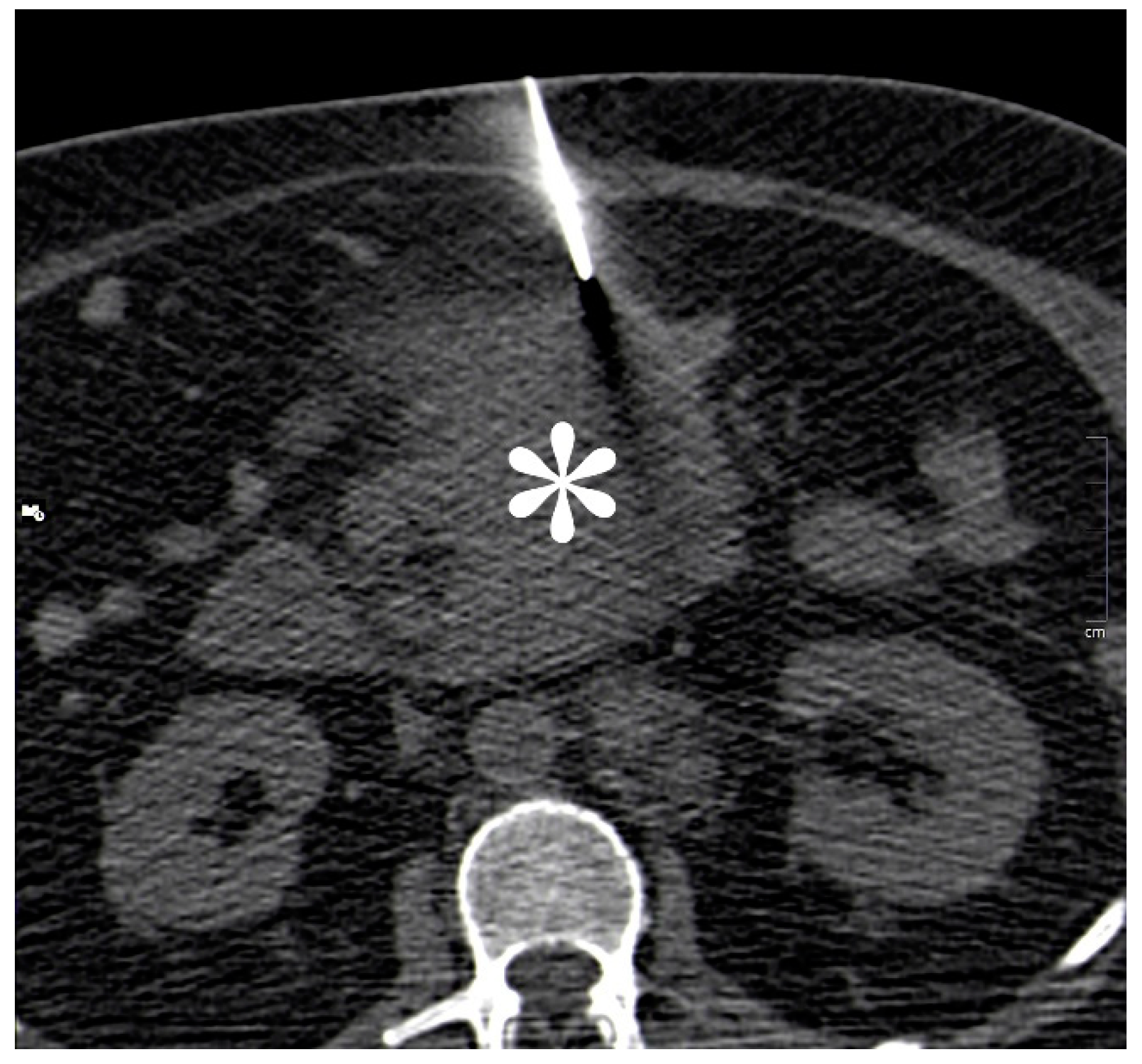
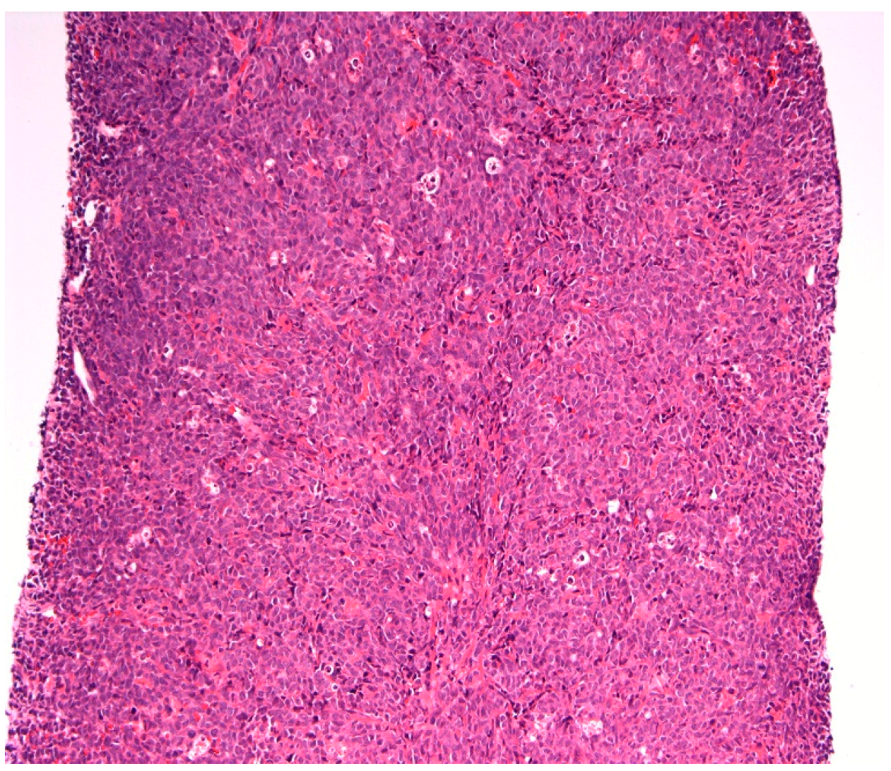
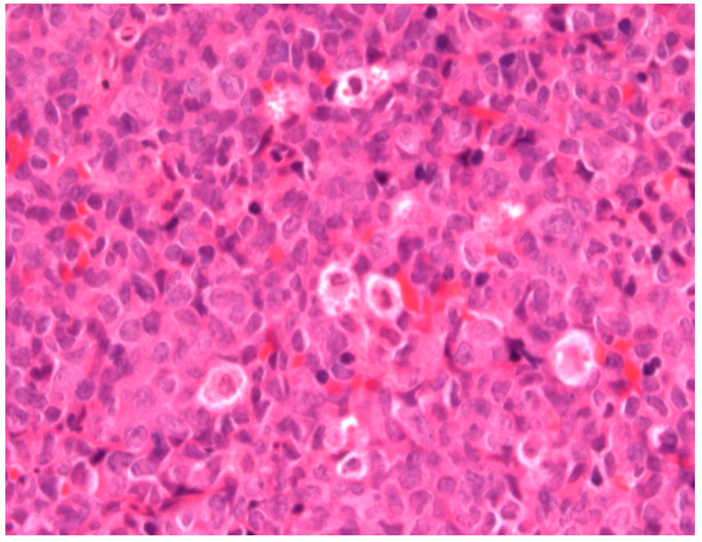
CT guided biopsy of the mesenteric mass (Figure 3) was performed to obtain a histological diagnosis. The analysis of this, demonstrated extensive infiltration of the tissue by immature blasts, with destruction of the native architecture (Figure 4 and Figure 5). Immunohistochemistry with myeloperoxidase immunostain (Figure 6) confirmed the myeloid lineage of these blasts, which had CD34− and CD117+, MPO+, PU+ and CD7− phenotype.
Intensive chemotherapy was deemed to be inappropriate because of his age, comorbidities and frailty. He received palliative chemotherapy in the form of cytarabine 20 mg subcutaneously twice daily for a period of 10 days every 4 weeks. This regime was effective as he was clinically improving: his symptoms of vomiting stopped after the fifth day of the first cycle, his appetite improved and he slowly started gaining weight. Another surrogate marker of clinical response to cytarabine was the lack of blood transfusion requirement. He received this chemotherapy regime for a total of 11 months when he then developed recurrent vomiting.
A repeat CT scan of his abdomen (Figure 7) confirmed secondary loss of response. Radiotherapy to the mesenteric mass was considered, however, he unfortunately developed gastrointestinal bleeding, which precluded this.
Consent: Written informed consent was obtained from the patient for publication of this case report and accompanying images. No Ethics approval required.

3. Discussion

Myeloid sarcoma is a tumour mass of myeloid blasts in any anatomical site other than the bone marrow [6]. It is also commonly known as “Chloroma” from the Greek word chloros (green) because of its classic green colour when the high levels of myeloperoxidase enzyme in the chloroma reacts with air [7]. It is also known as Granulocytic Sarcoma. However, the World Health Organisation (WHO) classification of myeloid neoplasms and AML renamed it as myeloid sarcoma. It is a pathological diagnosis defined as extra medullary proliferation of blasts, of one or more of the myeloid lineages, that disrupts the normal architecture of the tissue in which it is found [2]. Any anatomical site of the body can be affected, with the most common being soft tissue, bone, skin, lymph nodes and the periosteum [5]. The presenting symptoms of MS vary according to the anatomical site and the extent of disruption of normal tissue. In this report, we have discussed an unusual cause of persistent vomiting and weight loss.
MS is most often seen in acute myeloid leukemia (AML) but can also be seen in chronic myeloid leukemia (CML), myelodysplastic syndrome (MDS) and other myeloproliferative neoplasms (MPN) [8]. Most commonly, MS present in patients with known haematological malignancy but it can also be simultaneously diagnosed or precede the haematological malignancy [2]. It is a sign of relapse in patients with an existing haematological malignancy [9]. Our patient’s final diagnosis was a primary myeloid sarcoma, as his symptomatology started well before any abnormalities seen on his full blood count. In cases of isolated MS (i.e., without blood or bone marrow evidence of haematological malignancy), the diagnosis of MS should be considered as synonymous with AML and should be evaluated for morphologic, phenotypic and genetic features [2].
Immunohistochemistry is the most practical method for establishing the diagnosis of MS as it can differentiate between myeloid and non myeloid cells with monoclonal antibodies to myeloperoxidase. Myeloperoxidase staining is a quick way to rule out other tumors and establish the diagnosis of MS. The morphologic appearance on haematoxylin and eosin staining reveals a population of large cells with prominent nucleoli and fine nuclear chromatin [10].
Molecular mutations and genetic aberrations play an important role in assessing prognosis [10]. The presence of the NPM1 mutation confers a better prognosis. Additionally, patients with NPM1 mutation alone have a better prognosis than patients with mutations in both NPM1 and FLT3. Positivity for CD117 and negativity for CD34 carry a better prognosis in cases of acute myeloid leukemia. To our knowledge, there are no large studies characterizing the prognostic factors and treatment strategies in this group of patients. MS always warrants systemic treatment with conventional AML-type chemotherapy with regimen choice and dosing following standard age and cytogenetic-based risk profiling. The postremission chemotherapy has not been sufficiently reviewed in solitary MS and the role of haematopoietic stem cell transplantation has not been sufficiently explored. In general, radiotherapy is offered when there is extra medullary progression, marrow relapse, or rapid symptom relief is required. Radiotherapy can result in excellent local disease control and palliation of symptoms, without significant toxicity.
This case also highlights a diagnostic challenge endoscopically. The first line of investigation for persistent vomiting is endoscopic assessment using OGD. The two OGDs performed 2 weeks apart yielded different results in our patient. There have been recent concerns about missed upper GI malignancy at endoscopy, with recent meta-analysis involving 3787 patients with upper gastrointestinal cancer showing that 11.3% of them are missed at endoscopy 3 years before the diagnosis [11]. It is therefore important to consider repeat OGD in highly suspicious cases following discussion with the gastroenterology team. It also highlights the importance of cross-sectional imaging in diagnostic uncertainties, especially when patients present with signs of acute or subacute obstruction.

4. Conclusions

Primary myeloid sarcoma is a rare entity. MS of the gastrointestinal tract is even rarer, and is associated with serious complications including haemorrhage, perforation, necrosis, obstruction and intussusception. This case posed diagnostic challenges because of the anatomical location of the MS and the rarity of the condition. Prognostic factors and treatment strategies remain uncertain. Further studies are required to provide further information to guide treatment.

Funding

This research received no external funding.

Acknowledgments

We gratefully acknowledge the assistance and contribution of HMDS Leeds Haematological Malignancy Diagnostic Service.

Conflicts of Interest

The authors declare no conflict of interest.

References

  1. Byrd, J.C.; Edenfield, W.J.; Shields, D.J.; Dawson, N.A. Extramedullary myeloid cell tumors in acute nonlymphocytic leukemia: A clinical review. J. Clin. Oncol. 1995, 13, 1800–1816. [Google Scholar] [CrossRef] [PubMed]
  2. Vardiman, J.W.; Thiele, J.; Arber, D.A.; Brunning, R.D.; Borowitz, M.J.; Porwit, A.; Harris, N.L.; Le Beau, M.M.; Hellström-Lindberg, E.; Tefferi, A.; et al. The 2008 revision of the World Health Organization (WHO) classification of myeloid neoplasms and acute leukemia: Rationale and important changes. Blood 2009, 114, 937–951. [Google Scholar] [CrossRef] [PubMed]
  3. Benjazia, E.; Khalifa, M.; Benabdelkader, A.; Laatiri, A.; Braham, A.; Letaief, A.; Bahri, F. Granulocytic sarcoma of the rectum: Report of one case that presented with rectal bleeding. World J. Gastrointest. Pathophysiol. 2010, 1, 144–146. [Google Scholar] [CrossRef] [PubMed]
  4. Swedlow, S.H.; Campo, E.; Harris, N.L.; Jaffe, E.S.; Pileri, S.A.; Stein, H.; Thiele, J.; Vardiman, J.W. WHO Classification of Tumours of Haematopoietic and Lymphoid Tissues, 4th ed.; IARC Press: Lyon, France, 2008. [Google Scholar]
  5. Choi, E.K.; Ha, H.K.; Park, S.H. Granulocytic Sarcoma of Bowel: CT Findings. Radiology 2007, 243, 3. [Google Scholar] [CrossRef] [PubMed]
  6. Girelli, C.M.; Carsenzuola, V.; Latargia, M.; Aguzzi, A.; Serio, G. Small-bowel myeloid sarcoma: Report of a case with atypical presentation. Int. J. Surg. Case Rep. 2014, 5, 613–616. [Google Scholar] [CrossRef] [PubMed] [Green Version]
  7. Miliauskas, J.R.; Crowley, K.S. Granulocytic sarcoma (chloroma) of the small intestine with associated megakaryocytes. Pathology 1985, 17, 661–662. [Google Scholar] [CrossRef]
  8. John, S.; Ahmad, H.A.; Dunna, T.; Kern, W.; Holter, J.; Cherry, M. Unusual presentation of bladder myeloid sarcoma causing acute renal failure: Case report and review of the literature. Clin. Adv. Haematol. Oncol. 2013, 11, 388–392. [Google Scholar]
  9. Pileri, S.A.; Ascani, S.; Cox, M.C.; Campidelli, C.; Bacci, F.; Piccioli, M.; Piccaluga, P.P.; Agostinelli, C.; Asioli, S.; Novero, D.; et al. Myeloid sarcoma: Clinic-pathologic, phenotypic and cytogenetic analysis of 92 adult patients. Leukemia 2007, 21, 340–350. [Google Scholar] [CrossRef] [PubMed]
  10. Bakst, R.L.; Tallman, M.S.; Douer, D.; Yahalom, J. How I treat extramedullary acute myeloid leukemia. Blood 2011, 118, 3785–3793. [Google Scholar] [CrossRef] [PubMed] [Green Version]
  11. Menon, S.; Trudgill, N. How commonly is upper gastrointestinal cancer missed at endoscopy? A meta-analysis. Endosc. Int. Open 2014, 2, E46–E50. [Google Scholar] [CrossRef] [PubMed]
Figure 1. OGD images showing partial obstruction in the third part of the duodenum (D3) preventing the passage of the endoscope.
Reports 02 00002 g001
Figure 2. Axial (top row) and coronal (bottom row) CT images from an initial diagnostic portal venous phase contrast enhanced CT when patient presented with symptoms of gastric outlet obstruction. Fluid filled dilated stomach (white asterisk) and 2nd and 3rd part of duodenum. Ill-defined enhancing mass in the root of the mesentery (black asterisk) obstructing the 3rd part of the duodenum (white arrowhead).
Reports 02 00002 g002
Figure 3. Unenhanced CT image from CT guided biopsy procedure showing a 15 G coaxial introducer at the edge of the mass (white asterisk) through which two 16 G × 2 cm (Achieve®, CareFusion; San Diego, CA, USA) cores of tissue were obtained.
Reports 02 00002 g003
Figure 4. A core of tissue showing sheets of myeloblasts completely effacing the nodal architecture, with no normal lymphoid tissue present. (Haematoxylin and eosin, 200×).
Reports 02 00002 g004
Figure 5. A higher power view of the core, showing a population of large myeloblasts with prominent round to indented nuclei and fine nuclear chromatin. (Haematoxylin and eosin, 400×).
Reports 02 00002 g005
Figure 6. Immunohistochemistry with myeloperoxidase, reveals strong granular cytoplasmic staining in myeloblasts and confirms the myeloid lineage of the blasts. (Myeloperoxidase immunostain 200×).
Reports 02 00002 g006
Figure 7. Follow up axial (top row) and coronal (bottom row) venous phase contrast enhanced CT images performed 11 months after initial diagnostic CT when patient represented with symptoms of reduced appetite and abdominal swelling. The degree of gastric dilatation (white asterisk) is considerably less than on the initial CT. The mesenteric mass (black asterisk) has significantly reduced in volume following chemotherapy but the duodenum (D) is more dilated and obstructed at the 3rd part (white arrowhead), possibly due to post treatment fibrosis.
Reports 02 00002 g007
Table 1. Blood results on admission.
Full Blood CountPatient’s ResultsNormal Range
Haemoglobin (Hb)146 g/L130–180 g/L
White Cell Count (WCC)92.8 × 10⁹/L4.0 × 109/L–11.0 × 109/L
Platelets (Plts)60 × 109/L150× 109/L–450 × 109/L
Mean Corpuscular Volume (MCV)84/2 FL76.0–100.0 FL
Neutrophils21.6 × 109/L2.0 × 109/L–7.5 × 109/L
Lymphocytes10.6 × 109/L1.5 × 109/L–4.0 × 109/L
Monocytes25.3 × 109/L0.2 × 109/L–0.8 × 109/L
Eosinophils0.3 × 109/L0.0 × 109/L–0.4 × 109/L
Basophils4.0 × 109/L0.0 × 109/L–0.4 × 109/L
Renal Profile
Sodium136 mmol/L133–146 moml/L
Potassium3.3 mmol/L3.5–5.3 mmol/L
Urea12.7 mmol/L2.5–7.8 mmol/L
Creatinine111 mmol/L58–110 mmol/L
Liver Profile
Total Bilirubin10 umol/L0–21 umol/L
ALT21 IU/L3–53 IU/L
ALP109 IU/L30–130 IU/L
Albumin37 g/L35–50 g/L
Bone Profile
Calcium2.30 mmol/L2.20–2.60 mmol/L
Adjusted Calcium2.38 mmol/L2.20–2.60 mmol/L
Total Protein64 g/L60–80 g/L
Globulin27 g/L18–36 g/L
CRP53 mg/L0–10 mg/L
Lactate Dehydrogenase6104 IU/L313–619 IU/L