Systematic Review

Antineoplastic Effect of Metformin Against Glioblastoma Multiforme In Vitro and In Vivo: A Systematic Review and Meta-Analysis

by
Bhavya Vashi
,
Daniel Gonzales-Portillo
and
Jorge Cervantes
*
Kiran C. Patel College of Allopathic Medicine, Nova Southeastern University, Fort Lauderdale, FL 33328, USA
*
Author to whom correspondence should be addressed.
Neuroglia 2025, 6(4), 40; https://doi.org/10.3390/neuroglia6040040
Submission received: 17 August 2025 / Revised: 10 October 2025 / Accepted: 11 October 2025 / Published: 14 October 2025

Abstract

Background/Objectives: Glioblastoma multiforme (GBM) is a highly aggressive brain tumor associated with poor survival outcomes. Given the significant financial burden of cancer treatments, repurposing existing drugs can reduce costs and enhance therapeutic efficacy. Metformin, an antidiabetic medication, has been investigated for its antineoplastic effects against GBM. Here, we reviewed the in vitro and in vivo effects of metformin through GBM cell viability and overall animal survival, respectively. Methods: A systematic review was conducted following the Preferred Reporting Items for Systematic Reviews and Meta-Analyses (PRISMA) guidelines. Data extraction and statistical analyses were performed using Microsoft Excel, and R. Effect sizes were calculated as standard mean differences (SMDs) for in vitro studies assessing cell viability and hazard ratios (HRs) for in vivo mice survival analyses. Results: A total of two-hundred-thirty in vitro studies and five-hundred-sixty-six in vivo studies were screened. Of these, seven in vitro and eight in vivo studies were compatible for the meta-analysis. The random-effects model showed a reduction in cell viability (SMD [95% CI]: 3.70 [2.28, 5.12]). A pooled in vivo survival analysis suggests an increase in overall survival in mice receiving metformin (p-value = 0.055). A random-effects model for overall survival supports this pooled analysis (HR [95% CI]: 0.76 [0.39, 1.46]). Additionally, metformin also showed a reduction in cell viability (SMD [CI]; 2.27 [0.79, 3.75]) and an increase in overall animal survival (HR [CI], 0.23 [0.12, 0.45]) when it was added as an adjuvant to traditional GBM therapies. Conclusions: Our findings from in vitro and in vivo studies support the potential of metformin as an antineoplastic agent against GBM. We plan to extend our analyses into clinical studies to determine if these benefits extend to human patients. Metformin has the potential to revolutionize GBM therapy if a relationship exists due to its inexpensive nature.

1. Introduction

Glioblastoma multiforme (GBM) is the most prevalent and aggressive primary malignant brain tumor found in adults. GBM accounts for approximately half of all malignant tumors within the central nervous system, predominantly developing in the brain, but it may also arise in the brainstem, cerebellum, and spinal cord [1]. The incidence of glioblastoma begins to rise after age 40 and peaks between 75 and 84 years [2]. It has a poorly characterized familial inheritance pattern; however, it does have an association with exposure to ionizing radiation. This disease is more common in white non-Hispanic males and is associated with poorer outcomes in older patients [3].
The tumor is believed to arise from neural stem cells or glial progenitor cells, depending on the genetic mutations involved [4]. GBM typically arises from loss of cell cycle control from G1 to the S phase. Around 87% of patients with GBM will test positive for a mutation in p53, a key tumor suppressor gene that normally regulates the cell cycle [5]. Losing p53 means the cell’s capacity to undergo apoptosis is deeply hindered, leading to cell survival. Although aberrations with deoxyribonucleic acid (DNA) repair are typically present in cancer cells, paradoxically, the cancer cells require these mechanisms in order to proliferate and handle endogenous stress from metabolism. GBM is an energetically demanding cancer that requires the creation of new blood vessels to meet its demands for proliferation [6]. However, GBM is also capable of co-opting existing blood vessels in the brain to minimize interruptions in blood flow and leave the vessel walls intact [7]. Furthermore, GBM has various mechanisms to evade immune surveillance, such as suppressing dendritic cell function and secreting CD200, which promotes self-tolerance [5].
The current standard of care includes surgical resection with concurrent radiation therapy and chemotherapy. A gross total resection is first indicated, followed by radiation therapy for up to 6 months a few weeks after surgery. During this time, drugs such as temozolomide (TMZ) should be concurrently taken. This combination so far has demonstrated the best therapeutic approach in terms of median overall survival; however, the addition of TMZ does not impact quality of life any differently than radiation alone [2]. There is also the option of Tumor Treating Fields (TTFields) for when patients are unable to tolerate the effects of chemotherapy for recurrent GBM as this technology has shown similar response rates and reduction in risk of death compared to chemotherapy [8].
Despite intensive treatments, recurrence is high, and the prognosis remains poor for a majority of patients. Median survival remains limited to approximately 15 months following diagnosis, with only 5.5% of patients surviving beyond five years [9]. This poor outlook underscores an urgent need for novel therapeutic options.
New oncologic therapies, particularly targeted agents and immunotherapies, have demonstrated substantial clinical efficacy. However, their high and continuously rising costs pose significant challenges to patient accessibility [10]. There is growing concern regarding the financial burden associated with these modern treatment modalities. This has resulted in a call to strategically repurpose widely available low-cost pharmacologic agents with established safety profiles for cancer treatments.
Metformin, a first-line oral antihyperglycemic agent routinely prescribed for type 2 diabetes mellitus, has emerged as a promising candidate [11]. It originated from the Galega officinalis plant, also known as goat’s rue, which has been used in traditional medicine to treat diabetes symptoms. Its hypoglycemic properties were discovered in the 1920s, and it was approved for use as a drug in the United States in 1995. In recent years, beyond its role in glycemic control, metformin has demonstrated novel antineoplastic and immunomodulatory properties. Studies have revealed that metformin can slow or inhibit cellular proliferation and impair metastatic dissemination. These antineoplastic effects have been documented across multiple malignancies, including breast, liver, pancreatic, and bone [12]. In addition to its mechanistic activity, metformin has widespread availability and has a well-known safety profile, making it a candidate for drug repurposing initiatives.
At the cellular level, metformin exerts its antineoplastic effects through the activation of AMP-activated protein kinase (AMPK) and downregulation of the mechanistic target of rapamycin (mTOR) signaling pathway [12]. It disrupts mitochondrial respiration by inhibiting complex 1 of the electron transport chain, simultaneously preventing the release of reactive oxygen species (ROS) and the generation of adenosine triphosphate (ATP). ROS are activators of Hypoxia-Inducible Factor 1 (HIF-1), so, by stifling their creation, metformin can interfere with the microenvironment of cancer cells [13]. Furthermore, DNA damage is reduced by the elimination of genotoxic ROS. GBM is a hypoxic tumor that is highly dependent on anaerobic respiration to proliferate, and metformin’s capability of lowering cellular energy status by depleting the metabolites of the tricarboxylic acid (TCA) cycle can be useful [14]. Once the tumor cells are energetically depleted, the cells activate AMPK. AMPK is a critical compound that functions as an energy sensor in the cell. AMPK activation will promote ATP-generating catabolic processes and inhibit ATP-consuming processes. As a result, AMPK will suppress the mTOR pathway, a crucial signaling cascade for cell growth and proliferation [15,16]. Experimental studies have demonstrated that this metabolic reprogramming, mediated by AMPK activation and mTOR suppression, significantly impairs cancer cell growth and survival [17]. While mTOR inhibition is often a tumor-adaptive mechanism to conserve resources, it concurrently promotes cell cycle arrest, thus halting cell proliferation. These mechanisms are particularly relevant to GBM, a tumor characterized by high metabolic demand and resistance to conventional therapies.
Beyond the mechanism of action of metformin, there are other effects from the medication that could serve a role in antitumor activity [18]. Chiefly, its ability to lower the amount of glucose in circulation can slow the tumor’s ability to grow and proliferate [19]. Furthermore, hyperinsulinemia is associated with chronic inflammation in the body, a situation that is conducive to the development of cancer [20].
Although there is a growing body of literature that has examined metformin’s capability to serve as an antineoplastic agent, both in vivo and in vitro, many of these studies utilize different cell lines, animal models, and conditions when testing metformin. This fragmentation of data highlights the need for a meta-analysis to synthesize this preclinical evidence to justify further investigation for clinical use. We intend to pool this data to identify patterns, some of which may indicate a role for metformin in the treatment of GBM.
Here, we conducted a systematic review and meta-analysis to evaluate the antineoplastic effects of metformin, assessing its impact on GBM cell viability in vitro and overall survival outcomes in in vivo models.

2. Materials and Methods

2.1. Protocol and Registration

This meta-analysis followed the PRISMA (Preferred Reporting Items for Systematic Reviews and Meta-Analyses) guidelines.

2.2. Search Strategy and Information Sources

We searched several databases, including PubMed, EMBASE, and MEDLINE with Full Text, using the EBSCOhost platform. We also searched CINAHL Complete, Biomedical Reference Collection: Comprehensive, and Cochrane Clinical Answers. The search included all studies available up to January 2025.
Our initial search was broad, including all studies that mentioned metformin, glioblastoma multiforme, and either in vitro or animal models, with no limits on publication date or language. The search string used for in vitro studies was (‘metformin’/exp OR metformin) AND (‘glioblastoma multiforme’/exp OR ‘glioblastoma multiforme’ OR ((‘glioblastoma’/exp OR glioblastoma) AND multiforme) OR gbm) AND (‘in vitro’/exp OR ‘in vitro’ OR (in AND vitro)) AND ‘in vitro study’/de AND ‘article’/it. Search for animal models included the terms animal, mouse, rodent, or rat. The search string used for in vivo studies was (‘metformin’/exp OR metformin) AND (‘glioblastoma multiforme’/exp OR ‘glioblastoma multiforme’ OR ((‘glioblastoma’/exp OR glioblastoma) AND multiforme) OR gbm) AND (‘animal’/exp OR animal OR ‘mouse’/exp OR mouse OR ‘rodent’/exp OR rodent OR ‘rat’/exp OR rat) AND ‘in vivo study’/de AND ‘article’/it. Duplicate records were removed before additional screening. Systematic review articles were then removed, and the remaining studies were assessed independently by two reviewers for relevance based on the title and abstract. Full-text versions of the studies were then screened similarly. Studies that included a control group that did not receive metformin were chosen. Studies were selected based on whether they reported our defined outcomes of interest, which were cell viability for in vitro studies and overall animal survival for in vivo studies.

2.3. Bias and Quality Assessments

In vivo and in vitro studies were assessed using the SYRCLE tool and a structured checklist, respectively. The SYRCLE tool is adapted from the Cochrane Risk of Bias tool that caters to variables applicable to preclinical animal studies. For the in vitro studies, we examined cell line validation, random housing bias, blinding to outcomes, and reproducibility. In each of these categories, the studies are judged as adequate, unclear, or inadequate. The in vivo studies are judged in a similar manner.

2.4. Data Extraction and Statistical Analysis

Data was extracted by two reviewers using a spreadsheet in Microsoft Excel. We collected information on study design, type of treatment, metformin dosage, median overall survival, and hazard ratios. The main outcome of interest for murine trials was median overall survival, whereas, for in vitro studies, the main outcome was cell viability.
For in vitro studies, effect sizes of metformin were calculated using the standard mean difference (SMD). For murine survival studies, hazard ratios were estimated from the reported data or, when not directly available, extracted data from published Kaplan–Meier curves. These hazard ratios were then combined into a pooled Kaplan–Meier curve to generate a pooled analysis. A Cox proportional hazards model was used to estimate the effect of metformin on the survival of mice.
Additional analysis was conducted to assess the effect of metformin as an adjuvant in combination with standard treatments, which included chemotherapies such as TMZ and Dichloroacetate (DCA), as well as immunomodulators and radiation.
Statistical analysis and calculation of effect sizes were completed using R software (Version 4.4.1). A random-effects model was utilized.

3. Results

3.1. Characteristics of the Studies for Analysis

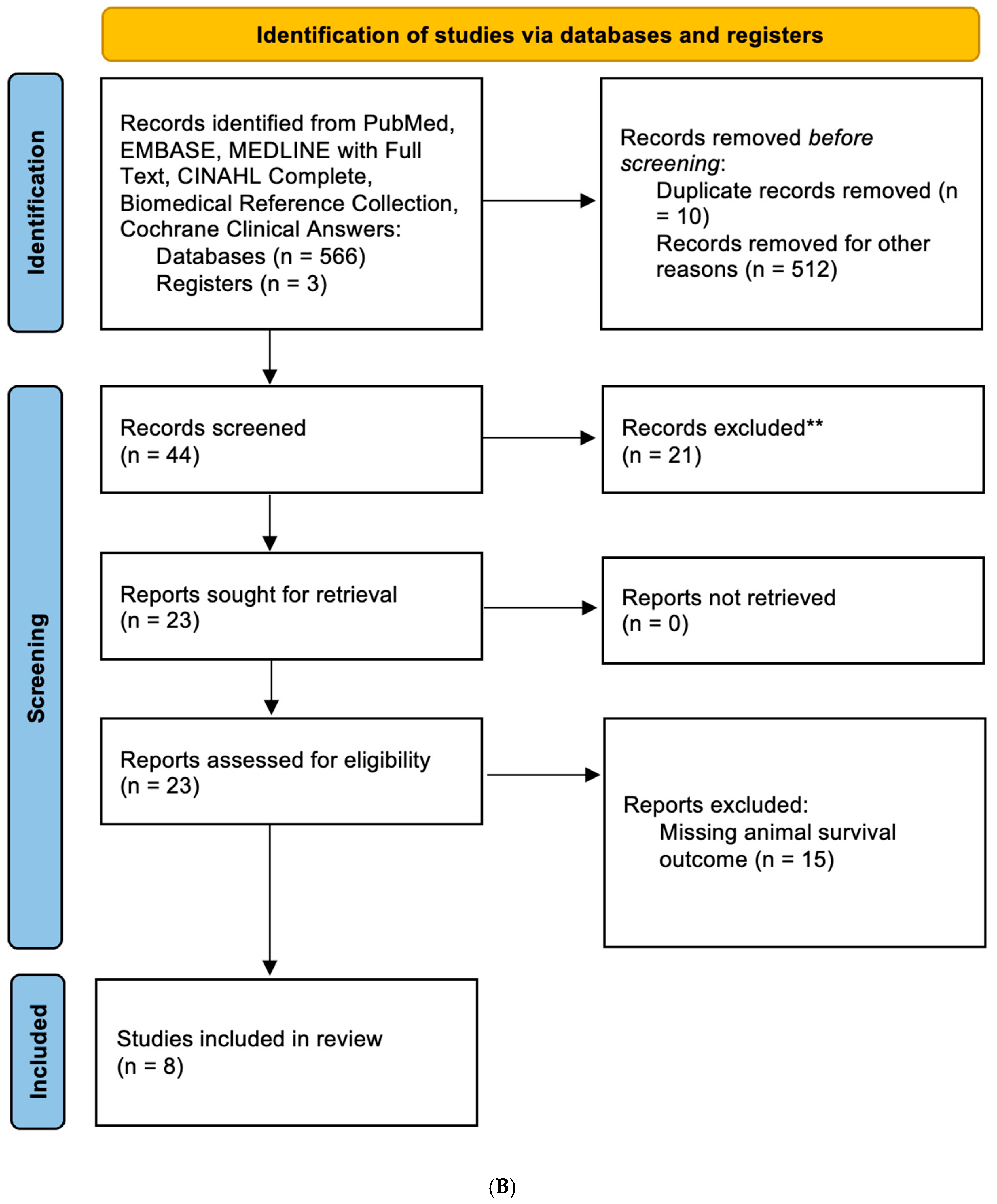
Our search yielded seven in vitro and eight in vivo studies, which were used for the analyses (Figure 1). These studies ranged from 2012 to 2024 and were all published in English. We found that the GBM cell lines utilized varied across the different studies and included EGFR1, EGFR2, L0627, U87, U251, A172, T98G, LN18, U87 glioblastoma stem-like cells (GSCs), and U251GSC. All of these were human cell lines.
A subset of studies also used metformin as an adjuvant to traditional therapies and were used in a separate analysis describing metformin’s role as an adjuvant. The characteristics of all the included studies are described in the table below (Table 1). The majority of the studies reported that metformin reduced cell viability or improved animal survival.

3.2. Metformin Reduced Cell Viability of GBM Cell Lines

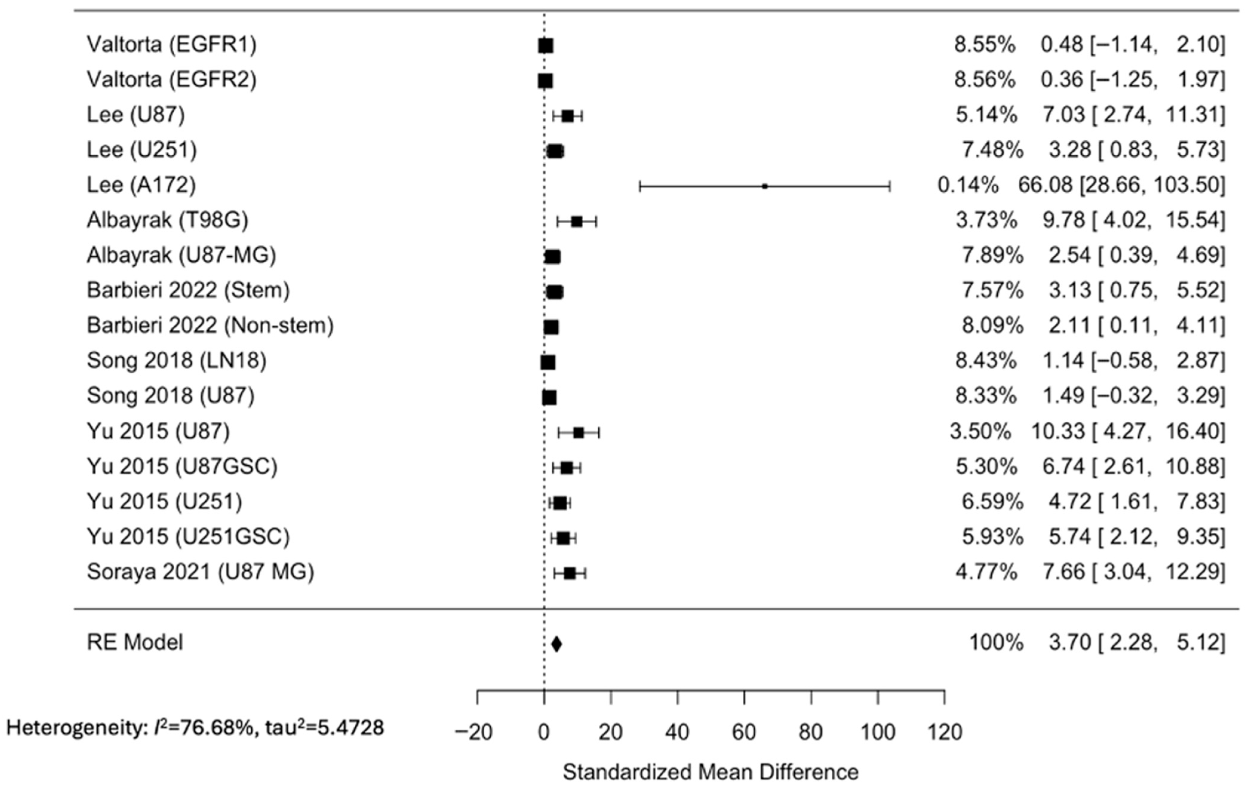
We first explored the effect of metformin on cell viability in vitro. The majority of the in vitro studies tested multiple GBM cell lines. Each cell line was quantified separately in our analyses. The SMD was calculated assuming 100% cell viability in the control groups; therefore, a positive number here indicates decreased viability. Most of the studies showed a reduction in cell viability with metformin treatment. There was one outlier identified that demonstrated markedly decreased cell viability compared to the other studies, but it did not highly contribute to the random-effects model. Overall, the random-effects model also showed a significant reduction in cell viability (SMD [CI]; 3.70 [2.28, 5.12]) (Figure 2).

3.3. Metformin Increases Survival of GBM In Vivo

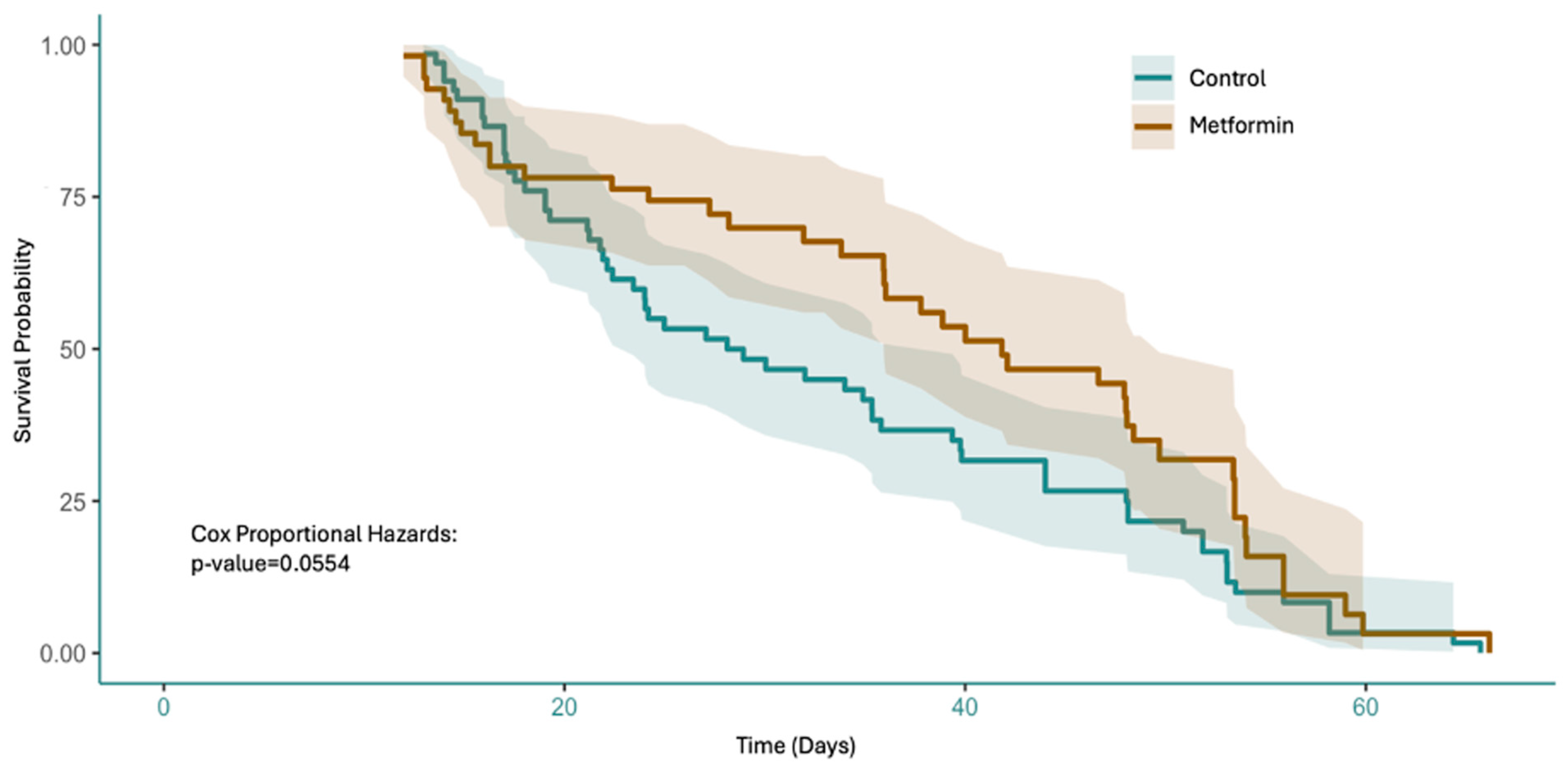
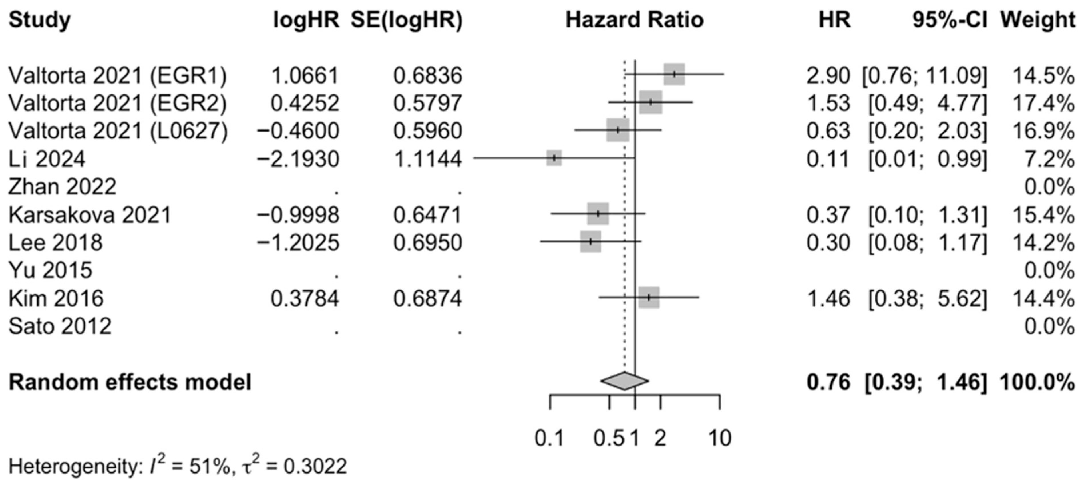
We then evaluated the effect of metformin on the survival of animals. The Cox proportional hazards model yielded a p-value of 0.0554 for pooled in vivo survival (Figure 3). Moreover, the metformin-treated group showed improved survival compared to the control groups, which was notable for up to around 60 days. Several studies and the overall random-effects model showed a low hazard ratio (HR [CI], 0.76 [0.39, 1.46]) (Figure 4).
A subset of the in vitro (n = 3) and in vivo (n = 5) studies reported the effects of the use of metformin as an adjuvant therapy compared to traditional chemotherapy agents such as TMZ. Many in vitro studies demonstrated decreased cell viability, with the overall random-effects model also showing a significant reduction (SMD [CI]; 2.27 [0.79, 3.75]) (Figure 5). Mice treated with metformin in addition to standard therapies showed improved survival rates in several in vivo studies. The overall random-effects model demonstrated significantly improved mortality in the groups where metformin was used as an adjuvant (HR [CI], 0.23 [0.12, 0.45]) (Figure 6).

3.4. Bias and Quality Assessments

In the eight in vivo studies, none reported procedures to address sequence generation, allocation concealment, blinding, or randomization of outcomes. Further, 63% of the studies disclosed the baseline characteristics of the rodents they used for their experiment, and 50% of the studies reported the conditions rodents were kept in for the experiments. For the seven in vitro experiments, none of the studies reported blinding procedures. One of seven studies explicitly confirmed their cell lines using short tandem repeat sequencing. All the studies described the conditions they kept their cells in. Moreover, 71% of the studies explicitly stated that their findings were replicated more than once. A table demonstrating the breakdown study –by study is available as a supplement. Summaries of the bias assessments for the in vitro and in vivo studies are available in Supplementary Table S1 and Supplementary Table S2 respectively.

4. Discussion

Our examination of the effects of metformin on GBM through a comprehensive meta-analysis of both in vitro and in vivo models revealed an overall effect on cell viability and median overall survival, respectively. Although the pooled analysis from animal studies did not demonstrate statistical significance, the in vitro analyses revealed a significant reduction in GBM cell viability following metformin treatment. These findings align with prior evidence suggesting that metformin exerts antitumor effects via induction of cell death, as well as an immunomodulatory effect [33]. Metformin also showed therapeutic promise as an adjuvant to standard therapy alone in both in vitro and in vivo models, significantly decreasing cell viability and improving animal survival. These standard therapies included chemotherapies such as TMZ and DCA, as well as immunotherapy and radiation.
An important consideration in interpreting these findings is the variety of GBM cell lines used across the studies, both in vitro and in vivo. The studies included a considerable range of established human glioblastoma cell lines, including EGFR1, EGFR2, U87, U251, A172, U87GSC, and U251GSC. Metformin’s mechanism of action can be differentially effective depending on the molecular profile of the tumor cells, such as that cell lines with high EGFR expression may be more susceptible to metformin. Additionally, a few studies included GSCs, which may be more resistant to therapy and have a role in recurrence [34]. Although this diversity introduces variability in drug sensitivity and treatment response, it also validates the overall effect of metformin as many of these cell lines are well-established and widely used in GBM research. While this variation enhances the generalizability of metformin’s antitumor effects, it also highlights the need for more standardized models or stratified analyses to identify subtypes that may exhibit the greatest therapeutic benefit.
An important aspect to highlight is that all the studies in this meta-analysis utilized a fixed concentration of metformin. For in vitro studies, this value was 10mM, which was shown to induce significant reductions in cell viability. However, there is evidence that metformin’s efficacy follows a dose-dependent pattern. A relatively recent study has shown that cell cultures treated with up to 50 mM showed lower cell viability compared to lower concentrations [33]. Dose selection will be critical for maximizing therapeutic efficacy.
A major barrier to effective GBM therapy lies in its complex tumor microenvironment, which actively contributes to therapeutic resistance, immune evasion, and disease progression. The role of immune cells in this tumor microenvironment, such as tumor-associated macrophages and microglia, along with secreted growth factors, cytokines, and chemokines, may ultimately promote tumor invasion, angiogenesis, and immune escape [33,35]. Given metformin’s potential role in immune modulation, its impact on the tumor microenvironment is a promising area for further investigation.
Importantly, metformin is a widely available low-cost medication with a favorable safety profile. The mean cost of modern GBM treatment, including craniotomy, radiation, and chemotherapy, is relatively expensive while achieving a median overall survival of approximately 16 months [36]. Enhancing cost-effective care is essential in the modern healthcare landscape, where resources are finite and expensive. Metformin could be used as an adjuvant therapy in this setting.
This manuscript has various strengths that enhance its reliability and relevance. Firstly, this meta-analysis was conducted in a manner compliant with the PRISMA guidelines to accomplish a systematic review. Two reviewers were utilized for data extraction to reduce bias. Both in vitro and in vivo studies were included, and all possible reports with extractable data were utilized to integrate a wide spectrum of preclinical evidence on metformin’s effect in GBM. The random-effects model was employed to account for heterogeneity between the studies. Further context to our main findings was discovered with our subgroup analysis that examined the efficacy of metformin as an adjuvant paired with standard therapies, such as TMZ, compared to metformin alone. This meta-analysis integrates findings across both in vitro and in vivo studies, providing a comprehensive perspective on metformin’s antineoplastic effects and highlighting how preclinical efficacy supports the rationale for advancing to human trials.
Despite these strengths, several limitations exist that must be considered when interpreting these results. Firstly, there is variability in the populations of cells and rats used in this analysis that can be a source of heterogeneity when comparing studies. There is also a lack of patient-derived xenografts that would better reflect the heterogeneity of the actual disease, limiting the generalizability of the results. Furthermore, we analyzed a narrow selection of outcomes. Although we were able to analyze the effects on survival, this study did not incorporate details about adverse reactions, long-term effects, or dose-dependent effects when utilizing metformin.

5. Conclusions

Our findings, nevertheless, support the evidence of metformin’s efficacy as an antineoplastic agent in vitro and suggest that it may be associated with improved median survival in animal models. Metformin’s established safety profile and low cost make it a promising candidate to join current glioblastoma treatment regimens. To fully assess its translational potential, similar analyses should be conducted using clinical data to evaluate metformin’s effectiveness in human GBM patients and determine its true utility in the treatment of GBM.

Supplementary Materials

The following supporting information can be downloaded at: https://www.mdpi.com/article/10.3390/neuroglia6040040/s1. Table S1: Summary table of Bias Analysis using a structured checklist for each in vitro study. Table S2: Summary table of Bias Analysis using the SYRCLE tool for each in vivo study.

Author Contributions

J.C. conceptualized the study; B.V. and D.G.-P. performed data search; B.V. analyzed data; J.C., B.V., and D.G.-P. wrote the manuscript. All authors have read and agreed to the published version of the manuscript.

Funding

This research received no external funding.

Institutional Review Board Statement

Not applicable.

Informed Consent Statement

Not applicable.

Data Availability Statement

No data generated.

Conflicts of Interest

The authors declare no conflicts of interest.

References

  1. Caverzan, M.D.; Beauge, L.; Oliveda, P.M.; Cesca Gonzalez, B.; Buhler, E.M.; Ibarra, L.E. Exploring Monocytes-Macrophages in Immune Microenvironment of Glioblastoma for the Design of Novel Therapeutic Strategies. Brain Sci. 2023, 13, 542. [Google Scholar] [CrossRef]
  2. Schaff, L.R.; Mellinghoff, I.K. Glioblastoma and Other Primary Brain Malignancies in Adults: A Review. JAMA 2023, 329, 574–587. [Google Scholar] [CrossRef] [PubMed]
  3. Zhao, Y.; Xu, Z.; Liu, Y.; Ye, M.; Chen, R.; Cao, Z.; Zhou, H.; Zhou, Y. Analysis of epidemiology and nomogram construction for prediction and clinical decision-making in gliomas. Front. Immunol. 2025, 16, 1624142. [Google Scholar] [CrossRef]
  4. Liu, C.; Sage, J.C.; Miller, M.R.; Verhaak, R.G.; Hippenmeyer, S.; Vogel, H.; Foreman, O.; Bronson, R.T.; Nishiyama, A.; Luo, L.; et al. Mosaic analysis with double markers reveals tumor cell of origin in glioma. Cell 2011, 146, 209–221. [Google Scholar] [CrossRef]
  5. Esemen, Y.; Awan, M.; Parwez, R.; Baig, A.; Rahman, S.; Masala, I.; Franchini, S.; Giakoumettis, D. Molecular Pathogenesis of Glioblastoma in Adults and Future Perspectives: A Systematic Review. Int. J. Mol. Sci. 2022, 23, 2607. [Google Scholar] [CrossRef]
  6. Bergers, G.; Benjamin, L.E. Tumorigenesis and the angiogenic switch. Nat. Rev. Cancer 2003, 3, 401–410. [Google Scholar] [CrossRef]
  7. Pouyan, A.; Ghorbanlo, M.; Eslami, M.; Jahanshahi, M.; Ziaei, E.; Salami, A.; Mokhtari, K.; Shahpasand, K.; Farahani, N.; Meybodi, T.E.; et al. Glioblastoma multiforme: Insights into pathogenesis, key signaling pathways, and therapeutic strategies. Mol. Cancer 2025, 24, 58. [Google Scholar] [CrossRef]
  8. Kotecha, R.; Odia, Y.; Khosla, A.A.; Ahluwalia, M.S. Key Clinical Principles in the Management of Glioblastoma. JCO Oncol. Pract. 2023, 19, 180–189. [Google Scholar] [CrossRef] [PubMed]
  9. Poon, M.T.C.; Sudlow, C.L.M.; Figueroa, J.D.; Brennan, P.M. Longer-term (≥2 years) survival in patients with glioblastoma in population-based studies pre- and post-2005: A systematic review and meta-analysis. Sci. Rep. 2020, 10, 11622. [Google Scholar] [CrossRef]
  10. Prasad, V.; De Jesus, K.; Mailankody, S. The high price of anticancer drugs: Origins, implications, barriers, solutions. Nat. Rev. Clin. Oncol. 2017, 14, 381–390. [Google Scholar] [CrossRef] [PubMed]
  11. Shah, S.; Mansour, H.M.; Aguilar, T.M.; Lucke-Wold, B. Advances in Anti-Cancer Drug Development: Metformin as Anti-Angiogenic Supplemental Treatment for Glioblastoma. Int. J. Mol. Sci. 2024, 25, 5694. [Google Scholar] [CrossRef]
  12. Lv, Z.; Guo, Y. Metformin and Its Benefits for Various Diseases. Front. Endocrinol. 2020, 11, 191. [Google Scholar] [CrossRef]
  13. Hua, Y.; Zheng, Y.; Yao, Y.; Jia, R.; Ge, S.; Zhuang, A. Metformin and cancer hallmarks: Shedding new lights on therapeutic repurposing. J. Transl. Med. 2023, 21, 403. [Google Scholar] [CrossRef]
  14. Bielecka-Wajdman, A.M.; Ludyga, T.; Smyk, D.; Smyk, W.; Mularska, M.; Świderek, P.; Majewski, W.; Mullins, C.S.; Linnebacher, M.; Obuchowicz, E. Glucose Influences the Response of Glioblastoma Cells to Temozolomide and Dexamethasone. Cancer Control 2022, 29, 10732748221075468. [Google Scholar] [CrossRef] [PubMed]
  15. Panwar, V.; Singh, A.; Bhatt, M.; Tonk, R.K.; Azizov, S.; Raza, A.S.; Sengupta, S.; Kumar, D.; Garg, M. Multifaceted role of mTOR (mammalian target of rapamycin) signaling pathway in human health and disease. Signal Transduct. Target. Ther. 2023, 8, 375. [Google Scholar] [CrossRef] [PubMed]
  16. He, L.; Cho, S.; Blenis, J. mTORC1, the maestro of cell metabolism and growth. Genes Dev. 2025, 39, 109–131. [Google Scholar] [CrossRef] [PubMed]
  17. Janzer, A.; German, N.J.; Gonzalez-Herrera, K.N.; Asara, J.M.; Haigis, M.C.; Struhl, K. Metformin and phenformin deplete tricarboxylic acid cycle and glycolytic intermediates during cell transformation and NTPs in cancer stem cells. Proc. Natl. Acad. Sci. USA 2014, 111, 10574–10579. [Google Scholar] [CrossRef]
  18. Pal, R.S.; Jawaid, T.; Rahman, M.A.; Verma, R.; Patra, P.K.; Vijaypal, S.V.; Pal, Y.; Upadhyay, R. Metformin’s anticancer odyssey: Revealing multifaceted mechanisms across diverse neoplastic terrains- a critical review. Biochimie 2025, 233, 109–121. [Google Scholar] [CrossRef]
  19. Chen, Y.C.; Li, H.; Wang, J. Mechanisms of metformin inhibiting cancer invasion and migration. Am. J. Transl. Res. 2020, 12, 4885–4901. [Google Scholar] [PubMed]
  20. Gonda, T.A.; Tu, S.; Wang, T.C. Chronic inflammation, the tumor microenvironment and carcinogenesis. Cell Cycle 2009, 8, 2005–2013. [Google Scholar] [CrossRef]
  21. Valtorta, S.; Lo Dico, A.; Raccagni, I.; Martelli, C.; Pieri, V.; Rainone, P.; Todde, S.; Zinnhardt, B.; De Bernardi, E.; Coliva, A.; et al. Imaging Metformin Efficacy as Add-On Therapy in Cells and Mouse Models of Human EGFR Glioblastoma. Front. Oncol. 2021, 11, 664149. [Google Scholar] [CrossRef] [PubMed]
  22. Lee, J.E.; Lim, J.H.; Hong, Y.K.; Yang, S.H. High-Dose Metformin Plus Temozolomide Shows Increased Anti-tumor Effects in Glioblastoma In Vitro and In Vivo Compared with Monotherapy. Cancer Res. Treat. 2018, 50, 1331–1342. [Google Scholar] [CrossRef] [PubMed]
  23. Albayrak, G.; Konac, E.; Dere, U.A.; Emmez, H. Targeting Cancer Cell Metabolism with Metformin, Dichloroacetate and Memantine in Glioblastoma (GBM). Turk. Neurosurg. 2021, 31, 233–237. [Google Scholar] [CrossRef] [PubMed]
  24. Barbieri, F.; Bosio, A.G.; Pattarozzi, A.; Tonelli, M.; Bajetto, A.; Verduci, I.; Cianci, F.; Cannavale, G.; Palloni, L.M.G.; Francesconi, V.; et al. Chloride intracellular channel 1 activity is not required for glioblastoma development but its inhibition dictates glioma stem cell responsivity to novel biguanide derivatives. J. Exp. Clin. Cancer Res. 2022, 41, 53. [Google Scholar] [CrossRef] [PubMed]
  25. Song, Y.; Chen, Y.; Li, Y.; Lyu, X.; Cui, J.; Cheng, Y.; Zhao, L.; Zhao, G. Metformin inhibits TGF-β1-induced epithelial-to-mesenchymal transition-like process and stem-like properties in GBM via AKT/mTOR/ZEB1 pathway. Oncotarget 2018, 9, 7023–7035. [Google Scholar] [CrossRef]
  26. Yu, Z.; Zhao, G.; Xie, G.; Zhao, L.; Chen, Y.; Yu, H.; Zhang, Z.; Li, C.; Li, Y. Metformin and temozolomide act synergistically to inhibit growth of glioma cells and glioma stem cells in vitro and in vivo. Oncotarget 2015, 6, 32930–32943. [Google Scholar] [CrossRef]
  27. Soraya, H.; Sani, N.A.; Jabbari, N.; Rezaie, J. Metformin Increases Exosome Biogenesis and Secretion in U87 MG Human Glioblastoma Cells: A Possible Mechanism of Therapeutic Resistance. Arch. Med. Res. 2021, 52, 151–162. [Google Scholar] [CrossRef]
  28. Li, Y.; Liu, B.; Cao, Y.; Cai, L.; Zhou, Y.; Yang, W.; Sun, T. Metformin-induced reduction of CCR8 enhances the anti-tumor immune response of PD-1 immunotherapy in glioblastoma. Eur. J. Pharmacol. 2024, 964, 176274. [Google Scholar] [CrossRef] [PubMed]
  29. Zhan, Q.; Yi, K.; Cui, X.; Li, X.; Yang, S.; Wang, Q.; Fang, C.; Tan, Y.; Li, L.; Xu, C.; et al. Blood exosomes-based targeted delivery of cPLA2 siRNA and metformin to modulate glioblastoma energy metabolism for tailoring personalized therapy. Neuro Oncol. 2022, 24, 1871–1883. [Google Scholar] [CrossRef]
  30. Korsakova, L.; Krasko, J.A.; Stankevicius, E. Metabolic-targeted Combination Therapy with Dichloroacetate and Metformin Suppresses Glioblastoma Cell Line Growth In Vitro and In Vivo. In Vivo 2021, 35, 341–348. [Google Scholar] [CrossRef]
  31. Kim, E.H.; Lee, J.H.; Oh, Y.; Koh, I.; Shim, J.K.; Park, J.; Choi, J.; Yun, M.; Jeon, J.Y.; Huh, Y.M.; et al. Inhibition of glioblastoma tumorspheres by combined treatment with 2-deoxyglucose and metformin. Neuro Oncol. 2017, 19, 197–207. [Google Scholar] [CrossRef] [PubMed]
  32. Sato, A.; Sunayama, J.; Okada, M.; Watanabe, E.; Seino, S.; Shibuya, K.; Suzuki, K.; Narita, Y.; Shibui, S.; Kayama, T.; et al. Glioma-initiating cell elimination by metformin activation of FOXO3 via AMPK. Stem Cells Transl. Med. 2012, 1, 811–824. [Google Scholar] [CrossRef]
  33. Hong, D.; Ambe, R.; Barragan, J.; Reyes, K.M.; Cervantes, J. Metformin Reduces Viability and Inhibits the Immunoinflammatory Profile of Human Glioblastoma Multiforme Cells. Neuroglia 2024, 5, 80–88. [Google Scholar] [CrossRef]
  34. Chen, J.; Li, Y.; Yu, T.S.; McKay, R.M.; Burns, D.K.; Kernie, S.G.; Parada, L.F. A restricted cell population propagates glioblastoma growth after chemotherapy. Nature 2012, 488, 522–526. [Google Scholar] [CrossRef]
  35. Hambardzumyan, D.; Gutmann, D.H.; Kettenmann, H. The role of microglia and macrophages in glioma maintenance and progression. Nat. Neurosci. 2016, 19, 20–27. [Google Scholar] [CrossRef] [PubMed]
  36. Goel, N.J.; Bird, C.E.; Hicks, W.H.; Abdullah, K.G. Economic implications of the modern treatment paradigm of glioblastoma: An analysis of global cost estimates and their utility for cost assessment. J. Med. Econ. 2021, 24, 1018–1024. [Google Scholar] [CrossRef] [PubMed]
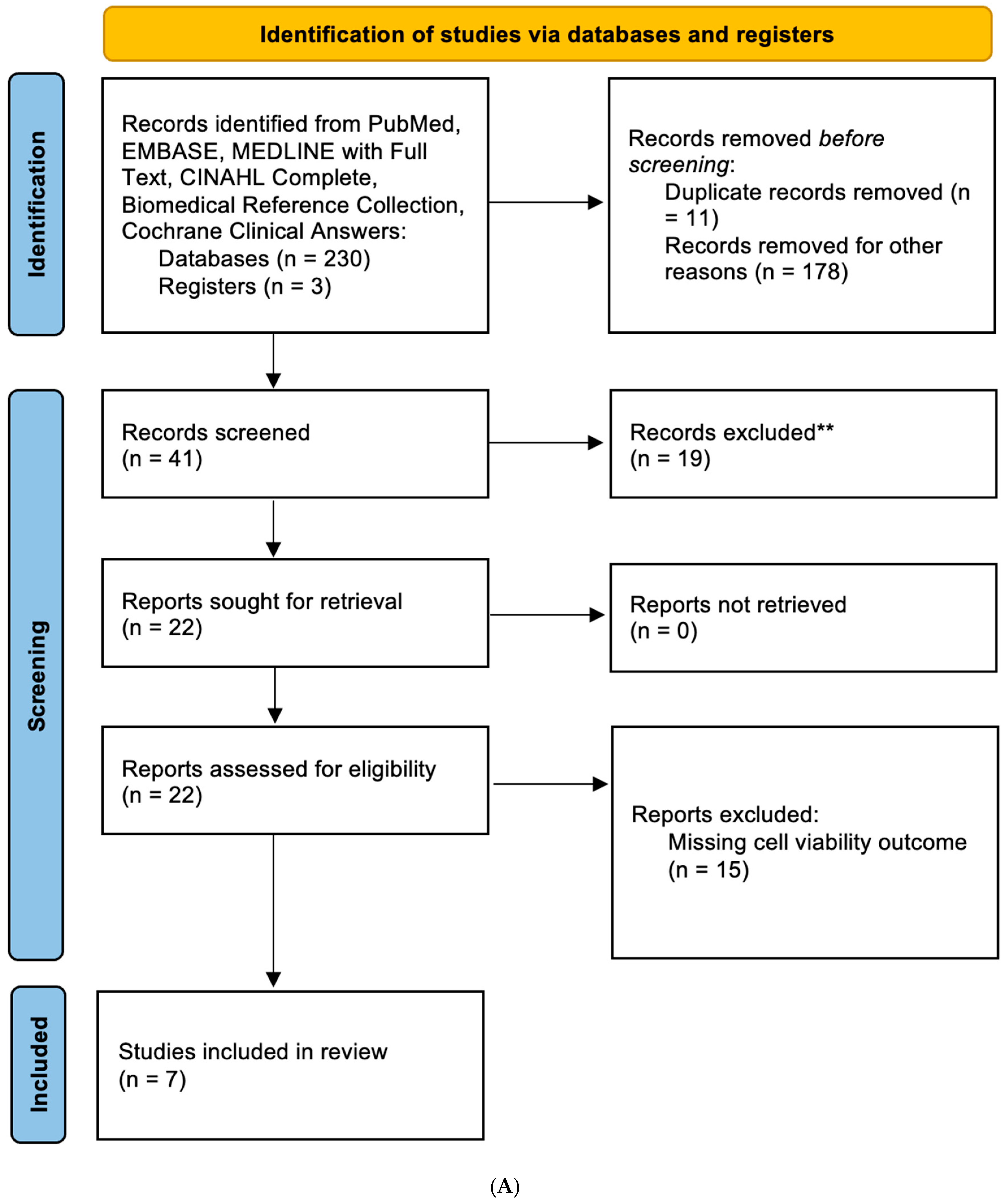
Figure 1. PRISMA flow diagram for systematic reviews for in vitro (A) and in vivo (B) studies. ** records were excluded based on relevance as screened by two independent reviewers.
Neuroglia 06 00040 g001aNeuroglia 06 00040 g001b
Figure 2. Standardized mean difference between metformin and no treatment in GBM cell viability using a random-effects model [21,22,23,24,25,26,27].
Neuroglia 06 00040 g002
Figure 3. Pooled survival curve of in vivo studies in mice treated with metformin compared to no treatment.
Neuroglia 06 00040 g003
Figure 4. Hazard ratio of metformin to overall mouse survival compared to no treatment in a random-effects model [21,22,26,28,29,30,31,32].
Neuroglia 06 00040 g004
Figure 5. Standardized mean difference between standard therapy plus metformin and standard therapy alone in GBM cell viability using a random-effects model [21,22,26].
Neuroglia 06 00040 g005
Figure 6. Hazard ratio of standard therapy plus metformin to overall mouse survival compared to standard therapy alone in a random-effects model [21,22,26,28,30].
Neuroglia 06 00040 g006
Table 1. Characteristics of included studies.
SourceType of StudyCell Line(s)Metformin Used as an Adjuvant Therapy?Results
Valtorta 2021 [21]In vitro, in vivoEGFR1, EGFR2, L0627YesMetformin addition improved TMZ efficacy in GBM preclinical models representative of classical molecular subtype, increasing survival time and reducing tumor relapsing rate.
Lee 2018 [22]In vitro, in vivoU87, U251, A172YesMetformin combined with TMZ enhanced apoptosis in GBM cell lines and prolonged survival in mouse models.
Albayrak 2021 [23]In vitroT98G, U87-MGNoMetformin reduced cell viability in a dose-dependent manner.
Barbieri 2022 [24]In vitroPatient-derived GBM culturesNoMetformin inhibited chloride intracellular channel 1 (CLIC1) activity, inhibiting GSC proliferation, although novel biguanide derivatives may demonstrate higher efficacy.
Song 2018 [25]In vitroLN18, U87NoMetformin suppressed transforming growth factor beta-1-induced (TGF-β1-induced) epithelial–mesenchymal transition-like processes, migration, and invasion in GBM cells by inhibiting the AKT/mTOR/ZEB1 pathway.
Yu 2015 [26]In vitro, in vivoU87, U87GSC, U251, U251GSCYesMetformin used with TMZ inhibited proliferation, self-renewal, and tumor growth of GBM cells in vitro and in vivo by suppressing AKT/mTOR signaling.
Soraya 2021 [27]In vitroU87-MGNoMetformin reduced cell survival while downregulating oncogenic microRNAs (miRNAs).
Li 2024 [28]In vivoGL261YesMetformin enhanced anti-programmed cell death protein-1 (anti-PD-1) immunotherapy in GBM mouse models by promoting T cell-mediated antitumor responses.
Zhan 2022 [29]In vivoPatient-derived GBM culturesNoExosome-mediated delivery of metformin disrupted GBM energy metabolism and significantly reduced tumor growth, prolonging survival in xenograft models.
Korsakova 2021 [30]In vivoGL261YesMetformin and DCA synergistically impaired GBM metabolism, reduced proliferation, and inhibited tumor growth in allograft models.
Kim 2017 [31]In vivoGSC11NoMetformin suppressed stemness and invasiveness, showing survival benefit in mouse xenograft models.
Sato 2012 [32]In vivoPatient-derived GBM culturesNoMetformin activated AMPK-FOXO3 signaling to promote differentiation of stem-like cells, depleting tumorigenic potential and inhibiting tumor formation to extend survival.
Disclaimer/Publisher’s Note: The statements, opinions and data contained in all publications are solely those of the individual author(s) and contributor(s) and not of MDPI and/or the editor(s). MDPI and/or the editor(s) disclaim responsibility for any injury to people or property resulting from any ideas, methods, instructions or products referred to in the content.