Review

An FGFR1-Altered Intramedullary Thoracic Tumor with Unusual Clinicopathological Features: A Case Report and Literature Review

1
Department of Pathology and Laboratory Medicine, KK Women’s and Children’s Hospital, 100 Bukit Timah Road, Singapore 229899, Singapore
2
Department of Neurosurgery, Vicente Sotto Memorial Medical Center, B. Rodriguez St., Cebu 6000, Philippines
3
Department of Diagnostic and Interventional Imaging Service, KK Women’s and Children’s Hospital, 100 Bukit Timah Road, Singapore 229899, Singapore
4
Hematology/Oncology Service, KK Women’s and Children’s Hospital, 100 Bukit Timah Road, Singapore 229899, Singapore
5
Neurosurgical Service, KK Women’s and Children’s Hospital, 100 Bukit Timah Road, Singapore 229899, Singapore
6
Department of Neurosurgery, National Neuroscience Institute, 11 Jalan Tan Tock Seng, Singapore 308433, Singapore
7
SingHealth Duke-NUS Neuroscience Academic Clinical Program, 11 Jalan Tan Tock Seng, Singapore 308433, Singapore
8
SingHealth Duke-NUS Pediatrics Academic Clinical Program, 100 Bukit Timah Road, Singapore 229899, Singapore
*
Author to whom correspondence should be addressed.
Neuroglia 2025, 6(4), 39; https://doi.org/10.3390/neuroglia6040039
Submission received: 11 August 2025 / Revised: 25 September 2025 / Accepted: 1 October 2025 / Published: 4 October 2025

Abstract

Background: Primary spinal gliomas are rare in the pediatric population. Separately, FGFR1 genomic aberrations are also uncommon in spinal cord tumors. We report a case of a previously well adolescent who presented with progressive symptoms secondary to an intramedullary tumor with unique radiological and molecular characteristics. Case Presentation: A previously well 17-year-old male presented with worsening mid-back pain associated with lower limb long-tract signs. Magnetic resonance imaging (MRI) of his neuro-axis reported a long-segment intramedullary lesion with enhancing foci and a multi-septate syrinx containing hemorrhagic components from C4 to T12. The largest enhancement focus was centered at T7. Additional MRI sequences observed no intracranial involvement or vascular anomaly. He underwent an emergent laminoplasty and excision of the thoracic lesion. Intraoperative findings demonstrated a soft, grayish intramedullary tumor associated with extensive hematomyelia that had multiple septations. Active fenestration of the latter revealed blood products in various stages of resolution. Postoperatively, the patient recovered well, with neurological improvement. Final histology reported a circumscribed low-grade glial neoplasm. Further molecular interrogation via next-generation sequencing panels showed FGFR1 p.K656E and V561M alterations. The unique features of this case are presented and discussed in corroboration with a focused literature review. Conclusions: We highlight an interesting case of an intramedullary tumor with unusual radiological and pathological findings. Emphasis is on the importance of tissue sampling in corroboration with genomic investigations to guide clinical management.

1. Introduction

Primary spinal cord neoplasms are rare in children, in comparison to their adult counterparts [1]. Broadly speaking, approximately 1% of all central nervous system (CNS) neoplasms are located within the spinal cord. Despite the heterogeneity of histological types, most of them tend to be glial in origin [2]. In the pediatric population, glial tumors are the most common intramedullary tumor and may occur throughout the length of the spinal cord [3]. Early clinical symptoms are often vague, leading to a significantly delayed diagnosis in this age group. Here, affected patients typically present with a progressive history of non-specific findings coupled with recurrent exacerbation and remission of symptoms that result from fluctuating degrees of peritumoral spinal cord edema [2]. Surgical resection is considered the mainstay of treatment for most spinal cord tumors. However, predictors of outcome include the tumor size and location, its histological grading, and the neurological status of the patient [4].
Separately, fibroblast growth factor receptors (FGFRs) are highly conserved transmembrane tyrosine kinase receptors (RTKs) integral in physiological processes of normal human cells. Upon the binding of fibroblast growth factor (FGF), associated FGFR kinases are implicated in activating multiple transduction pathways, including the Ras/mitogen-activated protein kinase (MAPK) pathway, the canonical and non-canonical Wnt pathway, and the phosphatidylinositol-4,5-bisphosphate 3-kinase (PI3K)–protein kinase B (AKT) pathway [4,5,6,7]. In cancer biology, FGFR aberrations have ubiquitous involvement in various neoplasms, including CNS tumors [8,9,10]. For example, a subset of adult malignant gliomas tends to harbor the FGFR3::TACC3 fusion gene [11]. In contrast, recent molecular insights report that FGFR aberrations are more commonly observed in low-grade spinal cord glial tumors in young children [12]. We herein report an interesting case of an adolescent who presented with progressive symptoms secondary to an intramedullary tumor with unique radiological and molecular characteristics. In view of the unexpected diagnosis, in combination with its atypical features, a focused literature review is performed to support the discussion.

2. Case Report

A previously well 17-year-old male presented with worsening mid-back pain associated with lower limb long-tract signs over a period of 2 months. Prior to this, there was no documented trauma, infection, or constitutional symptoms. At the time of his clinical presentation, there was reduced pin-prick sensation in the right L2 to L5 dermatomes and a loss of proprioception in his right big toe. His gross motor power was 5 out of 5 in all four limbs, and the gait assessment was unremarkable. There was no muscle wasting, dysmetria, or dysdiadochokinesis, and Rhomberg’s test was negative. A digital rectal examination demonstrated normal anal tone and no saddle anesthesia. However, a bedside post-void residual urine (PVRU) investigation showed persistently high residual urine volumes of more than 200 mL—an indication of possible inadequate bladder emptying. In view of the concerning neurological symptoms, urgent magnetic resonance imaging (MRI) of his neuro-axis was arranged. This reported a long-segment intramedullary lesion with enhancing foci and a multi-septate syrinx containing hemorrhagic components from C4 to T12. The largest enhancement focus was centered at the T7 level. No cerebellar tonsillar herniation was seen at the craniocervical junction. (Figure 1). Additional MRI brain and angiography images, no hydrocephalus, intracranial space-occupying lesions, or intracerebral vascular anomalies were observed. These scans included a time-resolved angiography with interleaved stochastic trajectories (TWIST) sequence which did not demonstrate the presence of a spinal arteriovenous malformation [13]. Blood investigations did not show underlying bleeding disorders or coagulopathy.
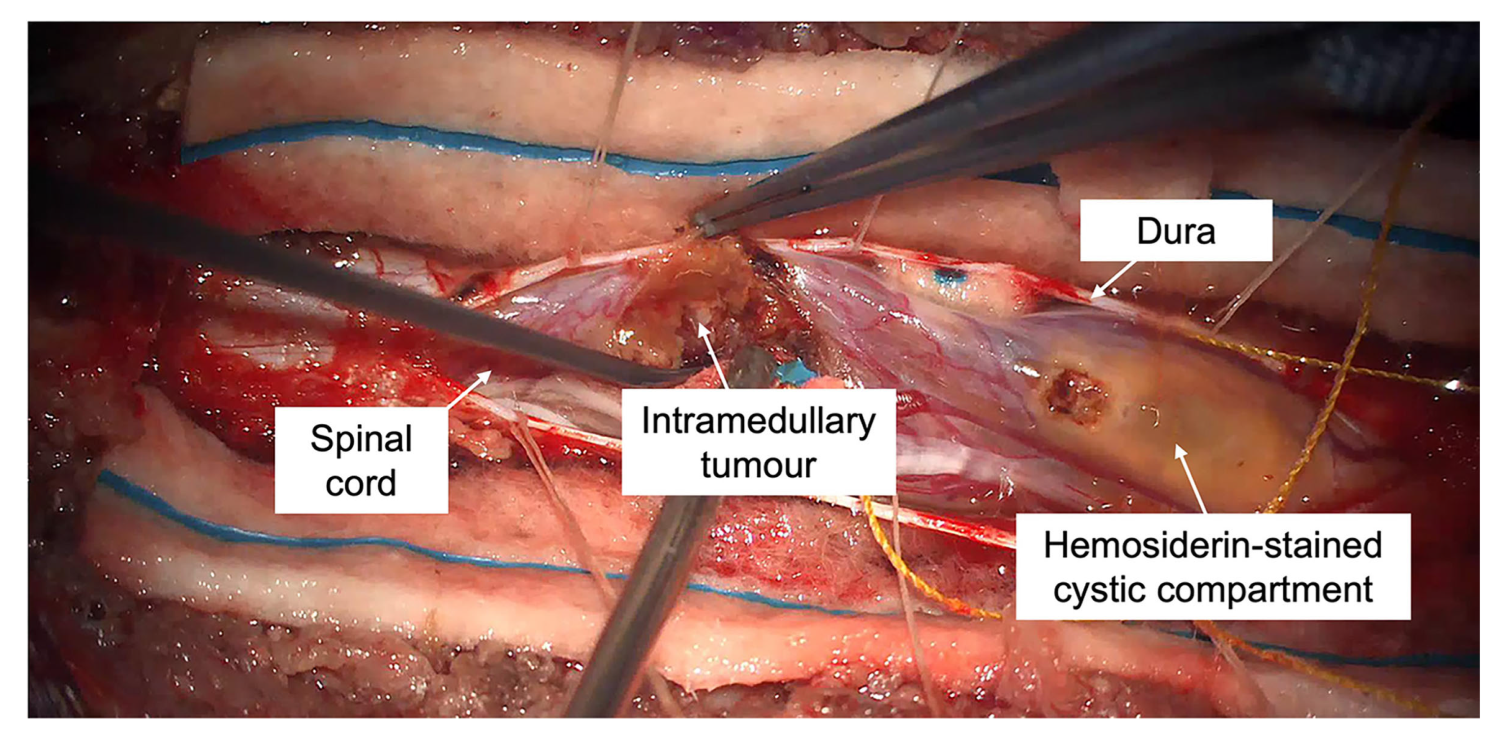
Based on the patient’s progressive clinical and radiological findings, the overall impression was that of an extensive, hemorrhagic intramedullary tumor causing significant mass effect—likely malignant in nature. Differential diagnoses were that of spinal ependymoma, high-grade glial neoplasm, and, less likely, metastases. He underwent an emergent T6-to-T8 laminoplasty and excision of the thoracic lesion under intraoperative neuromonitoring (IONM). As there were concerns of delineating the lesion from the normal spinal cord, intravenous fluorescein sodium (Na-Fl) was used—a method we previously described [14]. Briefly, after the induction of general anesthesia, a predefined intravenous dose of 2 mg/kg of 10% Na-Fl 10% is administered, with concurrent monitoring of vital signs. Subsequently, continuous monitoring is performed for the duration of the surgery, specifically looking out for cutaneous and/or physiological manifestations of an adverse drug reaction [14]. After placement of the patient in the prone position, his baseline motor evoked potentials (MEPs) and somatosensory evoked potentials (SSEPs) were documented. Despite good signal feedback for both MEPs and SSEPs, it was noted that the left lower limb (L2 to S3) had threshold readings lower by up to 10% in comparison to those on the right side. This finding was consistent with clinical examination of the patient. Prior to the dura opening, the mid-thoracic spinal cord was observed to be expanded. A midline myelotomy demonstrated a soft, grayish intrinsic lesion at the level of T7 associated with extensive hematomyelia that had multiple septations. These findings were congruent with the patient’s preoperative MRI findings. (Figure 2). Under the guidance of the YELLOW-560 nm filter, the fluorescing solid component was excised in piecemeal fashion. During the resection, the lesion was noted to be friable with areas of poorly defined cord–tumor interface. Following that, active fenestration of the septations in the surrounding hematomyelia revealed blood products in various stages of resolution. Upon tumor debulking and release of the observed blood products, the spinal cord was noted to be less swollen and visibly more pulsatile. Decision was made not to proceed with more extensive resection and fenestration when the IONM feedback showed a 50% drop in MEPs in the patient’s left L2 to S3 segments from the documented baseline at the time of skin incision. The SSEPs remained intact throughout the duration of surgery. A water-tight dura closure was completed after ensuring hemostasis in the surgical cavity. The laminoplasty bone graft was replaced, and the wound was closed in layers. At the end of the procedure, IONM checks showed return of the MEPs to the patient’s preoperative baseline. The frozen section reported that the specimen had features consistent with a glial neoplasm with hemosiderin and necrosis.
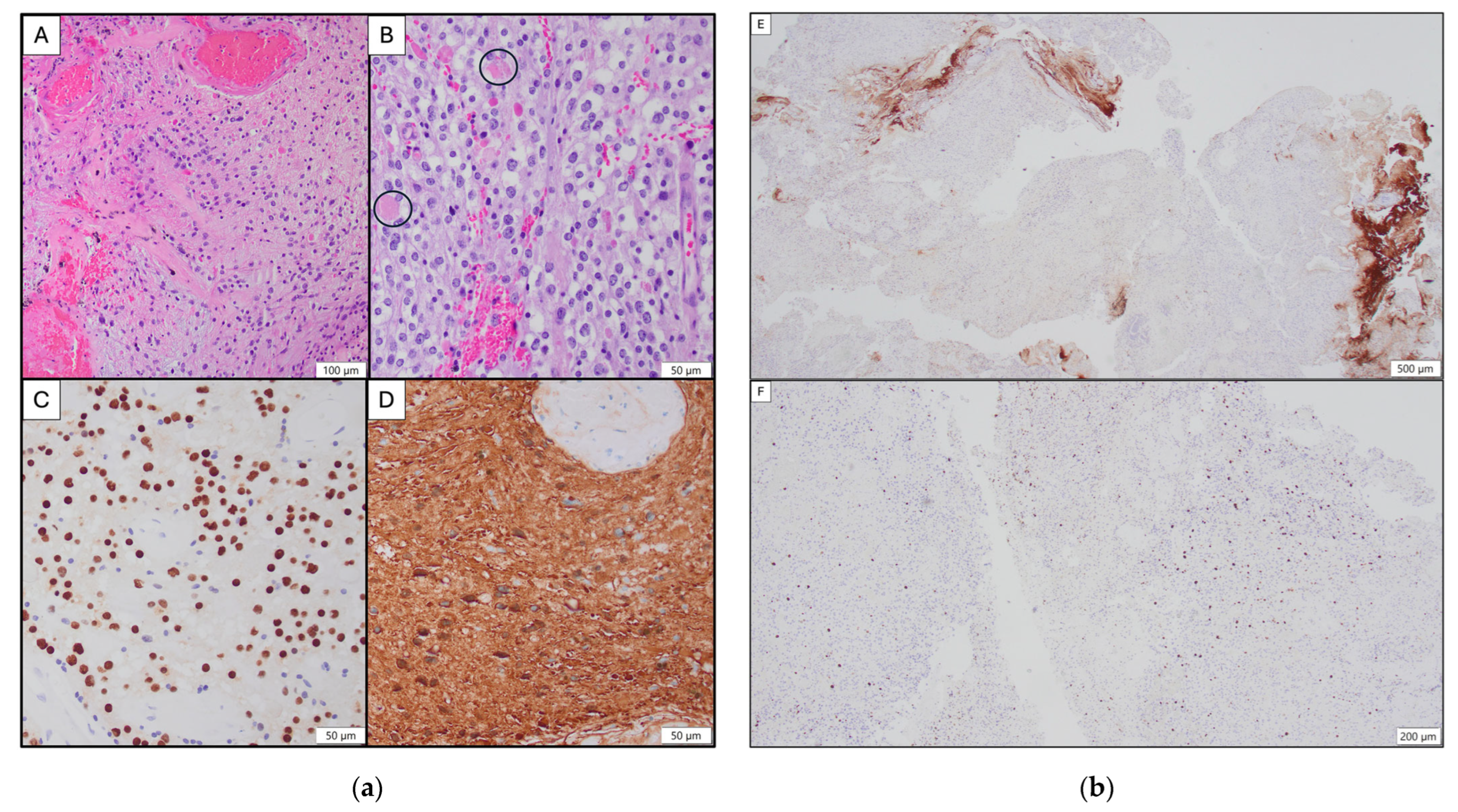
Postoperatively, the patient recovered well, with gradual improvement in his neurological symptoms, including his bladder function. A dedicated thoracic spine MRI the day after surgery reported an interval reduction in the size of the previous heterogeneously enhancing and multi-septate cystic areas. No new spinal cord canal stenosis, cord compression, or epidural hematoma was seen. He was discharged home uneventfully after a short period of intensive inpatient neurorehabilitation. There were no wound-related issues. Initial histology reported a circumscribed low-grade glial neoplasm which was positive for immunohistochemical staining with GFAP and OLIG2. Trimethylation of lysine 27 on histone H3 (H3K27me3) was retained. Eosinophilic granular bodies were present, though piloid cells and microcystic areas were a minority. Instead, the prominent proliferation of oligodendroglial-like cells with conspicuous nuclear variability and areas with a raised Ki67 index of up to 10% was striking, conferring a degree of atypia. Biological behavior was therefore deemed uncertain based on histology alone. (Figure 3).
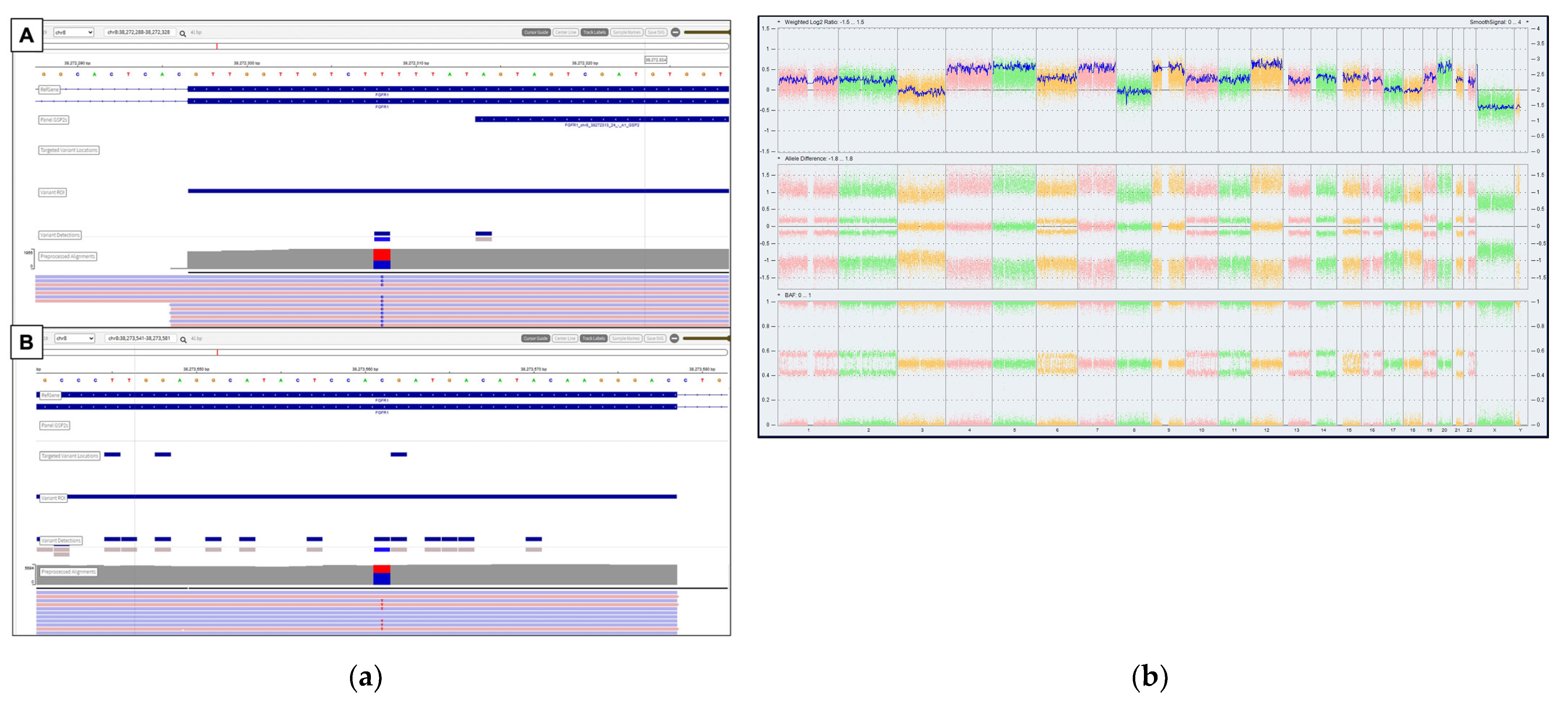
Molecular interrogation of the tumor tissue was subsequently performed via various platforms. Firstly, the Archer FusionPlex Pan-Solid Tumour V2 (Invitae, San Francisco, CA, USA) was used. This is a commercially available, high-throughput next-generation sequencing (NGS) panel that identifies gene translocations and internal tandem duplications across solid tumors and sarcomas in 137 genes. In addition, certain single-nucleotide changes are also identifiable if the region sequenced is covered by the panel’s primers. In brief, total RNA was extracted from the FFPE tissue section and quantified using a fluorometer. One hundred and fifty nanograms of RNA was used for library preparation, and the prepared library was sequenced using an Illumina MiniSeq sequencer (San Diego, CA, USA). The FASTQ data obtained were analyzed using the Archer Analysis (version 7.1.0) online portal. The AmpliSeqTM for Illumina® Childhood Cancer Panel kit (Illumina Inc., San Diego, CA, USA) was also applied. This panel covers single-nucleotide variants (SNVs) in hotspots of 86 genes, full exons of 44 genes, copy number variants (CNV) in 28 genes, and 97 gene fusions/translocations. Briefly, for DNA and RNA sequencing library construction and sequencing, DNA and RNA were extracted from macro-dissected, formalin-fixed, paraffin-embedded (FFPE) tumor sections. Library preparation was performed using the AmpliSeqTM for Illumina® Childhood Cancer Panel kit (Illumina Inc., San Diego, CA, USA). Sequencing results identified an activating FGFR1 K656E missense alteration in both sequencing runs (Archer FusionPlex allele fraction: 0.42; Ampliseq allele fraction: 0.28). An additional missense alteration, FGFR1 V561M, was also identified on the Archer FusionPlex platform (allele fraction: 0.40). This was not identified on the Ampliseq, as it occurred outside the panel’s hotspot region. Further validation of its presence was performed through Sanger sequencing. The occurrence of multiple FGFR1 mutations has previously been reported in pediatric low-grade gliomas (pLGGs) [15].
An additional chromosomal microarray was performed using the Thermo Fisher OncoScan CNV FFPE Assay, and its results were analyzed using the Chromosome Analysis Suite software v4.4. A near-triploid profile with multiple whole chromosomal gains was detected with notable absence of 1p/19q deletion, a loss of cyclin-dependent kinase inhibitors 2A and 2B (CDKN2A/B), and epidermal growth factor receptor (EGFR) amplification (Median of Absolute Pairwise Differences (MAPD): 0.235 (<0.3); normalized diploid SNP QC (ndSNPQC): 20.865 (>26). This near-triploid finding is regarded to be consistent with the description of aneuploidy states in low-grade gliomas with no established support for a higher-grade neoplasm [16] (Figure 4). The tumor tissue was also sent for DNA methylation. Briefly, the quality of the FFPE DNA was evaluated using an Infinium FFPE QC kit. The FFPE DNA was subjected to bisulfite conversion using the EZ DNA Methylation kit (Zymo Research Corporation, Irvine, CA, USA) and restoration using the Infinium HD FFPE DNA Restore Kit before hybridization onto the Infinium® MethylationEPIC v2.0 BeadChip (Illumina Inc., San Diego, CA, USA). All protocols were performed according to the manufacturer’s instructions. Unprocessed Intensity Data (IDAT) files generated from the array were uploaded onto an automated web-based DNA methylation profiling program website (https://app.epignostix.com/) and executed using the Heidelberg Epignostix CNS Tumor Classifier (v12.8) [17,18]. The calibrated match scores were 0.94678 for the low-grade glial/glioneuronal/neuroepithelial tumors superfamily and 0.90783 for the pilocytic astrocytoma family. Class and subclass scores were marginally below the match cut-off of 0.9 (Delta Cq value: −1.28 (<5)). (Figure 5). A summary of all of the molecular tests performed is available in Supplementary Table S1.
Put together, the following integrated diagnosis was made: ‘Pilocytic astrocytoma, circumscribed low-grade glioma, CNS WHO Grade 1, FGFR1 p.K656E and V561M-mutant, near-triploid profile without CDKN2A/B homozygous deletion, DNA methylation [Heidelberg Epignostix CNS Tumor Classifier (v12.8)] consistent with the pilocytic astrocytoma (PCA) family’.
The patient’s case was discussed at the multidisciplinary neuro-oncology tumor board (MDT). A consensus was made to withhold adjuvant treatment in view of the pLGG diagnosis and his postoperative neurological improvement. The rationale was to manage the disease expectantly and to keep the options of chemo- and/or radiotherapy available in the event of disease relapse. Surveillance MRI at the 9-month follow-up did not show recurrence of the excised T7 enhancing lesion, and the previously fenestrated multi-septate syrinx did not reaccumulate. The decision was made on 6-monthly outpatient clinical review and annual MRI scans to assess his tumor status. At the time of writing, he remains clinically stable.

3. Discussion

3.1. Pediatric Spinal Gliomas in the Era of WHO 2021 Classification

Central nervous system (CNS) tumors are the most common group of solid tumors and the leading cause of tumor-related mortality in children [19]. Here, spinal neoplasms account for an estimated up to 10% of all pediatric CNS neoplasms [20,21]. Although these tumors affect all age groups, they tend to be more prevalent towards the end of the first decade and at the beginning of the second decade of life with no gender predilection [9]. From a patient’s perspective, spine tumors can cause debilitating pain and sensorimotor deficits that impact an individual’s long-term neurological function [3]. This is especially significant in a young person, whereby the expected lifespan is comparatively longer than their adult counterparts. Historically, the subset of intramedullary spinal cord tumors are challenging to manage due to the lack of established care guidelines and limited treatment options [22]. Broadly speaking, management usually involves neurosurgical intervention (here, either resection or biopsy), chemotherapy, and/or radiotherapy with the goals of preserving neurological function and improving symptoms [23,24]. On a side note, spinal surgical procedures to preserve the posterior elements (such as laminoplasty in our patient) are preferred in pediatric patients due to the risk of postoperative spinal deformities [1,25,26].
The advent of modern molecular assays has identified important genomic alterations in several CNS tumors, including gliomas [27]. We are now aware that in the updated World Health Organization (WHO) 2021 classification of glial neoplasms, pediatric gliomas are now considered as clinically, radiologically, and molecularly distinct entities [8,19,27]. In the context of pediatric low-grade gliomas (pLGGs), each tumor subtype’s prognosis is dependent on patient age, tumor location, histology, and molecular profile [19]. For example, most cases of PCA have genetic alterations in the proto-oncogene B-raf (BRAF) [28]. Next, hotspot mutations in N546 or K656 in FGFR1 are the second most common major genetic alterations in PCA, after BRAF alterations [29]. Overall, the prognosis of pLGG is expected to be good, with an extended long-term survival, especially in contrast to other malignant childhood CNS tumors. The downside of a pLGG diagnosis is that affected children are at risk of lifetime recurrences and disease-related morbidities due to its indolent nature [30,31,32]. Under such circumstances, mortality tends to be associated with complications secondary to treatment or, occasionally, as a direct consequence of the primary tumor [33].

3.2. Overview of FGFR Alterations in Pediatric Primary CNS Tumors

The fibroblast growth factor receptor (FGFR) family consists of four transmembrane tyrosine kinase receptors (FGFR1 to 4). They are involved in a myriad of critical cellular processes, including cell proliferation, angiogenesis, cell survival, migration, and differentiation [34]. Here, FGFR1 is a tyrosine kinase receptor for which ligand binding triggers downstream signaling in several pathways, including the MAPK and PI3K/AKT pathways [35]. Alterations in FGFR1 encompass amplifications, gene fusions, tandem duplications, and single-nucleotide variations in various cancer types [10]. Notable examples include tyrosine kinase domain duplication (e.g., FGFR1 TKDD), fusions (e.g., FGFR1::TACC1), and FGFR1 hotspot mutations (e.g., N546K and K656E) [36]. Alterations in FGFR (especially FGFR1) account for the third most frequent alterations in pLGGs after BRAF and NF1 alterations [37]. In addition, FGFR signaling has pivotal roles in physiological processes such as embryonic development, cell differentiation, proliferation, migration, angiogenesis, and endocrine-related signaling [38]. Accordingly, aberrant FGFR signaling in cancer leads to antiapoptotic, mutagenic, angiogenic, and chemoresistance responses in cells [9,39]. Put together, oncogenic FGFR dysregulation is well established in many types of solid tumors, including primary brain tumors [40,41].
In cancer biology, FGFR1 to 4 mutations, fusions, and amplifications can activate the downstream MAPK-ERK, PI3K/AKT, and/or JAK-STAT pathways—all of which are clinically relevant [38,39,42,43]. Mechanistically, single-nucleotide changes in FGFR1 concentrate within the protein tyrosine kinase domain and functionally result in an increased ligand binding affinity or ligand-independent receptor activation [10]. FGFR1 fusions and tandem duplications also tend to involve the protein tyrosine kinase domain, with the former resulting in constitutive activation through the addition of C-terminal dimerization domains [10]. A recent large-scale genomic analysis of pediatric and adult gliomas reported that 4.5% of all pediatric and adult gliomas harbored FGFR alterations, with an incidence of 9.6% in the pediatric cohort. FGFR1 alterations were most frequent in pediatric gliomas, particularly for low-grade subtypes [44]. These FGFR1 fusions are notably distinct from the FGFR3::TACC3 fusions which tend to occur in adult glioblastomas, rather than pLGGs [45]. For unclear reasons at the time of writing, FGFR1 single-nucleotide-mutated low-grade gliomas appear to be more clinically aggressive as opposed to their fusion/tandem duplication counterparts and have been dubbed an ‘intermediate risk’ as opposed to a ‘low risk’ in the context of clinical progression and overall survival [37]. Of interest, the contemporary literature reports that FGFR1 aberrations tend to be more commonly highlighted in low-grade neuroepithelial brain tumors than spinal tumors. Furthermore, FGFR1 alterations in pediatric spinal PCAs are observed to bear a different methylation signature compared to that in their intracranial counterparts, therefore suggesting that they are likely to arise from distinct region-specific progenitors [12].
Separately, FGFR1-altered pLGGs appear to be distinct from other molecular drivers of the same entity. For instance, the presence of FGFR1 alterations has been associated with spontaneous intracranial hemorrhage (ICH) of the brain tumor, although the underlying mechanism of this observation remains unelucidated [46,47,48]. In the context of pediatric populations, the neurological injury caused by a severe ICH is devastating. Based on our current understanding, up to 1/3 of children die due to a severe ICH, and those that survive tend to suffer from permanent neurodevelopmental and cognitive impairments that impact their extended lifespans [46,49]. Of interest, FGFR1 has been shown to be essential in the expression and function of vascular endothelial growth factor A (VEGFA)—a potent player in angiogenesis [50]. However, the exact tumor-specific mechanisms underlying spontaneous bleeding in FGFR1-altered pLGGs have yet to be fully elucidated [48]. We hope that our case will first be a useful contribution to the existing knowledge on FGFR1-altered pLGG with spontaneous hemorrhage and next be incorporated into future meta-analyses on this topic. A summary of FGFR1-altered pLGG cases presenting with ICH is featured in Table 1.

3.3. Targeting FGFR in Pediatric Primary CNS Tumors

In this era of modern medicine, there have been significant breakthroughs for novel therapies for pLGGs. First and foremost, we are now aware that pLGG is considered a single-pathway disease with a lack of secondary alterations in most cases. These tumors often have molecular driver alterations centered within the MAPK pathway. Here, the most commonly altered gene is BRAF, in up to 2/3 of patients [15,51,52]. The identification of these drivers has led to the development of therapies targeting gene mutations and dysregulated pathways involving mitogen-activated protein kinase kinase (MEK), BRAF, and, more recently, FGFR [52,53]. A recent study demonstrated that activating FGFR alterations are found in approximately 3% of pediatric solid tumors [43]. Pertaining to CNS tumors, activated FGF signaling is demonstrated in 4% of childhood CNS malignancies and in up to 11% within the pLGG cohort [30,54]. Under such circumstances, targeting FGFR is hence a feasible treatment strategy for selected tumors in this group of children [4]. To date, clinical trials for various cancers using novel agents to target FGF/FGFR signaling have tended to be adult-centric, whereby very few of them actively enroll pediatric patients [4,43,55]. Nonetheless, there have been promising results from anecdotal cases reported in the literature [30]. One example is the use of erdafitinib, a selective FGFR1 to 4 tyrosine kinase inhibitor already approved for the treatment of adult urothelial cancer harboring FGFR2 or FGFR3 alterations [56]. In Stepien et al.’s study, the use of erdafitinib treatment in a patient with pLGG harboring ITD in the FGFR1 gene demonstrated efficacy [30]. Thus, for clinical cases of intramedullary tumors (like our patient), the role of FGFR1-targeted therapy may be the way forward in the event of tumor relapse or progression. This approach can potentially mitigate the sequelae of an iatrogenic neurological deficit due to aggressive surgery and/or toxicity effects from radiotherapy. Despite the purported benefits, there are reported adverse effects associated with FGFR-targeted therapy—likely due to the multiple physiological functions that the FGF/FGFR signaling pathway is involved in [57]. Examples include hyperphosphatemia, fatigue, and various dermatologic toxicities, such as hair loss, nail bed infections, onycholysis, dry skin, and xerostomia, which may lead to altered taste and stomatitis [57]. Similarly to other targeted therapies, the extent of the complications from prolonged use of FGFR inhibitors is still not yet fully elucidated at the time of writing. Put together, judicious clinical monitoring of the use of these medications should be mandatory within a supervised framework.

3.4. Study Reflections and Future Directions

In present times, molecular diagnostics is recommended for the routine classification of pediatric CNS tumors and to establish appropriate treatment plans for affected patients. This is especially relevant in our patient, as the initial assumption was that he had an aggressive, high-grade neoplasm given his short clinical history and the neuroimaging findings. Nonetheless, we are cognizant that advanced molecular techniques and/or bioinformatic expertise and tools may not be readily available in all healthcare institutions. In addition, the reality of integrated histomolecular diagnostic work-up adds substantial costs and can financially impact some healthcare settings [58]. Under such circumstances, the lack of access to these important investigations implies that not all children with a neuro-oncological diagnosis can benefit from personalized approaches to management [59]. As the way forward, there is a need for global collaborative efforts amongst clinicians and researchers to develop accessible and cost-effective approaches in pediatric neuro-oncology. Emerging considerations include new artificial intelligence (AI)-driven models that leverage clinical, imaging, and molecular features of brain tumors combinatorially for large-scale data that allow targeted therapies to become more feasible as they are being developed [59].

4. Conclusions

We highlight a case of an adolescent with an intramedullary tumor with unusual radiological and unexpected pathological findings. In the context of this patient, who presented with progressive neurological symptoms, we advocate the role of spinal cord decompression and, at minimum, an IONM-guided biopsy to preserve neurological function. Here, the emphasis is on the importance of tissue sampling in corroboration with genomic investigations to guide clinical management via an MDT consensus. This report adds to the growing body of literature on pediatric spinal tumors with unique clinical and molecular features and underscores the need for clinicians to be mindful of this sub-entity of intrinsic spinal tumors.

Supplementary Materials

The following supporting information can be downloaded at https://www.mdpi.com/article/10.3390/neuroglia6040039/s1. Table S1: Summary of molecular tests and results.

Author Contributions

Conceptualization: S.J.A. and S.Y.Y.L.; methodology: S.J.A., J.Y.G. and S.Y.Y.L.; software: S.J.A., J.Y.G. and S.Y.Y.L.; validation: S.J.A., J.Y.G., T.S.E.T., E.E.K.T., J.M.E. and S.Y.Y.L.; formal analysis: S.J.A., J.Y.G., T.S.E.T. and S.Y.Y.L.; investigation: S.J.A., J.Y.G., T.S.E.T., E.E.K.T., J.M.E. and S.Y.Y.L.; resources: S.J.A., J.Y.G., T.S.E.T. and S.Y.Y.L.; data curation: S.J.A. and S.Y.Y.L.; writing—original draft preparation: S.J.A. and S.Y.Y.L.; writing—review and editing: S.J.A. and S.Y.Y.L.; visualization: S.J.A., T.S.E.T., J.M.E. and S.Y.Y.L.; supervision: S.Y.Y.L.; project administration: S.J.A. and S.Y.Y.L. All authors have read and agreed to the published version of the manuscript.

Funding

This research was funded by the VIVA-KKH Pediatric Brain and Solid Tumors Programme. This is a philanthropic grant that was awarded to the institution (KK Women’s and Children’s Hospital) where this study was conducted.

Institutional Review Board Statement

This study was conducted in accordance with the Declaration of Helsinki and approved by the SingHealth Centralised Institutional Review Board (CIRB) (Reference: 2014/2079).

Informed Consent Statement

Informed consent to participate in this study was provided by the participants’ legal guardian/next of kin. Informed consent to publication and the publication of clinical images was part of the written consent form to participate in this study. A copy of the consent form is available from the corresponding author upon reasonable request.

Data Availability Statement

The original contributions presented in this study are included in the article. Further inquiries can be directed to the corresponding author.

Acknowledgments

The preliminary results of this paper were accepted as an electronic poster presentation (PP065) for the 29th Congress of the European Society for Pediatric Neurosurgery (ESPN) held in London, the UK, from 11 to 14 May 2025 (https://doi.org/10.1007/s00381-025-06865-7).

Conflicts of Interest

The authors declare no conflicts of interest. The funders had no role in the design of this study; in the collection, analyses, or interpretation of the data; in the writing of the manuscript; or in the decision to publish the results.

References

  1. Caroli, E.; Salvati, M.; Ferrante, L. Spinal glioblastoma with brain relapse in a child: Clinical considerations. Spinal Cord. 2005, 43, 565–567. [Google Scholar] [CrossRef]
  2. Huisman, T.A. Pediatric tumors of the spine. Cancer Imaging 2009, 9, S45–S48. [Google Scholar] [CrossRef]
  3. Kumar, N.; Tan, W.L.B.; Wei, W.; Vellayappan, B.A. An overview of the tumors affecting the spine-inside to out. Neurooncol. Pract. 2020, 7, i10–i17. [Google Scholar] [CrossRef]
  4. Zuo, W.; He, Y.; Li, W.; Wu, H.; Zhao, Z.; Zhang, Y.; Chen, S.; Yin, Y. Landscape of FGF/FGFR Alterations in 12,372 Chinese Cancer Patients. J. Cancer 2020, 11, 6695–6699. [Google Scholar] [CrossRef]
  5. Katoh, M. Fibroblast growth factor receptors as treatment targets in clinical oncology. Nat. Rev. Clin. Oncol. 2019, 16, 105–122. [Google Scholar] [CrossRef] [PubMed]
  6. Katoh, M.; Nakagama, H. FGF receptors: Cancer biology and therapeutics. Med. Res. Rev. 2014, 34, 280–300. [Google Scholar] [CrossRef]
  7. Lemmon, M.A.; Schlessinger, J. Cell signaling by receptor tyrosine kinases. Cell 2010, 141, 1117–1134. [Google Scholar] [CrossRef]
  8. Dono, A.; El Achi, H.; Bundrant, B.E.; Goli, P.S.; Zhu, P.; Ozkizilkaya, H.I.; Esquenazi, Y.; Ballester, L.Y. Infiltrating gliomas with FGFR alterations: Histologic features, genetic alterations, and potential clinical implications. Cancer Biomark. Sect. A Dis. Markers 2023, 36, 117–131. [Google Scholar] [CrossRef] [PubMed]
  9. Chioni, A.M.; Grose, R.P. Biological Significance and Targeting of the FGFR Axis in Cancer. Cancers 2021, 13, 5681. [Google Scholar] [CrossRef] [PubMed]
  10. Brown, L.M.; Ekert, P.G.; Fleuren, E.D.G. Biological and clinical implications of FGFR aberrations in paediatric and young adult cancers. Oncogene 2023, 42, 1875–1888. [Google Scholar] [CrossRef]
  11. Picca, A.; Sansone, G.; Santonocito, O.S.; Mazzanti, C.M.; Sanson, M.; Di Stefano, A.L. Diffuse Gliomas with FGFR3-TACC3 Fusions: Oncogenic Mechanisms, Hallmarks, and Therapeutic Perspectives. Cancers 2023, 15, 5555. [Google Scholar] [CrossRef]
  12. Métais, A.; Bouchoucha, Y.; Kergrohen, T.; Dangouloff-Ros, V.; Maynadier, X.; Ajlil, Y.; Carton, M.; Yacoub, W.; Saffroy, R.; Figarella-Branger, D.; et al. Pediatric spinal pilocytic astrocytomas form a distinct epigenetic subclass from pilocytic astrocytomas of other locations and diffuse leptomeningeal glioneuronal tumours. Acta Neuropathol. 2023, 145, 83–95. [Google Scholar] [CrossRef] [PubMed]
  13. Blackham, K.A.; Passalacqua, M.A.; Sandhu, G.S.; Gilkeson, R.C.; Griswold, M.A.; Gulani, V. Applications of Time-Resolved MR Angiography. Am. J. Roentgenol. 2011, 196, W613–W620. [Google Scholar] [CrossRef] [PubMed]
  14. Tan, A.J.L.; Tey, M.L.; Seow, W.T.; Low, D.C.Y.; Chang, K.T.E.; Ng, L.P.; Looi, W.S.; Wong, R.X.; Tan, E.E.K.; Low, S.Y.Y. Intraoperative Fluorescein Sodium in Pediatric Neurosurgery: A Preliminary Case Series from a Singapore Children’s Hospital. NeuroSci 2023, 4, 54–64. [Google Scholar] [CrossRef]
  15. Ryall, S.; Tabori, U.; Hawkins, C. Pediatric low-grade glioma in the era of molecular diagnostics. Acta Neuropathol. Commun. 2020, 8, 30. [Google Scholar] [CrossRef]
  16. Fontebasso, A.M.; Shirinian, M.; Khuong-Quang, D.A.; Bechet, D.; Gayden, T.; Kool, M.; De Jay, N.; Jacob, K.; Gerges, N.; Hutter, B.; et al. Non-random aneuploidy specifies subgroups of pilocytic astrocytoma and correlates with older age. Oncotarget 2015, 6, 31844–31856. [Google Scholar] [CrossRef] [PubMed]
  17. Capper, D.; Jones, D.T.W.; Sill, M.; Hovestadt, V.; Schrimpf, D.; Sturm, D.; Koelsche, C.; Sahm, F.; Chavez, L.; Reuss, D.E.; et al. DNA methylation-based classification of central nervous system tumours. Nature 2018, 555, 469–474. [Google Scholar] [CrossRef]
  18. Sill, M.; Schrimpf, D.; Patel, A.; Sturm, D.; Jäger, N.; Sievers, P.; Schweizer, L.; Banan, R.; Reuss, D.; Suwala, A.; et al. Advancing CNS tumor diagnostics with expanded DNA methylation-based classification. medRxiv 2025. [Google Scholar] [CrossRef]
  19. AlRayahi, J.; Alwalid, O.; Mubarak, W.; Maaz, A.U.R.; Mifsud, W. Pediatric Brain Tumors in the Molecular Era: Updates for the Radiologist. Semin. Roentgenol. 2023, 58, 47–66. [Google Scholar] [CrossRef]
  20. Wilson, P.E.; Oleszek, J.L.; Clayton, G.H. Pediatric spinal cord tumors and masses. J. Spinal Cord. Med. 2007, 30 (Suppl. S1), S15–S20. [Google Scholar] [CrossRef]
  21. Shweikeh, F.; Quinsey, C.; Murayi, R.; Randle, R.; Nuño, M.; Krieger, M.D.; Patrick Johnson, J. Treatment patterns of children with spine and spinal cord tumors: National outcomes and review of the literature. Childs Nerv. Syst. 2017, 33, 1357–1365. [Google Scholar] [CrossRef]
  22. Tobin, M.K.; Geraghty, J.R.; Engelhard, H.H.; Linninger, A.A.; Mehta, A.I. Intramedullary spinal cord tumors: A review of current and future treatment strategies. Neurosurg. Focus FOC 2015, 39, E14. [Google Scholar] [CrossRef]
  23. Kalayci, M.; Cagavi, F.; Gül, Ş.; Yenidünya, S.; Açikgöz, B. Intramedullary spinal cord metastases: Diagnosis and treatment—An illustrated review. Acta Neurochir. 2005, 146, 1347–1354, discussion 1354. [Google Scholar] [CrossRef]
  24. Catran, A.J.; Jalil, M.F.A. Haemorrhagic intramedullary spinal cord lesion. J. Clin. Neurosci. 2018, 48, 85. [Google Scholar] [CrossRef]
  25. Kaufman, B.A.; Park, T.S. Congenital spinal cord astrocytomas. Child’s Nerv. Syst. 1992, 8, 389–393. [Google Scholar] [CrossRef]
  26. Gutierrez, F.; Oi, S.; McLone, D. Intraspinal tumors in children: Clinical review, surgical results and follow-up in 51 cases. Concepts Pediatr. Neurosurg. 1983, 4, 1983-405. [Google Scholar]
  27. WHO Classification of Tumours Editorial Board. WHO Classification of Tumours, 5th edition: Central Nervous System Tumours, 5th ed.; WHO Classification of Tumours Editorial Board, Ed.; International Agency for Research on Cancer: Lyon, France, 2021; p. 568. [Google Scholar]
  28. Collins, V.P.; Jones, D.T.; Giannini, C. Pilocytic astrocytoma: Pathology, molecular mechanisms and markers. Acta Neuropathol. 2015, 129, 775–788. [Google Scholar] [CrossRef] [PubMed]
  29. Jones, D.T.; Hutter, B.; Jäger, N.; Korshunov, A.; Kool, M.; Warnatz, H.J.; Zichner, T.; Lambert, S.R.; Ryzhova, M.; Quang, D.A.; et al. Recurrent somatic alterations of FGFR1 and NTRK2 in pilocytic astrocytoma. Nat. Genet. 2013, 45, 927–932. [Google Scholar] [CrossRef]
  30. Stepien, N.; Mayr, L.; Schmook, M.T.; Raimann, A.; Dorfer, C.; Peyrl, A.; Azizi, A.A.; Schramm, K.; Haberler, C.; Gojo, J. Feasibility and antitumour activity of the FGFR inhibitor erdafitnib in three paediatric CNS tumour patients. Pediatr. Blood Cancer 2024, 71, e30836. [Google Scholar] [CrossRef]
  31. Armstrong, G.T.; Conklin, H.M.; Huang, S.; Srivastava, D.; Sanford, R.; Ellison, D.W.; Merchant, T.E.; Hudson, M.M.; Hoehn, M.E.; Robison, L.L.; et al. Survival and long-term health and cognitive outcomes after low-grade glioma. Neuro Oncol. 2011, 13, 223–234. [Google Scholar] [CrossRef]
  32. Bandopadhayay, P.; Bergthold, G.; London, W.B.; Goumnerova, L.C.; Morales La Madrid, A.; Marcus, K.J.; Guo, D.; Ullrich, N.J.; Robison, N.J.; Chi, S.N.; et al. Long-term outcome of 4,040 children diagnosed with pediatric low-grade gliomas: An analysis of the Surveillance Epidemiology and End Results (SEER) database. Pediatr. Blood Cancer 2014, 61, 1173–1179. [Google Scholar] [CrossRef]
  33. Upadhyaya, S.A.; Ghazwani, Y.; Wu, S.; Broniscer, A.; Boop, F.A.; Gajjar, A.; Qaddoumi, I. Mortality in children with low-grade glioma or glioneuronal tumors: A single-institution study. Pediatr. Blood Cancer 2018, 65, e26717. [Google Scholar] [CrossRef]
  34. Touat, M.; Ileana, E.; Postel-Vinay, S.; André, F.; Soria, J.C. Targeting FGFR Signaling in Cancer. Clin. Cancer Res. 2015, 21, 2684–2694. [Google Scholar] [CrossRef]
  35. Bale, T.A. FGFR- gene family alterations in low-grade neuroepithelial tumors. Acta Neuropathol. Commun. 2020, 8, 21. [Google Scholar] [CrossRef]
  36. Yvone, G.M.; Breunig, J.J. Pediatric low-grade glioma models: Advances and ongoing challenges. Front. Oncol. 2024, 13, 1346949. [Google Scholar] [CrossRef]
  37. Ryall, S.; Zapotocky, M.; Fukuoka, K.; Nobre, L.; Guerreiro Stucklin, A.; Bennett, J.; Siddaway, R.; Li, C.; Pajovic, S.; Arnoldo, A.; et al. Integrated Molecular and Clinical Analysis of 1,000 Pediatric Low-Grade Gliomas. Cancer Cell 2020, 37, 569–583.e565. [Google Scholar] [CrossRef] [PubMed]
  38. Ornitz, D.M. FGFs, heparan sulfate and FGFRs: Complex interactions essential for development. BioEssays 2000, 22, 108–112. [Google Scholar] [CrossRef]
  39. Turner, N.; Grose, R. Fibroblast growth factor signalling: From development to cancer. Nat. Rev. Cancer 2010, 10, 116–129. [Google Scholar] [CrossRef]
  40. Du, S.; Zhang, Y.; Xu, J. Current progress in cancer treatment by targeting FGFR signaling. Cancer Biol. Med. 2023, 20, 490–499. [Google Scholar] [CrossRef] [PubMed]
  41. Helsten, T.; Elkin, S.; Arthur, E.; Tomson, B.N.; Carter, J.; Kurzrock, R. The FGFR Landscape in Cancer: Analysis of 4,853 Tumors by Next-Generation Sequencing. Clin. Cancer Res. 2016, 22, 259–267. [Google Scholar] [CrossRef] [PubMed]
  42. Friesel, R.E.; Maciag, T. Molecular mechanisms of angiogenesis: Fibroblast growth factor signal transduction. FASEB J. 1995, 9, 919–925. [Google Scholar] [CrossRef]
  43. Lazo De La Vega, L.; Comeau, H.; Sallan, S.; Al-Ibraheemi, A.; Gupta, H.; Li, Y.Y.; Tsai, H.K.; Kang, W.; Ward, A.; Church, A.J.; et al. Rare FGFR Oncogenic Alterations in Sequenced Pediatric Solid and Brain Tumors Suggest FGFR Is a Relevant Molecular Target in Childhood Cancer. JCO Precis. Oncol. 2022, 6, e2200390. [Google Scholar] [CrossRef]
  44. Morin, E.; Apfelbaum, A.A.; Sturm, D.; Ayoub, G.; DiGiacomo, J.; Bahadur, S.; Chandarana, B.; Power, P.C.; Cusick, M.M.; Novikov, D.; et al. A diverse landscape of FGFR alterations and co-mutations defines novel therapeutic strategies in pediatric low-grade gliomas. bioRxiv 2024, 2024.08.27.609922. [Google Scholar] [CrossRef]
  45. Louis, D.N.; Aldape, K.D.; Capper, D.; Gianni, C.; Horbinski, C.M.; Ng, H.K.; Perry, A.; Reifenberger, G.; Sarkar, C.; Soffietti, R.; et al. Glioblastoma, IDH-wildtype. In Central Nervous System Tumours, 5th ed.; WHO Classification of Tumours Series; WHO Classification of Tumours Editorial Board, Ed.; International Agency for Research on Cancer: Lyon, France, 2021; Volume 6. [Google Scholar]
  46. Gonzalez-Vega, M.; Lebert, B.M.; Campion, S.; Wagner, A.; Aguilar-Bonilla, A.; Smith, A.A. Fibroblast growth factor receptor 1 gene mutation as a potential risk factor for spontaneous intracranial hemorrhage in pediatric low-grade glioma patients. Neurooncol Adv. 2024, 6, vdae074. [Google Scholar] [CrossRef] [PubMed]
  47. Campion, S.; Aguilar-Bonilla, A.E.; Elbabaa, S.; Smith, A.A. LGG-31. Pediatric low-grade gliomas with FGFR1 mutations and spontaneous hemorrhage: Case series. Neuro-Oncology 2022, 24, i95. [Google Scholar] [CrossRef]
  48. Ishi, Y.; Yamaguchi, S.; Hatanaka, K.C.; Okamoto, M.; Motegi, H.; Kobayashi, H.; Terasaka, S.; Houkin, K. Association of the FGFR1 mutation with spontaneous hemorrhage in low-grade gliomas in pediatric and young adult patients. J. Neurosurg. 2021, 134, 733–741. [Google Scholar] [CrossRef] [PubMed]
  49. Tan, A.P.; Svrckova, P.; Cowan, F.; Chong, W.K.; Mankad, K. Intracranial hemorrhage in neonates: A review of etiologies, patterns and predicted clinical outcomes. Eur. J. Paediatr. Neurol. 2018, 22, 690–717. [Google Scholar] [CrossRef]
  50. Lee, C.; Chen, R.; Sun, G.; Liu, X.; Lin, X.; He, C.; Xing, L.; Liu, L.; Jensen, L.D.; Kumar, A.; et al. VEGF-B prevents excessive angiogenesis by inhibiting FGF2/FGFR1 pathway. Signal Transduct. Target. Ther. 2023, 8, 305. [Google Scholar] [CrossRef]
  51. Jones, D.T.; Kocialkowski, S.; Liu, L.; Pearson, D.M.; Bäcklund, L.M.; Ichimura, K.; Collins, V.P. Tandem duplication producing a novel oncogenic BRAF fusion gene defines the majority of pilocytic astrocytomas. Cancer Res. 2008, 68, 8673–8677. [Google Scholar] [CrossRef]
  52. Lassaletta, A.; Zapotocky, M.; Bouffet, E. Chemotherapy in pediatric low-grade gliomas (PLGG). Childs Nerv. Syst. 2024, 40, 3229–3239. [Google Scholar] [CrossRef]
  53. Nguyen, T.; Mueller, S.; Malbari, F. Review: Neurological Complications From Therapies for Pediatric Brain Tumors. Front. Oncol. 2022, 12, 853034. [Google Scholar] [CrossRef]
  54. Sturm, D.; Capper, D.; Andreiuolo, F.; Gessi, M.; Kölsche, C.; Reinhardt, A.; Sievers, P.; Wefers, A.K.; Ebrahimi, A.; Suwala, A.K.; et al. Multiomic neuropathology improves diagnostic accuracy in pediatric neuro-oncology. Nat. Med. 2023, 29, 917–926. [Google Scholar] [CrossRef] [PubMed]
  55. Zhang, P.; Yue, L.; Leng, Q.; Chang, C.; Gan, C.; Ye, T.; Cao, D. Targeting FGFR for cancer therapy. J. Hematol. Oncol. 2024, 17, 39. [Google Scholar] [CrossRef] [PubMed]
  56. Loriot, Y.; Necchi, A.; Park, S.H.; Garcia-Donas, J.; Huddart, R.; Burgess, E.; Fleming, M.; Rezazadeh, A.; Mellado, B.; Varlamov, S.; et al. Erdafitinib in Locally Advanced or Metastatic Urothelial Carcinoma. N. Engl. J. Med. 2019, 381, 338–348. [Google Scholar] [CrossRef] [PubMed]
  57. Kommalapati, A.; Tella, S.H.; Borad, M.; Javle, M.; Mahipal, A. FGFR Inhibitors in Oncology: Insight on the Management of Toxicities in Clinical Practice. Cancers 2021, 13, 2968. [Google Scholar] [CrossRef]
  58. Sahm, F.; Brandner, S.; Bertero, L.; Capper, D.; French, P.J.; Figarella-Branger, D.; Giangaspero, F.; Haberler, C.; Hegi, M.E.; Kristensen, B.W.; et al. Molecular diagnostic tools for the World Health Organization (WHO) 2021 classification of gliomas, glioneuronal and neuronal tumors; an EANO guideline. Neuro Oncol. 2023, 25, 1731–1749. [Google Scholar] [CrossRef]
  59. Jagasia, S.; Tasci, E.; Zhuge, Y.; Camphausen, K.; Krauze, A.V. Cost Matrix of Molecular Pathology in Glioma-Towards AI-Driven Rational Molecular Testing and Precision Care for the Future. Biomedicines 2022, 10, 3029. [Google Scholar] [CrossRef]
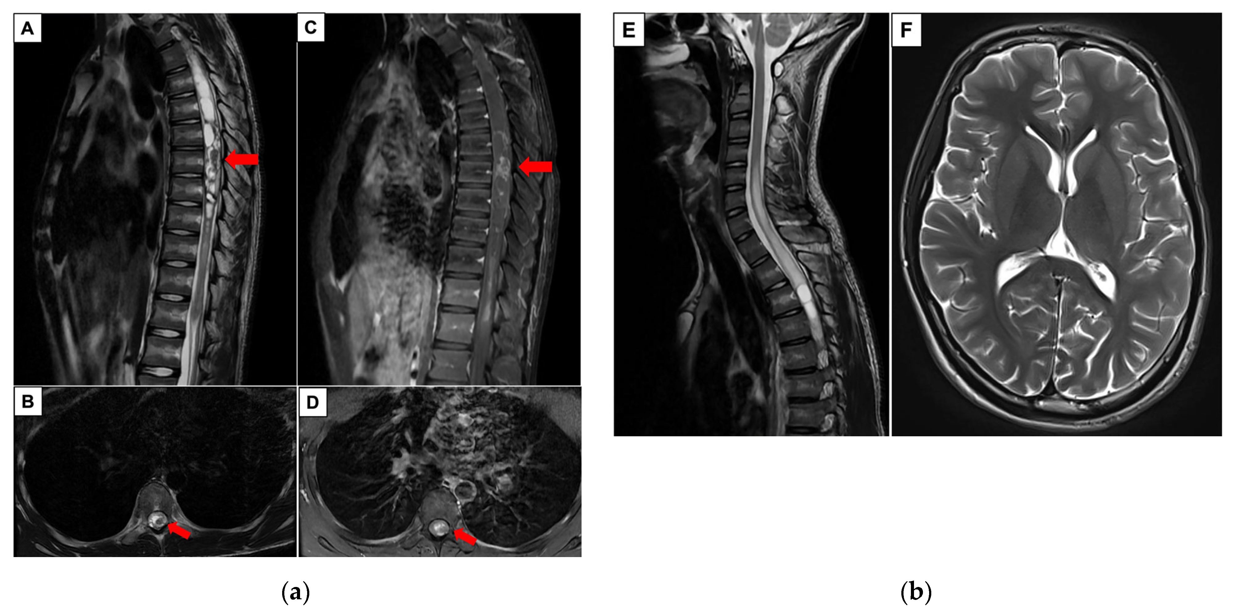
Figure 1. (a) Representative MRI images: (A) T2-weighted image in sagittal view that shows an expanded, long-thoracic-segment intramedullary lesion with hypointense foci (red arrow) and a multi-septate syrinx with hemorrhagic components; (B) T2-weighted image in axial view at T7 level (the red arrow depicts the lesion of interest). (C) T1-weighted post-contrast image in sagittal view [corresponding to (A)] showing a heterogeneously enhancing intramedullary lesion at the level of T7 (red arrow); (D) T1-weighted post-contrast image in axial view at T7 level (the red arrow depicts the lesion of interest). (b) Representative MRI images in T2-weighted sequences: (E) sagittal view of the craniocervical junction that depicts no evidence of low-lying cerebellar tonsils. Of note, there are hyperintense changes in the spinal cord commencing from C4 downwards; (F) axial view of the patient’s brain that shows no hydrocephalus.
Neuroglia 06 00039 g001
Figure 2. An intraoperative photo depicting the finding of a hemorrhagic, intramedullary lesion with relevant labels.
Neuroglia 06 00039 g002
Figure 3. (a) Histopathology images of the tumor specimen. (A) The hematoxylin and eosin slide (10×) shows a tumor with hyalinized vessels and areas of hemorrhage. (B) The hematoxylin and eosin slide (20×) shows parts of the tumor with oligodendrogliocyte-like cells with eosinophilic granular bodies (emphasized in circles). Immunohistochemical stains are positive for (C) OLIG2 (20×) and (D) GFAP (20×); (b) additional immunohistochemical stains performed. (E) Neurofilaments (2×) are positive mainly at the tumor edges and negative within the tumor, suggestive of a circumscribed glioma. (F) For Ki67 (4×), most areas show a positivity of up to 5%, though focal areas measuring 10% can be seen.
Neuroglia 06 00039 g003
Figure 4. (a) (A,B) show the respective FGFR1 single-nucleotide changes detected on the complementary strand. In A, a T>C change is identified, corresponding to FGFR1 c.1966A>G [FGFR1 p.Lys656Glu (K656E)]. In B, a C>T change is identified, corresponding to FGFR1 c.1681G>A [FGFR1 p.Val561Met (V561M)]; (b) screenshot of OncoScan Whole Genome View of the tumor showing log2 ratio gains (2–3×) in multiple chromosomes (1, 2, 4, 5, 6, 7, 9, 10, 11, 12, 13, 14, 15, 16, 19, 20, 21, and 22), alongside the B-allele frequency plot.
Neuroglia 06 00039 g004
Figure 5. (a) A screenshot of the patient’s Illumina® Childhood Cancer Panel Description (Illumina Inc., San Diego, CA, USA) that confirms an FGFR1 K656E hotspot mutation and no other targetable mutations. (b) A screenshot of the patient’s DNA methylation result. Of note, the tumor showed prediction scores of >0.9 for low-grade glial/glioneuronal/neuroepithelial tumors and pilocytic astrocytoma.
Neuroglia 06 00039 g005
Table 1. Summary of FGFR1-altered pLGG cases presenting with spontaneous ICH.
Year/AuthorsPatient NumberAge (Years)Tumor LocationMolecular Alteration
2021/Ishi et al. [48]42 to 193 hypothalamus and 1 thalamus1 FGFR1 p.K656E, 2 FGFR1 p.K656E + p.D652G, and 1 FGFR1 p.N546K
2022/Campion et al. [47]4Not available1 brainstem, 1 optic pathway, and 2 suprasellarAll FGFR1 p.N546K
2024/Gonzalez-Vega et al. [46]51.4 to 171 hypothalamus, 3 optic pathway, and 1 suprasellar4 FGFR1 p.N546K and 1 FGFR1 Dup.Exon9-18
2025/our case117Intramedullary thoracic spineFGFR1 p.K656E and V561M
Disclaimer/Publisher’s Note: The statements, opinions and data contained in all publications are solely those of the individual author(s) and contributor(s) and not of MDPI and/or the editor(s). MDPI and/or the editor(s) disclaim responsibility for any injury to people or property resulting from any ideas, methods, instructions or products referred to in the content.