Review

The Role of Oral Microbiota and Glial Cell Dynamics in Relation to Gender in Cardiovascular Disease Risk

1
Department of Biochemistry, Saraswati Dental College and Hospital, Lucknow 226028, India
2
Department of Molecular Medicine and Biotechnology, Sanjay Gandhi Post Graduate Institute of Medical Sciences, Lucknow 226014, India
*
Author to whom correspondence should be addressed.
Neuroglia 2025, 6(3), 30; https://doi.org/10.3390/neuroglia6030030
Submission received: 22 July 2025 / Revised: 17 August 2025 / Accepted: 19 August 2025 / Published: 22 August 2025

Abstract

The oral microbiota, long recognized for their role in local pathologies, are increasingly implicated in systemic disorders, particularly cardiovascular disease (CVD). This review focuses on emerging evidence linking oral dysbiosis to neuroglial activation and autonomic dysfunction as key mediators of cardiovascular pathology. Pathogen-associated molecular patterns, as well as gingipains and leukotoxin A from Porphyromonas gingivalis, Fusobacterium nucleatum, Treponema denticola, Aggregatibacter actinomycetemcomitans, etc., disrupt the blood–brain barrier, activate glial cells in autonomic centers, and amplify pro-inflammatory signaling. This glia driven sympathetic overactivity fosters hypertension, endothelial injury, and atherosclerosis. Crucially, sex hormones modulate these neuroimmune interactions, with estrogen and testosterone shaping microbial composition, glial reactivity, and cardiovascular outcomes in distinct ways. Female-specific factors such as early menarche, pregnancy, adverse pregnancy outcomes, and menopause exert profound influences on oral microbial ecology, systemic inflammation, and long-term CVD risk. By mapping this oral–brain–heart axis, this review highlights the dual role of oral microbial virulence factors and glial dynamics as mechanistic bridges linking periodontal disease to neurogenic cardiovascular regulation. Integrating salivary microbiome profiling with glial biomarkers [e.g., GFAP (Glial Fibrillary Acidic Protein) and sTREM2 (soluble Triggering Receptor Expressed on Myeloid cells 2)] offers promising avenues for sex-specific precision medicine. This framework not only reframes oral dysbiosis as a modifiable cardiovascular risk factor, but also charts a translational path toward gender tailored diagnostics and therapeutics to reduce the global CVD burden.

1. Introduction

1.1. Oral Microbiota, Glial Interaction, and Their Significance in Health and Disease

1.1.1. Overview of the Oral Microbiota

The microbiota comprise living microbes (bacteria, archaea, fungi, protists, and algae), while the microbiome includes these organisms along with their metabolites, structural components, host-derived molecules, and non-living genetic elements such as phages, viruses, plasmids, and extracellular DNA [1]. Among the various microbial communities within the human body, the oral microbiota are the second largest [2]. The oral cavity comprises several ecological niches, including the saliva, tongue, tooth surfaces, gingiva, buccal mucosa, palate, and sub-/supragingival plaque, that host largely similar, yet subtly distinct, microbial communities. Environmental shifts (e.g., pH), genetic changes, and microbial interactions can markedly alter diversity and activity. Five phyla, Firmicutes, Proteobacteria, Bacteroidetes, Actinobacteria, and Fusobacteria, dominate among over 600 species. A “core” microbiome, including Streptococcus, Veillonella, Neisseria, and Actinomyces, underpins oral and systemic health through its stability [3,4]. Microbial communities across oral and gut sites cluster into the following four groups: (i) buccal mucosa, keratinized gingiva, and hard palate; (ii) saliva, tongue, tonsils, and throat; (iii) subgingival and supragingival plaques; and (iv) stool. Dental plaque communities are distinct from other oral surfaces, yet share core families such as Porphyromonadaceae, Veillonellaceae, and Lachnospiraceae, whose genus-level distributions differ markedly by site [5]. The American Heart Association (AHA) and American Dental Association (ADA) recognize a significant association between periodontal disease and cardiovascular conditions. PCR-based studies demonstrate that dental plaque and saliva host distinct bacterial communities; plaque is dominated by Actinobacteria, Fusobacteria, and Spirochaetes, while saliva is richer in Bacteroidetes, Firmicutes, and Proteobacteria [6]. When shifts in plaque composition disrupt the oral microbial equilibrium, the resultant dysbiosis elicits a host immune reaction, driving chronic inflammation and tissue destruction characteristic of periodontal disease. The condition oscillates between active and dormant stages until biofilm removal or tooth loss permits resolution of the inflammatory process [7].

1.1.2. Oral Microbiota and Gliotransmission: Implications for Systemic Health

Oral microbes release pathogen-associated molecular patterns (PAMPs), for example, lipopolysaccharide (LPS) from Gram-negative bacteria, that engage innate receptors such as TLR4 on endothelial and immune cells, spurring cytokine release (TNF-α, IL-1β, and IL-6). These mediators, along with small microbial products, enter the brain through circumventricular organs (CVOs) or paracellular routes, where they activate endothelial cells and perivascular macrophages to produce secondary inflammatory signals [8,9]. LPS can alter blood–brain barrier (BBB) integrity and engage microglia and other CNS immune cells [10,11]. Glial cells in key autonomic centers sense these signals. Activated microglia in the hypothalamic paraventricular nucleus (PVN) produce pro-inflammatory cytokines that enhance sympathetic outflow. In the PVN, microglial IL-1β and TNF-α increase the firing of presympathetic neurons, raising heart rate and blood pressure [12,13]. In the brainstem, the nucleus tractus solitarius (NTS) integrates baroreceptor signals and transmits them to the rostral ventrolateral medulla (RVLM), which tonically drives sympathetic outflow. Under normal baroreflex function, elevated arterial pressure activates the NTS, inhibiting the RVLM and reducing sympathetic tone. However, systemic inflammation and associated glial dysfunction disrupt cytokine-sensitive NTS/RVLM circuits, blunting the baroreflex and shifting the autonomic balance toward sympathetic overactivity, thereby promoting hypertension [14,15].
Oral pathogens such as Porphyromonas gingivalis (P. gingivalis), Fusobacterium nucleatum (F. nucleatum), and Aggregatibacter actinomycetemcomitans (A. actinomycetemcomitans) influence CNS immunity through the following two main routes relevant to CVD and sex-specific outcomes: (1) PAMP- and cytokine-driven vagal activation of the nucleus tractus solitarius, priming microglia [16], and (2) BBB disruption from systemic inflammation and virulence factors like gingipains, LPS, and leukotoxin A [17,18]. Peptidyl arginine deiminase (PAD) and gingipains are unique virulence factors of P. gingivalis that synergistically drive periodontal and systemic inflammation. PAD citrullinates host proteins, potentially breaking immune tolerance and linking P. gingivalis to autoimmune diseases such as rheumatoid arthritis, while gingipains degrade structural and immune proteins, disrupt epithelial barriers, and facilitate nutrient acquisition. Their combined role in immune evasion, dysbiosis maintenance, and the promotion of vascular inflammation is relevant to CVD [19,20]. The Socransky complexes categorize subgingival bacteria into distinct color-coded groups according to their correlation with periodontal health or disease. Among these, the red complex comprising P. gingivalis, T. forsythia, and Treponema denticola is particularly associated with the progression and severity of periodontitis. Sex hormones modulate these effects, with estrogen often dampening and testosterone variably amplifying microglial reactivity [21,22]. Additionally, outer membrane vesicles (OMVs) from oral pathogens act as nanoscale delivery systems for virulence factors that can cross the BBB. These vesicles bypass traditional size and permeability constraints, enabling the direct transport of pro-inflammatory and neurotoxic cargo to glial cells, thereby amplifying neuroinflammation and potentially contributing to neurodegenerative and cardiovascular–brain axis pathology [23,24]. Among neuroglia cells, astrocytes harness intracellular Ca2+ signals to release glutamate, ATP, GABA, and cytokines, a form of gliotransmission that actively shapes synaptic activity and neuronal excitability [25,26]. In autonomic centers ike the PVN and RVLM, astrocytes release ATP, converted to adenosine, to tune neuronal excitability, while microglia secrete proinflammatory cytokines and ROS with similar effects. Oral dysbiosis, by raising PAMPs and cytokines, may prime glia in the PVN, NTS, and RVLM, amplifying sympathetic outflow (the “heart” axis) [27,28,29].

1.2. CVD: Risk Factors and Impact of Oral Dysbiosis on Pathophysiology

CVD covers disorders of the heart and blood vessels, such as coronary artery disease, stroke, and peripheral artery disease, characterized by impaired blood flow and oxygen delivery, and is a leading global cause of morbidity and mortality [30]. Major risk factors include hypertension, which exerts excessive strain on the heart and arterial walls, and dyslipidemia-elevated LDL (“bad”) cholesterol coupled with low HDL (“good”) cholesterol, which promotes arterial plaque formation and vessel obstruction [31]. Tobacco use, both active smoking and second-hand exposure, damages vessels, promotes clotting, and accelerates arterial narrowing. Diabetes mellitus, marked by unstable glucose levels, fosters atherosclerosis and impairs cardiac function [32,33]. Obesity and inactivity exacerbate risk via metabolic imbalance and inflammation, while diets high in saturated fats, refined sugars, and sodium drive disease progression. Excessive alcohol and chronic stress further undermine cardiovascular health [34,35,36]. Frank Billings first proposed that oral infections via bacteria or their byproducts from dental plaque entering the bloodstream might underlie systemic conditions like rheumatoid arthritis, nephritis, and endocarditis [37]. Alterations in the oral microbiota have since been linked to systemic disease progression, positioning the oral microbiome as a promising biomarker for early detection and prognosis. Moreover, dysbiosis can trigger transient bacteremia after dental procedures such as extractions, subgingival cleanings, endodontic treatments, third-molar surgeries, and tonsillectomies, further implicating oral microbes in systemic health [38]. Periodontal infections can cause transient bacteremia, enabling oral bacteria to invade endothelial cells and contribute to atherosclerotic plaques; low-antibody titers against Tannerella forsythia also predict higher CVD mortality [39]. Intensive periodontal therapy disrupts the IL-6–gp130–CRP axis, lowering IL-6, CRP, and blood pressure and improving lipids, and reduces E-selectin, myeloperoxidase, ICAM-1, and VCAM-1, key mediators of leukocyte adhesion and vascular inflammation linked to CVD [40].

1.3. Rationale for Exploring Glial Cells in Oral Dysbiosis–CVD Link

Oral dysbiosis fuels systemic inflammation and atherosclerosis in CVD. In stroke patients, reduced oral diversity with low Streptococcus predicting stroke-associated pneumonia and high Actinobacteriota signaling worse outcomes underscore its impact on post-stroke prognosis [41]. The presence of oral- and gut-associated pathogens, including Proteobacteria, Bacteroidetes, Firmicutes, and Actinobacteria, has been confirmed in atheromatous plaques obtained from patients undergoing post-endarterectomy procedures using PCR analysis [42]. In acute ischemic stroke, both early and long-term poor outcomes have been linked to higher oral levels of Bacteroidales, Prevotellaceae, and pathogens such as Enterobacteriaceae and Solobacterium [43,44]. While oral health’s role in CVD is well-established [45,46], the influence of gender-specific factors remains underexplored. Differences in hormonal regulation, immune responses, oral hygiene practices, and lifestyle factors can shape the composition of the oral microbiota and CVD risk. Glia in CVOs and hypothalamic nuclei sense PAMPs and cytokines from dysbiosis, with estrogen and androgen signaling driving sex-specific glial activation, neurovascular coupling, and autonomic control, potentially explaining CVD sex differences [47,48]. This review underscores the critical link between oral microbiota dysbiosis, glial dynamics, and cardiovascular disease, as depicted in Figure 1.
The review also explores how sex-specific hormonal, behavioral, and socioeconomic factors shape microbial profiles and modulate CVD risk. By weaving together oral microbial dynamics, systemic inflammation, and neuroglial regulation in precision medicine frameworks, it charts a transformative path toward gender-tailored risk stratification [46,49] and targeted therapies, offering a powerful strategy to curb the global CVD burden.

2. Gender Differences in CVD Risk

CVD is the leading cause of death globally as per World Health Organization. Although women have a lower incidence than men, they face a higher mortality and poorer post-event outcomes. This risk underestimation in women leads to less aggressive treatment and fewer diagnostic procedures, highlighting the need for gender-specific therapies. Emerging evidence also shows sex-based disparities in diagnostic criteria and interventions such as percutaneous coronary intervention (PCI) and coronary artery bypass grafting (CABG) [50,51]. After menopause, blood pressure rises ~0.5 mm Hg per year. Lowering systolic pressure to 120 mm Hg (vs. 130 mm Hg) more effectively prevents CHD events in women than in men. Moreover, diabetes boosts CVD risk three- to seven-fold in women versus two- to three-fold in men [52,53]. Women have roughly twice the CVD mortality of men, with angina, heart failure, and stroke becoming increasingly common. Depression, affecting 20–25% of women, also poses a non-traditional CVD risk, driven by sympathetic overactivity and HPA axis dysfunction [54,55]. The 2019 American College of Cardiology (ACC)/American Heart Association (AHA) CVD prevention guidelines emphasize female-specific risk factors, such as early menopause, pre-eclampsia, early menarche, PCOS, multiparity, adverse pregnancy outcomes, and hormone therapy, as key contributors to lifetime CVD risk, stressing that addressing these factors is essential for improving prevention, diagnosis, and treatment in women [56].
There are some unique female-specific risk-enhancing factors related to CVD which men do not experience [56]. These include early menarche, PCOS, multiparity, adverse pregnancy outcomes, premature menopause, and other hormonal factors, as mentioned in Table 1. Early menarche, marked by prolonged estrogen exposure, alters the oral microbiome, promoting pathogenic bacteria like Porphyromonas and Prevotella, increasing the risk of gingival inflammation and periodontitis [57,58]. Additionally, it is linked to systemic inflammation and endothelial dysfunction, early markers of atherosclerosis, which may elevate CVD risk [59,60]. PCOS is linked to elevated androgen levels, contributing to periodontal inflammation and poorer oral health, including increased bleeding, deeper pockets, and higher plaque indices. While PCOS is associated with increased periodontal pathogens, evidence for T. denticola remains inconclusive. Chronic inflammation in PCOS, marked by elevated TNF-α and IL-6, promotes insulin resistance and atherosclerosis, heightening CVD risk [61,62,63]. Hormonal fluctuations during pregnancy, particularly elevated estrogen and progesterone levels, influence the oral microbiome and immune response. These changes promote the growth of bacteria like Prevotella intermedia and P. gingivalis, contributing to gingival inflammation [64]. PAD and gingipains are unique virulence factors of P. gingivalis that synergistically drive periodontal and systemic inflammation. PAD citrullinates host proteins, potentially breaking immune tolerance and linking P. gingivalis to autoimmune diseases such as rheumatoid arthritis, while gingipains degrade structural and immune proteins, disrupt epithelial barriers, and facilitate nutrient acquisition. Their combined role in immune evasion, dysbiosis maintenance, and the promotion of vascular inflammation is relevant to CVD [19,20].
Gingival inflammation can alter the subgingival biofilm, exacerbate periodontal issues [65], and may trigger systemic inflammation, potentially leading to adverse pregnancy outcomes. Periodontal pathogens and their inflammatory byproducts can reach the placenta, further increasing pregnancy-related risks. Thus, maintaining oral hygiene during pregnancy is crucial for both maternal and fetal health [76]. Research suggests that F. nucleatum contributes to adverse pregnancy outcomes (APOs) like pre-eclampsia and gestational diabetes by migrating from the oral cavity to the placenta by activating Toll-like receptors (TLRs) and triggering inflammation. This placental inflammation, linked to poor periodontal health, increases the risk of preterm birth and hypertensive disorders [64,67]. Additionally, APOs heighten the likelihood of long-term cardiovascular issues, including hypertension and heart disease. These findings highlight the lasting impact of pregnancy-related inflammation on heart health, emphasizing the importance of maintaining oral hygiene to reduce associated risks [68]. Menopausal estrogen decline impairs oral and cardiovascular health: it makes the mucosal epithelium prone to infections (candidiasis), burning mouth syndrome, oral lichen planus, and neuropathy; reduces salivary flow, leading to dry mouth, caries, and periodontal disease; and alters the oral microbiome [69]. Additionally, the menopausal transition is associated with heightened oxidative stress and inflammation, which contribute to arterial stiffening, raising cardiovascular risks. These changes highlight the importance of oral and systemic health management during menopause [70,71]. HRT shapes microbiota, immunity, and cardiovascular health, with effects driven by one’s metabolic profile. It can enhance the gut microbiome by reducing pro-inflammatory bacteria and modulating immune responses. Cardiovascular outcomes—mixed in studies—vary by hormone type, delivery method, and individual health, making personalized regimens essential to optimize benefit and limit risk [72,73,74,75].
Testosterone exhibits a complex, sometimes paradoxical relationship with inflammation and CVD risk in men. On one hand, physiological testosterone levels have been shown to exert anti-inflammatory effects by downregulating pro-inflammatory cytokines such as TNF-α and IL-6, enhancing endothelial nitric oxide production, and modulating immune cell activation, mechanisms that could theoretically protect against atherogenesis [77,78]. Conversely, epidemiological and clinical data reveal that both low and supraphysiological testosterone levels are associated with increased CVD risk, but through different pathways: hypogonadism correlates with metabolic syndrome, visceral adiposity, and low-grade systemic inflammation, while excessive testosterone (endogenous or from supplementation) can promote thrombosis, erythrocytosis, and dyslipidemia, potentially accelerating vascular events [79,80]. This bidirectional relationship may explain conflicting findings in the literature, highlighting the importance of context, dose, and patient-specific factors in interpreting testosterone’s cardiovascular impact.

3. Gender-Specific Features of Oral Microbiota

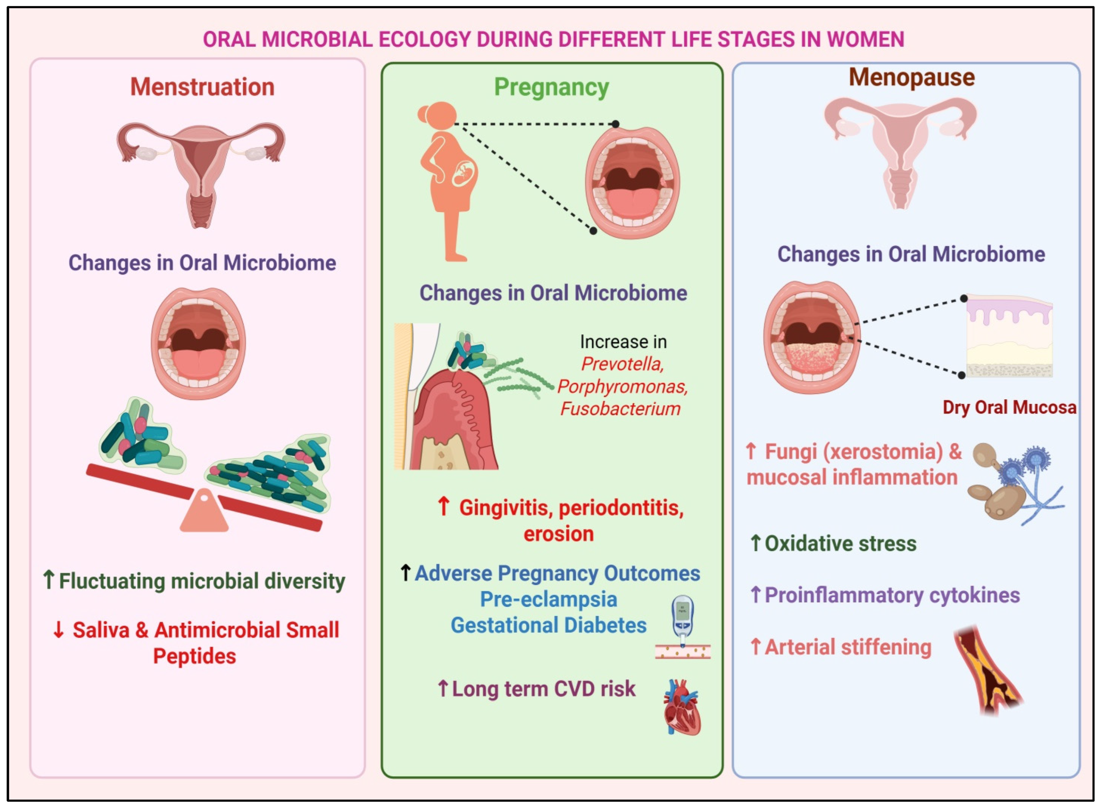
Fluctuations in sex steroids across the female lifespan drive pronounced shifts in oral microbial ecology. During pregnancy, when estrogen and progesterone levels peak, gingival tissues become hyperemic and crevicular fluid increases, predisposing to gingivitis, periodontitis, and dental erosion [81,82]. Systemic complications such as gestational diabetes and pre-eclampsia further perturb the oral flora, whereas regular prenatal dental care reduces Streptococcus mutans and subgingival P. gingivalis, correlating with term delivery; notably, periodontal status often rebounds postpartum [82,83]. Beyond pregnancy, cyclical drops in estrogen during menstruation and the precipitous decline at menopause diminish salivary flow and antimicrobial peptides, fostering an ecological niche for keystone pathogens. Indeed, postmenopausal women exhibit altered oral mycobiomes, with increased colonization by Candida, Malassezia, and Aspergillus species linked to xerostomia and mucosal inflammation [84], and hormone replacement therapy has been shown to restore salivary flow, pH, and buffering capacity, mitigating dysbiosis and oral discomfort [85]. Sex hormones also modulate neuro-immune crosstalk in the periodontium: estrogen enhances the production of pro-inflammatory cytokines (IL-6 and IL-17), amplifying pathogen clearance but exacerbating tissue inflammation, whereas androgen-predominant environments elicit dampened cytokine responses, altering microbial colonization patterns [86,87]. Behavioral factors compound these biological effects: women’s higher rates of routine oral hygiene and healthcare engagement foster commensal-rich communities, while men’s greater tobacco and alcohol use promotes dysbiotic overgrowth [88,89]. Emerging microbiome-targeted interventions hold promise for gender-tailored care. Probiotic and prebiotic strategies such as Lactobacillus reuteri lozenges and specialized mouth rinses have demonstrated efficacy in sustaining beneficial taxa and reducing gingival inflammation in hormonally altered niches [90]. Together, these findings underscore a complex, hormone-driven interplay between host, behavior, and oral microbes that shape women’s oral and systemic health across reproductive and menopausal transitions. Significant aspects of changes in the oral microbiota during menstruation, pregnancy, and menopause are summarized in Figure 2.

4. Glial Dynamics in Cardiovascular Disease

4.1. Microglial Activation via LPS, Blood–Brain Barrier Disruption, and the Paraventricular Nucleus (PVN)

Periodontal pathogens (e.g., P. gingivalis and T. denticola) shed LPS and other virulence factors into the blood during infection. LPS may cross the BBB via regions lacking tight junctions, such as CVOs like the organum vasculosum laminae terminalis (OVLT), and by active transport mechanisms [91]. Systemic LPS also activates peripheral immune cells to release IL-1β, TNF-α, etc., which can signal to the brain by binding their brain endothelial receptors or via vagal afferents. IL-1β and TNF-α are known to activate the hypothalamic PVN; PVN neurons express receptors for cytokines and angiotensin II. Critically, the PVN receives inputs from CVOs (e.g., subfornical organs) and can be affected by circulating AngII and endotoxin [91]. Rodent studies have shown that intracerebral minocycline (microglial inhibitor) blocks LPS-induced sympathetic increases [92].
Microglia in the PVN express TLR4 and purinergic receptors like P2Y12. LPS binding to TLR4 on microglia triggers NF-κB and MAPK cascades, leading to TNF-α/IL-1β release and oxidative stress [91,92]. LPS upregulates microglial P2Y12, and P2Y12 antagonists reduce LPS-induced IL-1β and TNF production [93]. Thus, systemic LPS may both directly (via TLR4) and indirectly (via ATP release from damaged cells signaling through P2Y12) activate microglia. Activated microglia enlarge, migrate, and secrete pro-inflammatory mediators that act locally. In the PVN, elevated microglial TNF-α/IL-1β increases presympathetic neuronal excitability and diminishes inhibitory GABA tone [92], thereby raising sympathetic outflow. Thus, chronic LPS or periodontal inflammation “primes” PVN microglia to be hyper-responsive. Microglia express sex hormone receptors that modulate their response. Estradiol signaling via microglial ERβ is generally anti-inflammatory: ERβ agonists suppress NF-κB activity and iNOS expression in microglia [94]. Estrogen can dampen TLR4 signaling pathways, reducing microglial cytokine output. In contrast, microglial androgen receptors and testosterone tend to promote an anti-inflammatory state. For example, testosterone profoundly reduces microglial activation and astrocyte reactivity after injury. Clinically, higher testosterone in men correlates with lower TNF-α and IL-6 and higher IL-10 [95]. Thus, male hormones may blunt PAMP-induced inflammation, whereas lower testosterone (or fluctuating estrogens) in women could allow for greater neuroinflammation. Indeed, neonatal estrogen manipulations alter microglial gene expression and later neuroinflammatory sensitivity [95].

4.2. Role of Astrocytes in the Rostral Ventrolateral Medulla (RVLM) and Microglial Priming

In the RVLM, astrocytes and microglia jointly regulate sympathetic tone. Astrocytes sense circulating LPS and AngII via TLR4 and AT1R, altering their physiology through NF-κB activation. Astrocytic IKKβ-driven NF-κB activation raises blood pressure by triggering IL-1β release and reducing hypothalamic GABA, leading to hypertension [96]. Astrocytes also modulate RVLM neurons via gliotransmission, as upon inflammation-induced Ca2+ signals, astrocytes release ATP and glutamate. This “gliotransmission” increases extracellular glutamate in the RVLM, potentiating sympathetic neuron firing. ATP from astrocytes can also be converted to adenosine, which generally inhibits synaptic activity; thus, alterations in astrocyte signaling can tip the balance of excitatory/inhibitory tone in the RVLM [97,98].
Chronic periodontal inflammation “primes” microglia toward a pro-inflammatory (M1-like) metabolic state. Normally, resting microglia rely on oxidative phosphorylation (OXPHOS) for energy. When primed (by LPS or systemic inflammation), microglia increase glycolysis: glucose uptake is upregulated (via GLUT1), glycolytic flux rises, and lactate production surges. This metabolic shift (Warburg-like) quickly generates ATP and the biosynthetic precursors needed for massive cytokine synthesis. Blocking glycolysis (GLUT1 inhibition) forces microglia back to OXPHOS and reduces their inflammatory output. Thus, in a hypertensive context, microglial priming involves increased NF-κB/MAPK signaling and a glycolytic switch, fueling the persistent production of IL-1β, TNF-α, and reactive oxygen [99,100]. TLR2/4 and cytokine receptors in microglia trigger the NF-κB and MAPK (p38, ERK, and JNK) pathways. In rats, chronic AngII infusion activates NF-κB in PVN microglia, raising the TNF-α and IL-1β effects reversed by minocycline or IL-10, which also lower blood pressure. NF-κB drives proinflammatory gene transcription, MAPKs boost cytokine release, and activated microglia generate NO (via iNOS) and superoxide, directly exciting neurons and impairing NO-dependent baroreflexes. Released cytokines like IL-1β then perpetuate NF-κB activation in nearby glia and neurons, fueling a self-amplifying neurogenic inflammation [91]. Furthermore A. actinomycetemcomitans and P. gingivalis activate NLRP3, driving IL-1β release that links oral dysbiosis to neuroinflammation, making IL-1β a key therapeutic target [101].
The NF-κB/MAPK response differs by sex. Estrogen (via ERβ) interferes with NF-κB signaling: ERβ agonists recruit co-repressors to NF-κB target genes and increase IκB (the NF-κB inhibitor) [94]. This yields lower TNF-α and iNOS expressions in microglia. Testosterone (via AR) also suppresses microglial activation: as noted, it upregulates anti-inflammatory IL-10 and downregulates multiple proinflammatory cytokines [95]. Thus, male microglia tend to skew toward an “M2-like” (tissue-healing) state, whereas female microglia, lacking androgen protection and facing estrogen fluctuation, may be more M1-prone. This sex dimorphism suggests that the same oral inflammatory insult could produce stronger microgliosis and sympathetic excitation in females. In summary, in the RVLM and PVN, astrocyte NF-κB activation and microglial priming (via glycolysis and NF-κB/MAPK) amplify sympathetic drive; estrogen and testosterone modulate these glial responses to inflammatory signals [94,96].

4.3. Microglial Priming and Its Contribution to Neurogenic Hypertension

Chronic low-grade inflammation (e.g., from periodontitis or obesity) “primes” the microglia in cardiovascular nuclei, predisposing to exaggerated neurogenic hypertension. Primed microglia exhibit heightened NF-κB and MAPK signaling and a metabolic shift to glycolysis. Studies of neurogenic hypertension (e.g., chronic angiotensin II infusion) in rodent models show that blocking microglial activation attenuates hypertension, as intracerebroventricular minocycline reduces mean arterial pressure, cardiac hypertrophy, and norepinephrine levels in AngII-treated rats, coincident with lower PVN IL-1β and TNF-α. Conversely, microglial depletion or IL-1β infusions demonstrate that microglial cytokines alone can drive blood pressure. Mechanistically, AngII-induced hypertension engages PVN microglial TLRs to trigger NF-κB and ROS [91,102,103].
Activated NF-κB translocates to the nucleus, upregulating genes for TNF-α, IL-1β, IL-6, and iNOS. These cytokines feed back onto PVN and RVLM neurons: IL-1β in the PVN robustly increases sympathetic outflow and blood pressure. MAPKs (p38 and JNK) are also activated in this process, further amplifying cytokine production and stabilizing inflammatory transcripts [104,105].
At the same time, primed microglia switch their metabolism. LPS or AngII stimulation upregulates glycolytic enzymes (GLUT1 and hexokinase) and downregulates mitochondrial respiration. This “aerobic glycolysis” provides rapid ATP and NADPH for cytokine synthesis and ROS generation. Indeed, inhibiting glycolysis (or promoting OXPHOS) limits microglial cytokine release in vitro. Glycolytic microglia also produce more lactate; excess lactate can itself modulate neural activity and barrier function. Thus, metabolic reprogramming is a core feature of microglial priming in hypertension [99]. Male and female mice show different baseline microglial gene expression. Estrogen increases IL-10 in males, but can be pro-inflammatory in females. Testosterone (AR) profoundly inhibits microglial activation: it reduces NF-κB-dependent transcription and boosts anti-inflammatory IL-10 [106,107]. In hypertensive models, male rodents often show blunted microgliosis compared to females. Clinically, this may underlie sex differences in neurogenic hypertension prevalence: premenopausal women (lower testosterone) might have more pronounced neuroinflammation from the same oral insult. In summary, microglial priming in neurogenic hypertension involves TLR4/NF-κB–driven cytokine loops and a glycolytic phenotype, both of which are tempered by sex steroids [94].

5. Impact of Oral Microbiota on Cardiovascular Health

5.1. Oral Microbiota Dysbiosis: Driving Inflammation, Endothelial Dysfunction, and Atherosclerosis

Studies have shown that procedures like tooth extraction, subgingival cleaning, endodontic therapy, third-molar surgery, and tonsillectomy can lead to low-grade bacteremia. Based on this, it is hypothesized that periodontal infections may similarly trigger transient bacteremia, allowing bacteria to directly invade endothelial cells and potentially contribute to CVD [38,108]. Periodontal disease is linked to atherosclerosis, as PCR analyses of post-endarterectomy and thrombo-endarterectomy specimens have detected DNA from the oral and gut pathogens Proteobacteria, Bacteroidetes, Firmicutes, and Actinobacteria within atherosclerotic plaques [42]. Epidemiological, clinical, and experimental evidence supports a pro-atherogenic role for periodontal pathogens: oral bacteria enter the bloodstream most often via bacteremia from routine activities or dental procedures and are carried to vascular tissues by various mechanisms [109]. Another confirmed mechanism involves the transcellular migration of bacteria through gingival microcapillaries located near periodontal pockets [110]. Additionally, the “Trojan horse” hypothesis, though not yet conclusively proven, suggests that pathogens may evade immune detection by hijacking immune cells through phagocytosis-mediated transport, thereby gaining access to vascular tissues [111,112]. Genetic material and antigens from key periodontal pathogens such as P. gingivalis, A. actinomycetemcomitans [113], and Veillonella species [42] have been identified in atheromatous plaques. Once in circulation, periodontal bacteria and their components (LPS and peptidoglycans) activate TLRs, triggering T cell responses and the release of pro-inflammatory mediators (cytokines, chemokines, ROS, and RNS); the presence of viable P. gingivalis and A. actinomycetemcomitans in atheromatous tissue confirms their ability to invade cardiovascular cells [114]. Experimental models further show that P. gingivalis accelerates atherosclerosis in mice [115], induces vascular lesions in pigs, and promotes fatty streak formation in rabbits [116]. A. actinomycetemcomitans produces the leukotoxin LtxA, which induces potassium efflux in immune cells, triggering NLRP3 inflammasome activation and the release of IL-1β/IL-18 [117]. As NLRP3-driven inflammation is linked to neurodegeneration, LtxA may connect oral dysbiosis to brain disorders via systemic cytokine surges, BBB disruption, and glial priming, though direct in vivo evidence remains limited.

5.2. Gender-Specific Links Between Oral Microbiota and Cardiovascular Risk Markers

The interplay between the oral microbiota and cardiovascular health has gained increasing attention over the past decade. Emerging evidence suggests that not only does the overall composition of the oral microbiome influence systemic inflammation and vascular function, but that these relationships may differ markedly between men and women.
A 10.4-year prospective study of postmenopausal women (mean age 63) using subgingival plaque 16S rRNA sequencing linked specific bacteria with both baseline blood pressure and incident hypertension. Ten taxa, e.g., Streptococcus anginosus, Selenomonas infelix, and Prevotella spp., were positively associated with hypertension risk, while five, including Neisseria subflava and Aggregatibacter segnis, showed inverse associations. These associations held after adjusting for demographic, clinical, and lifestyle factors, suggesting a causal role for oral bacteria in women’s blood pressure regulation [118].
In a northern Finland birth cohort study of 869 participants (mean age 46), oral microbiome diversity (Shannon index and Bray–Curtis β-diversity) correlated with carotid intima–media thickness (cIMT) in men but not women. After adjusting for traditional CVD factors, higher abundances of Prevotella, Megasphaera, and Veillonella were positively linked to cIMT, whereas Capnocytophaga, Gemella, and Neisseria were inversely related; men with a greater cIMT also showed reduced anti-phosphorylcholine IgA and IgG, suggesting that impaired mucosal immunity may mediate these microbiome vascular interactions [119]. A meta-analysis of subgingival biofilms in periodontitis found marked sexual dimorphism: six taxa were enriched in females versus two in males (healthy controls showed fewer differences). A “microbial sex index” (MSI), the log-ratio of female to male-enriched taxa, was inversely correlated with species richness in disease, but not in health. This amplified divergence suggests that women and men harbor distinct periodontal microbiomes that may differentially influence systemic inflammation and cardiovascular risk [49].
Sex hormones likely shape the oral microbes affecting cardiovascular health: estrogen boosts nitrate-reducing bacteria that produce vasodilatory nitric oxide, perhaps explaining the lower hypertension in premenopausal women, while androgens may skew immunity toward pro-atherogenic taxa in men. Though much evidence comes from gut microbiome studies (e.g., FMO3-regulated TMAO production), analogous oral pathways via microbial metabolites like lipopolysaccharides and short-chain fatty acids triggering endothelial dysfunction and low-grade inflammation probably operate in a sex-dependent manner [120].

6. Mechanisms Underlying Gender Differences in the Oral Microbiota–CVD Axis

6.1. Potential Mechanisms Through Which Gender-Specific Factors Modulate the Relationship Between Oral Microbiota and CVD Risk

The oral microbiota are influenced by host genetic factors, leading to site- and sex-specific variations in microbial composition. Nitrate-reducing bacteria such as Streptococcus salivarius, S. mitis, S. bovis, Veionella spp., Staphyloccocus aureus, S. epidermidis, Nocordia spp., and Corynebacterium sp. in the oral cavity play a crucial role in converting nitrate into nitric oxide, which is essential for endothelial function, promotes vasodilation, and helps to regulate blood pressure [121,122,123]. Notably, studies indicate that using mouthwash can reduce oral nitrate-reducing bacteria, leading to an increase in blood pressure (BP) [124,125]. Several periodontal pathogens, particularly Prevotella and Streptococcus species, which showed a significant association with BP in this study, have also been implicated in vascular disease. In addition to activating inflammatory pathways, bacterial infections may contribute to the progression or worsening of peripheral artery disease by increasing platelet reactivity, promoting von Willebrand factor binding, elevating factor VIII and fibrinogen levels, activating P-selectin, altering plasma lipid profiles, enhancing oxidative stress, and inducing insulin resistance [123,126].
Systolic and diastolic BP are positively correlated with pathogenic oral bacteria (P. gingivalis, Tannerella forsythia, T. denticola, and F. nucleatum), but not with beneficial species. In postmenopausal women with elevated BP or on antihypertensives, 65 bacterial species differed in abundance versus normotensive women; Prevotella oral and Streptococcus oralis remained significantly lower in those taking medication [127].
Periodontal disease is associated with systemic inflammatory conditions such as diabetes, arthritis, and cardiovascular disease, and some studies report elevated oral P. gingivalis and Tannerella forsythia in these patients, though results are inconsistent. After adjusting for active caries, smoking, age, and gender, individuals with the highest multi-morbidity actually showed a lower salivary abundance of these pathogens [128].

6.2. Hormonal Modulation, Immune Responses, and Gene–Environment Interactions

Using UK Biobank data, researchers have quantified how genetic factors drive sex differences across numerous cardiovascular-related traits, revealing that heredity contributes differently to disease susceptibility in men and women [129]. A separate GWAS with targeted metabolomics highlighted a female-specific link between coronary artery disease and the mitochondrial enzyme carbamoyl-phosphate synthase 1, pointing toward a potential avenue for sex-tailored CAD therapies [130].
Sex hormones further modulate post-infarction remodeling and risk. In animal models, testosterone promotes early eccentric cardiac changes after myocardial ischemia, worsening function and increasing rupture risk, whereas estrogen shows little acute benefit [131]. Clinically, postmenopausal elevations in ovarian testosterone driven by rising gonadotropins may underlie the surge in women’s cardiovascular mortality [132]. Conversely, obese and elderly men often exhibit raised estradiol levels, sometimes exceeding those of age-matched women [133]. Immune-regulatory genes also display sex-biased expression: women mount stronger innate and adaptive responses, yet are more prone to autoimmunity, alterations that can influence vascular and myocardial inflammation [134].

6.3. Clinical and Experimental Trials Targeting the Oral–Brain–Heart Axis

Anti-inflammatory agents that penetrate the BBB, such as minocycline, statins, and IL-1β antagonists, have been evaluated chiefly in acute stroke and dementia, rather than for blood pressure control [135,136]. A Cochrane review of statin therapy in Alzheimer’s disease highlighted anti-inflammatory effects, yet inconclusive cognitive benefits [137]. In cardiovascular populations, the CANTOS trial showed that canakinumab, an IL-1β monoclonal antibody, reduced recurrent myocardial infarction by targeting systemic inflammation, implying that modulating neuroimmune pathways might influence heart disease outcomes, although that trial did not assess any glial-specific biomarkers [138]. Blood measurements of glial markers have gained traction. Plasma glial fibrillary acidic protein (GFAP), reflective of astrocytic injury, rises sharply in intracerebral hemorrhage and correlates with bleed volume [139,140]. Microglial activation, as indicated by TREM2 expression, has been observed in white-matter regions of post-stroke dementia brains, with increased densities of TREM2+ cells in frontal-lobe perivascular areas [141] To date, no randomized clinical trial has validated GFAP or soluble TREM2 (sTREM2) as surrogate endpoints for cardiovascular risk. Incorporating these glial biomarkers as exploratory outcomes in future interventional studies could clarify their prognostic and mechanistic roles in CVD. In the PAVE trial (Circulation 2009), periodontal therapy improved gum health in post-myocardial infarction patients, but failed to reduce recurrent cardiovascular events. A 2022 Cochrane review likewise concluded that existing randomized controlled trials are too small and heterogeneous to demonstrate cardiovascular benefits from periodontal treatment. In a metabolic syndrome cohort of roughly 165 participants, aggressive scaling yielded no advantage over standard care in mortality or myocardial infarction rates. By contrast, a pilot study of 13 patients showed that adding probiotic supplementation to non-surgical periodontal therapy lowered HbA1c by 0.3% and reduced systolic blood pressure by 9.6 mmHg over 12 weeks, findings that underscore the need for larger, more definitive trials [142].

7. Clinical Implications and Future Directions

7.1. Sex-Specific Oral–Cardiovascular Prevention: Microbiota- and Glial-Marker-Guided Screening and Early Intervention

Oral microbial imbalance is now recognized as a changeable contributor to cardiovascular risk and should be considered during routine CVD evaluations. Key periodontal bacteria such as P. gingivalis, F. nucleatum, and A. actinomycetemcomitans promote endothelial injury, plaque formation, and systemic inflammation, all of which underlie hypertension, coronary disease, and heart failure [46,143] Combining periodontal exams (pocket depths and bleeding on probing) with salivary microbiome profiling may uncover subclinical cardiovascular risk in those with borderline hypertension or metabolic syndrome, while circulating glial activation markers (serum GFAP and soluble TREM2) could reflect central neuroinflammation driving autonomic imbalance and sympathetic overactivity [144,145]. Classifying patients by combined oral microbiome and neuroglial biomarker profiles could sharpen CVD prevention strategies. Salivary metagenomic testing reveals sex-linked microbial patterns such as a greater Streptococcal abundance in women and Prevotella enrichment in men, tying these signatures to impaired baroreflex function and heightened sympathetic activity through glial-driven cytokine release [46,143]. Measuring circulating glial exosomes alongside inflammatory cytokines (IL-1β and TNF-α) may reveal microglial priming that triggers blood pressure spikes during periodontal flares, and incorporating these markers into risk models could outperform Framingham or SCORE, enabling earlier, personalized interventions in sex-specific high-risk groups [145]. Recent studies indicate that periodontal treatment can lower systemic systolic blood pressure by approximately 2–3 mm Hg, likely through reduced endotoxin spillover and diminished neurogenic inflammation [146,147]. Enhancing oral care with antimicrobial rinses and specialized probiotics or prebiotics aimed at re-establishing a healthy microbiome may also curb glial activation. On the neuroglial side, agents that boost interleukin-10 signaling in the brain and drugs targeting the purinergic P2Y12 receptor have shown the ability to dampen microglia-driven sympathetic overactivity in neurogenic hypertension in animal models [144,145]. Bringing these approaches into clinical practice will demand BBB-permeable formulations and tailored dosing strategies that account for sex differences in microglial androgen and estrogen receptor modulation.
Sex hormones modulate glial cells, warranting sex-specific therapies: in premenopausal women, estradiol via ERβ/GPER1 shifts astrocytes and microglia to an anti-inflammatory, M2-like state, protecting against periodontal-induced neurogenic hypertension; in men, androgen receptor activation drives a pro-inflammatory, M1-dominant glial phenotype. In postmenopausal women, HRT could rebalance glial responses, reduce sympathetic tone, and lower CVD risk, ideally tested in periodontal care plus HRT trials with glial biomarkers, while selective androgen receptor modulators might similarly modulate microglia in men without systemic androgen side effects [144,145]. Collaborative cardiology dentistry care is essential: patients with persistent, treatment-resistant hypertension should be evaluated for periodontal disease, with salivary microbiome profiling (using gender-specific panels) to gauge inflammatory burden. If glial activation biomarkers (GFAP and sTREM2) are elevated, referral for TSPO-PET neuroimaging can assess microglial activation and central neuroinflammation in cardiovascular-regulating brain regions.

7.2. Bridging Cardiology and Dentistry: Neuroinflammatory Diagnostics

Emerging evidence highlights a complex and bidirectional relationship between oral microbial dysbiosis, neuroglial activation, and CVD. To unravel the mechanisms underpinning these interactions, future research must prioritize the role of glial cells as central mediators of neuroimmune signaling and cardiovascular regulation.
Large, longitudinal, sex-stratified cohort studies are essential to map the temporal and causal dynamics across the oral–brain–heart axis. These studies should integrate routine dental evaluations, high-resolution salivary microbiome profiling, sex hormone analysis, and serial measurements of circulating and CNS-derived glial biomarkers such as GFAP and sTREM2. Such data could clarify how oral microbial shifts influence glial activity over time and whether these interactions differ by sex. Integration of multi-omics approaches metagenomics, transcriptomics, and metabolomics with advanced neuroimaging modalities such as TSPO-PET and functional MRI will provide mechanistic insight into how specific microbial taxa modulate glial gene expression and inflammatory signaling within cardiovascular regulatory centers of the brain [148,149]. This systems-level understanding could uncover novel molecular targets for both diagnostic and therapeutic intervention. Randomized controlled trials are needed to evaluate whether periodontal treatments (e.g., scaling and root planning and targeted antimicrobial therapy) combined with pharmacologic glial modulators such as P2Y12 inhibitors or IL-10 analogues can attenuate neuroinflammation and improve cardiovascular outcomes. These studies should include sex-specific endpoints to capture potential differences in blood pressure regulation and glial reactivity between males and females.
From a precision medicine perspective, integrating microbiome and glial biomarkers into CVD risk assessment could improve patient stratification, particularly regarding gender differences in neuroimmune vascular regulation. Oral dysbiosis, especially the enrichment of P. gingivalis and F. nucleatum, is linked to systemic inflammation, vagal afferent activation, and microglial priming, influencing neurogenic cardiovascular control [150,151]. Peripheral inflammation can disrupt the BBB, enabling microbial metabolites such as LPS to directly activate glia and amplify neuroinflammation, affecting autonomic heart regulation [152]. Biomarkers including serum S100B, soluble TREM2, and microbial diversity indices could be incorporated into predictive models alongside hormone profiling to identify neuroimmune–vascular phenotypes [101,153]. Clinically, salivary microbiome sequencing and plasma neuroinflammatory markers could be integrated into cardiovascular workups for high-risk groups, particularly men with low or borderline testosterone, while interventions such as periodontal therapy and microbiome modulation have shown reductions in systemic inflammation and improved vascular reactivity [154,155]. Therefore, precision microbiome therapies represent a promising frontier. Gender-tailored probiotic formulations aimed at restoring beneficial oral taxa and suppressing lipopolysaccharide-producing pathogens may reduce systemic inflammation and preempt neuroglial priming. Additionally, bacteriophage-based interventions targeting keystone periodontal pathogens could offer highly specific, minimally disruptive alternatives to broad-spectrum antimicrobials. Also, with more people retaining their natural teeth into advanced age, understanding oral–systemic connections gains critical importance. This knowledge can guide preventive and therapeutic strategies to improve health outcomes in aging populations [156].
There is a pressing need for policy and regulatory frameworks that formally recognize periodontitis as a modifiable risk factor for cardiovascular disease. Clinical guidelines should evolve to include glial biomarker screening, particularly in cases of treatment-resistant hypertension. The implementation of science and health economics analyses will be critical in demonstrating the feasibility, cost-effectiveness, and public health impact of integrating oral health, neuroimmunology, and cardiovascular care, especially within gender-sensitive precision medicine models.

Author Contributions

D.G.: conceptualization, methodology, literature search, writing the original manuscript draft, illustration preparation and finalizing the manuscript; A.K.: editing, reviewing, and finalizing the manuscript. All authors have read and agreed to the published version of the manuscript.

Funding

This research received no external funding.

Data Availability Statement

Not applicable.

Acknowledgments

The figures have been prepared in https://BioRender.com.

Conflicts of Interest

The authors declare no conflict of interest.

Abbreviations

CVDCardiovascular Disease
PAMPsPathogen-Associated Molecular Patterns
LPSLipopolysaccharide
TLRToll Like Receptors
ILsInterleukins
CVOsCircumventricular Organs
CNSCentral Nervous System
BBBBlood–Brain Barrier
PVNParaventricular Nucleus
RVLMRostral Ventrolateral Medulla
GABAGamma-Aminobutyric acid
NTSNucleus Tractus Solitarius
CRPC-Reactive Protein
ICAM-1Intercellular Adhesion Molecule 1
VCAM-1Vascular Cell Adhesion Molecule 1
LDLLow-Density Lipoprotein
HDLHigh-Density Lipoprotein
CHDCoronary Heart Disease
PCOSPolycystic Ovarian Syndrome
APOsAdverse Pregnancy Outcomes
OLPOral Lichen Planus
HRTHormone Replacement Therapy
cIMTCarotid Intima Media Thickness
GWASGenome-Wide Association Study
NLRP3NLR family pyrin domain containing 3
PADPeptidyl Arginine Deiminase

References

  1. Berg, G.; Rybakova, D.; Fischer, D.; Cernava, T.; Vergès, M.C.C.; Charles, T.; Chen, X.; Cocolin, L.; Eversole, K.; Corral, G.H. Microbiome definition re-visited: Old concepts and new challenges. Microbiome 2020, 8, 103. [Google Scholar] [PubMed]
  2. Deo, P.N.; Deshmukh, R. Oral microbiome: Unveiling the fundamentals. J. Oral Maxillofac. Pathol. 2019, 23, 122–128. [Google Scholar] [CrossRef] [PubMed]
  3. Shade, A.; Handelsman, J. Beyond the Venn diagram: The hunt for a core microbiome. Environ. Microbiol. 2012, 14, 4–12. [Google Scholar] [CrossRef]
  4. Jenkinson, H.F. Beyond the oral microbiome. Environ. Microbiol. 2011, 13, 3077–3087. [Google Scholar] [CrossRef]
  5. Segata, N.; Haake, S.K.; Mannon, P.; Lemon, K.P.; Waldron, L.; Gevers, D.; Huttenhower, C.; Izard, J. Composition of the adult digestive tract bacterial microbiome based on seven mouth surfaces, tonsils, throat and stool samples. Genome Biol. 2012, 13, R42. [Google Scholar] [CrossRef]
  6. Keijser, B.J.F.; Zaura, E.; Huse, S.M.; van der Vossen, J.M.B.M.; Schuren, F.H.J.; Montijn, R.C.; ten Cate, J.M.; Crielaard, W. Pyrosequencing analysis of the oral microflora of healthy adults. J. Dent. Res. 2008, 87, 1016–1020. [Google Scholar] [CrossRef]
  7. Kinane, D.F.; Stathopoulou, P.G.; Papapanou, P.N. Periodontal diseases. Nat. Rev. Dis. Primers 2017, 3, 17038. [Google Scholar] [CrossRef]
  8. Zhu, X.; Huang, H.; Zhao, L. PAMPs and DAMPs as the Bridge Between Periodontitis and Atherosclerosis: The Potential Therapeutic Targets. Front. Cell Dev. Biol. 2022, 10, 856118. [Google Scholar] [CrossRef]
  9. Mallard, C. Innate immune regulation by toll-like receptors in the brain. ISRN Neurol. 2012, 2012, 701950. [Google Scholar] [CrossRef] [PubMed]
  10. Kalyanaraman, B.; Cheng, G.; Hardy, M. Gut microbiome, short-chain fatty acids, alpha-synuclein, neuroinflammation, and ROS/RNS: Relevance to Parkinson’s disease and therapeutic implications. Redox Biol. 2024, 71, 103092. [Google Scholar] [CrossRef]
  11. Müller, L.; Di Benedetto, S. Bridging the brain and gut: Neuroimmune mechanisms of neuroinflammation and therapeutic insights. Front. Cell. Neurosci. 2025, 19, 1590002. [Google Scholar] [CrossRef]
  12. Hanani, M. Satellite glial cells in sympathetic and parasympathetic ganglia: In search of function. Brain Res. Rev. 2010, 64, 304–327. [Google Scholar] [CrossRef] [PubMed]
  13. Wang, M.; Pan, W.; Xu, Y.; Zhang, J.; Wan, J.; Jiang, H. Microglia-Mediated Neuroinflammation: A Potential Target for the Treatment of Cardiovascular Diseases. J. Inflamm. Res. 2022, 15, 3083–3094. [Google Scholar] [CrossRef] [PubMed]
  14. Colombari, E.; Sato, M.A.; Cravo, S.L.; Bergamaschi, C.T.; Campos, R.R.J.; Lopes, O.U. Role of the medulla oblongata in hypertension. Hypertension 2001, 38 Pt 2, 549–554. [Google Scholar] [CrossRef]
  15. Suarez-Roca, H.; Mamoun, N.; Sigurdson, M.I.; Maixner, W. Baroreceptor Modulation of the Cardiovascular System, Pain, Consciousness, and Cognition. Compr. Physiol. 2021, 11, 1373–1423. [Google Scholar] [CrossRef]
  16. Wang, L.; Liu, T.; Wang, X.; Tong, L.; Chen, G.; Zhou, S.; Zhang, H.; Liu, H.; Lu, W.; Wang, G.; et al. Microglia-derived TNF-α contributes to RVLM neuronal mitochondrial dysfunction via blocking the AMPK-Sirt3 pathway in stress-induced hypertension. J. Neuroinflamm. 2023, 20, 137. [Google Scholar] [CrossRef]
  17. Ilievski, V. Chronic oral application of a periodontal pathogen results in brain inflammation, neurodegeneration, and amyloid beta production in mice. PLoS ONE 2018, 13, 204941. [Google Scholar] [CrossRef]
  18. Pritchard, A.B.; Fabian, Z.; Lawrence, C.L.; Morton, G.; Crean, S.; Alder, J.E. An Investigation into the Effects of Outer Membrane Vesicles and Lipopolysaccharide of Porphyromonas Gingivalis on Blood-Brain Barrier Integrity, Permeability, and Disruption of Scaffolding Proteins in a Human in Vitro Model. J. Alzheimers Dis. 2022, 86, 343–364. [Google Scholar] [CrossRef]
  19. Gabarrini, G. The peptidylarginine deiminase of P. gingivalis is a virulence factor that modulates the immune response and contributes to dysbiosis. J. Clin. Periodontol. 2018, 49, 736–747. [Google Scholar]
  20. Nakayama, M.; Ohara, N. Molecular Mechanisms of Porphyromonas Gingivalis -Host Cell Interaction on Periodontal Diseases. Jpn. Dent. Sci. Rev. 2017, 53, 134–140. [Google Scholar] [CrossRef]
  21. Tariq, M.B.; Lee, J.; McCullough, L.D. Sex Differences in the Inflammatory Response to Stroke. Semin. Immunopathol. 2023, 45, 295–313. [Google Scholar] [CrossRef]
  22. Lynch, M.A. Exploring Sex-Related Differences in Microglia May Be a Game-Changer in Precision Medicine. Front. Aging Neurosci. 2022, 14, 868448. [Google Scholar] [CrossRef]
  23. Cecil, J.D.; Sirisaengtaksin, N.; O’Brien-Simpson, N.M.; Krachler, A.M. Outer membrane vesicle-host cell interactions. Microbiol. Spectr. 2019, 7, 10-1128. [Google Scholar] [CrossRef]
  24. Lai, D.; Ma, W.; Wang, J.; Zhang, L.; Shi, J.; Lu, C.; Gu, X. Immune Infiltration and Diagnostic Value of Immune-related Genes in Periodontitis Using Bioinformatics Analysis. J. Periodontal. Res. 2023, 58, 369–380. [Google Scholar] [CrossRef]
  25. de Ceglia, R.; Ledonne, A.; Litvin, D.G.; Lind, B.L.; Carriero, G.; Latagliata, E.C.; Bindocci, E.; Di Castro, M.A.; Savtchouk, I.; Vitali, I.; et al. Specialized Astrocytes Mediate Glutamatergic Gliotransmission in the CNS. Nature 2023, 622, 120–129. [Google Scholar] [CrossRef] [PubMed]
  26. Li, M.; Chen, M.; Li, H.; Gao, D.; Zhao, L.; Zhu, M. Glial cells improve Parkinson’s disease by modulating neuronal function and regulating neuronal ferroptosis. Front. Cell Dev. Biol. 2024, 12, 1510897. [Google Scholar] [CrossRef]
  27. Ferreira-Neto, H.C.; Antunes, V.R.; Stern, J.E. ATP stimulates rat hypothalamic sympathetic neurons by enhancing AMPA receptor-mediated currents. J. Neurophysiol. 2015, 114, 159–169. [Google Scholar] [CrossRef]
  28. Li, D.P.; Chen, S.R.; Pan, H.L. Adenosine inhibits paraventricular pre-sympathetic neurons through ATP-dependent potassium channels. J. Neurochem. 2010, 113, 530–542. [Google Scholar] [CrossRef] [PubMed]
  29. Wang, X.; Chen, Z.; Geng, B.; Cai, J. The Bidirectional Signal Communication of Microbiota-Gut-Brain Axis in Hypertension. Int. J. Hypertens. 2021, 2021, 8174789. [Google Scholar] [CrossRef]
  30. Roth, G.A.; Mensah, G.A.; Johnson, C.O.; Addolorato, G.; Ammirati, E.; Baddour, L.M.; Barengo, N.C.; Beaton, A.Z.; Benjamin, E.J.; Benziger, C.P. Global Burden of Cardiovascular Diseases and Risk Factors, 1990–2019: Update From the GBD 2019 Study. J. Am. Coll. Cardiol. 2020, 76, 2982–3021. [Google Scholar] [CrossRef] [PubMed]
  31. White-Al Habeeb, N.M.A.; Higgins, V.; Wolska, A.; Delaney, S.R.; Remaley, A.T.; Beriault, D.R. The Present and Future of Lipid Testing in Cardiovascular Risk Assessment. Clin. Chem. 2023, 69, 456–469. [Google Scholar] [CrossRef]
  32. Wong, N.D.; Sattar, N. Cardiovascular risk in diabetes mellitus: Epidemiology, assessment and prevention. Nat. Rev. Cardiol. 2023, 20, 685–695. [Google Scholar] [CrossRef]
  33. Tang, C.; Pang, T.; Dang, C.; Liang, H.; Wu, J.; Shen, X.; Wang, L.; Luo, R.; Lan, H.; Zhang, P. Correlation between the Cardiometabolic Index and Arteriosclerosis in Patients with Type 2 Diabetes Mellitus. BMC Cardiovasc. Disord. 2024, 24, 186. [Google Scholar] [CrossRef]
  34. Gomez-Delgado, F.; Torres-Peña, J.D.; Gutierrez-Lara, G.; Romero-Cabrera, J.L.; Perez-Martinez, P. Artificial sweeteners and cardiovascular risk. Curr. Opin. Cardiol. 2023, 38, 344–351. [Google Scholar] [CrossRef]
  35. Kaminsky, L.A.; German, C.; Imboden, M.; Ozemek, C.; Peterman, J.E.; Brubaker, P.H. The importance of healthy lifestyle behaviors in the prevention of cardiovascular disease. Prog. Cardiovasc. Dis. 2022, 70, 8–15. [Google Scholar] [CrossRef] [PubMed]
  36. Kim, Y.; An, H.J.; Seo, Y.G. The Relationship between Breakfast and Sleep and Cardiovascular Risk Factors. Nutrients 2023, 15, 4596. [Google Scholar] [CrossRef]
  37. Billings, F. Mouth Infection as a Source of Systemic Disease. J. Am. Med. Assoc. 1914, 63, 2024–2025. [Google Scholar] [CrossRef]
  38. Lockhart, P.B.; Brennan, M.T.; Sasser, H.C.; Fox, P.C.; Paster, B.J.; Bahrani-Mougeot, F.K. Bacteremia associated with toothbrushing and dental extraction. Circulation 2008, 117, 3118–3125. [Google Scholar] [CrossRef]
  39. Lund Håheim, A.L. Oral anaerobe bacteria-a common risk for cardiovascular disease and mortality and some forms of cancer? Front. Oral Health 2024, 5, 1348946. [Google Scholar] [CrossRef]
  40. Ramírez, J.H.; Parra, B.; Gutierrez, S.; Arce, R.M.; Jaramillo, A.; Ariza, Y.; Contreras, A. Biomarkers of Cardiovascular Disease Are Increased in Untreated Chronic Periodontitis: A Case Control Study. Aust. Dent. J. 2014, 59, 29–36. [Google Scholar] [CrossRef] [PubMed]
  41. Ren, Y.; Liang, J.; Li, X.; Deng, Y.; Cheng, S.; Wu, Q.; Song, W.; He, Y.; Zhu, J.; Zhang, X.; et al. Association between Oral Microbial Dysbiosis and Poor Functional Outcomes in Stroke-Associated Pneumonia Patients. BMC Microbiol. 2023, 23, 305. [Google Scholar] [CrossRef] [PubMed]
  42. Koren, O.; Spor, A.; Felin, J.; Fåk, F.; Stombaugh, J.; Tremaroli, V.; Behre, C.J.; Knight, R.; Fagerberg, B.; Ley, R.E.; et al. Human Oral, Gut, and Plaque Microbiota in Patients with Atherosclerosis. Proc. Natl. Acad. Sci. USA 2011, 108 (Suppl. 1), 4592–4598. [Google Scholar] [CrossRef]
  43. Liang, J.; Ren, Y.; Zheng, Y.; Lin, X.; Song, W.; Zhu, J.; Zhang, X.; Zhou, H.; Wu, Q.; He, Y.; et al. Functional Outcome Prediction of Acute Ischemic Stroke Based on the Oral and Gut Microbiota. Mol. Neurobiol. 2025, 62, 5413–5431. [Google Scholar] [CrossRef]
  44. Johansson, A.; Johansson, I.; Eriksson, M.; Ahrén, A.M.; Hallmans, G.; Stegmayr, B. Systemic antibodies to the leukotoxin of the oral pathogen Actinobacillus actinomycetemcomitans correlate negatively with stroke in women. Cerebrovasc. Dis. 2005, 20, 226–232. [Google Scholar] [CrossRef]
  45. Zhen, W.; Wang, Z.; Wang, Q.; Sun, W.; Wang, R.; Zhang, W.; Zhang, Y.; Qin, W.; Li, B.; Wang, Q.; et al. Cardiovascular Disease Therapeutics via Engineered Oral Microbiota: Applications and Perspective. iMeta 2024, 3, e197. [Google Scholar] [CrossRef]
  46. Tonelli, A.; Lumngwena, E.N.; Ntusi, N.A.B. The oral microbiome in the pathophysiology of cardiovascular disease. Nat. Rev. Cardiol. 2023, 20, 386–403. [Google Scholar] [CrossRef] [PubMed]
  47. Kerr, N.; Dietrich, D.W.; Bramlett, H.M.; Raval, A.P. Sexually dimorphic microglia and ischemic stroke. CNS Neurosci. Ther. 2019, 25, 1308–1317. [Google Scholar] [CrossRef] [PubMed]
  48. Nelson, L.H.; Lenz, K.M. The immune system as a novel regulator of sex differences in brain and behavioral development. J. Neurosci. Res. 2017, 95, 447–461. [Google Scholar] [CrossRef]
  49. Del Pinto, R.; Ferri, C.; Giannoni, M.; Cominelli, F.; Pizarro, T.T.; Pietropaoli, D. Meta-analysis of oral microbiome reveals sex-based diversity in biofilms during periodontitis. JCI Insight 2024, 9, e171311. [Google Scholar] [CrossRef]
  50. Di Giosia, P.; Passacquale, G.; Petrarca, M.; Giorgini, P.; Marra, A.M.; Ferro, A. Gender differences in cardiovascular prophylaxis: Focus on antiplatelet treatment. Pharmacol. Res. 2017, 119, 36–47. [Google Scholar] [CrossRef]
  51. Kim, C.; Redberg, R.F.; Pavlic, T.; Eagle, K.A. A systematic review of gender differences in mortality after coronary artery bypass graft surgery and percutaneous coronary interventions. Clin. Cardiol. 2007, 30, 491–495. [Google Scholar] [CrossRef]
  52. Rajendran, A.; Minhas, A.S.; Kazzi, B.; Varma, B.; Choi, E.; Thakkar, A.; Michos, E.D. Sex-Specific Differences in Cardiovascular Risk Factors and Implications for Cardiovascular Disease Prevention in Women. Atherosclerosis 2023, 384, 117269. [Google Scholar] [CrossRef]
  53. Gao, Z.; Chen, Z.; Sun, A.; Deng, X. Gender differences in cardiovascular disease. Med. Nov. Technol. Devices 2019, 4, 100025. [Google Scholar] [CrossRef]
  54. Hiremath, P.G.; Aversano, T.; Spertus, J.A.; Lemmon, C.C.; Naiman, D.Q.; Czarny, M.J. Sex Differences in Health Status and Clinical Outcomes After Nonprimary Percutaneous Coronary Intervention. Circ. Cardiovasc. Interv. 2022, 15, e011308. [Google Scholar] [CrossRef] [PubMed]
  55. Bucciarelli, V.; Caterino, A.L.; Bianco, F.; Caputi, C.G.; Salerni, S.; Sciomer, S.; Maffei, S.; Gallina, S. Depression and Cardiovascular Disease: The Deep Blue Sea of Women’s Heart. Trends Cardiovasc. Med. 2020, 30, 170–176. [Google Scholar] [CrossRef] [PubMed]
  56. Elder, P.; Sharma, G.; Gulati, M.; Michos, E.D. Identification of female-specific risk enhancers throughout the lifespan of women to improve cardiovascular disease prevention. Am. J. Prev. Cardiol. 2020, 2, 100028. [Google Scholar] [CrossRef] [PubMed]
  57. Okoth, K.; Chandan, J.S.; Marshall, T.; Thangaratinam, S.; Thomas, G.N.; Nirantharakumar, K.; Adderley, N.J. Association between the Reproductive Health of Young Women and Cardiovascular Disease in Later Life: Umbrella Review. BMJ 2020, 371, m3502. [Google Scholar] [CrossRef]
  58. AlHarbi, S.G.; Almushayt, A.S.; Bamashmous, S.; Abujamel, T.S.; Bamashmous, N.O. The oral microbiome of children in health and disease—A literature review. Front. Oral Health 2024, 5, 1477004. [Google Scholar] [CrossRef]
  59. Pacinella, G.; Ciaccio, A.M.; Tuttolomondo, A. Endothelial Dysfunction and Chronic Inflammation: The Cornerstones of Vascular Alterations in Age-Related Diseases. Int. J. Mol. Sci. 2022, 23, 15722. [Google Scholar] [CrossRef]
  60. Souilhol, C.; Harmsen, M.C.; Evans, P.C.; Krenning, G. Endothelial-mesenchymal transition in atherosclerosis. Cardiovasc. Res. 2018, 114, 565–577. [Google Scholar] [CrossRef]
  61. Dou, Y.; Xin, J.; Zhou, P.; Tang, J.; Xie, H.; Fan, W.; Zhang, Z.; Wu, D. Bidirectional Association between Polycystic Ovary Syndrome and Periodontal Diseases. Front. Endocrinol. 2023, 14, 1008675. [Google Scholar] [CrossRef]
  62. Akcalı, A.; Bostanci, N.; Özçaka, Ö.; Öztürk-Ceyhan, B.; Gümüş, P.; Buduneli, N.; Belibasakis, G.N. Association between Polycystic Ovary Syndrome, Oral Microbiota and Systemic Antibody Responses. PLoS ONE 2014, 9, e108074. [Google Scholar] [CrossRef]
  63. González, F. Inflammation in Polycystic Ovary Syndrome: Underpinning of insulin resistance and ovarian dysfunction. Steroids 2012, 77, 300–305. [Google Scholar] [CrossRef] [PubMed]
  64. Nannan, M.; Xiaoping, L.; Ying, J. Periodontal disease in pregnancy and adverse pregnancy outcomes: Progress in related mechanisms and management strategies. Front. Med. 2022, 9, 963956. [Google Scholar] [CrossRef]
  65. Carrillo-de-Albornoz, A.; Figuero, E.; Herrera, D.; Bascones-Martínez, A. Gingival changes during pregnancy: II. Influence of hormonal variations on the subgingival biofilm. J. Clin. Periodontol. 2010, 37, 230–240. [Google Scholar] [CrossRef]
  66. Han, Y.W. Fusobacterium nucleatum: A commensal-turned pathogen. Curr. Opin. Microbiol. 2015, 23, 141–147. [Google Scholar] [CrossRef] [PubMed]
  67. Vander Haar, E.L.; So, J.; Gyamfi-Bannerman, C.; Han, Y.W. Fusobacterium nucleatum and adverse pregnancy outcomes: Epidemiological and mechanistic evidence. Anaerobe 2018, 50, 55–59. [Google Scholar] [CrossRef] [PubMed]
  68. Lane-Cordova, A.D.; Khan, S.S.; Grobman, W.A.; Greenland, P.; Shah, S.J. Long-Term Cardiovascular Risks Associated With Adverse Pregnancy Outcomes: JACC Review Topic of the Week. J. Am. Coll. Cardiol. 2019, 73, 2106–2116. [Google Scholar] [CrossRef]
  69. Ciesielska, A.; Kusiak, A.; Ossowska, A.; Grzybowska, M.E. Changes in the Oral Cavity in Menopausal Women-A Narrative Review. Int. J. Environ. Res. Public Health 2021, 19, 253. [Google Scholar] [CrossRef] [PubMed]
  70. Tramice, A.; Paris, D.; Manca, A.; Guevara Agudelo, F.A.; Petrosino, S.; Siracusa, L.; Carbone, M.; Melck, D.; Raymond, F.; Piscitelli, F. Analysis of the Oral Microbiome during Hormonal Cycle and Its Alterations in Menopausal Women: The “AMICA” Project. Sci. Rep. 2022, 12, 22086. [Google Scholar] [CrossRef]
  71. Hildreth, K.L.; Kohrt, W.M.; Moreau, K.L. Oxidative stress contributes to large elastic arterial stiffening across the stages of the menopausal transition. Menopause 2014, 21, 624–632. [Google Scholar] [CrossRef]
  72. Wu, Z.; Sun, Y.; Huang, W.; Jin, Z.; You, F.; Li, X.; Xiao, C. Direct and Indirect Effects of Estrogens, Androgens and Intestinal Microbiota on Colorectal Cancer. Front. Cell. Infect. Microbiol. 2024, 14, 1458033. [Google Scholar] [CrossRef]
  73. Martinelli, S.; Nannini, G.; Cianchi, F.; Coratti, F.; Amedei, A. The Impact of Microbiota-Immunity-Hormone Interactions on Autoimmune Diseases and Infection. Biomedicines 2024, 12, 616. [Google Scholar] [CrossRef]
  74. Johansson, T.; Karlsson, T.; Bliuc, D.; Schmitz, D.; Ek, W.E.; Skalkidou, A.; Center, J.R.; Johansson, Å. Contemporary Menopausal Hormone Therapy and Risk of Cardiovascular Disease: Swedish Nationwide Register Based Emulated Target Trial. BMJ 2024, 387, e078784. [Google Scholar] [CrossRef]
  75. Villa, P.; Amar, I.D.; Shachor, M.; Cipolla, C.; Ingravalle, F.; Scambia, G. Cardiovascular Risk/Benefit Profile of MHT. Medicina 2019, 55, 571. [Google Scholar] [CrossRef] [PubMed]
  76. Fischer, L.A.; Demerath, E.; Bittner-Eddy, P.; Costalonga, M. Placental colonization with periodontal pathogens: The potential missing link. Am. J. Obstet. Gynecol. 2019, 221, 383–392.e3. [Google Scholar] [CrossRef]
  77. Kelly, D.M.; Jones, T.H. Testosterone and obesity. Obes. Rev. 2015, 16, 581–606. [Google Scholar] [CrossRef]
  78. Yeap, B.B. Testosterone and cardiovascular disease risk. Curr. Opin. Endocrinol. Diabetes Obes. 2015, 22, 193–202. [Google Scholar] [CrossRef] [PubMed]
  79. Budoff, M.J. Testosterone treatment and coronary artery plaque volume in older men with low testosterone. JAMA 2017, 317, 708–716. [Google Scholar] [CrossRef] [PubMed]
  80. Baillargeon, J. Risk of myocardial infarction in men receiving testosterone therapy. J. Am. Heart Assoc. 2014, 11, 23538. [Google Scholar] [CrossRef]
  81. Sachelarie, L.; Iman, A.E.H.; Romina, M.V.; Huniadi, A.; Hurjui, L.L. Impact of Hormones and Lifestyle on Oral Health During Pregnancy: A Prospective Observational Regression-Based Study. Medicina 2024, 60, 1773. [Google Scholar] [CrossRef]
  82. González-Jaranay, M.; Téllez, L.; Roa-López, A.; Gómez-Moreno, G.; Moreu, G. Periodontal status during pregnancy and postpartum. PLoS ONE 2017, 12, e0178234. [Google Scholar] [CrossRef]
  83. Jang, H.; Patoine, A.; Wu, T.T.; Castillo, D.A.; Xiao, J. Oral microflora and pregnancy: A systematic review and meta-analysis. Sci. Rep. 2021, 11, 16870. [Google Scholar] [CrossRef]
  84. Bogdan-Andreescu, C.F.; Bănățeanu, A.-M.; Albu, C.-C.; Poalelungi, C.-V.; Botoacă, O.; Damian, C.M.; Dȋră, L.M.; Albu, Ş.-D.; Brăila, M.G.; Cadar, E.; et al. Oral Mycobiome Alterations in Postmenopausal Women: Links to Inflammation, Xerostomia, and Systemic Health. Biomedicines 2024, 12, 2569. [Google Scholar] [CrossRef] [PubMed]
  85. Mahesh, D.R.; Komali, G.; Jayanthi, K.; Dinesh, D.; Saikavitha, T.V. Evaluation of Salivary Flow Rate, pH and Buffer in Pre, Post & Post Menopausal Women on HRT. J. Clin. Diagn. Res. 2014, 8, 233–236. [Google Scholar]
  86. Bowland, G.B.; Weyrich, L.S. The Oral-Microbiome-Brain Axis and Neuropsychiatric Disorders: An Anthropological Perspective. Front. Psychiatry 2022, 13, 810008. [Google Scholar] [CrossRef]
  87. Grover, V.; Jain, A.; Kapoor, A.; Malhotra, R.; Singh Chahal, G. The Gender Bender effect in Periodontal Immune Response. Endocr. Metab. Immune Disord.-Drug Targets 2016, 16, 12–20. [Google Scholar] [CrossRef] [PubMed]
  88. Sangalli, L.; Souza, L.C.; Letra, A.; Shaddox, L.; Ioannidou, E. Sex as a Biological Variable in Oral Diseases: Evidence and Future Prospects. J. Dent. Res. 2023, 102, 1395–1416. [Google Scholar] [CrossRef]
  89. Di Spirito, F.; Amato, A.; Romano, A.; Dipalma, G.; Xhajanka, E.; Baroni, A.; Serpico, R.; Inchingolo, F.; Contaldo, M. Analysis of Risk Factors of Oral Cancer and Periodontitis from a Sex- and Gender-Related Perspective: Gender Dentistry. Appl. Sci. 2022, 12, 9135. [Google Scholar] [CrossRef]
  90. Barrea, L.; Verde, L.; Auriemma, R.S.; Vetrani, C.; Cataldi, M.; Frias-Toral, E.; Pugliese, G.; Camajani, E.; Savastano, S.; Colao, A.; et al. Probiotics and Prebiotics: Any Role in Menopause-Related Diseases? Curr. Nutr. Rep. 2023, 12, 83–97. [Google Scholar] [CrossRef]
  91. Shi, P.; Diez-Freire, C.; Jun, J.Y.; Qi, Y.; Katovich, M.J.; Li, Q.; Sriramula, S.; Francis, J.; Sumners, C.; Raizada, M.K. Brain Microglial Cytokines in Neurogenic Hypertension. Hypertension 2010, 56, 297–303. [Google Scholar] [CrossRef]
  92. Han, T.H.; Lee, H.W.; Kang, E.A.; Song, M.S.; Lee, S.Y.; Ryu, P.D. Microglial activation induced by LPS mediates excitation of neurons in the hypothalamic paraventricular nucleus projecting to the rostral ventrolateral medulla. BMB Rep. 2021, 54, 620–625. [Google Scholar] [CrossRef]
  93. Jing, F.; Zhang, Y.; Long, T.; He, W.; Qin, G.; Zhang, D.; Chen, L.; Zhou, J. P2Y12 Receptor Mediates Microglial Activation via RhoA/ROCK Pathway in the Trigeminal Nucleus Caudalis in a Mouse Model of Chronic Migraine. J. Neuroinflammation 2019, 16, 217. [Google Scholar] [CrossRef] [PubMed]
  94. Wu, W.; Tan, X.; Dai, Y.; Krishnan, V.; Warner, M.; Gustafsson, J.Å. Targeting estrogen receptor β in microglia and T cells to treat experimental autoimmune encephalomyelitis. Proc. Natl. Acad. Sci. USA 2013, 110, 3543–3548. [Google Scholar] [CrossRef]
  95. O’Connor, J.L.; Nissen, J.C. The Pathological Activation of Microglia Is Modulated by Sexually Dimorphic Pathways. Int. J. Mol. Sci. 2023, 24, 4739. [Google Scholar] [CrossRef] [PubMed]
  96. Zhang, Y.; Reichel, J.M.; Han, C.; Zuniga-Hertz, J.P.; Cai, D. Astrocytic Process Plasticity and IKKβ/NF-κB in Central Control of Blood Glucose, Blood Pressure, and Body Weight. Cell Metab. 2017, 25, 1091–1102.e4. [Google Scholar] [CrossRef] [PubMed]
  97. Harada, K.; Kamiya, T.; Tsuboi, T. Gliotransmitter Release from Astrocytes: Functional, Developmental, and Pathological Implications in the Brain. Front. Neurosci. 2015, 9, 499. [Google Scholar] [CrossRef]
  98. Marina, N.; Tang, F.; Figueiredo, M.; Mastitskaya, S.; Kasimov, V.; Mohamed-Ali, V.; Roloff, E.; Teschemacher, A.G.; Gourine, A.V.; Kasparov, S. Purinergic Signalling in the Rostral Ventro-Lateral Medulla Controls Sympathetic Drive and Contributes to the Progression of Heart Failure Following Myocardial Infarction in Rats. Basic Res. Cardiol. 2013, 108, 317. [Google Scholar] [CrossRef]
  99. Lauro, C.; Limatola, C. Metabolic Reprograming of Microglia in the Regulation of the Innate Inflammatory Response. Front. Immunol. 2020, 11, 493. [Google Scholar] [CrossRef]
  100. Almarhoumi, R.; Alvarez, C.; Harris, T.; Tognoni, C.M.; Paster, B.J.; Carreras, I.; Dedeoglu, A.; Kantarci, A. Microglial Cell Response to Experimental Periodontal Disease. J. Neuroinflammation 2023, 20, 142. [Google Scholar] [CrossRef]
  101. Heneka, M.T.; Carson, M.J.; Khoury, J.E.; Landreth, G.E.; Brosseron, F.; Feinstein, D.L.; Jacobs, A.H.; Wyss-Coray, T.; Vitorica, J.; Ransohoff, R.M.; et al. Neuroinflammation in Alzheimer’s disease. Lancet Neurol. 2015, 14, 388–405. [Google Scholar] [CrossRef]
  102. Dange, R.B.; Agarwal, D.; Masson, G.S.; Vila, J.; Wilson, B.; Nair, A.; Francis, J. Central Blockade of TLR4 Improves Cardiac Function and Attenuates Myocardial Inflammation in Angiotensin II-Induced Hypertension. Cardiovasc. Res. 2014, 103, 17–27. [Google Scholar] [CrossRef]
  103. Dange, R.B.; Agarwal, D.; Teruyama, R.; Francis, J. Toll-like Receptor 4 Inhibition within the Paraventricular Nucleus Attenuates Blood Pressure and Inflammatory Response in a Genetic Model of Hypertension. J. Neuroinflammation 2015, 12, 31. [Google Scholar] [CrossRef]
  104. Wu, W.Y.; Wu, Y.Y.; Huang, H.; He, C.; Li, W.Z.; Wang, H.L.; Chen, H.Q.; Yin, Y.Y. Biochanin A Attenuates LPS-Induced pro-Inflammatory Responses and Inhibits the Activation of the MAPK Pathway in BV2 Microglial Cells. Int. J. Mol. Med. 2015, 35, 391–398. [Google Scholar] [CrossRef]
  105. Shi, Z.; Gan, X.-B.; Fan, Z.D.; Zhang, F.; Zhou, Y.-B.; Gao, X.Y.; De, W.; Zhu, G.Q. Inflammatory Cytokines in Paraventricular Nucleus Modulate Sympathetic Activity and Cardiac Sympathetic Afferent Reflex in Rats. Acta Physiol. 2011, 203, 289–297. [Google Scholar] [CrossRef]
  106. Barreto, G.; Veiga, S.; Azcoitia, I.; Garcia-Segura, L.M.; Garcia-Ovejero, D. Testosterone decreases reactive astroglia and reactive microglia after brain injury in male rats: Role of its metabolites, oestradiol and dihydrotestosterone. Eur. J. Neurosci. 2007, 25, 3039–3046. [Google Scholar] [CrossRef] [PubMed]
  107. Crain, J.M.; Nikodemova, M.; Watters, J.J. Microglia express distinct M1 and M2 phenotypic markers in the postnatal and adult central nervous system in male and female mice. J. Neurosci. Res. 2013, 91, 1143–1151. [Google Scholar] [CrossRef] [PubMed]
  108. Ricciardi, R.M.; Cipollone, A.; D’Ardes, D.; Di Giacomo, D.; Pignatelli, P.; Cipollone, F.; Curia, M.C.; Magni, P.; Bucci, M. Risk Factors and Immunoinflammatory Mechanisms Leading to Atherosclerosis: Focus on the Role of Oral Microbiota Dysbiosis. Microorganisms 2023, 11, 1479. [Google Scholar] [CrossRef]
  109. Li, X.; Kolltveit, K.M.; Tronstad, L.; Olsen, I. Systemic diseases caused by oral infection. Clin. Microbiol. Rev. 2000, 13, 547–558. [Google Scholar] [CrossRef] [PubMed]
  110. Takeuchi, H.; Furuta, N.; Morisaki, I.; Amano, A. Exit of intracellular Porphyromonas gingivalis from gingival epithelial cells is mediated by endocytic recycling pathway. Cell. Microbiol. 2011, 13, 677–691. [Google Scholar] [CrossRef]
  111. Carrion, J.; Scisci, E.; Miles, B.; Sabino, G.J.; Zeituni, A.E.; Gu, Y.; Bear, A.; Genco, C.A.; Brown, D.L.; Cutler, C.W. Microbial Carriage State of Peripheral Blood Dendritic Cells (DCs) in Chronic Periodontitis Influences DC Differentiation, Atherogenic Potential. J. Immunol. 2012, 189, 3178–3187. [Google Scholar] [CrossRef]
  112. Zeituni, A.E.; Jotwani, R.; Carrion, J.; Cutler, C.W. Targeting of DC-SIGN on Human Dendritic Cells by Minor Fimbriated Porphyromonas gingivalis Strains Elicits a Distinct Effector T Cell Response. J. Immunol. 2009, 183, 5694–5704. [Google Scholar] [CrossRef]
  113. Olsen, I.; Potempa, J. Strategies for Porphyromonas gingivalis gingipain inhibition for treatment of periodontitis and associated systemic diseases. J. Oral Microbiol. 2014, 13, 1881364. [Google Scholar]
  114. Kozarov, E.V.; Dorn, B.R.; Shelburne, C.E.; Dunn, W.A.; Progulske-Fox, A. Human Atherosclerotic Plaque Contains Viable Invasive Actinobacillus actinomycetemcomitans and Porphyromonas gingivalis. Arterioscler. Thromb. Vasc. Biol. 2005, 25, e17–e18. [Google Scholar] [CrossRef] [PubMed]
  115. Lalla, E.; Lamster, I.B.; Hofmann, M.A.; Bucciarelli, L.; Jerud, A.P.; Tucker, S.; Lu, Y.; Papapanou, P.N.; Schmidt, A.M. Oral Infection With a Periodontal Pathogen Accelerates Early Atherosclerosis in Apolipoprotein E–Null Mice. Arterioscler. Thromb. Vasc. Biol. 2003, 23, 1405–1411. [Google Scholar] [CrossRef]
  116. Jain, A.; Batista, E.L.; Serhan, C.; Stahl, G.L.; Van Dyke, T.E. Role for Periodontitis in the Progression of Lipid Deposition in an Animal Model. Infect. Immun. 2003, 71, 6012–6018. [Google Scholar] [CrossRef]
  117. Kelk, P.; Moghbel, N.S.; Hirschfeld, J.; Johansson, A. Aggregatibacter actinomycetemcomitans Leukotoxin Activates the NLRP3 Inflammasome and Cell-to-Cell Communication. Pathogens 2022, 11, 159. [Google Scholar] [CrossRef]
  118. LaMonte, M.J.; Gordon, J.H.; Diaz-Moreno, P.; Andrews, C.A.; Shimbo, D.; Hovey, K.M.; Buck, M.J.; Wactawski-Wende, J. Oral Microbiome Is Associated With Incident Hypertension Among Postmenopausal Women. J. Am. Heart Assoc. 2022, 11, e021930. [Google Scholar] [CrossRef]
  119. Akhi, R.; Lavrinienko, A.; Hakula, M.; Tjäderhane, L.; Hindström, R.; Nissinen, A.; Wang, C.; Auvinen, J.; Kullaa, A.M.; Ylöstalo, P.; et al. Oral Microbiome Diversity Associates with Carotid Intima Media Thickness in Middle-Aged Male Subjects. Commun. Med. 2025, 5, 66. [Google Scholar] [CrossRef] [PubMed]
  120. Razavi, A.C.; Potts, K.S.; Kelly, T.N.; Bazzano, L.A. Sex, gut microbiome, and cardiovascular disease risk. Biol. Sex Differ. 2019, 10, 29. [Google Scholar] [CrossRef]
  121. Hezel, M.P.; Weitzberg, E. The oral microbiome and nitric oxide homoeostasis. Oral Dis. 2015, 21, 7–16. [Google Scholar] [CrossRef] [PubMed]
  122. Kapil, V.; Haydar, S.M.A.; Pearl, V.; Lundberg, J.O.; Weitzberg, E.; Ahluwalia, A. Physiological role for nitrate-reducing oral bacteria in blood pressure control. Free Radic. Biol. Med. 2013, 55, 93–100. [Google Scholar] [CrossRef]
  123. Bryan, N.S.; Tribble, G.; Angelov, N. Oral Microbiome and Nitric Oxide: The Missing Link in the Management of Blood Pressure. Curr. Hypertens. Rep. 2017, 19, 33. [Google Scholar] [CrossRef]
  124. Bondonno, C.P.; Liu, A.H.; Croft, K.D.; Considine, M.J.; Puddey, I.B.; Woodman, R.J.; Hodgson, J.M. Antibacterial Mouthwash Blunts Oral Nitrate Reduction and Increases Blood Pressure in Treated Hypertensive Men and Women. Am. J. Hypertens. 2015, 28, 572–575. [Google Scholar] [CrossRef] [PubMed]
  125. Mei, X.; Mell, B.; Cheng, X.; Yeo, J.-Y.; Yang, T.; Chiu, N.; Joe, B. Beyond the Gastrointestinal Tract: Oral and Sex-Specific Skin Microbiota Are Associated with Hypertension in Rats with Genetic Disparities. Physiol Genom. 2022, 54, 242–250. [Google Scholar] [CrossRef]
  126. Budzyński, J.; Wiśniewska, J.; Ciecierski, M.; Kędzia, A. Association between Bacterial Infection and Peripheral Vascular Disease: A Review. Int. J. Angiol. 2016, 25, 3–13. [Google Scholar]
  127. Gordon, J.H.; LaMonte, M.J.; Genco, R.J.; Zhao, J.; Li, L.; Hovey, K.M.; Tsompana, M.; Buck, M.J.; Andrews, C.A.; Mcskimming, D.I.; et al. Is the Oral Microbiome Associated with Blood Pressure in Older Women? High Blood Press. Cardiovasc. Prev. 2019, 26, 217–225. [Google Scholar] [CrossRef]
  128. Shen, M.T.; Shahin, B.; Chen, Z.; Adami, G.R. Unexpected lower level of oral periodontal pathogens in patients with high numbers of systemic diseases. PeerJ 2023, 11, e15502. [Google Scholar] [CrossRef]
  129. Bernabeu, E.; Canela-Xandri, O.; Rawlik, K.; Talenti, A.; Prendergast, J.; Tenesa, A. Sex differences in genetic architecture in the UK Biobank. Nat. Genet. 2021, 53, 1283–1289. [Google Scholar] [CrossRef]
  130. Hartiala, J.A.; Wilson Tang, W.H.; Wang, Z.; Crow, A.L.; Stewart, A.F.R.; Roberts, R.; McPherson, R.; Erdmann, J.; Willenborg, C.; Hazen, S.L.; et al. Genome-Wide Association Study and Targeted Metabolomics Identifies Sex-Specific Association of CPS1 with Coronary Artery Disease. Nat. Commun. 2016, 7, 10558. [Google Scholar] [CrossRef] [PubMed]
  131. Cavasin, M.A.; Tao, Z.Y.; Yu, A.L.; Yang, X.P. Testosterone enhances early cardiac remodeling after myocardial infarction, causing rupture and degrading cardiac function. Am. J. Physiol.-Heart Circ. Physiol. 2006, 290, H2043–H2050. [Google Scholar] [CrossRef]
  132. Sluijmer, A.V.; Heineman, M.J.; De Jong, F.H.; Evers, J.L. Endocrine activity of the postmenopausal ovary: The effects of pituitary down-regulation and oophorectomy. J. Clin. Endocrinol. Metab. 1995, 80, 2163–2167. [Google Scholar]
  133. Carlson, L.E.; Sherwin, B.B. Higher levels of plasma estradiol and testosterone in healthy elderly men compared with age-matched women may protect aspects of explicit memory. Menopause 2000, 7, 168–177. [Google Scholar] [CrossRef]
  134. Regitz-Zagrosek, V.; Kararigas, G. Mechanistic Pathways of Sex Differences in Cardiovascular Disease. Physiol. Rev. 2017, 97, 1–37. [Google Scholar] [CrossRef]
  135. Malhotra, K.; Chang, J.J.; Khunger, A.; Blacker, D.; Switzer, J.A.; Goyal, N.; Hernandez, A.V.; Pasupuleti, V.; Alexandrov, A.V.; Tsivgoulis, G. Minocycline for Acute Stroke Treatment: A Systematic Review and Meta-Analysis of Randomized Clinical Trials. J. Neurol. 2018, 265, 1871–1879. [Google Scholar] [CrossRef]
  136. Kohler, E.; Prentice, D.A.; Bates, T.R.; Hankey, G.J.; Claxton, A.; van Heerden, J.; Blacker, D. Intravenous Minocycline in Acute Stroke: A Randomized, Controlled Pilot Study and Meta-Analysis. Stroke 2013, 44, 2493–2499. [Google Scholar] [CrossRef] [PubMed]
  137. McGuinness, B.; Craig, D.; Bullock, R.; Malouf, R.; Passmore, P. Statins for the treatment of dementia. Cochrane Database Syst. Rev. 2014, 2014, CD007514. [Google Scholar] [CrossRef]
  138. Ridker, P.M.; Everett, B.M.; Thuren, T.; MacFadyen, J.G.; Chang, W.H.; Ballantyne, C.; Fonseca, F.; Nicolau, J.; Koenig, W.; Anker, S.D.; et al. Antiinflammatory Therapy with Canakinumab for Atherosclerotic Disease. N. Engl. J. Med. 2017, 377, 1119–1131. [Google Scholar] [CrossRef] [PubMed]
  139. Cabezas, J.A.; Bustamante, A.; Giannini, N.; Pecharroman, E.; Katsanos, A.H.; Tsivgoulis, G.; Rozanski, M.; Audebert, H.; Mondello, S.; Llombart, V.; et al. Discriminative Value of Glial Fibrillar Acidic Protein (GFAP) as a Diagnostic Tool in Acute Stroke. Individual Patient Data Meta-Analysis. J. Investig. Med. 2020, 68, 1379–1385. [Google Scholar] [CrossRef]
  140. Kyme, C. Serum GFAP: An early indicator of intracerebral hemorrhage in acute stroke. Nat. Clin. Pract. Neurol. 2006, 2, 237. [Google Scholar] [CrossRef]
  141. Hase, Y.; Ameen-Ali, K.E.; Waller, R.; Simpson, J.E.; Stafford, C.; Mahesh, A.; Ryan, L.; Pickering, L.; Bodman, C.; Hase, M.; et al. Differential Perivascular Microglial Activation in the Deep White Matter in Vascular Dementia Developed Post-stroke. Brain Pathology 2022, 32, e13101. [Google Scholar] [CrossRef]
  142. Ye, Z.; Cao, Y.; Miao, C.; Liu, W.; Dong, L.; Lv, Z.; Iheozor-Ejiofor, Z.; Li, C. Periodontal Therapy for Primary or Secondary Prevention of Cardiovascular Disease in People with Periodontitis. Cochrane Database Syst. Rev. 2022, 10, CD009197. [Google Scholar] [CrossRef]
  143. Wang, Z.; Kaplan, R.C.; Burk, R.D.; Qi, Q. The Oral Microbiota, Microbial Metabolites, and Immuno-Inflammatory Mechanisms in Cardiovascular Disease. Int. J. Mol. Sci. 2024, 25, 12337. [Google Scholar] [CrossRef]
  144. Cheng, L.; Correia, M.L.d.G. More Evidence Links Microglia and Neuroinflammation With Hypertension. Am. J. Hypertens. 2022, 35, 787–789. [Google Scholar] [CrossRef]
  145. Shen, X.Z.; Li, Y.; Li, L.; Shah, K.H.; Bernstein, K.E.; Lyden, P.; Shi, P. Microglia Participate in Neurogenic Regulation of Hypertension. Hypertension 2015, 66, 309–316. [Google Scholar] [CrossRef]
  146. Pietropaoli, D.; Del Pinto, R.; Ferri, C.; Wright, J.T.; Giannoni, M.; Ortu, E.; Monaco, A. Poor Oral Health and Blood Pressure Control Among US Hypertensive Adults. Hypertension 2018, 72, 1365–1373. [Google Scholar] [CrossRef] [PubMed]
  147. Surma, S.; Romańczyk, M.; Witalińska-Łabuzek, J.; Czerniuk, M.R.; Łabuzek, K.; Filipiak, K.J. Periodontitis, Blood Pressure, and the Risk and Control of Arterial Hypertension: Epidemiological, Clinical, and Pathophysiological Aspects-Review of the Literature and Clinical Trials. Curr. Hypertens. Rep. 2021, 23, 27. [Google Scholar] [CrossRef] [PubMed]
  148. Sheng, C.; Du, W.; Liang, Y.; Xu, P.; Ding, Q.; Chen, X.; Jia, S.; Wang, X. An Integrated Neuroimaging-Omics Approach for the Gut-Brain Communication Pathways in Alzheimer’s Disease. Front. Aging Neurosci. 2023, 15, 1211979. [Google Scholar] [CrossRef] [PubMed]
  149. Zhang, M.; Qian, X.-H.; Hu, J.; Zhang, Y.; Lin, X.; Hai, W.; Shi, K.; Jiang, X.; Li, Y.; Tang, H.-D.; et al. Integrating TSPO PET Imaging and Transcriptomics to Unveil the Role of Neuroinflammation and Amyloid-β Deposition in Alzheimer’s Disease. Eur. J. Nucl. Med. Mol. Imaging 2024, 51, 455–467. [Google Scholar] [CrossRef]
  150. Pritchard, A.B.; Crean, S.; Olsen, I.; Singhrao, S.K. Periodontitis, microbiomes and their role in Alzheimer’s disease. Front. Aging Neurosci. 2017, 9, 336. [Google Scholar] [CrossRef]
  151. Leblhuber, F.; Geisler, S.; Steiner, K.; Fuchs, D.; Schütz, B. Elevated fecal calprotectin in patients with Alzheimer’s dementia indicates leaky gut. J. Neural Transm. 2015, 126, 559–564. [Google Scholar] [CrossRef] [PubMed]
  152. Banks, W.A.; Erickson, M.A. The blood-brain barrier and the regulation of immune function in the central nervous system. Neurobiol. Dis. 2010, 37, 26–32. [Google Scholar] [CrossRef]
  153. Varatharaj, A.; Galea, I. The blood-brain barrier in systemic inflammation. Brain Behav. Immun. 2017, 60, 1–12. [Google Scholar] [CrossRef]
  154. Tonetti, M.S.; D’Aiuto, F.; Nibali, L.; Donald, A.; Storry, C.; Parkar, M.; Suvan, J.; Hingorani, A.D.; Vallance, P.; Deanfield, J. Treatment of Periodontitis and Endothelial Function. N. Engl. J. Med. 2007, 356, 911–920. [Google Scholar] [CrossRef]
  155. Vanhatalo, A.; Blackwell, J.R.; L’Heureux, J.E.; Williams, D.W.; Smith, A.; Giezen, M.; Wylie, L.J.; Kelly, J.; Jones, A.M. Nitrate-Responsive Oral Microbiome Modulates Nitric Oxide Homeostasis and Blood Pressure in Humans. J. Am. Heart Assoc. 2018, 9, 14313. [Google Scholar] [CrossRef] [PubMed]
  156. Vemulapalli, A.; Mandapati, S.R.; Kotha, A.; Rudraraju, H.; Aryal, S. Prevalence of complete edentulism among US adults 65 years and older: A Behavioral Risk Factor Surveillance System study from 2012 through 2020. J. Am. Dent. Assoc. 2024, 155, 399–408. [Google Scholar] [CrossRef] [PubMed]
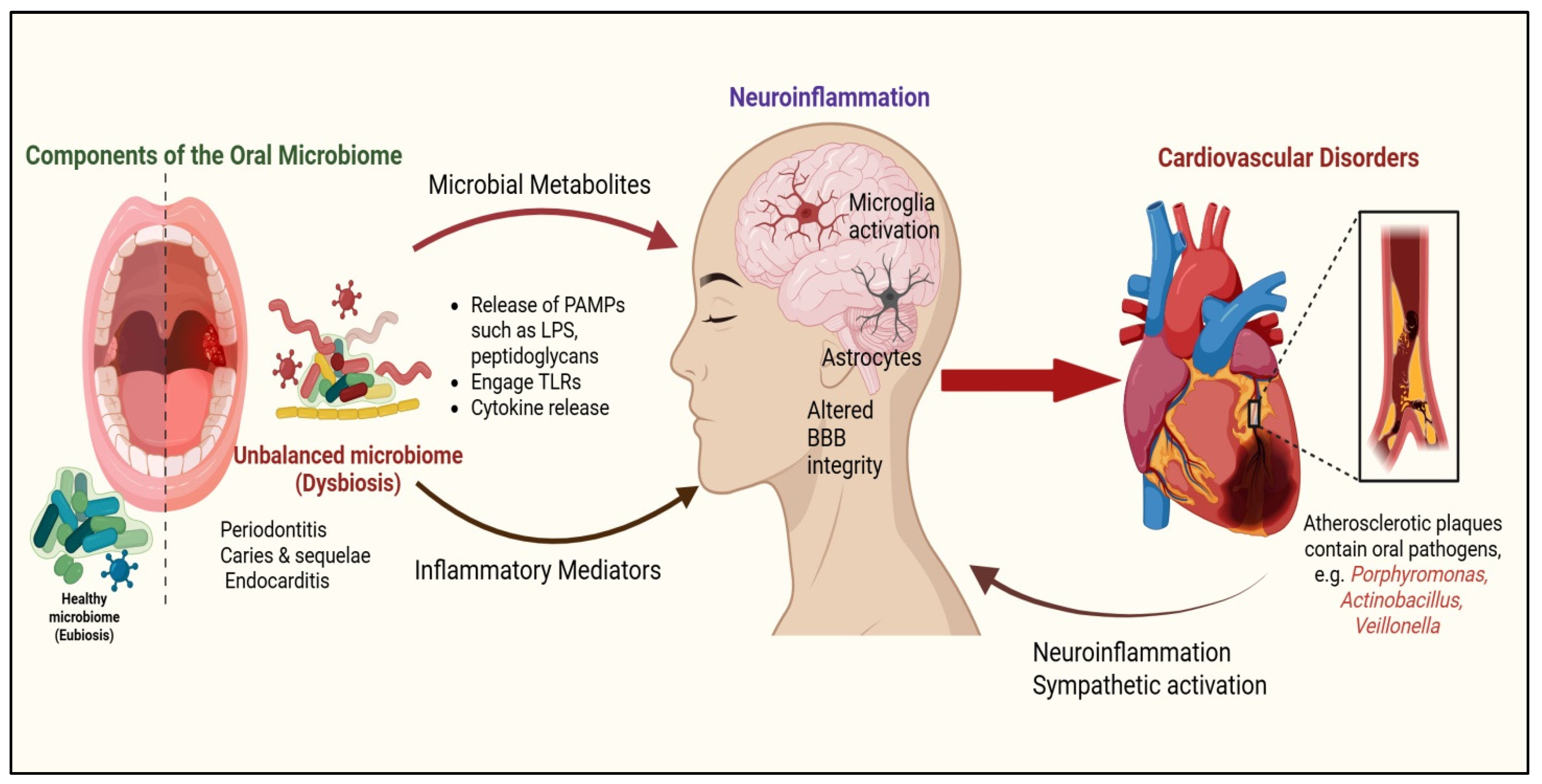
Figure 1. Proposed mechanistic link between oral microbiota dysbiosis, neuroinflammation, and CVD.
Neuroglia 06 00030 g001
Figure 2. Oral microbial ecology during different life stages in women. Hormonal changes during menstruation, pregnancy, and menopause alter the oral microbiome, with systemic implications. During menstruation, microbial diversity fluctuates while saliva and antimicrobial peptides decrease. Pregnancy shifts the microbiome toward Prevotella, Porphyromonas, and Fusobacterium, increasing the risk of gingivitis, periodontitis, adverse pregnancy outcomes, and long-term CVD. Menopause leads to dry oral mucosa, fungal overgrowth, inflammation, oxidative stress, and vascular stiffening, linking oral health to systemic aging.
Neuroglia 06 00030 g002
Table 1. Overview of female-specific factors influencing the composition and function of the oral microbiota, with potential implications for CVD risk.
Female Specific FactorsImpact on Oral MicrobiotaPotential Link to CVDReferences
Early MenarcheAltered estrogen levels affect oral microbial composition, favoring pathogenic bacteria (e.g., Porphyromonas, Prevotella, Fusobacterium)Increased systemic inflammation and endothelial dysfunction[57,58,59,60]
Polycystic Ovary Syndrome (PCOS)Elevated androgen levels lead to periodontal inflammation and increased T. denticolaChronic inflammation contributes to insulin resistance and atherosclerosis[61,62,63]
Pregnancy and MultiparityHormonal shifts enhance Prevotella and Porphyromonas growth, worsening gingival inflammationIncreased vascular stress and systemic inflammation[64,65]
Adverse Pregnancy Outcomes (e.g., Pre-eclampsia and Gestational Diabetes)Increased F. nucleatum linked to placental inflammation and poor periodontal healthHigher long-term risk of hypertension and cardiovascular dysfunction[64,66,67,68]
Menopause and Estrogen DeficiencyLoss of estrogen reduces salivary flow, leading to dysbiosis (Actinomyces increase, Lactobacillus decrease) and worsening periodontal diseaseElevated oxidative stress and pro-inflammatory cytokines contribute to arterial stiffening[69,70,71]
Hormone Replacement Therapy (HRT)Influences microbiota composition, reducing pro-inflammatory bacteria but also altering immune responseVariable impact on cardiovascular risk depending on individual metabolic profiles[72,73,74,75]
Disclaimer/Publisher’s Note: The statements, opinions and data contained in all publications are solely those of the individual author(s) and contributor(s) and not of MDPI and/or the editor(s). MDPI and/or the editor(s) disclaim responsibility for any injury to people or property resulting from any ideas, methods, instructions or products referred to in the content.