Article

The Effect of Frankincense and Myrrh on the Sealing Ability and Hardness of Glass Ionomer Cement

1
College of Dentistry, Hawler Medical University, Kurdistan Region, Iraq
2
Kurdistan Higher Council of Medical Specialties, Kurdistan Region, Iraq
*
Author to whom correspondence should be addressed.
Ceramics 2025, 8(3), 101; https://doi.org/10.3390/ceramics8030101
Submission received: 12 June 2025 / Revised: 31 July 2025 / Accepted: 4 August 2025 / Published: 6 August 2025

Abstract

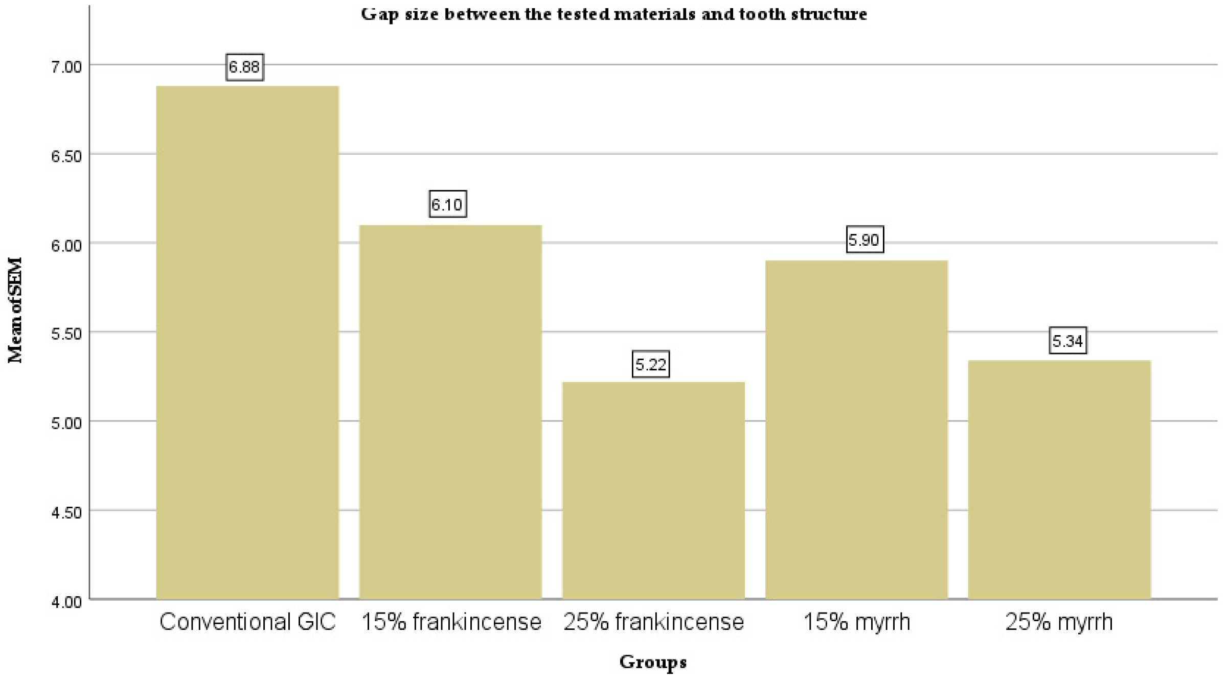
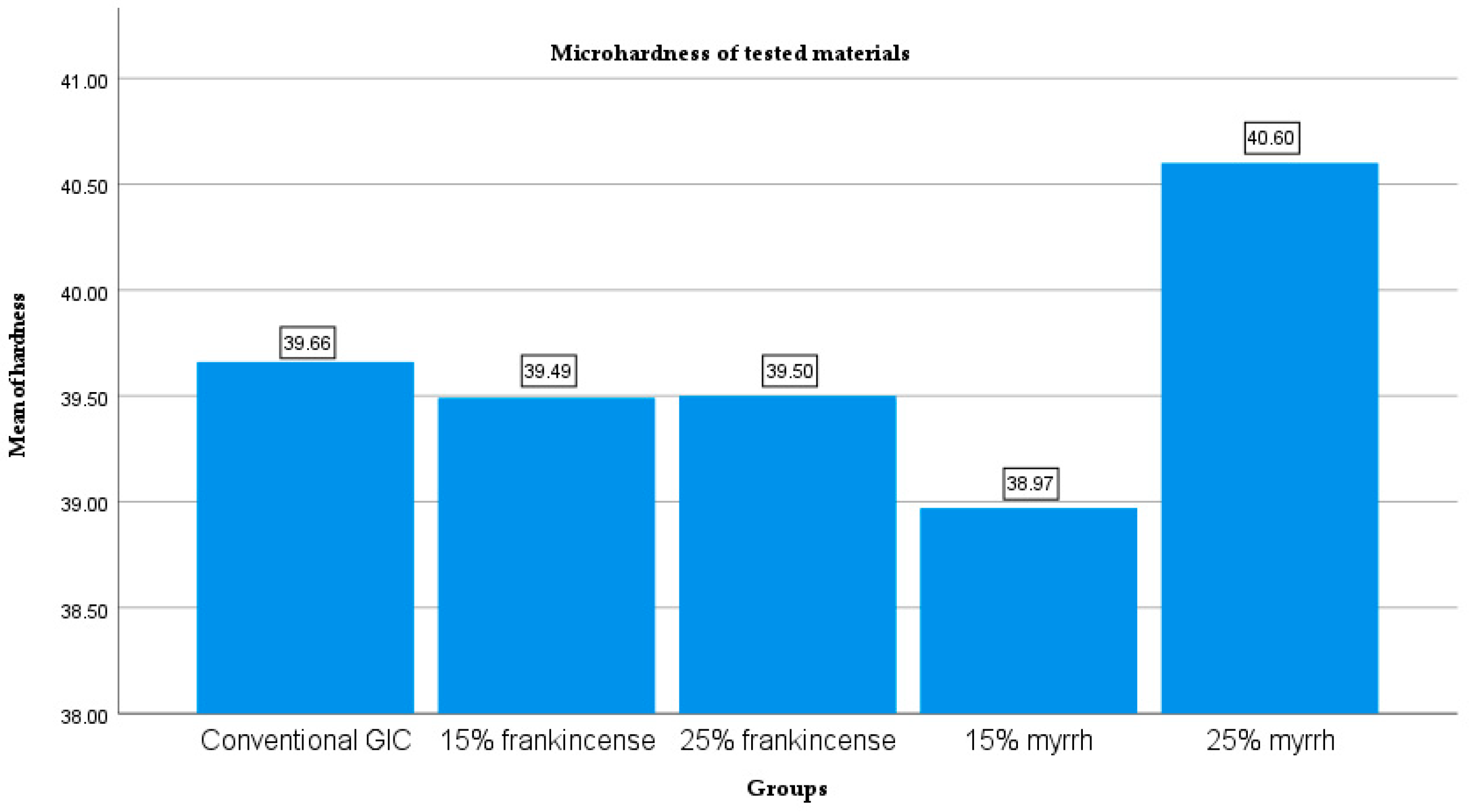
Efforts to enhance the mechanical and physicochemical properties of conventional glass ionomer cement (GIC) are ongoing. This study aimed to evaluate the effect of incorporating varying concentrations of frankincense and myrrh liquids into conventional GIC on its microhardness and sealing ability. Frankincense and myrrh liquids were prepared by dissolving 25 g of each ground resin in 50 mL of distilled water at 60 °C and allowing the solutions to stand for 8 h. Five experimental groups were evaluated: Group A (conventional GIC), Group B (15% frankincense-modified GIC), Group C (25% frankincense-modified GIC), Group D (15% myrrh-modified GIC), and Group E (25% myrrh-modified GIC). Microhardness was evaluated using a Vickers hardness tester, and sealing ability was evaluated via interfacial gap measurements using scanning electron microscopy (SEM). SEM analysis revealed that all modified GIC groups exhibited significantly smaller interfacial gap sizes (Groups B–E: 6.1, 5.22, 5.9, and 5.34 µm, respectively) compared to conventional GIC (Group A: 6.88 µm). However, there were no statistically significant differences in microhardness among the groups (p > 0.5). The incorporation of 15% and 25% concentrations of frankincense or myrrh liquids into conventional GIC significantly improved sealing ability without compromising hardness.

1. Introduction

Despite significant developments in restorative materials, the primary function of GIC is still restoration and pediatric dentistry, including restorative material in primary dentition, such as liners and bases, luting agents, pit and fissure sealants, and bonding agents for orthodontic brackets [1,2]. This is due to its unique ability to bond chemically to the tooth structure. In addition, it has biocompatibility and anti-cariogenic properties resulting from the release of fluoride, and heat compatibility with dental enamel [3]. The main composition of conventional GIC is calcium–alumino-silicate glass powder and an aqueous solution of an acrylic acid homopolymer or copolymer [4].
In spite of its many advantages, GIC also has some drawbacks, such as brittleness, low fracture toughness, poor wear resistance, short working times, and extended setting durations [3,4]. These mechanical and physicochemical properties of GIC depend on several factors, such as the composition of the powder and liquid, the size of the particle, the ratio of constituents in the cement mix, and the process of mixing. To improve its mechanical properties and clinical performance, numerous studies have attempted to modify commercially available GIC by adding inorganic or organic components to powders or liquid constituents of GIC [5,6,7,8,9,10].
Frankincense is an oleo-gum resin derived from a deep longitudinal incision in the trunks of trees belonging to the Boswellia genus [11]. Boswellia sacra gum resin is recognized to help in curing wounds, sores, ulcers, carbuncles, and hemorrhoids [12]. Furthermore, Boswellia sacra has therapeutic effects as an anti-inflammatory, antibacterial, and analgesic, and when added to bioactive glass materials, it is a promising candidate for phytotherapeutic-loaded antibacterials and the development of tissue healing and regenerative materials [13,14]. In dentistry, it was found that 10% of the Boswellia sacra water extract has a chelating potential similar to that of 17% EDTA [12]. Also, the inclusion of Frankincense liquid results in the improved hardness of GICs [3].
Myrrh, on the other hand, is a yellowish, resin-like substance derived from various species of Commiphora, primarily Commiphora molmol and Engler of the Burseraceae family, and is harvested from the tree’s phloem. It consists mainly of three components: resin (25–40%), gum (30–60%), and volatile oil (2–10%). It has wound-healing, antibacterial, and antiviral properties, making it useful as a mouthwash to treat ulcers, gingivitis, and tonsillitis. Additionally, it is as effective as sodium hypochlorite for antibacterial action against Enterococcus faecalis and Fusobacterium nucleatum when used as a root canal irrigant [15,16,17,18]. In addition, it was found that the solubility and antibacterial activity of myrrh-modified glass ionomer cement increased with the corresponding increase in myrrh concentration [15].
In this study, two natural organic resins—frankincense and myrrh—were incorporated to enhance the mechanical properties of conventional GIC. Both resins are naturally derived and possess multiple therapeutic benefits in dental applications. In the literature, only two studies were found using frankincense and myrrh for the modification of conventional GIC [3,15]. In addition, Abdallah et al. (2016) used myrrh powder rather than myrrh liquid for modification [15]. Moreover, none of these studies measure the gap size between tooth enamel/dentine and GIC with frankincense and myrrh liquids. Thus, the aims of this study were to evaluate the influence of incorporating varying proportions of liquid frankincense and liquid myrrh into GIC on its hardness and sealing ability. We also compared frankincense-added GIC and myrrh-added GIC in relation to their hardness and sealing ability.

2. Materials and Methods

2.1. Preparation of Frankincense and Myrrh Extracts

Frankincense extract was prepared by dissolving 25 g of ground frankincense resin (Dhofar, Oman). in 50 mL of distilled water at 60 °C. The solution was left to stand for approximately 8 h to allow complete dissolution. Following this, the mixture was filtered using cellulose filter paper to obtain a clear, purified solution. The same procedure was followed for the preparation of the myrrh extract, using 25 g of ground myrrh in 50 mL of distilled water, which was also maintained at 60 °C for 8 h and subsequently filtered [3].

2.2. Tooth Selection and Cavity Preparation

A total of twenty-five freshly extracted human upper and lower premolars, extracted for orthodontic reasons, were selected for this study. All teeth were free of cracks, caries, and structural defects (checked under stereo microscopes). They were cleaned using a hand-scaling instrument followed by polishing with a rubber cup and pumice. The teeth were disinfected using 0.5% chloramine solution and stored in artificial saliva at 37 °C until use [19].
Standardized Class V cavities were prepared on the buccal and lingual surfaces of each crown, with dimensions of a 3 mm width, 4 mm length, and 2 mm depth [20]. A high-speed handpiece with air–water spray and #4 spherical carbide burs (Jet, Ontario, Canada), as well as #2082 diamond burs (KG Sorensen, Barueri, SP, Brazil), were used for cavity preparation, with one bur allocated per group to maintain cutting efficiency.
The specimens were randomly divided into five groups (n = 10 cavities per group):
  • Group A (Control): Cavities restored with conventional GIC using a 1:1 powder-to-liquid ratio as per the manufacturer’s instructions.
  • Group B: Cavities restored with 15% frankincense, using a 1:1 powder-to-modified liquid ratio (17 drops of GIC liquid mixed with 3 drops of frankincense extract = modified liquid).
  • Group C: Cavities restored with 25% frankincense, using a 1:1 powder-to-modified liquid ratio (3 drops of GIC liquid mixed with 1 drop of frankincense extract = modified liquid).
  • Group D: Cavities restored with 15% myrrh, using a 1:1 powder-to-modified liquid ratio (17 drops of GIC liquid mixed with 3 drops of myrrh extract = modified liquid).
  • Group E: Cavities restored with 25% myrrh, using a 1:1 powder-to-modified liquid ratio (3 drops of GIC liquid mixed with 1 drop of myrrh extract = modified liquid).
All restorations were finished and polished using soft Lex discs (3M ESPE, St. Paul, MN, USA) and Poliglass polishing paste (TDV Dental Ltda, Pomerode, SC, Brazil).

2.3. SEM Specimen Preparation and Evaluation

The crowns were separated from the roots at the cemento-enamel junction using a diamond disc. Each crown was then sectioned buccolingually through the center of the restoration using a low-speed diamond disc (Isomet, Buehler Ltd., Bluff, IL, USA. The sectioned specimens were etched with 37% orthophosphoric acid for 5 s, rinsed with water for 15 s, and air-dried [21].
Samples were mounted on metallic stubs using silver paste and gold-coated in a vacuum using a sputtering device. They were then examined under a scanning electron microscope (Vega Easy Probe, Schiltach, Germany) at an accelerating voltage of 10–30 kV. The interfacial gap between the GIC restoration and cavity walls (enamel and dentin) was evaluated at 2000× magnification. Measurements were taken at multiple points, and the mean gap size was calculated in micrometers.

2.4. Specimen Preparation for Microhardness Testing

Disk-shaped samples were prepared using a stainless-steel mold of 8 mm diameter and 2 mm depth. The mold was positioned on a glass slab, and GIC was mixed and placed as per the group specifications (n = 10 samples per group):
  • Group A: Conventional GIC (control group).
  • Group B: GIC with 15% frankincense extract.
  • Group C: GIC with 25% frankincense extract.
  • Group D: GIC with 15% myrrh extract.
  • Group E: GIC with 25% myrrh extract.
All samples were stored in artificial saliva at 37 °C for one month to simulate aging prior to testing.

2.5. Microhardness Testing

Surface hardness was evaluated using a Vickers microhardness tester (HMV-G31 Series, Shimadzu Corporation, Kyoto, Japan) equipped with a pyramidal diamond indenter and a 50 g load. Measurements were recorded at multiple points on each sample surface, and values were expressed in kg/mm2. The data were subsequently used for statistical analysis.

2.6. Statistical Analysis

The data were first tested for normality using the Shapiro–Wilk and Kolmogorov–Smirnov tests. Interfacial gap measurements were analyzed using the Kruskal–Wallis test, followed by pairwise comparisons with the Mann–Whitney U test. Microhardness data were analyzed using one-way analysis of variance (ANOVA). Statistical analysis was performed using SPSS version 27 (IBM Corp., Armonk, NY, USA), with the significance level set at p < 0.05.

3. Results

The gap size between the tested materials and the tooth structure was measured using SEM (Figure 1). The results indicated significant differences between the groups (p < 0.001). Pairwise comparisons using the Mann–Whitney U-test revealed significant differences between most groups, except for the comparisons between 15% frankincense-modified GIC and 15% myrrh-modified GIC and 25% frankincense-modified GIC and 25% myrrh-modified GIC (p = 0.241) (Table 1 and Table 2, and Figure 2). On the other hand, a one-way ANOVA that was conducted to assess the differences in microhardness between the materials revealed no significant difference (p = 0.662) (Table 3 and Table 4, and Figure 3).

4. Discussion

The success of restorative materials largely relies on their ability to prevent recurrent caries and their possession of high mechanical strength. Conventional GIC, when utilized as a restorative material, demonstrates undesirable physical and mechanical properties, including low strength and polishing abilities, vulnerability to dehydration and moisture contamination during the initial setting phase, weak wear resistance, and a tendency to develop cracks and gaps [3,15,22]. To address some of these limitations, this study mixed conventional GIC with varying concentrations of frankincense and myrrh liquids and then measured both the microhardness and interfacial gap size between the tooth structure and GIC using a Vickers microhardness tester and SEM, respectively. Unlike previous researchers who predominantly focused on modifications to the powder component of conventional GIC [5,10], the present study explored the effect of incorporating varying concentrations of frankincense and myrrh liquids into the GIC liquid component.
SEM is widely recognized for its effectiveness in evaluating the detailed morphology of dental hard tissues and restorative materials, including surface topography, roughness, and marginal integrity at the interface between restorative materials and dental substrates such as enamel and dentin [22]. Marginal gaps are of clinical concern, as they can lead to restoration failure by permitting bacterial infiltration, fluid leakage, and subsequent material degradation [23]. A well-sealed interface is, therefore, critical for the longevity and clinical success of restorations [22,24].
The SEM results revealed that both the 15% and 25% concentrations of frankincense and myrrh groups significantly reduced the interfacial spaces between the tooth structure and GIC, with no significant difference observed between the two materials. However, a significant difference was found between both high and low concentrations, with smaller gaps observed at the 25% concentrations. According to previous studies, the gap size and marginal sealing of GIC varied when using GIC with a variety of components. In addition, incorporating resin components into GIC enhanced both its in vitro and vivo performance by improving mechanical properties and decreasing the material’s sensitivity to early fluid contamination compared to traditional GICs [23,25].
In this study, the notable decrease in the interfacial gap size in the groups of modified frankincense and myrrh could be related to the chemical resin component of the frankincense and myrrh liquid. This enables stronger bonding, better adhesion of the tooth structure and marginal sealing, limited cracking, the inhibition of demineralization, and less solubility [26,27,28]. Incorporating resins such as frankincense and myrrh could improve the flowability of GIC. This improved flow allows the material to better conform to the tooth’s surface, aiding in the filling of microvoids and minimizing the formation of gaps between the restoration and tooth structure [29]. On the other hand, both frankincense and myrrh have antibacterial properties [13,16,17], which can contribute to the reduction in the number of residual bacteria in cavities as well as the remineralization of softened dentin [30].
Microhardness, which is a measure of a material’s resistance to localized plastic deformation, serves as an indirect indicator of its structural durability and wear resistance under oral conditions [7,31,32]. In this study, no statistically significant differences in surface microhardness were observed between the modified GIC groups and the conventional GIC group (p > 0.05). These findings are consistent with previous studies. Abdallah et al. (2016) reported a similar non-significant decrease in microhardness when GIC was modified with 1% and 5% myrrh powder [15], and Khadim and Ghalib (2023) found that while 75% frankincense liquid improved microhardness, a 25% concentration mainly influenced surface roughness [3].
The slight reduction in microhardness observed in the modified groups may be due to the dilution effect of the added extracts, which do not participate in the GIC’s acid-base setting reaction. As the particles in frankincense and myrrh liquid may not react chemically with the GIC matrix, they could weaken the bulk structure and reduce resistance to indentation [5,15,33]. Conversely, the modest increase in hardness seen in the 25% myrrh group (mean = 40.60) might be attributed to the enhanced homogeneity and better dispersion of the extract within the matrix, leading to improved cross-linking, polysalt bridge formation, and load distribution [6,10,15,34]. Additionally, the mechanical performance of GIC is influenced by several factors, including particle size, interfacial bonding between glass fillers, the polymer matrix, and the presence of voids or defects in the set material [9,35,36].

5. Conclusions

In summary, the incorporation of frankincense and myrrh extracts into conventional GIC significantly decreases the gap size and improves the marginal adaptation of modified GIC with tooth structure without significantly compromising surface hardness. These findings support the potential of natural resin extracts to act as bioactive modifiers for enhancing the clinical performance of GIC restorations. Further studies on the other physical, mechanical properties, and antibacterial characteristics of this modified material are advocated.

Author Contributions

Conceptualization, H.H. and N.A.; methodology, N.A.; validation, M.S., H.H., N.A., and D.K.B.; writing—original draft preparation, M.S. and H.H. writing—review and editing, M.S. and D.K.B.; supervision, D.K.B. All authors have read and agreed to the published version of the manuscript.

Funding

This research received no external funding.

Data Availability Statement

No new data were created or analyzed in this study.

Conflicts of Interest

The authors declare no conflicts of interest.

Abbreviations

The following abbreviations are used in this manuscript:
GICGlass ionomer cement
gGram
mLMilliliter
SEMScanning electron microscopy
µmMicrometers

References

  1. Garcia-Contreras, R.; Vilchis, R.J.; Scougall Contreras-Bulnes, R.; Sakagami, H.; Morales-Luckie, R.A.; Nakajima, H. Mechanical, antibacterial and bond strength properties of nano-titanium-enriched glass ionomer cement. J. Appl. Oral Sci. 2015, 23, 321–328. [Google Scholar] [CrossRef] [PubMed] [PubMed Central]
  2. Sidhu, S.K.; Nicholson, J.W. A Review of Glass-Ionomer Cements for Clinical Dentistry. J. Funct. Biomater. 2016, 7, 16. [Google Scholar] [CrossRef] [PubMed]
  3. Khadim, H.J.; Ghalib, L. The effect of frankincense chewing gum on the surface roughness and hardness of GIC. J. Eng. Sustain. Dev. 2023, 148–156. [Google Scholar] [CrossRef]
  4. Upadhya, N.; Kishore, G. Glass ionomer cement: The different generations. Trends Biomater. Artif. Organs. 2005, 18, 158–165. [Google Scholar]
  5. Prentice, L.H.; Tyas, M.J.; Burrow, M.F. The effect of ytterbium fluoride and barium sulphate nanoparticles on the reactivity and strength of a glass-ionomer cement. Dent. Mater. 2006, 22, 746–751. [Google Scholar] [CrossRef]
  6. Moshaverinia, A.; Ansari, S.; Moshaverinia, M.; Roohpour, N.; Darr, J.A.; Rehman, I. Effects of incorporation of hydroxyapatite and fluoroapatite nanobioceramics into conventional glass ionomer cements (GIC). Acta Biomater. 2008, 4, 432–440. [Google Scholar] [CrossRef]
  7. Silva, R.M.; Pereira, F.V.; Mota, F.A.; Watanabe, E.; Soares, S.M.; Santos, M.H. Dental glass ionomer cement reinforced by cellulose microfibres and cellulose nanocrystals. Mater. Sci. Eng. 2016, 58, 389–395. [Google Scholar] [CrossRef]
  8. Bilić-Prcić, M.; Rajić, V.B.; Ivanišević, A.; Pilipović, A.; Gurgan, S.; Miletić, I. Mechanical Properties of Glass Ionomer Cements after Incorporation of Marine Derived Hydroxyapatite. Materials 2020, 13, 3542. [Google Scholar] [CrossRef] [PubMed]
  9. Chen, J.; Zhao, Q.; Peng, J.; Yang, X.; Yu, D.; Zhao, W. Antibacterial and mechanical properties of reduced graphene-silver nanoparticle nanocomposite modified glass ionomer cements. J. Dent. 2020, 96, 103332. [Google Scholar] [CrossRef] [PubMed]
  10. Rini, A.D.K.; Juwita, F.T.; Bagjana, R.W.; Octivany, S.; Purnama, R.B.; Rijal, M.S.; Anwar, A.M.; Purwasasmita, B.S.; Asri, L.A.T. Improving the mechanical properties of glass ionomer cement with nanocrystalline cellulose from rice husk. J. Biomed. Mater. Res. B Appl. Biomater. 2024, 112, e35472. [Google Scholar] [CrossRef]
  11. Singer, L.; Bourauel, C. Herbalism and glass-based materials in dentistry: Review of the current state of the art. J. Mater. Sci. Mater. Med. 2023, 34, 60. [Google Scholar] [CrossRef] [PubMed]
  12. Ahmad, N.A.; Elsheikh, H.M.; Elhousiny, M.A. Effect of Using Boswellia Sacra Extract as Final Irrigant on Removal of Smear Layer. Al-Azhar J. Dent. 2021, 8, 55–63. [Google Scholar] [CrossRef]
  13. Al-Hazzaa, A.M.; Badr, A.E.; Kataia, E.M. Physical properties of root canal sealers. Mansoura J. Dent. 2020, 7, 62–70. [Google Scholar] [CrossRef]
  14. Ilyas, K.; Singer, L.; Akhtar, M.A.; Bourauel, C.P.; Boccaccini, A.R. Boswellia sacra extract-loaded mesoporous bioactive glass nano particles: Synthesis and biological effects. Pharmaceutics 2022, 14, 126. [Google Scholar] [CrossRef]
  15. Abdallah, R.M.; Abdelghany, A.M.; Aref, N.S. Myrrh Addition to a conventional Glass-Ionomer Cement: Influence on Physical and Antibacterial Properties. Egypt. Dent. J. 2016, 62, 1491. [Google Scholar] [CrossRef]
  16. Al-Madi, E.M.; Almohaimede, A.A.; Al-Obaida, M.I.; Awaad, A.S. Comparison of the Antibacterial Efficacy of Commiphora molmol and Sodium Hypochlorite as Root Canal Irrigants against Enterococcus faecalis and Fusobacterium nucleatum. Evid.-Based Complement. Altern. Med. 2019, 6916795. [Google Scholar] [CrossRef]
  17. Kim, J.W.; Park, S.; Sung, Y.W.; Song, H.J.; Yang, S.W.; Han, J.; Jo, J.W.; Lee, I.S.; Lee, S.H.; Choi, Y.K. Evaluation of antibacterial and antiviral compounds from Commiphora myrrha (T.Nees) engl. resin and their promising application with biochar. Appl. Sci. 2023, 13, 10549. [Google Scholar] [CrossRef]
  18. Refaey, M.S.; Abosalem, E.F.; Yasser El-Basyouni, R.; Elsheriri, S.E.; Elbehary, S.H.; Fayed, M.A. Exploring the therapeutic potential of medicinal plants and their active principles in dental care: A comprehensive review. Heliyon 2024, 10, e37641. [Google Scholar] [CrossRef]
  19. Coelho, A.; Amaro, I.; Rascão, B.; Marcelino, I.; Paula, A.; Saraiva, J.; Spagnuolo, G.; Ferreira, M.M.; Marto, C.M.; Carrilho, E. Effect of cavity disinfectants on dentin bond strength and clinical success of composite restorations—A systematic review of In Vitro, In Situ and Clinical Studies. Int. J. Mol. Sci. 2021, 22, 353. [Google Scholar] [CrossRef] [PubMed]
  20. Panahandeh, N.; Sheikholeslamian, M.; Farzaneh, H. Microleakage in Class V Cavities Restored with Sandwich Technique: Self-Etch versus Total-Etch Bonding Systems. J. Dent. Sch. 2015, 33, 74–79. [Google Scholar]
  21. Kharouf, N.; Reitzer, F.; Ashi, T.; Daras, N.; Haikel, Y.; Mancino, D. Effectiveness of etching with phosphoric acid when associated with rubbing technique. J. Stomatol. 2021, 74, 16–21. [Google Scholar] [CrossRef]
  22. Samy, F.M.; El-Kholany, N.R.; Hamama, H.H. Micromorphological Analysis of Different Bioactive Restorative Materials/Dentin interface: A Comparative In Vitro Study. Egypt. Dent. J. 2024, 70, 3909–3916. [Google Scholar] [CrossRef]
  23. Ibrahim, B.; Elkholany, N.; Enab, T.; Zaghloul, N. Marginal adaptation of self adhesive bulk-fill resin composite vs conventional resin composite and resin modified glass- ionomer: A comparative study. Egypt. Dent. J. 2024, 70, 3879–3886. [Google Scholar] [CrossRef]
  24. Elgezawi, M.; Haridy, R.; Abdalla, M.A.; Heck, K.; Draenert, M.; Kaisarly, D. Current Strategies to Control Recurrent and Residual Caries with Resin Composite Restorations: Operator- and Material-Related Factors. J. Clin. Med. 2022, 11, 6591. [Google Scholar] [CrossRef]
  25. Pontes, D.G.; Guedes-Neto, M.V.; Cabral, M.F.; Cohen-Carneiro, F. Microleakage evaluation of class V restorations with conventional and resin-modified glass ionomer cements. Oral Health Dent. Manag. 2014, 13, 642–646. [Google Scholar]
  26. Ching, H.S.; Luddin, N.; Kannan, T.P.; AbRahman, I.; Abdul Ghani, N.R.N. Modification of glass ionomer cements on their physical-mechanical and antimicrobial properties. J. Esthet. Restor. Dent. 2018, 30, 557–571. [Google Scholar] [CrossRef]
  27. Ruengrungsom, C.; Burrow, M.F.; Parashos, P.; Palamara, J.E.A. Comprehensive characterisation of flexural mechanical properties and a newclassification for porosity of 11 contemporary ion-leaching dental restorative materials. J. Mech. Behav. Biomed. Mater. 2021, 121, 104615. [Google Scholar] [CrossRef]
  28. Santos, M.J.M.C.; Leon, L.; Siddique, I.; Butler, S. Retrospective Clinical Evaluation of RMGIC/GIC Class V Restorations. Dent. J. 2023, 11, 225. [Google Scholar] [CrossRef] [PubMed]
  29. Baroudi, K.; Mahmoud, S. Improving Composite Resin Performance Through Decreasing its Viscosity by Different Methods. Open Dent. J. 2015, 9, 235–242. [Google Scholar] [CrossRef] [PubMed]
  30. Takahashi, Y.; Imazato, S.; Kaneshiro, A.V.; Ebisu, S.; Frencken, J.E.; Tay, F.R. Antibacterial effects and physical properties of glass-ionomer cements containing chlorhexidine for the ART approach. Dent. Mater. 2006, 22, 647–652. [Google Scholar] [CrossRef]
  31. Nicholson, J.W.; Sidhu, S.K.; Czarnecka, B. Enhancing the mechanical properties of glass-ionomer dental cements: A review. Materials 2020, 13, 2510. [Google Scholar] [CrossRef]
  32. Shivanna, S.; Roshan, S.; Sameera, S.; Sivakumar, M.; Ravi, M.; Ram, L.S. Comparative evaluation of hardness in ceramic reinforced, resin modified glass ionomer cement and conventional glass ionomer cement. J. Pharm. Bioall. Sci. 2025, 17, S1916–S1919. [Google Scholar] [CrossRef]
  33. Saadat, M.; Moradian, M.; Mirshekari, B. Evaluation of the surface hardness and roughness of a resin-modified glass ionomer cement containing bacterial cellulose nanocrystals. Int. J. Dent. 2021, 8231473. [Google Scholar] [CrossRef] [PubMed]
  34. Siddiqui, A.Z.; Sultana, N.; Mirani, Z.A.; Alkhureif, A.A.; Rehan, F.; Siddiqui, I.A. Enhancing the antibacterial and surface hardness of glass ionomer cement modified with Salvadora persica and Chlorhexidine: An in vitro study. Pak. J. Med. Sci. 2024, 40, 1808–1812. [Google Scholar] [CrossRef] [PubMed]
  35. Xie, D.; Brantley, W.A.; Culbertson, B.M.; Wang, G. Mechanical properties and microstructures of glass-ionomer cements. Dent. Mater. 2000, 16, 129–138. [Google Scholar] [CrossRef] [PubMed]
  36. Sharafeddin, F.; Shirani, M.M.; Jowkar, Z. Assessing the Impact of Nano-Graphene Oxide Addition on Surface Microhardness and Roughness of Glass Ionomer Cements: A Laboratory Study. Int. J. Dent. 2024, 5597367. [Google Scholar] [CrossRef]
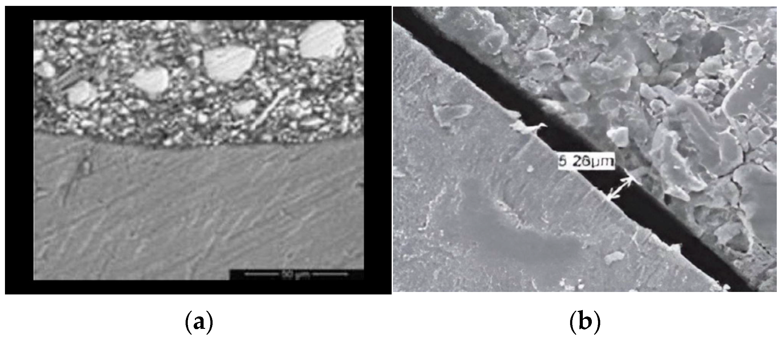
Figure 1. (a) SEM image showing the clear interfacial gap line between the tested material and tooth structure at 2000×; (b) the gap size measurement between 25% frankincense-modified GIC and tooth structure in (µm).
Ceramics 08 00101 g001
Figure 2. Mean value of the gap size between the tested materials and tooth structure.
Ceramics 08 00101 g002
Figure 3. The mean value of the microhardness of tested materials.
Ceramics 08 00101 g003
Table 1. Descriptive statistics of the gap size between the tested materials and tooth structure.
GroupsNMean (µm)Std. DeviationStd. Error95% Confidence Interval for MeanMinimumMaximum
LowerUpper
Conventional GIC106.88±0.370.126.627.146.237.44
15% frankincense106.1±0.410.135.806.405.166.73
25% frankincense105.22±0.290.095.015.434.915.91
15% myrrh105.9±0.460.145.576.234.916.52
25% myrrh105.34±0.260.085.165.524.995.81
Table 2. Mann–Whitney U-test results (p-value) for the difference in the gap size between the tested materials and tooth structure using SEM. The mean difference is significant at the p < 0.05 level.
GroupsConventional GIC15% Frankincense25% Frankincense15% Myrrh25% Myrrh
Conventional GIC-<0.001<0.001<0.001<0.001
15% frankincense<0.001-<0.0010.2410.001
25% frankincense<0.001<0.001-<0.010.241
15% myrrh<0.0010.241<0.01-<0.01
25% myrrh<0.0010.0010.241<0.01-
Table 3. Descriptive statistics of microhardness of tested materials.
GroupsNMeanStd. DeviationStd. Error95% Confidence Interval for MeanMinimumMaximum
Lower BoundUpper Bound
Conventional GIC1039.66±1.980.6338.2441.0837.1043.30
15% frankincense1039.49±2.950.9337.3841.6034.1044.10
25% frankincense1039.50±2.830.8937.4841.5234.1044.40
15% myrrh1038.97±1.870.5937.6340.3136.3042.10
25% myrrh1040.60±2.280.7238.9742.2337.2044.20
Table 4. One way ANOVA (p-value) for the difference in the microhardness between tested materials. The mean difference is significant at the p < 0.05 level.
Sum of SquaresDfMean SquareFSig.
Between Groups14.12943.5320.6030.662
Within Groups263.494455.855
Total277.62349
Disclaimer/Publisher’s Note: The statements, opinions and data contained in all publications are solely those of the individual author(s) and contributor(s) and not of MDPI and/or the editor(s). MDPI and/or the editor(s) disclaim responsibility for any injury to people or property resulting from any ideas, methods, instructions or products referred to in the content.