Article

Pretreatment of Hybrid Ceramics Using Ho: YAG, Low-Level Laser Therapy Activated Malachite Green, and Non-Thermal Plasma on Surface Roughness, Bond Strength, and Color Change, SEM and EDX Analysis

by
Fahad Alkhudhairy
* and
Yasser F. AlFawaz
Restorative Dental Sciences Department, College of Dentistry, King Saud University, Riyadh 11421, Saudi Arabia
*
Author to whom correspondence should be addressed.
Ceramics 2024, 7(3), 944-957; https://doi.org/10.3390/ceramics7030061
Submission received: 27 May 2024 / Revised: 30 June 2024 / Accepted: 4 July 2024 / Published: 9 July 2024

Abstract

:
The study aimed to assess the effects of different surface conditionings on hybrid ceramics (HBC). Hydrofluoric acid was combined with a silane (HFA+S), low-level laser therapy activated Malachite green (LLLT-MG), Ho: YAG laser, and non-thermal plasma (NTP) as surface conditioning methods for HBC. Eighty-four HBC discs were prepared and divided into four groups according to surface conditioning methods. The total number of samples (n = 21) for each group was further split into two for the non-thermocycling and thermocycling subgroups. After surface treatment, all samples were examined to study the effect of color change and surface roughness. The shear bond strength (SBS) test of HBC was performed on thermo-cycled samples. Statistical analysis using ANOVA with Tukey post hoc was performed to observe any significant difference among tested groups, p > 0.05. The HFA+S and Ho: YAG surface-treated samples showed higher SBS than other surface-treated samples due to higher surface roughness. All surface conditioning methods, except NTP, induced noticeable color change, making them less suitable for aesthetical purposes in clinical settings. Overall, surface conditioning methods are critical in affecting shear bond strength through surface roughness and color change.

1. Introduction

Hybrid ceramics, also referred to as resin matrix ceramics (RMCs) for indirect restorations, have gained popularity recently. They are favored for their effective blend of aesthetic appeal, durability, and biocompatibility [1]. Bonding hybrid ceramics (HBC) to human teeth can pose challenges, often resulting in reduced shear bond strength (SBS) to the tooth substrate. Consequently, one essential step in the bonding process of dental ceramics to human teeth involves altering the surface texture of the ceramics [2,3]. This modification primarily aims to enhance the micromechanical retention of the cement by creating a rougher surface texture. Two main methods, chemical and mechanical, are commonly employed to improve both the surface roughness (Ra) and SBS of hybrid ceramics to the dental substrate [3,4]. Additionally, there are limited data available on how the optical properties of cemented ceramics evolve following the application of various surface conditioning techniques [5,6,7].
Among the different chemicals employed, hydrofluoric acid (HFA) combined with a silane coupling agent (S) is regarded as the gold standard for enhancing both the adhesion strength and surface roughness (Ra) of resin matrix ceramics (RMCs) [8]. Alsunbul and colleagues have noted that of the various pretreatment methods used to improve Ra and bond strength, the sequence of applying HFA followed by silane yielded the most effective and satisfactory results [9]. However, researchers have reported that HFA is corrosive and possesses a propensity for human tissue harm [10]. Therefore, scholars are trying to find better alternatives that positively affect the mechanical properties without causing any damage to the human tissues.
Lasers, photodynamic treatment (PDT), and low-level laser therapy (LLLT) are non-invasive techniques increasingly used in various dental procedures. PDT involves the use of a photoactive dye and a specific light wavelength to generate reactive oxygen species (ROS), which effectively eliminate bacterial cells [11,12]. In the past, indexed literature has identified that when LLLT is employed as a pre-cementation conditioning protocol, it displays a positive influence on the adhesion strength of ceramic to the resin cement [13]. Among the several photosensitizers (PS) employed, Malachite green (MG), also known as brilliant green, has piqued the curiosity of academicians and researchers [14,15]. The cationic dye absorbs extremely well in the red area of the visible spectrum [16]. Similarly, Ho: YAG (Holmium: yttrium aluminum garnet crystal) or holmium laser has been studied extensively [17,18].
The laser, emitting light in the near-infrared region, is widely used in surgical applications across various medical and dental fields. It has shown promising results in conditioning the dentin surface. However, the impact of LLLT-activated malachite green (LLLT-MG) and Holmium: yttrium-aluminum-garnet (Ho: YAG) laser as surface conditioners on color stability (∆E), Ra, and SBS of HBC to resin cement remains unexplored and warrants further investigation.
Non-thermal plasma (NTP) technology offers a precise and consistent approach to improving the surface characteristics of several indirect restorative materials [19]. NTP, composed of electrons, neutral molecules, and charged particles, modifies the surface of the material by forming functional groups while maintaining its mechanical properties [20]. The application of this treatment is recognized for its ability to enhance the surface energy of ceramics and facilitate chemical interactions, thereby reinforcing the adhesion with resin cement [19]. Although NTP has several benefits, there is yet little evidence of its effect on ∆E, Ra, and SBS of HBC.
The present study aimed to examine ∆E and Ra values achieved through the latest pretreatment techniques; NTP, LLLT-MG, and Ho: YAG laser, applied to HBC, would be similar to those obtained with conventional control methods. Furthermore, it was postulated that SBS of HBC adhered to resin cement utilizing these contemporary conditioning approaches would not exhibit a substantial disparity compared to that achieved with HFA+S. The objective of this study is to investigate the impact of different conditioning techniques on the ∆E, Ra, and SBS of RMCs.

2. Materials and Methods

2.1. CAD-CAM Disc Preparation

The current study followed all the guidelines laid out in the checklist for reporting in vitro study (CRIS) guidelines. Utilizing a diamond cutting saw (Isomet, Buehler, Düsseldorf, North Rhine, Germany) in a water-cooled environment, eighty-four plates measuring 10 mm in diameter and 2 mm in thickness were cut from the Vita Enamic CAD-CAM blocks. The next step was to fix these plates in self-cure acrylic resin within cylindrical plastic molds. To achieve a uniform surface finish, the plates were ground using silicon carbide paper with grits of 600, 800, and 1200 (Agar Scientific, Stansted, Essex, UK). After that, the discs were submerged in 96% isopropanol (Oil Chem Manufacturing Company, Gujrat, India) for three minutes and air-dried. The discs were then classified into four categories according to the surface conditioning methods employed (n = 21). Five samples from each group were assessed for Ra, five samples from each group were assessed for color stability, one sample from each group was assessed for surface characteristics of ceramics after conditioning, and ten samples from each group underwent aging-only bonding and were tested for SBS.
Group 1: Pretreatment using HFA gel + S.
The HBC discs were conditioned using a 9.6% HFA gel (Yellow Porcelain Etch, Cerkamed, Dehli, India) for 1 min. After the conditioning process, the samples were washed with a forceful water jet for 10 s to remove any residual acid. All the discs were then dried by exposing them to air, allowing any remaining moisture to evaporate gradually. This was followed by Silane (S) coupling agent (Clearfil Porcelain Bond Activator and Clearfil SE Bond Primer, Kuraray Medical Inc., Okayama, Japan) application for 1 min using an applicator brush [21].
Group 2: Pretreatment using LLLT-activated MG.
The HBC discs were treated with a 0.01% w/v solution of MG for 5 min, which is known as the pre-irradiation period. Next, the surface was exposed to laser irradiation for around 3 min, with energy levels of 5.4 J/cm2 [13].
Group 3: Pretreatment using Ho: YAG laser.
A Ho: YAG laser (StoneLight Holmium Laser System, AMS Inc., Minnetonka, MN, USA) was employed to etch RMC plates along with the assistance of water cooling for 20 s. The laser energy used in pulse mode was 0.5 J with a frequency of 8 Hz, power output of 6 W, and a wavelength of 2090 nm1s [17].
Group 4: Pretreatment using NTP.
The specimens were exposed to NTP using a plasma generating (Neoplas Greifswald, Berlin, Germany) with argon (Ar) gas. Operating parameters for the device were 3 kVpp, 4.80 mA, and a flow rate of 5.0 slm. The specimens were subjected to NTP treatment for 1 min at a height of 10 mm above the conditioning surface [22].

2.2. Measurement of Ra

Using a non-contact 3D surface profilometry system (Contour GT-K 3D Optical microscope, Bruker, Tucson, AZ, USA), five samples from each cohort underwent Ra evaluation. This method employs vertical scan interferometry along with a Gaussian regression filter, which is highly efficient in measuring objects with different surface topography and pixel height differences greater than 135 nm. The test parameters included a Michelson lens magnification of 5×, a field of view measuring 1  mm  ×  1  mm, a thresholding value set at 4, and a scan speed of 1×. The device’s settings were controlled using Vision 64 (Control and Analysis software Bruker Tucson, AZ, USA). The samples were carefully positioned on the stage of the optical microscope using a jig and precisely adjusted until a clear image was visible on the monitor. The surface of each sample was scanned three times. The three measurements were averaged to calculate the Ra surface roughness value [23,24].

2.3. Assessment of Color Stability

Five samples from each group were analyzed for color stability measurements using a spectrophotometer device (Vita Easyshade Advance 4.0, Vita Zahnfabrik, Germany) before and after surface conditioning under conventional daylight conditions, specifically D65 illumination. The color of each item was quantified using the Commission Internationale de l’Eclairage (CIE) system, which assigns numerical values to several color attributes. These attributes include b* (representing the blue-yellow chromatic coordinate), L* (representing lightness, with 100 indicating white and 0 indicating black), and a* (representing the red-green chromatic coordinate). The experimental setup involved positioning the samples at 4 mm from each other while ensuring that they were oriented at a 45-degree angle relative to the tip of the spectrophotometer. During the spectrophotometric measurement process, a small quantity of distilled water was introduced to improve optical contact and minimize light attenuation at the edges of the disc. A section measuring 2 mm in thickness was taken from the mid-labial region of each disc. Before each measurement, the spectrophotometer underwent recalibration using a calibration plate. Following the completion of the surface conditioning process, an evaluation was conducted to determine any color changes exhibited by the samples. This assessment was carried out using the standardized method mentioned earlier [25,26].
∆E = [(∆L*) 2 + (∆a*) 2 + (∆b*) 2] 1/2
∆E value > 1.2 was considered perceptible, and ∆E value > 2.7 was clinically unacceptable.

2.4. Scanning Electron Microscope (SEM)

To evaluate the surface characteristics of RMCs, one specimen from each group underwent SEM (Zeiss Leo 1430, Carl Zeiss, Oberkochen, Germany) analysis. The samples were dried in the air and then attached to copper stubs. They were then coated with a thin layer of gold using a sputter coating evaporator (SPI-Sputter Coater, IL, USA) and analyzed under a scanning electron microscope (SEM) (JEOL.JSM.6510LV, Tokyo, Japan) operating at a magnification of ×3000 [27,28].
Resin cement application.
An Ultradent jig (Ultradent Products Inc., South Jordan, UT, USA) measuring 2 mm in diameter and 3 mm in height was used to bond a dual-cure cement system (Panavia V5, Kuraray; Tokyo, Japan) to the HBC discs, ensuring standardization. The resin cement was then subjected to photopolymerization using LED curing light t (BA Optima 10 Boses 20, BA International Ltd., Manchester, UK) at a power density of 1000–1200 mW/cm2 for 20 s. Afterward, the specimens were placed in distilled water at a temperature of 37 °C for 24 h [4].

2.5. Artificial Aging

Ten samples from each group were subjected to thermocycling (Robota, Alexandria, Egypt) consisting of 6000 cycles, with temperatures ranging from 5 °C to 55 °C. The dwell period and transfer time for each cycle were set at 30 s and 10 s, respectively.

2.6. Shear Adhesion Test between HBC Discs to Resin Cement

The SBS between HBC and resin cement was determined using a universal testing machine (UTM) (Lloyds, LF, plus, Ametek Inc., London, Great Britain, UK) (n = 10 each). The testing was conducted at a crosshead speed of 1 mm/min and a force of 2.5 KN. The Instron machine’s jaws were moving in a counter-direction and constant motion until bond failure occurred. The measurement of SBS was conducted in Megapascal (MPa).

2.7. Evaluation of the Nature of Bond Failure under a Stereomicroscope

Following shear bond testing, the samples were analyzed under a stereomicroscope (Wild M3B, Heerbrugg, Switzerland) at a magnification of 40× to determine the kind of bond failure. The bond failures were classified into three categories: (1) Cohesive failure occurring within the adhesive layer itself rather than at the interface between the adhesive and the tooth or restorative material; (2) Adhesive failure where the bond fails at the interface between the adhesive and the tooth structure or between the adhesive and the restorative material; (3) Admixed failure, which is a combination of both failure types.

2.8. Statistical Analysis

For analysis (SPSS version 19, Inc., Chicago, IL, USA) was expended. The mean and standard deviation (SD) of ∆E, Ra, and SBS findings were examined using a one-way analysis of variance (ANOVA). Tukey post hoc test was used to perform multiple comparisons among different tested groups p ˂ 0.05.

3. Results

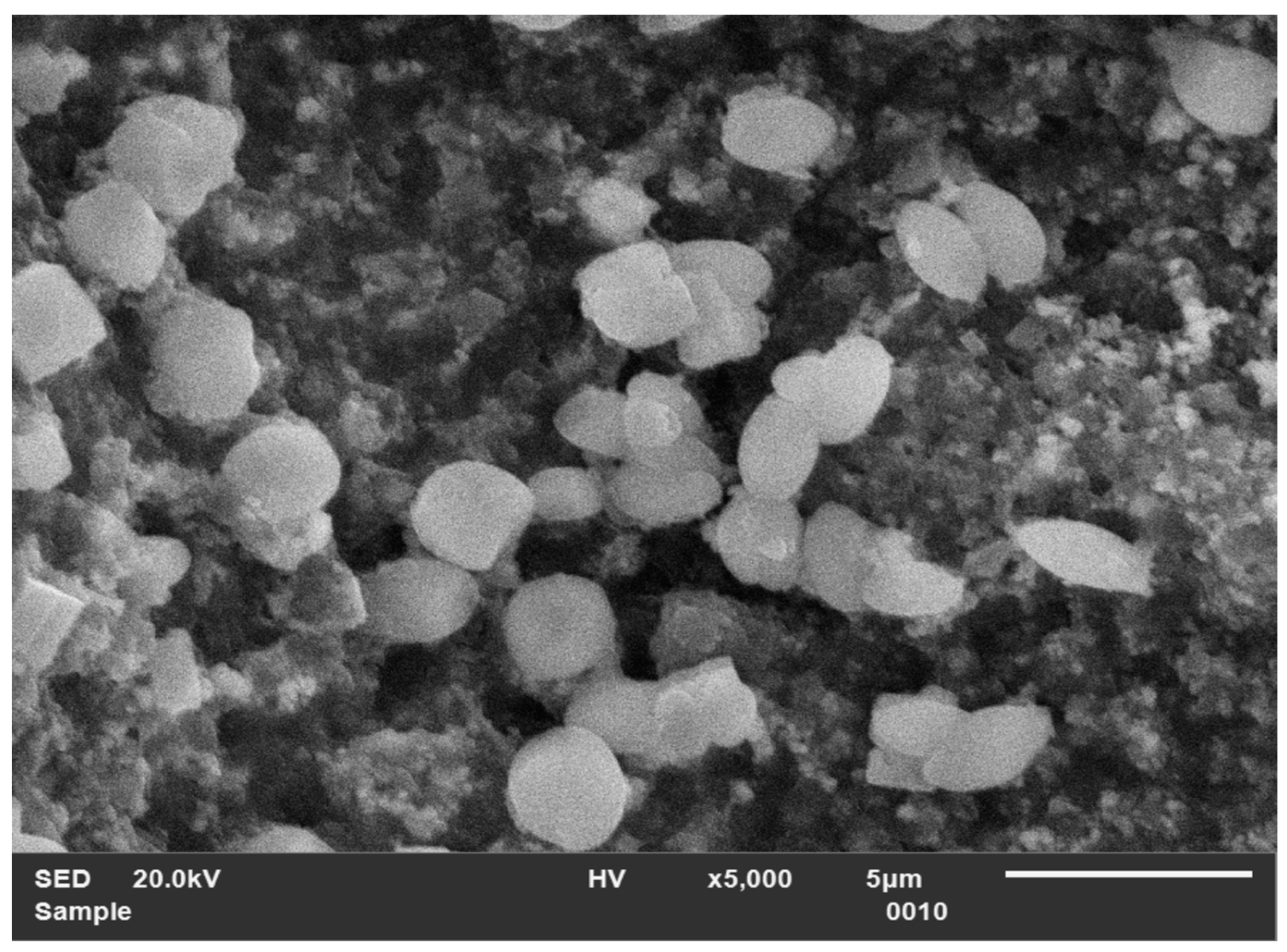
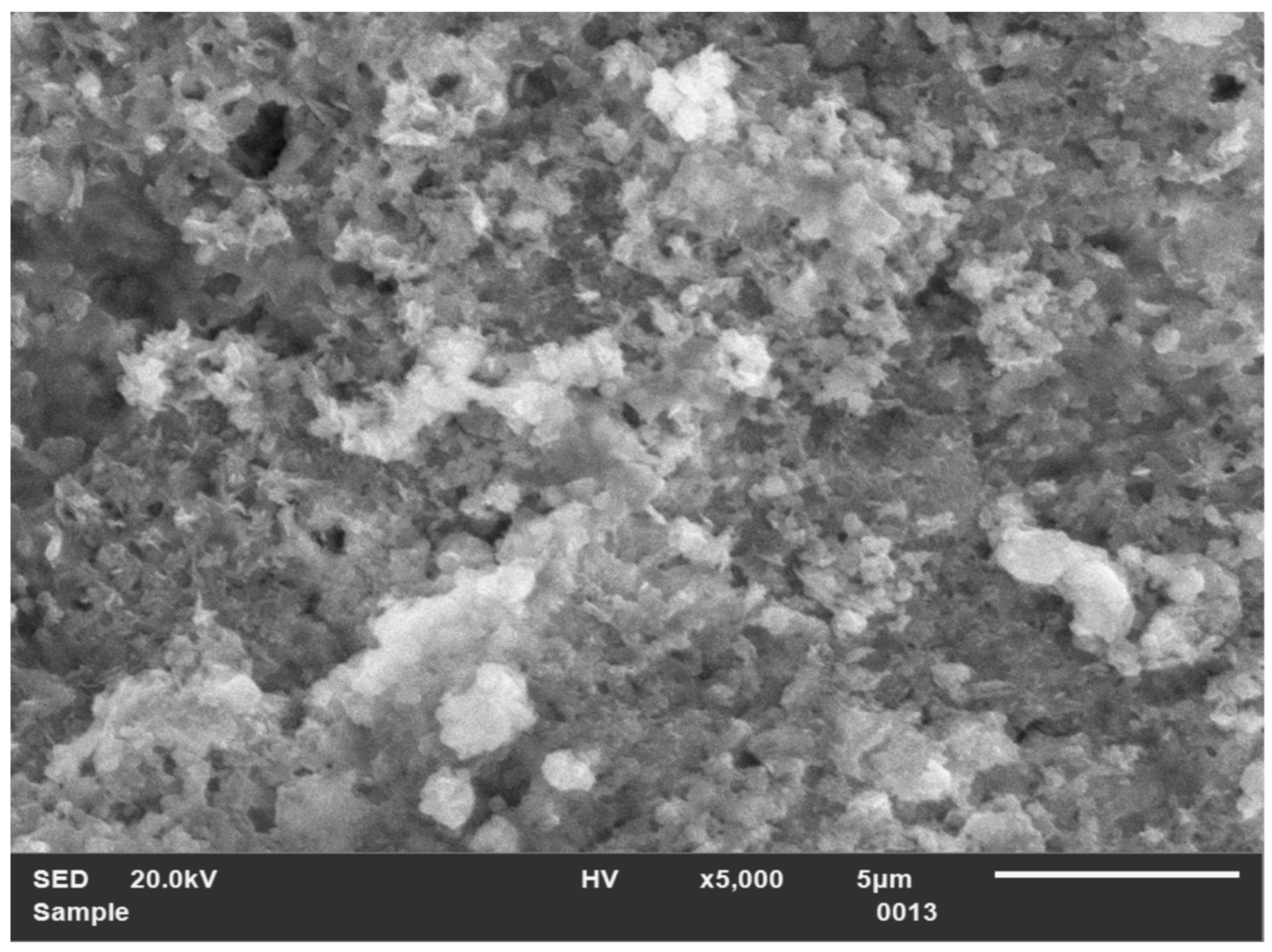
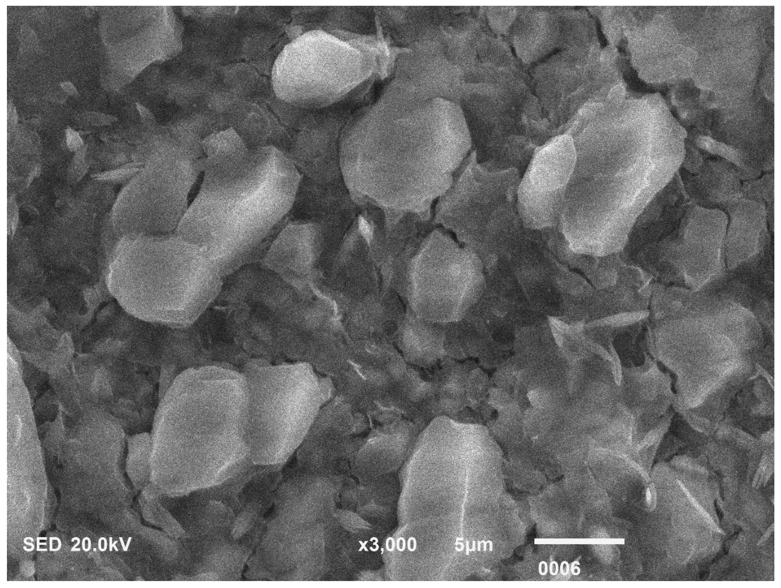
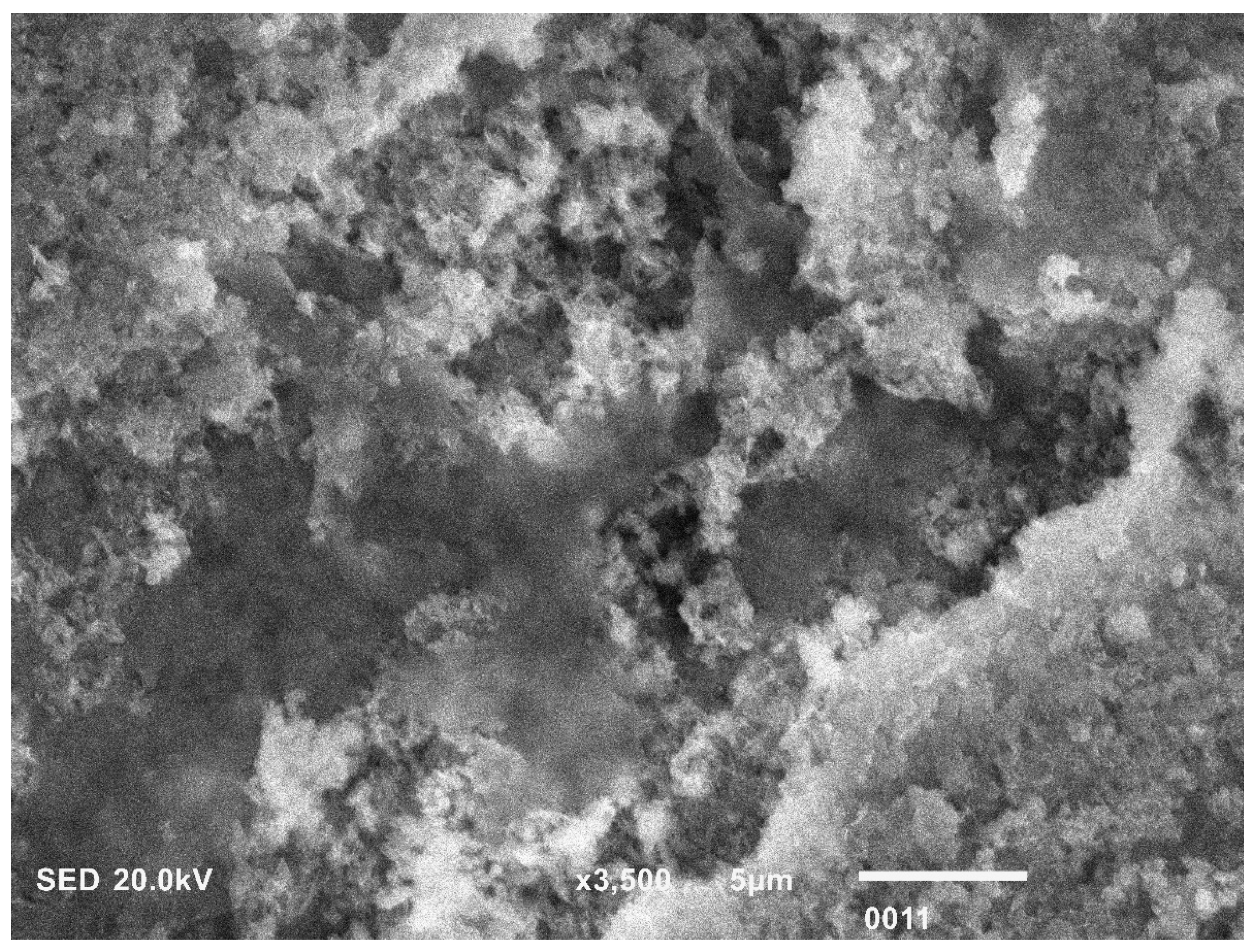
The disk was cut from Vita Enamic CAD-CAM blocks, measuring 10 mm in diameter and 2 mm in thickness (Figure 1A). The SEM image illustrated the typical structure of hybrid ceramic before surface treatment, displaying an even, homogeneous surface with fine particles (×1500) (Figure 1B). EDX analysis shows a varied elemental distribution within the hybrid ceramics (Figure 1C). Post HFA+S treatment of hybrid ceramics, the micrograph reveals surface porosity and matrix dissolution. The surface exhibits uneven particle sizes with visible gaps between particles, indicating increased surface roughness (Figure 2). Hybrid ceramics pretreated with LLLT-activated MG exhibit an uneven surface with particle agglomeration. There is no matrix dissolution, and the surface is free of cracks and porosity (×5000) (Figure 3). Surface treatment of hybrid ceramics with Ho: YAG laser displays varied particle sizes with distinct porosity and cracks between the particles, leading to increased surface roughness. Each particle appears unique with observable dissolution of the matrix (×3000) (Figure 4). Hybrid ceramics subjected to NTP pretreatment show no significant surface alterations. The surface has a cloud-like appearance, with particle structure obscured and no visible dissolution of the matrix (×3500) (Figure 5).

3.1. Ra Evaluation

The mean Ra scores of HBC discs after using different pretreatment regimes are exhibited in Table 1. The surface roughness of HBC without surface treatment is 464.18 ± 0.013. The findings of the contemporary research revealed that group 3 (Ho: YAG laser) exhibited the maximum Ra scores (1282.88 ± 0.031 µm). However, group 4 HBC surface treated with NTP showed minimum Ra values (880.32 ± 0.012 µm). The results of the intergroup comparisons showed that there was no statistically significant variation in the surface Ra values between group 1 (HF acid+ S) (1115.44 ± 0.023 µm) and group 3 (p > 0.05). Further analysis revealed that group 4 and group 2 LLLT-MG (890.17 ± 0.015 µm) treated samples also exhibited similar Ra scores (p > 0.05).

3.2. SBS Analysis

After pretreating HBC with various surface modifiers, the mean SBS scores are demonstrated in Table 2. The SBS outcomes exhibited by group 3 (Ho: YAG laser) (11.22 ± 6.2 MPa) were highest in comparison to other experimental groups, whereas the lowest values of bond integrity were recorded in group 4 (NTP) (8.22 ± 2.9 MPa) study samples. The comparative analysis between different investigated groups revealed that group 1 (HF acid+ S) (10.87 ± 5.5 MPa) and group 3 established no significant dissimilarity in the bond strength results (p > 0.05). In the same vein, it was also noted that group 2 LLLT (MG) (8.41 ± 3.2 MPa) and group 4 treated samples also revealed comparable SBS (p > 0.05) (Figure 6).

3.3. Nature of Bond Failure

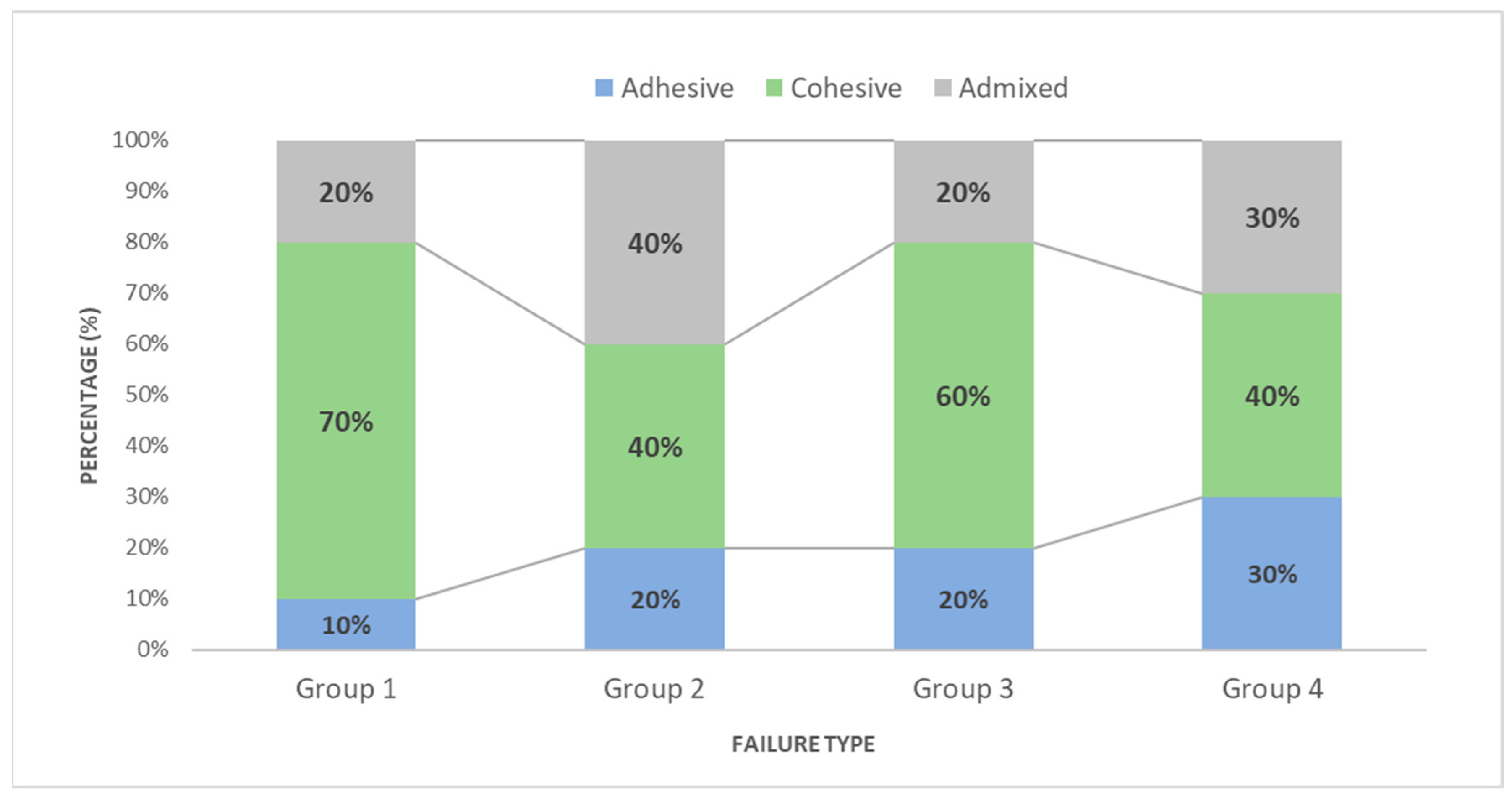
Group 1 and group 3 specimens exhibited the cohesive type of failure pattern. Group 2 and group 4 predominantly displayed bond failure of all three types (Figure 7).

3.4. Evaluation of ΔE

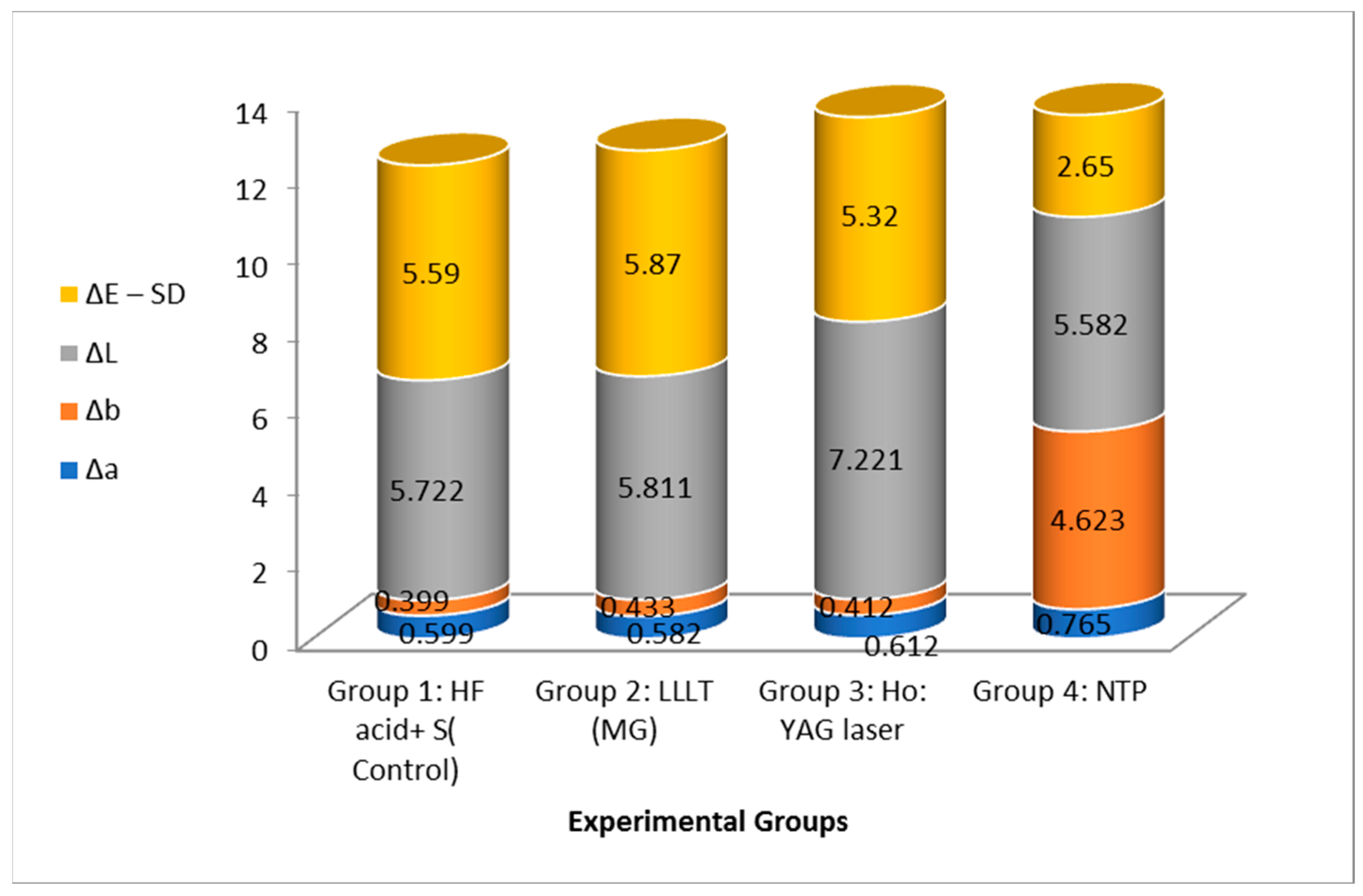
∆E value > 1.2 was considered perceptible, and ∆E value > 2.7 was clinically unacceptable. The mean and SD of ΔE among different experimental groups are presented in Figure 8. The highest perceptible ΔE was observed in group 1 treated samples (HF acid+ S) (5.59 ± 1.2). However, the minimum scores of ΔE were demonstrated by group 4 (NTP) (2.65 ± 0.07) treated discs. Intergroup comparison analysis revealed that group 1, group 2 LLLT (MG) (5.87 ± 0.9), and group 3 (Ho: YAG laser) (5.32 ± 0.7) displayed comparable scores in color change (p > 0.05). However, group 4 treated samples presented significantly lower scores of ΔE (p ˂ 0.05).

4. Discussion

The current work originally examined the hypothesis that the ∆E values of HBC, following the application of the latest pretreatment methods (NTP, LLLT (MG), and Ho: YAG laser), would be similar to the standard HFA+S pretreatment approach. Furthermore, it was expected that the Ra and SBS of HBC attached to the resin cement would show no significant difference when modern conditioning techniques were employed, as compared to HFA+S. The main hypothesis that was proposed was partially confirmed, indicating that the combination of HFA+S, Ho: YAG laser, and LLLT (MG) conditioning of HBC resulted in a ΔE value that surpassed the clinically tolerable threshold range, leading to a significant shift (∆E value > 1.2 was considered perceptible and ∆E value > 2.7 was clinically unacceptable). In addition, it was observed that the secondary stated hypothesis was partially disproven since the samples treated with NTP and LLLT (MG) showed lower levels of Ra and SBS compared to the control group. Resin cement’s bond strength with different CAD/CAM materials was evaluated using the SBS test. This test offers a larger percentage of the adhesive specimen diameter, which decreases the chances of complex stress development and leads to a higher proportion of adhesive failure rather than mixed failure [29,30].
Regarding the Ra and SBS analysis, it was determined that the HBC discs, which were treated with a Ho: YAG laser and HFA, exhibited the highest levels of Ra and adhesion strength when bonded with the resin cement. Albakri and colleagues conducted an in vitro study, which found that the Ho: YAG laser exhibits properties of both the CO2 and Nd: YAG lasers [31]. Their discovery explains the observed rise in Ra and bond integrity scores. The use of Nd: YAG laser enhances the surface roughness (Ra) of ceramics by inducing the formation of surface cracks and molten regions [31]. Additionally, CO2 laser irradiation contributes to the rise in Ra by inducing thermomechanical melting of the HBC surface. The process of melting leads to the expansion of the surface of HBC, whereas solidification results in rapid contraction [32]. Surface cracks are formed due to the generation of stress caused by abrupt changes in the physical condition of the material, as previously described [33]. These fissures are responsible for improving the shear bond strength (SBS) of the resin cement by mechanical interlocking [32]. In addition, the positive results regarding roughness and SBS achieved through HFA surface modification can be explained based on the research conducted by Alsunbul and his colleagues [34].
One potential explanation for this observation is the formation of hexafluorosilicates, a process that occurs when the HFA etchant interacts with the glass matrix. This chemical reaction roughens the surface and increases the surface energy, thereby enhancing the adhesive bond [35]. Additionally, the Ra score obtained post-HFA conditioning can be further explained by the studies conducted by Papadopoulos et al. According to their findings, pretreating RMC with HFA modifies the surface micromorphology, generating a pattern of pores and grooves of varying widths [36]. Conversely, research by Salem et al. has reported findings that contradict these outcomes [37].
LLLT combined with MG and NTP demonstrated lower, yet similar, Ra scores and bond integrity compared to other methods. The reduced bonding strength of HBC, when exposed to LLLT in conjunction with MG PS, may be attributed to various factors. Maawadh and colleagues conducted a study that revealed that water absorption on the surface of HBC discs following MG treatment adversely affects their bonding capacity [4]. This phenomenon arises due to the presence of a positive charge and the hydrophilic nature of the dye. Henningsen et al. and Vechiato et al., in their research, asserted that NTP does not alter the roughness parameters or modify the microstructure of conventional ceramics. This observation helps to explain the low shear bond strength (SBS) noted in this group, which aligns with their findings [38,39]. Additionally, a study by Jasmin and colleagues demonstrated that while NTP treatment significantly increased the SBS of resin cement to zirconia, it did not significantly impact the SBS of resin nano-ceramic [40]. These results suggest that the effects of NTP vary depending on the material type. Therefore, further research is needed to substantiate the conclusions of existing investigations and to fully understand the material-specific impacts of NTP.
Hybrid ceramics containing Triethylene glycol di-methacrylate (TEGDMA), a resin monomer, exhibited elevated levels of discoloration compared to other RMCs suggesting that the hydrophilic character of TEGDMA is responsible for the color change observed. In the present study, the CIELAB color space, a standardized three-dimensional color order system, was utilized for assessing color changes. To avoid the edge loss phenomenon highlighted in studies by Hejazy et al. and Pohjola et al., measurements were performed at the center of the buccal surface [41,42]. The findings from the recent study indicate that Non-Thermal Plasma (NTP) treatment does not significantly alter the ΔE (color change) of HBC discs. This result aligns with laboratory research by Sasany and associates, who demonstrated that argon gas disrupts oxygen bonding on the ceramic surface, thereby enhancing color stability by preventing oxidation [43].
Conversely, conditioning with HFA, Ho: YAG laser, and LLLT (MG) also resulted in ΔE values surpassing the clinically acceptable threshold, leading to significant color change. Literature indicates that HFA etching adversely affects the translucency of ceramics. The etching process reduces the glass matrix, diminishing translucency and consequently decreasing the amount of light that passes through the ceramic material. These observations are consistent with the findings of a study by Hafez and colleagues [44]. Furthermore, this phenomenon can be explained by the behavior of light on different surface textures. On a smooth surface, light reflects uniformly, maintaining the same angle and direction. However, a rough surface induces diffuse reflection, where light scatters in various directions, leading to a noticeable change in color perception [45].
When analyzing the fracture patterns, it was noted that the groups treated with HFA and Ho: YAG laser primarily showed cohesive failures, indicating that the fractures occurred within a single material component [46]. In contrast, the samples treated with NTP and LLLT (MG) exhibited all three types of fracture patterns. The diversity of fracture types suggests that the cement was predominantly concentrated at the outer edge of the bonding interface. This distribution indicates that stress concentration typically occurs in the central region of the interface between the hybrid ceramics and resin cement [8,47].
The current study underscores several inherent limitations. The SBS results, while informative, may not fully replicate clinical conditions, which are influenced by a multitude of factors. Consequently, these findings should be interpreted with caution. Moreover, additional studies are required to explore the before and after effects of conditioning on HBC discs utilizing atomic force microscopy (AFM). It is also imperative to conduct dispersive spectroscopy on debonded surfaces to gain further insights. Additionally, comprehensive evaluations are needed to assess the impact of varying concentrations of MG, along with different irradiation protocols, on the color stability, roughness average (Ra), and SBS of Resin Matrix Ceramics (RMCs).

5. Conclusions

Ho: YAG laser and HFA+S pretreatment demonstrated the highest surface roughness and shear bond strength. A direct correlation was observed between surface roughness and SBS. HFA+S, Ho: YAG laser, and LLLT (MG) conditioning of HBC resulted in an ΔE value that surpassed the clinically tolerable threshold range, making them less suitable for use in aesthetically critical areas. Consequently, there is a pressing need to identify alternative surface conditioners that can improve roughness and bond strength without altering the color of the materials, thus ensuring both functional integrity and aesthetic appeal.

Author Contributions

Conceptualization, F.A. and Y.F.A.; methodology, F.A. and Y.F.A.; software, F.A. and Y.F.A.; validation, F.A. and Y.F.A.; formal analysis, F.A. and Y.F.A.; investigation, F.A. and Y.F.A.; resources, F.A. and Y.F.A.; data curation, F.A.; writing—original draft preparation, F.A. and Y.F.A.; writing—review and editing, Y.F.A.; visualization, F.A.; supervision, F.A.; project administration, F.A.; funding acquisition, Y.F.A. and F.A. All authors have read and agreed to the published version of the manuscript.

Funding

The Project funded by Researchers Supporting Program (RSPD) at King Saud University, Riyadh, Saudi Arabia, through project number (RSPD2024R815).

Institutional Review Board Statement

The study was approved by the ethical board of King Saud University under IRB # FC-475-55.

Informed Consent Statement

Not applicable.

Data Availability Statement

The data can be made available on request to the authors.

Acknowledgments

The authors are grateful to the researchers supporting the project at King Saud University for funding through the project (RSPD2024R815), Riyadh, Saudi Arabia.

Conflicts of Interest

The authors declare no conflicts of interest.

References

  1. Coldea, A.; Swain, M.V.; Thiel, N. Mechanical Properties of Polymer-Infiltrated-Ceramic-Network Materials. Dent. Mater. 2013, 29, 419–426. [Google Scholar] [CrossRef] [PubMed]
  2. Jung, S.N.; Rüttermann, S. Influence of Mechanical and Chemical Pre-Treatments on the Repair of a Hybrid Ceramic. Dent. Mater. 2022, 38, 1140–1148. [Google Scholar] [CrossRef]
  3. Bayraktar, Y.; Arslan, M.; Demirtag, Z. Repair Bond Strength and Surface Topography of Resin-Ceramic and Ceramic Restorative Blocks Treated by Laser and Conventional Surface Treatments. Microsc. Res. Tech. 2021, 84, 1145–1154. [Google Scholar] [CrossRef] [PubMed]
  4. Maawadh, A.M.; Almohareb, T.; Al-Hamdan, R.S.; Al Deeb, M.; Naseem, M.; Alhenaki, A.M.; Vohra, F.; Abduljabbar, T. Repair Strength and Surface Topography of Lithium Disilicate and Hybrid Resin Ceramics with LLLT and Photodynamic Therapy in Comparison to Hydrofluoric Acid. J. Appl. Biomater. Funct. Mater. 2020, 18, 2280800020966938. [Google Scholar] [CrossRef] [PubMed]
  5. Kilinc, E.; Antonson, S.A.; Hardigan, P.C.; Kesercioglu, A. Resin Cement Color Stability and Its Influence on the Final Shade of All-Ceramics. J. Dent. 2011, 39, e30–e36. [Google Scholar] [CrossRef] [PubMed]
  6. Vohra, F.; Labban, N.; Al-Hussaini, A.; Al-Jarboua, M.; Zawawi, R.; Alrahlah, A.; Naseem, M. Influence of Er;Cr:YSGG Laser on Shear Bond Strength and Color Stability of Lithium Disilicate Ceramics: An in Vitro Study. Photobiomodulation Photomed. Laser Surg. 2019, 37, 483–488. [Google Scholar] [CrossRef]
  7. Turgut, S.; Bagis, B.; Ayaz, E.A.; Korkmaz, F.M.; Ulusoy, K.U.; Bagis, Y.H. Erratum to How Will Surface Treatments Affect the Translucency of Porcelain Laminate Veneers? J. Adv. Prosthodont. 2014, 6, 150. [Google Scholar] [CrossRef]
  8. Panah, F.G.; Rezai, S.M.M.; Ahmadian, L. The Influence of Ceramic Surface Treatments on the Micro-Shear Bond Strength of Composite Resin to IPS Empress 2. J. Prosthodont. 2008, 17, 409–414. [Google Scholar] [CrossRef]
  9. Alsunbul, H.; Almutairi, B.; Aljanakh, M.; Abduljabbar, T. Hybrid Ceramic Repair Strength, Surface Roughness, and Bond Failure, Using Methylene Blue-Activated Low-Level Laser Therapy, Carbon Dioxide, and Ti: Al2O3 Laser. Photodiagnosis Photodyn. Ther. 2023, 43, 103693. [Google Scholar] [CrossRef]
  10. Campos, F.; Almeida, C.S.; Rippe, M.P.; De Melo, R.M.; Valandro, L.F.; Bottino, M.A. Resin Bonding to a Hybrid Ceramic: Effects of Surface Treatments and Aging. Oper. Dent. 2016, 41, 171–178. [Google Scholar] [CrossRef]
  11. Al Deeb, L.; Bin-Shuwaish, M.S.; Abrar, E.; Naseem, M.; Al-Hamdan, R.S.; Maawadh, A.M.; Al Deeb, M.; Almohareb, T.; Al Ahdal, K.; Vohra, F.; et al. Efficacy of Chlorhexidine, Er Cr YSGG Laser and Photodynamic Therapy on the Adhesive Bond Integrity of Caries Affected Dentin. An in-Vitro Study. Photodiagnosis Photodyn. Ther. 2020, 31, 101875. [Google Scholar] [CrossRef] [PubMed]
  12. Akram, Z. How Effective Is Adjunctive Antimicrobial Photodynamic Therapy in Treating Deep Periodontal Pockets in Periodontal Disease? A Systematic Review. J. Investig. Clin. Dent. 2018, 9, e12345. [Google Scholar] [CrossRef] [PubMed]
  13. Al Ahdal, K.; Maawadh, A.M.; Al Deeb, L.; Alshamrani, A.S.; Almohareb, T.; Alrahlah, A. Effect of Malachite Green, Ocimum Sanctum, and Er, Cr: YSGG Laser on Antimicrobial Activity against S.Mutans and CAD Disinfection Bonded to Resin Restoration. Photodiagnosis Photodyn. Ther. 2023, 42, 103571. [Google Scholar] [CrossRef] [PubMed]
  14. Alqahtani, W.M.S.; Qamar, Z.; Yousief, S.A.; Abdul, N.S.; Reddy, R.N.; Alghufaili, S.; Khuthija Khanam, H.M.; Barakat, A.; Niazi, F.H. Use of Final Irrigants MTAD, Salvedora Perscia, Malachite Green, and Ti-Sapphire Laser on Push-out Bond Strength of Zirconia Post. Photodiagnosis Photodyn. Ther. 2023, 43, 103605. [Google Scholar] [CrossRef] [PubMed]
  15. Alrahlah, A.; Naseem, M.; Tanveer, S.A.; Abrar, E.; Charania, A.; AlRifaiy, M.Q.; Vohra, F. Influence of Disinfection of Caries Effected Dentin with Different Concentration of Silver Diamine Fluoride, Curcumin and Er, Cr:YSGG on Adhesive Bond Strength to Resin Composite. Photodiagnosis Photodyn. Ther. 2020, 32, 102065. [Google Scholar] [CrossRef] [PubMed]
  16. Bartlett, J.A.; Indig, G.L. Spectroscopic and Photochemical Properties of Malachite Green Noncovalently Bound to Bovine Serum Albumin. Dye. Pigment. 1999, 43, 219–226. [Google Scholar] [CrossRef]
  17. Gorler, O.; Dogan, D.O.; Ulgey, M.; Goze, A.; Hubbezoğlu, I.; Zan, R.; Ozdemir, A.K. The Effects of Er:YAG, Nd:YAG, and Ho:YAG Laser Surface Treatments to Acrylic Resin Denture Bases on the Tensile Bond Strength of Silicone-Based Resilient Liners. Photomed. Laser Surg. 2015, 33, 409–414. [Google Scholar] [CrossRef] [PubMed]
  18. Gorler, O.; Ozdemir Dogan, D.; Ulgey, M.; Goze, A.; Zan, R.; Hubbezoğlu, I.; Kemal Ozdemir, A. Dual Effect of Ho: YAG Laser on Tensile Bond Strength of Acrylate and Soft Liner Materials. Int. J. Acad. Res. 2016, 8, 10–15. [Google Scholar] [CrossRef]
  19. Vechiato Filho, A.J.; Dos Santos, D.M.; Goiato, M.C.; De Medeiros, R.A.; Moreno, A.; Bonatto, L.D.R.; Rangel, E.C. Surface Characterization of Lithium Disilicate Ceramic after Nonthermal Plasma Treatment. J. Prosthet. Dent. 2014, 112, 1156–1163. [Google Scholar] [CrossRef] [PubMed]
  20. Attia, M.A.; Shokry, T.E.; Abdel-Aziz, M. Effect of Different Surface Treatments on the Bond Strength of Milled Polyetheretherketone Posts. J. Prosthet. Dent. 2022, 127, 866–874. [Google Scholar] [CrossRef]
  21. AlShahrani, I.; Kamran, M.A.; Almoammar, S.; Alhaizaey, A. Photosensitization of Lithium Di-Silicate Ceramic by Er, Cr: YSGG and Fractional Carbon Dioxide Laser Bonded to Orthodontic Bracket. Photodiagnosis Photodyn. Ther. 2019, 28, 273–276. [Google Scholar] [CrossRef] [PubMed]
  22. Lanza, M.D.S.; Rodrigues Lanza, F.J.S.; Manso, A.P.; Matinlinna, J.P.; Carvalho, R.M. Innovative Surface Treatments for Improved Ceramic Bonding: Lithium Disilicate Glass Ceramic. Int. J. Adhes. Adhes. 2018, 82, 60–66. [Google Scholar] [CrossRef]
  23. Dilber, E.; Yavuz, T.; Kara, H.B.; Ozturk, A.N. Comparison of the Effects of Surface Treatments on Roughness of Two Ceramic Systems. Photomed. Laser Surg. 2012, 30, 308–314. [Google Scholar] [CrossRef] [PubMed]
  24. Pham, C.M.; Chen, C.Y.; Kim, D.M. The Effects of Using Erbium, Chromium-Doped:Yttrium-Scandium-Gallium-Garnet Laser on the Surface Modification, Bacterial Decontamination, and Cell Adhesion on Zirconia Discs: An in Vitro Study. Lasers Med. Sci. 2021, 36, 1701–1708. [Google Scholar] [CrossRef] [PubMed]
  25. Alkhudhairy, F.; Naseem, M.; Ahmad, Z.H.; Alnooh, A.N.; Vohra, F. Efficacy of Phototherapy with Different Conventional Surface Treatments on Adhesive Quality of Lithium Disilicate Ceramics. Photodiagnosis Photodyn. Ther. 2019, 25, 292–295. [Google Scholar] [CrossRef]
  26. Alkhudhairy, F.; Vohra, F.; Naseem, M.; Owais, M.M.; Amer, A.H.B.; Almutairi, K.B. Color Stability and Degree of Conversion of a Novel Dibenzoyl Germanium Derivative Containing Photo-Polymerized Resin Luting Cement. J. Appl. Biomater. Funct. Mater. 2020, 18, 2280800020917326. [Google Scholar] [CrossRef] [PubMed]
  27. Alkhudhairy, F.; AlRefeai, M.H. Self-Etch Adhesive-Loaded ZrO2/Ag3PO4 Nanoparticles on Caries-Affected Dentin: A Tensile Bond Strength, Scanning Electron Microscopy, Energy Dispersive X-Ray Spectroscopy, Survival Rate Assessment of S. Mutans, and Degree of Conversion Analysis. Appl. Sci. 2024, 14, 563. [Google Scholar] [CrossRef]
  28. Aljamhan, A.S.; Alrefeai, M.H.; Alhabdan, A.; Alhusseini, S.A.; Farooq, I.; Vohra, F.; Naseem, M.; Alkhudhairy, F. Influence of ER-CR-YSGG Laser and Photodynamic Therapy on the Dentin Bond Integrity of Nano-Hydroxyapatite Containing Resin Dentin Adhesive: SEM-EDX. Polymers 2021, 13, 1903. [Google Scholar] [CrossRef] [PubMed]
  29. Almuammar, M.F.; Schulman, A.; Salama, F.S. Shear Bond Strength of Six Restorative Materials. J. Clin. Pediatr. Dent. 2001, 25, 221–225. [Google Scholar] [CrossRef]
  30. De Melo, R.M.; Travassos, A.C.; Neisser, M.P. Shear Bond Strengths of a Ceramic System to Alternative Metal Alloys. J. Prosthet. Dent. 2005, 93, 64–69. [Google Scholar] [CrossRef]
  31. Albakri, A.S. Pretreatment of Hybrid Ceramics Using Alumina (Al2O3) Nanoparticles, Hydrofluoric Acid, and Holmium: YAG Laser for Optimizing Surface Roughness, Shear Bond Strength, and Topography. J. Biomater. Tissue Eng. 2024, 13, 1133–1138. [Google Scholar] [CrossRef]
  32. Yildirim, A.Z.; Unver, S.; Mese, A.; Bayram, C.; Denkbas, E.B.; Cevik, P. Effect of Argon Plasma and Er:YAG Laser on Tensile Bond Strength between Denture Liner and Acrylic Resin. J. Prosthet. Dent. 2020, 124, 799.e1–799.e5. [Google Scholar] [CrossRef] [PubMed]
  33. Bahar, A. The Use of Holmium-Yttrium Aluminum Garnet Laser as Pit and Fissure Cleaner. Dent. J. (Majalah Kedokt. Gigi) 2009, 42, 147. [Google Scholar] [CrossRef]
  34. Alkhudhairy, F.; Aljamhan, A.S. Surface conditioning of PEEK post using Nd: YVO4 laser, photodynamic therapy, and sulfuric acid on the pushout bond strength to canal dentin. Photodiagnosis Photo Dyn. Ther. 2023, 42, 103601. [Google Scholar] [CrossRef] [PubMed]
  35. Çelik, E.; Sahin, S.C.; Dede, D.Ö. Shear Bond Strength of Nanohybrid Composite to the Resin Matrix Ceramics after Different Surface Treatments. Photomed. Laser Surg. 2018, 36, 424–430. [Google Scholar] [CrossRef] [PubMed]
  36. Papadopoulos, K.; Pahinis, K.; Saltidou, K.; Dionysopoulos, D.; Tsitrou, E. Evaluation of the Surface Characteristics of Dental CAD/CAM Materials after Different Surface Treatments. Materials 2020, 13, 981. [Google Scholar] [CrossRef] [PubMed]
  37. Salem, A.; Mohsen, C. Atomic Force Microscope Surface Roughness Analysis of Surface Treated Ceramics. Open Access Maced. J. Med. Sci. 2020, 8, 134–138. [Google Scholar] [CrossRef]
  38. Henningsen, A.; Smeets, R.; Heuberger, R.; Jung, O.T.; Hanken, H.; Heiland, M.; Cacaci, C.; Precht, C. Changes in Surface Characteristics of Titanium and Zirconia after Surface Treatment with Ultraviolet Light or Non-Thermal Plasma. Eur. J. Oral Sci. 2018, 126, 126–134. [Google Scholar] [CrossRef]
  39. Liebermann, A.; Keul, C.; Bähr, N.; Edelhoff, D.; Eichberger, M.; Roos, M.; Stawarczyk, B. Impact of plasma treatment of PMMA-based CAD/CAM blanks on surface properties as well as on adhesion to self-adhesive resin composite cements. Dent. Mater. 2013, 29, 935–944. [Google Scholar] [CrossRef]
  40. Adımcı, P.; İbiş, F.; Ercan, U.K.; Bagis, B. Evaluation of effects of non-thermal plasma treatment on surface properties of CAD/CAM materials. J. Adhes. Sci. Technol. 2019, 33, 35–49. [Google Scholar] [CrossRef]
  41. Huang, P.Y.; Masri, R.; Romberg, E.; Driscoll, C.F. The Effect of Various Disinfectants on Dental Shade Guides. J. Prosthet. Dent. 2014, 112, 613–617. [Google Scholar] [CrossRef]
  42. Hejazy, M.; Shebl, A.; Desoky, M.; El Gabrouny, M. Effect of Laser Debonding on Color Change of Three Different Ceramic Materials. Dent. Sci. Updates 2023, 4, 301–312. [Google Scholar] [CrossRef]
  43. Sasany, R.; Saraç, D.; Özcan, M. Effect of Different Liner Techniques and Argon Plasma Treatment of Zirconia Base on the Adhesion and Color Change of Veneering Ceramic. J. Adhes. Sci. Technol. 2021, 35, 1981–1994. [Google Scholar] [CrossRef]
  44. Abdelhamid Hafez, P.; Elgabarouny, M.; Abdelgawad, L.; Shebl, A. Effect of Surface Treatments on the Color Change and Shear Bond Strength of Porcelain Laminate Veneer. Dent. Sci. Updates 2020, 1, 15–22. [Google Scholar] [CrossRef]
  45. Gré, C.P.; de Ré Silveira, R.C.; Shibata, S.; Lago, C.T.R.; Vieira, L.C.C. Effect of Silanization on Microtensile Bond Strength of Different Resin Cements to a Lithium Disilicate Glass Ceramic. J. Contemp. Dent. Pract. 2016, 17, 149–153. [Google Scholar] [CrossRef] [PubMed]
  46. Maruo, Y.; Nishigawa, G.; Irie, M.; Yoshihara, K.; Matsumoto, T.; Minagi, S. Does Acid Etching Morphologically and Chemically Affect Lithium Disilicate Glass Ceramic Surfaces? J. Appl. Biomater. Funct. Mater. 2017, 15, 93–100. [Google Scholar] [CrossRef]
  47. Matani, J.D.; Kheur, M.; Jambhekar, S.S.; Bhargava, P.; Londhe, A. Evaluation of Experimental Coating to Improve the Zirconia-Veneering Ceramic Bond Strength. J. Prosthodont. 2014, 23, 626–633. [Google Scholar] [CrossRef]
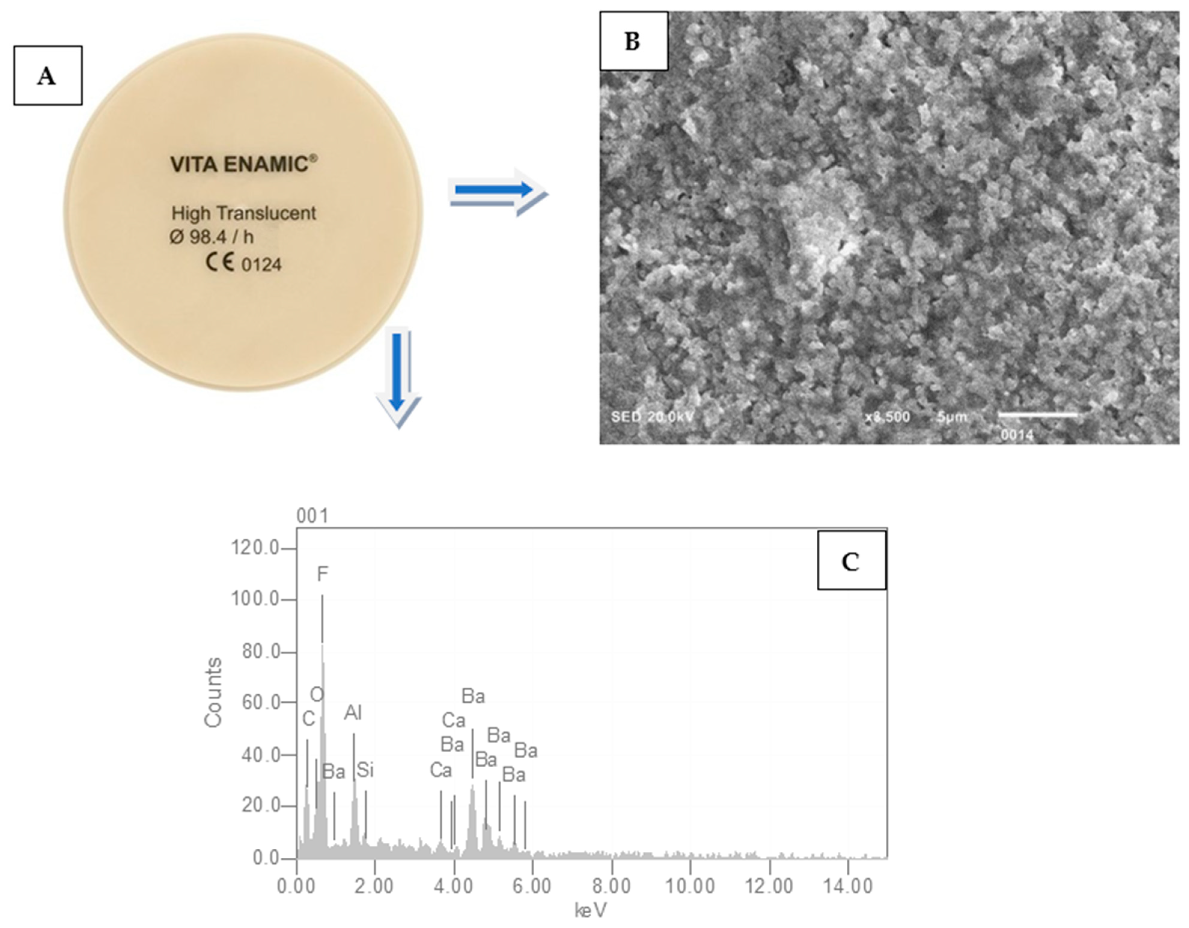
Figure 1. (A) Disc measuring 10 mm in diameter and 2 mm in thickness cut from the Vita Enamic CAD-CAM blocks. (B) SEM image shows the normal structure of hybrid ceramic before surface treatment. Even homogenous surfaces show fine particles (×3500). (C) EDX analysis demonstrates the different distribution of elements in hybrid ceramics.
Ceramics 07 00061 g001
Figure 2. Pretreatment of hybrid ceramics after HFA+S. The above micrograph clearly shows porosity on the surface along with the dissolution of the matrix. Uneven particle size with inter-particle gaps visible showing increased surface roughness.
Ceramics 07 00061 g002
Figure 3. Hybrid ceramics pretreated with LLLT-activated malachite green. An uneven surface with particle agglomeration. There is no dissolution of the matrix. No surface cracks and porosity are visible (×5000).
Ceramics 07 00061 g003
Figure 4. Surface treatment of hybrid ceramics with Ho: YAG laser. The surface shows uneven particle size. Between particles, porosity and cracks are distinct, increasing surface roughness. Each particle is different with the dissolution of the matrix (×3000).
Ceramics 07 00061 g004
Figure 5. Hybrid surface pretreatment with NTP. No prominent changes on the surface. The surface shows a cloud-like appearance. Particle structure is not visible. The surface does not show the dissolution of the matrix (×3500).
Ceramics 07 00061 g005
Figure 6. Descriptive data of surface roughness (Ra) and shear bond strength (SBS) of hybrid ceramics to resin cement after using different pretreatment protocols. The figure depicts that an increase in Ra resulted in improved SBS.
Ceramics 07 00061 g006
Figure 7. Distribution of failure mode in percentages across various study groups.
Ceramics 07 00061 g007
Figure 8. Mean and standard deviation (SD) of color change (ΔE) among different investigational groups after using different pretreatment protocols.
Ceramics 07 00061 g008
Table 1. Mean and SD of surface roughness (Ra) after using different conditioning regimes on HBC.
Investigated GroupsMean ± SD (µm)p-Value!
Group 1: HFA+S (Control)1115.44 ± 0.023 a<0.05
Group 2: LLLT (MG)890.17 ± 0.015 b
Group 3: Ho: YAG laser1282.88 ± 0.031 a
Group 4: NTP880.32 ± 0.012 b
Hydrofluoric acid + Silane (HFA+S), Low-level laser therapy-Malachite green LLLT-MG, Holmium: yttrium-aluminum-garnet (Ho: YAG laser), Non-thermal plasma (NTP), HBC (Hybrid ceramics). The different superscript denotes a statistically significant difference (p < 0.05).
Table 2. Descriptive data of shear bond strength (SBS) of hybrid ceramics to resin cement after using different pretreatment protocols.
Investigated GroupsMean ± SD
(MPa)
p-Value!
Group 1: HF acid+ S (Control)10.87 ± 5.5 a<0.05
Group 2: LLLT (MG)8.41 ± 3.2 b
Group 3: Ho: YAG laser11.22 ± 6.2 a
Group 4: NTP8.22 ± 2.9 b
Hydrofluoric acid + Silane (HF acid+ S), Low-level laser therapy (Malachite green) LLLT (MG), Holmium: yttrium-aluminum-garnet (Ho: YAG laser), non-thermal plasma (NTP). The different superscript denotes a statistically significant difference (p < 0.05).
Disclaimer/Publisher’s Note: The statements, opinions and data contained in all publications are solely those of the individual author(s) and contributor(s) and not of MDPI and/or the editor(s). MDPI and/or the editor(s) disclaim responsibility for any injury to people or property resulting from any ideas, methods, instructions or products referred to in the content.