Article

Different Techniques of Creating Bone Digital 3D Models from Natural Specimens

1
Institute of Anatomy and Anthropology, Rīga Stradiņš University, LV-1010 Riga, Latvia
2
Department of Computer Graphics and Computer Vision, Riga Technical University, LV-1048 Riga, Latvia
*
Author to whom correspondence should be addressed.
Appl. Syst. Innov. 2022, 5(4), 85; https://doi.org/10.3390/asi5040085
Submission received: 29 July 2022 / Revised: 17 August 2022 / Accepted: 18 August 2022 / Published: 22 August 2022
(This article belongs to the Special Issue Applied Systems on Emerging Technologies and Educational Innovations)

Abstract

:
The choice of technique for the creation of a 3D digital human bone model from natural specimens has a critical impact on the final result and usability of the obtained model. The cornerstone factor in 3D modeling is the number of faces of polygon mesh, along with topological accuracy, as well as resolution and level of detail of the texture map. Three different techniques (3D scanning, photogrammetry, and micro-computed tomography) have been used to create a digital 3D model of the human zygomatic bone. As implementation and use of 3D models can be divided into three main categories—visualization, simulation, and physical replication to obtain a functioning model (implant or prothesis)—the obtained models have been evaluated by the density and topological accuracy of the polygonal mesh, as well as by visual appearance by inspecting the obtained texture map. The obtained data indicate that for biomedical applications and computer biomechanical simulation the most appropriate technique of 3D model obtainment is micro-computed tomography, in its turn for visualization and educational purposes, the photogrammetry technique is a more preferable choice.

1. Introduction

Today, three-dimensional (3D) printing technology and models play an irreplaceable and significant role in different areas of medicine, including the education process, anatomical images and modeling, preclinical, and clinical studies [1]. 3D printing technology offers researchers the opportunity to create patient-specific models from medical images that represent anatomical structures in complex cases of congenital or multiple anomalies [2]. This allows a more accurate assessment of different and unique surgical procedures, their preintervention, or planning. 3D printing with various possibilities, materials, and challenges, allows it to become increasingly popular in the fields of medical implant design, manufacturing, tissue engineering, and biomedicine, including the fabrication of scaffolds called patient-specific tissue regeneration lattice structures [3]. Therefore, the new industrial revolution of 3D printing technologies requires a high level of expertise to achieve acceptable results, notably in modern STEM educational practices (Science, Technology, Engineering, Arts, and Mathematics) [4]. In addition, an essential part of the medical field is the development of the fourth industrial revolution, or Industry 4.0, with technologies that perform special functions, solve various medical problems with the interdisciplinary approach, increase the precision of surgical procedures, and create high-quality medical devices and components using advanced manufacturing technique [5]. In conjunction with autonomous, interconnected, and intelligent systems, portability, and a wide variety of materials, 3D printing has become one of the leading emerging technologies with a rapidly expanding market, where different innovative products are being created, from prototypes to fully functional complexes [6].
It is difficult to imagine teaching and learning human anatomy without any visualization tools to understand complex details, their dislocations, and relationships. Several authors recommend the integration of 3D models into teaching and evaluation for educational alignment, anatomical knowledge, and selection of a model for specific purposes [7,8,9]. 3D printed anatomical models are excellent tools that can be used for training and developing practical skills, not only for students, but also for medical professionals. Related to this, these models can also increase the potential interest and attention of different specialists in the development of new techniques, methods, and printing materials. Models can be replicated in varied sizes, colors, and quantities, according to the demand. It should be noted that the quality of models can be affected by thermal deformation of materials, removal of support structures, or other factors [10]. Today, the use of 3D models by educators in basic medical studies, practitioners in hospitals, and clinics, has exciting potential and should continue with a focus on sustainability. Furthermore, 3D computer-assisted reconstruction may improve treatment results, increase safety, and education, and improve surgeons’ ability to perform complex operations [11]. At the same time, there are many challenges in adopting this process [12]. Medical students can also emphasize the artistic role of 3D models in education, and the positive impact they have on their observation skills [13]. Another advantage of 3D models is the possibility of recreating different anatomical variations of any complexity, preoperative planning, development, and simulation of specific procedures [14]. 3D printed models, especially bones, can also make the necessity of natural human bones obsolete, encouraging virtual and remote education, helping to plan surgical interventions, and stimulating the implementation of cutting-edge technologies [15]. In addition to this, traditional anatomy teaching that incorporates 3D models is more involving and modern than the use of only computed tomography (CT) and/or magnetic resonance imaging (MRI) data, which can improve an understanding of the specific clinical manipulations and improve patient outcomes [16]. In the future, novel anatomical models can offer many possibilities for sectional anatomy, due to their nature as a multilayered and functional dissectible unit, as well as the ability to publish/share them for free access for educators, scientists, and students [17].
In anatomy education, 3D printed anatomical models can be used as a teaching tool as well as additional content to the curriculum and complement established learning methods, such as dissection-based teaching [18]. Furthermore, 3D models can offer new online teaching possibilities, reshaping teaching and learning spaces and providing a new experience for tutors and students [19]. The design of specific models for more complicated anatomical topics and microstructures is a major step in the development of modern study courses. In this research, we used three different techniques (3D scanning, photogrammetry, and micro-computed tomography) to produce a digital 3D model of zygomatic bone from a natural specimen. As zygomatic bone poses an intricate structure in terms of morphology, as well as its well-established protocol of replacement by an implant in case of severe injury, this makes zygomatic bone an ideal candidate for the creation of a precise and accurate human bone 3D digital model. Three techniques have been chosen as the most effective approaches in 3D model creation from natural specimens, which are being actively used at the Department of Morphology of Rīga Stradiņš University (RSU).

2. Materials and Methods

The photo images used for the photogrammetry technique were acquired with the help of a Sony ILCE-7RM2 camera and a Sigma 70 mm F2.8 DG MACRO Art lens (image capture parameters: f/8.0, 1/640s, ISO 100). The most important characteristic of the camera in terms of photogrammetry is the sensors’ pixel count (the used model has 42.3 megapixels; Table 1).
The higher the pixel count, the higher the quality/resolution of the obtained texture of the 3D model. Related to photogrammetry, the most crucial factor is the sharpness and clearness of the images, which require the use of a tripod and additional light sources. For the final image postprocessing (RAW data conversion, saturation, contrast), Adobe Photoshop and Capture One software have been used, followed by processing in RealityCapture (Table 2).
For micro-computed tomography scanning the µCT50 machine (Table 2) has been used, along with the following parameters:
Dimensions of the scanned area (cylinder): diameter = 48 mm, height= 110 mm;
Energy: 90 kVp;
Intensity: 88 uA;
Filter: Al 0.5 mm;
Preset: High resolution,
Field of view (FOV): 49.6 mm;
Voxel size: 24.2 um;
Integration time: 1500.
The software for raw data processing has been shipped along with the machine. The post-processed data have been exported in a form of DICOM (Digital Imaging and Communications in Medicine) files, and then imported into 3D Slicer (Table 1) software for the creation of a 3D model according to the Segmentation Protocol.
For mesh simplification and optimization, MeshLab software has been used (Table 1) according to the Simplification and Optimization Protocol.
The acquired photo images were post-processed with the help of RealityCapture software (Table 1). The program requires purchasing a license, but for academic and research staff is being provided free of charge. The process is quite straightforward and intuitive, due to the simple GUI (graphical user interface) of the software, as well as the built-in ‘Quick Start’ tool, which guides you through the entire creation process of the textured 3D model. The standard settings have been changed according to Photogrammetry Protocol to maximize the quality and precision of the final result. The obtained texture has been corrected with the help of Adobe Photoshop software (Table 1) to make it isotropic in terms of contrast, brightness, saturation, and shadow presence. The model was then simplified and optimized according to the Simplification and Optimization Protocol (to reduce the file size, since the Sketchfab platform, which is being used to publish and present the model, has a limitation of 100 MB per model (for a base account)). Simplification has been carried out, while keeping the original models’ topology and level of detail of the original models.
For 3D scanning, EinScan-S (Table 2) has been used, along with the following parameters:
Picture number: 36
Turntable Speed: 1
Turntable turns: half turn
The obtained 3D model has been optimized according to the Simplification and Optimization Protocol (the Simplification: quadric edge collapse decimation step has been skipped due to the low number of faces, in comparison to models created by other techniques).

2.1. Photogrammetry Protocol|Reality Capture

  • Launch the RealityCapture program.
  • Import images into the program.
  • Adjust alignment settings:
    • Max feature per mpx.: 20,000;
    • Max features per image: 80,000;
    • Preselector features: 20,000;
    • Image overlap: low;
    • Force component rematch: yes;
    • Detector sensitivity: high.
  • Launch the alignment process.
  • Define the reconstruction region.
  • Use reconstruction with High detail option to initialize the meshing process.
  • Use Clean Model tool to remove topology defects (non-manifold vertices, non-manifold edges, holes, isolated vertices).
  • Use the Texture instrument with the following setting to create a texture for the model:
    • Imported-model default texture resolution: 16,384 × 16,384;
    • Correct colors: Yes.
  • Export the 3D model along with the texture as an OBJ (file format) object.

2.2. Segmentation Protocol|3D Slicer

  • Launch the 3D Slicer program.
  • Import CT data into the program:
    • Set the image contrast to ensure better visibility.
  • Add a new segment using the tool:
    • Segment Editor:
      Set up the Threshold tool;
      Using Scissors, Draw, Islands, manually segment the required structure.
  • When segmentizing one structure is complete, proceed to the second one by adding a new segment and repeat the segmentation procedure if needed.
  • Export the completed segment or segments as a 3D model in the OBJ (file format) file format.

2.3. Simplification and Optimization Protocol|MeshLab

  • Launch the MeshLab program.
  • Import a 3D model into the program.
  • Remove artifacts, simplify, and optimize the model using tools (all the default values with modifications indicated below):
    • Remove isolated pieces (wrt diameter);
    • Remove duplicated faces;
    • Remove duplicated vertex;
    • Remove zero area faces;
    • Repair non-manifold edges by removing faces;
    • Repair non-manifold vertices by splitting;
    • Remove unreferenced vertices;
    • Simplification: quadric edge collapse decimation:
      Preserve boundary of the mesh: on;
      Preserve normal: on;
      Preserve topology: on;
      Planar simplification: on.
    • Remeshing: isotropic explicit remeshing:
      Adaptive remeshing: on;
      Collapse step: off.
  • Export the completed segment or segments as a 3D model in the binary PLY file format.
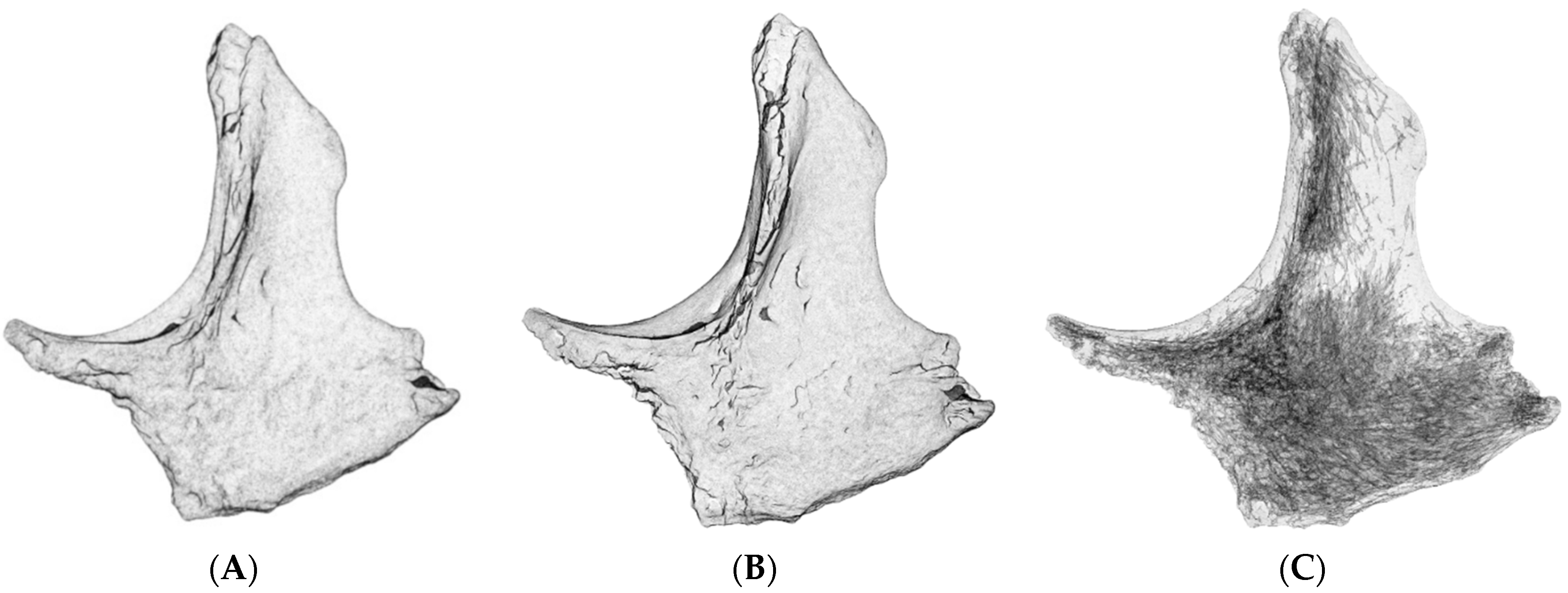
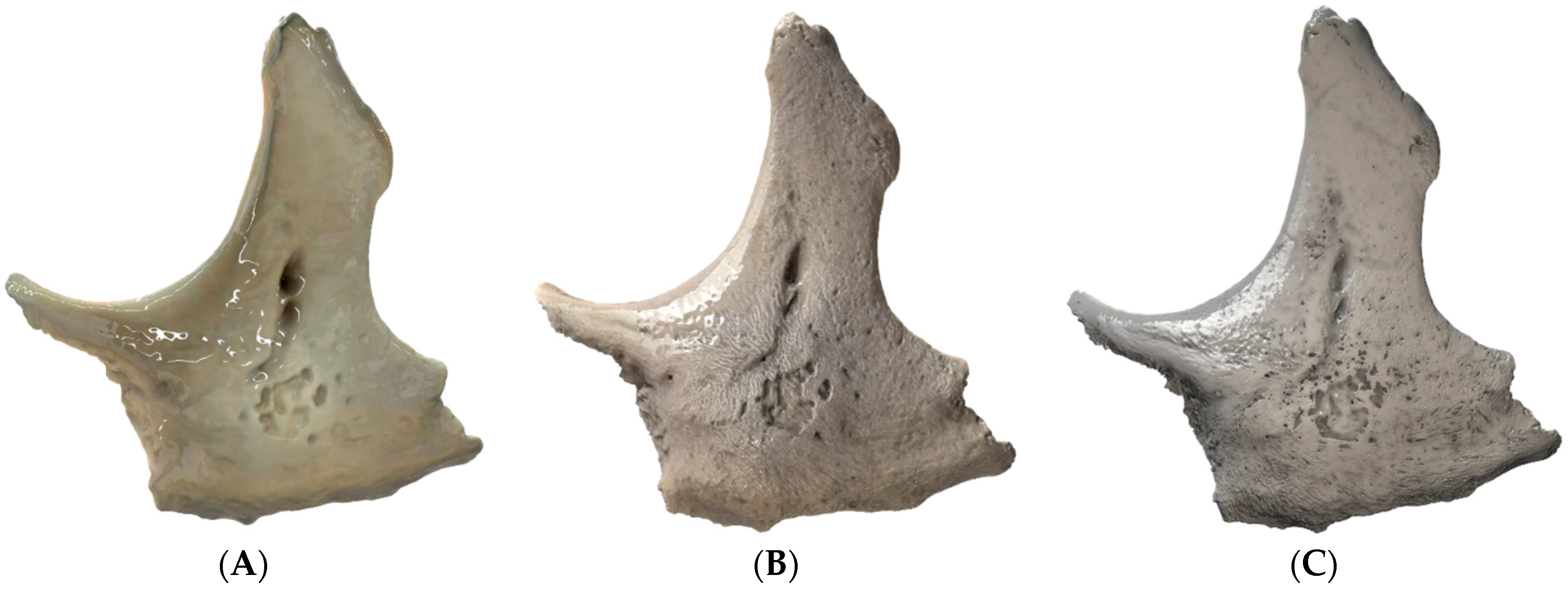
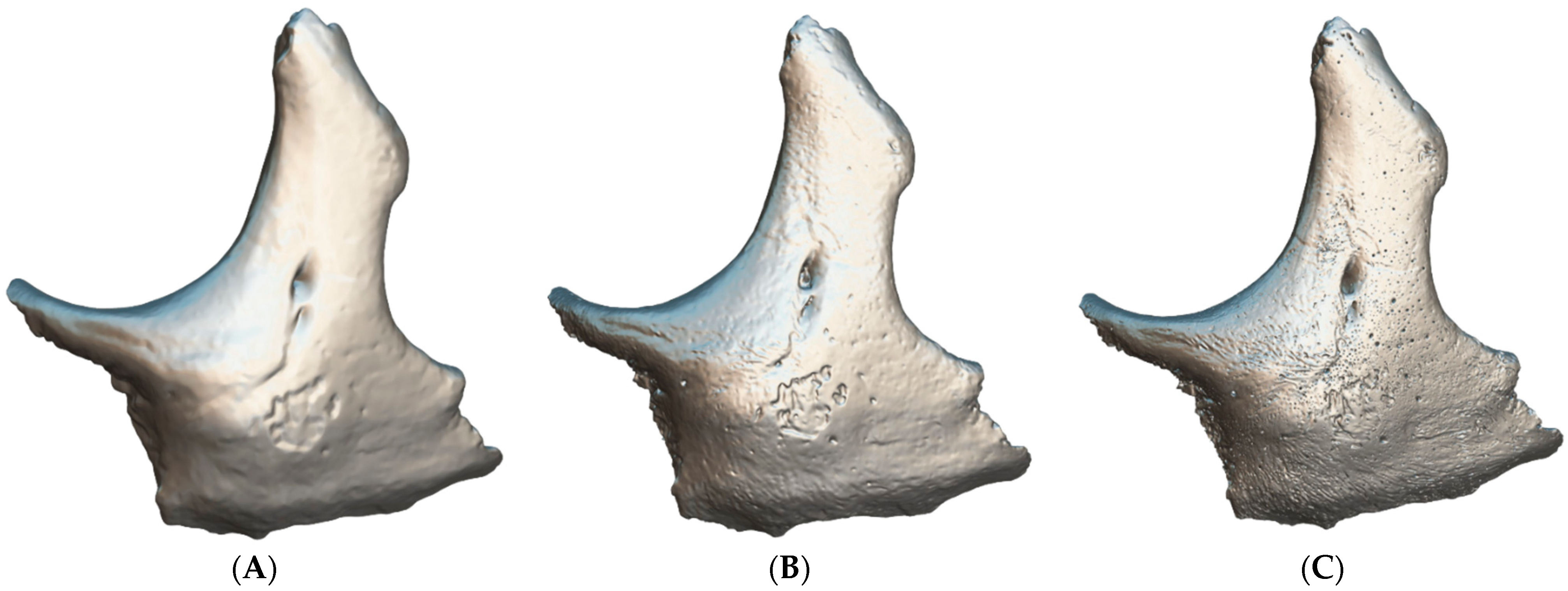
3. Results

Three models have been created in total using different techniques—3D scanning, photogrammetry, and micro-computed tomography (Figure 1).
All models have been simplified and optimized except the model created by the 3D scanning technique, which was only optimized.
The original number of faces and vertices for 3D models before and after the application of the Simplification and Optimization Protocol can be observed in Table 3.
The size of an obtained texture map of 3D models can be observed in Table 4.
In Figure 2, models with an applied X-ray shader allowing to see the inner structures can be observed.
Next, Figure 3, presents the 3D models with an applied shader, light sources, global illumination, ambient occlusion, and post-process filters, on the Sketchfab platform. All the models, except the one obtained with the help of the micro-computed tomography technique, are provided with textures.
All the models can be observed in 3D, downloaded, and used under the Attribution 4.0 International (CC BY 4.0) license at the Sketchfab platform—https://sketchfab.com/edler/models (accessed on 17 August 2022).

4. Discussion

Different techniques for the creation of 3D models allow their greater application in medical training and biomedical sciences [20]. Without a doubt, digital technologies have revolutionized anatomy education through 3D visual reconstruction of the human body, and have stimulated the teaching/learning experience of clinicians in different fields and levels [21]. Following an overview of new technological developments for anatomy education, this process raises several ethical and technical questions, at the same time providing proof in favor of 3D printing, and its advantages over other teaching resources [22,23]. Some studies have demonstrated the benefits of using 3D printed models in complex cases in gynecology, orthopedics, neuroanatomy, and radiology [24,25,26,27]. Researchers underline the potential of 3D printing to create more detailed and complex models, including regional, vascular, and nervous system structures [28]. Therefore, various materials may be needed to reproduce particular anatomical structures, or precise physical properties of human tissue, especially in scaffold engineering [29]. Furthermore, different anatomical structures have different mechanical properties, and they need special materials to meet the required performance of the printed object. Related to this, the most important factor is the correct selection of materials which is related to the selection of the 3D printing process, printer, and the mechanical properties of the model [30]. Widely used 3D printing technologies and specially designed models allow students to study complex anatomical regions and areas that are difficult for them to comprehend, requiring various levels of expertise [31,32]. In the medical field, 3D printed models serve as a great visualization tool that can represent the anatomy of patients, with great potential in presurgical planning and surgery [33,34,35,36]. A realistic 3D printed anatomical model can increase the effectiveness of surgery, can be used for determining the safest treatment strategy, and for designing patient-specific surgical instruments [37]. The process of developing a bone prototype with the help of modern 3D technologies can be an alternative to conventional model manufacturing [38].
The construction of 3D models from two-dimensional (2D) images depends on the user’s needs, possibilities, the time needed to create the models, and costs. In the process of anatomical models, the cornerstone is the methodology used for their development [39]. Using inexpensive materials and/or low-cost equipment can decrease the quality of the models. The choice of the 3D printing technique is the most crucial step that affects the accuracy and quality [40]. Moreover, early detection of errors and flaws during the design creation and fabrication processes of 3D printed products can reduce waste, manufacturing time, and cost [41]. Several studies report that the accuracy of 3D printed bones is key in the production of representative prints, but it can be affected by printer technology [42,43]. Visualization of 3D anatomic structures offers different interactive tools for teaching students, or patients, about the anatomy of the body; however, it requires a knowledgeable specialist in both teaching and anatomy [44,45]. Students should develop an understanding of complex anatomical structures after a limited time of practical classes. In these cases, access to 3D models can help them better prepare for tests and exams, improve anatomical knowledge, and prepare for clinical study courses. It is important to mention that our students can study the basic steps of 3D printing in the first study year of the Human Anatomy course at RSU. Based on our experience, we believe that created and printed anatomical models are especially useful in the teaching and learning of anatomy for undergraduate students [46]. Several studies show that models can serve not only as an addition to cadaver dissection but also can serve as a valuable alternative to natural specimens when access to the cadaveric material is limited [47,48]. Some authors reported that different medical specialists have focused on 3D scanning and printing technologies to enrich the medical education process with modern technologies [49,50]. All 3D technologies complement each other, and their integration can open new possibilities for the creation of new, valuable, and revolutionizing products [51,52].
The implementation and application of 3D models can be divided into three main categories: visualization, simulation, and physical replication to obtain a functioning model (implant or prosthesis). The visualization aspect prioritizes the overall appearance over the topological accuracy of the model. There are different areas, such as education, where the original look of a scanned bone is significant for the education process. Such an application requires a 3D model creation technique to capture not only the topological data, but also the visual ones. As can be seen in Figure 3, no texture is captured during microcomputed tomography scanning, and the final result consists only of topological data. Two other techniques, photogrammetry, and 3D scanning can capture visual data; however, their capabilities differ. As the camera image sensor is important for the photogrammetry process, it is for 3D scanning. In this study, a mid-level 3D scanner was used, which negatively impacted the resolution of the obtained texture map; it cannot compete with the texture map obtained with the help of the photogrammetry technique (Table 4). More advanced, industrial-grade 3D scanners capable of competing with photogrammetry in terms of the resolution of 3D model texture maps, are far more expensive and can cost up to 30,000 euros. In its turn, the equipment for photogrammetry can be obtained for less than 3000 euros. Photogrammetry is a relatively simple and inexpensive method of creating realistic 3D digital models that can be used in almost any setting to produce digital models [53]. Following special guidelines, these models can be metrically accurate, promoting self-directed learning and a greater understanding of complex anatomical structures [54]. However, creating a series of models through photogrammetry can be time-consuming [55,56].
In relation to the simulation and physical replication aspects, the density and accuracy of the polygonal mesh are more important factors than the resolution, or presence of the texture map. The data from Table 3 demonstrate that the microcomputed tomography technique is the most preferable way to obtain high density polygonal mesh, as well as this technique produces the most accurate results in terms of topology. Another crucial aspect is the presence of inner structures that are impossible to capture using any other technique (Figure 2). These data are necessary for implant production, or correct computer biomechanical simulation.
It is important to note that the first requirement for the creation of an anatomically accurate 3D printed model is a high-resolution volumetric dataset. Most models are created from computed tomography (CT) data, but it is possible to create models from datasets with modalities such as magnetic resonance imaging (MRI) and ultrasound [57]. Today, digital platforms and data storage include many massive medical image datasets [58]. Access to high-quality virtual images can offer a unique possibility for acquiring information about normal anatomy and/or anatomical variations. Differentiation of tissue density (from low to high) allows the segmentation technique to be used for different anatomical structures [59]. Depending on the imaging modality, different features can be observed, and different image segmentation algorithms can be applied. These segmentation algorithms and the availability of 3D printers stimulated the development of 3D printing in medical education and research, which had a positive impact on the precision of 3D printing technology and the costs of patient-specific 3D printed implants and prostheses [60,61]. Our experience and practice do not stand aside from these processes. In practical classes, 3D printed specimens help academic personnel and students to explain and/or understand anatomical structures.
Despite the many advantages of the use of 3D anatomical structures, the results of some studies were inconsistent in their effectiveness in improving anatomical knowledge and expertise for students with low spatial visualization ability, knowledge acquisition, and knowledge retention in a long-term perspective [62,63]. More research and analysis are necessary to better assess the role of anatomical models in the educational process. Although several models can be suitable for simple teaching/learning purposes, special 3D prints should be prepared for detailed topographical anatomy, and for a wide range of human anatomy pathologies [64]. Furthermore, every model should be morphologically accurate and qualitative. Some authors recommend using high-quality 3D scanners only because techniques such as photogrammetry are more time consuming and possess an increased risk of the occurrence of different artifacts in the 3D models [65].
Different technical parameters and the morphological variability of each model should also be considered. High-resolution and geometrically accurate medical images have been recommended in the literature [66]. Many studies have focused on best practices, optimal methodologies for conversion of patient radiographic scans to printable 3D models, specific software, and mathematical algorithms for optimization of 3D models’ polygonal mesh [67,68].
According to our research, it is important to note that the results can be affected by several factors in the 3D model creation process. Due to the possible errors in each step of the creation and/or printing of the anatomical 3D model, the choice of size and verification techniques depends on unique and specific requirements, guidelines, preferences, and the expertise of technical personnel [69]. Finally, the accuracy of printed models should be compared with that of the original samples [70]. In this study, one of the limitations is the choice of the bones from natural specimens, and their complicity and sizes because of the type and specifications of the 3D scanner/micro-computed tomography machine. In this case, it was only possible to use bones of a small size. Some technical difficulties and artifacts during scanning occurred, thus several steps were repeated several times as a countermeasure to presume the morphological accuracy and quality of the texture map. Furthermore, the choice of techniques and software used depends on the available equipment and possibilities available in the Department of Morphology. Finally, the background, anatomical, and technical expertise of researchers has a significant impact on the accuracy and quality of bone digital 3D models.

5. Conclusions

In this study, three different techniques have been presented and compared for the creation of the digital 3D zygomatic bone model from natural specimens. The results demonstrate that these techniques varied in their precision, complexity, quality, and accuracy, which distinguishes the techniques in terms of possible applications of the created 3D model. For morphological accuracy, the most preferable technique is micro-computed tomography; for visualization and demonstration of the original texture, the photogrammetry technique is the most optimal choice. The 3D scanning technique requires a professional, advanced 3D scanner to be able to compete with the other two techniques.
In addition, we have provided an improved strategy for the creation of digital 3D bone models from natural specimens and their assessment, in terms of practical use for educational purposes in the Human Anatomy course at Rīga Stradiņš University, also underlining the scientific and practical potential of digital models within the medical field in future work.

Author Contributions

Conceptualization, E.E.; methodology, E.E.; software, E.E.; validation, E.E. and K.B.; formal analysis, E.E.; investigation, E.E.; resources, K.B. and E.E.; writing—original draft preparation, E.E. and D.K.; writing—review and editing, E.E., D.K., K.B. and M.P.; visualization, E.E.; supervision, E.E. All authors have read and agreed to the published version of the manuscript.

Funding

This research received no external funding.

Institutional Review Board Statement

The study was conducted in accordance with the Declaration of Helsinki, and the protocol was approved by the Ethics Committee of Rīga Stradiņš University (2-PĒK-4/97/2022).

Informed Consent Statement

Not applicable.

Data Availability Statement

The simplified models are published on the Sketchfab platform—https://sketchfab.com/edler/models (accessed on 17 August 2022).

Acknowledgments

Authors acknowledge the support and assistance of Janis Locs and Marika Mosina from Riga Technical University.

Conflicts of Interest

The authors declare no conflict of interest.

References

  1. Ye, Z.; Dun, A.; Jiang, H.; Nie, C.; Zhao, S.; Wang, T.; Zhai, J. The role of 3D printed models in the teaching of human anatomy: A systematic review and meta-analysis. BMC Med. Educ. 2020, 20, 335. [Google Scholar] [CrossRef] [PubMed]
  2. Schrot, J.; Pietila, T.; Sahu, A. State of the art: 3D printing for creating compliant patient-specific congenital heart defect models. J. Cardiovasc. Magn. Reason. 2014, 16 (Suppl. 1), W19. [Google Scholar] [CrossRef] [Green Version]
  3. Kantaros, A.; Piromalis, D. Fabricating lattice structures via 3d printing: The case of porous bio-engineered scaffolds. Appl. Mech. 2021, 2, 289–302. [Google Scholar] [CrossRef]
  4. Kantaros, A.; Diegel, O.; Piromalis, D.; Tsaramirsis, G.; Khadidos, A.O.; Khadidos, A.O.; Khan, F.Q.; Jan, S. 3D printing: Making an innovative technology widely accessible through makerspaces and outsourced services. Mater. Today Proc. 2022, 49, 2712–2723. [Google Scholar] [CrossRef]
  5. Javaid, M.; Haleem, A. Industry 4.0 applications in medical field: A brief review. Curr. Med. Res. Pract. 2019, 9, 102–109. [Google Scholar] [CrossRef]
  6. Tsaramirsis, G.; Kantaros, A.; Al-Darraji, I.; Piromalis, D.; Apostolopoulos, C.; Pavlopoulou, A.; Alrammal, M.; Ismail, Z.; Buhari, S.M.; Stojmenovic, M.; et al. A modern approach towards an industry 4.0 model: From driving technologies to management. J. Sens. 2022, 2022, 5023011. [Google Scholar] [CrossRef]
  7. Hammerton, C.; Yip, S.W.L.; Manobharath, N.; Myers, G.; Sturrock, A. Are 3D printed models acceptable in assessment? Clin. Teach. 2022, 19, 221–228. [Google Scholar] [CrossRef]
  8. Yuen, J. What is the role of 3d printing in undergraduate anatomy education? A scoping review of current literature and recommendations. Med. Sci. Edu. 2020, 30, 1321–1329. [Google Scholar] [CrossRef]
  9. Garas, M.; Vaccarezza, M.; Newland, G.; McVay-Doornbusch, K.; Hasani, J. 3D-Printed specimens as a valuable tool in anatomy education: A pilot study. Ann. Anat. 2018, 219, 57–64. [Google Scholar] [CrossRef]
  10. Zou, Y.; Han, Q.; Weng, X.; Zou, Y.; Yang, Y.; Zhang, K.; Yang, K.; Xu, X.; Wang, C.; Qin, Y.; et al. The precision and reliability evaluation of 3-dimensional printed damaged bone and prosthesis models by stereo lithography appearance. Medicine 2018, 97, e9797. [Google Scholar] [CrossRef]
  11. Day, K.M.; Kelley, P.K.; Harshbarger, R.J.; Dorafshar, A.H.; Kumar, A.R.; Steinbacher, D.M.; Patel, P.; Combs, P.D.; Levine, J.P. Advanced three-dimensional technologies in craniofacial reconstruction. Plast. Reconstr. Surg. 2021, 148, 94e–108e. [Google Scholar] [CrossRef] [PubMed]
  12. Bastawrous, S.; Wu, L.; Liacouras, P.C.; Levin, D.B.; Ahmed, M.T.; Strzelecki, B.; Amendola, M.F.; Lee, J.T.; Coburn, J.; Ripley, B. Establishing 3d printing at the point of care: Basic principles and tools for success. Radiographics 2022, 42, 451–468. [Google Scholar] [CrossRef] [PubMed]
  13. Bell, L.T.O.; Evans, D.J.R. Art, anatomy, and medicine: Is there a place for art in medical education? Art, Anatomy, and Medical Education. Anat. Sci. Edu. 2014, 7, 370–378. [Google Scholar] [CrossRef] [PubMed]
  14. Govsa, F.; Yagdi, T.; Ozer, M.A.; Eraslan, C.; Alagoz, A.K. Building 3D anatomical model of coiling of the internal carotid artery derived from CT angiographic data. Eur. Arch. Otorhinolaryngol. 2017, 274, 1097–1102. [Google Scholar] [CrossRef] [PubMed]
  15. Pugalendhi, A.; Arumugam, S.; Ranganathan, R.; Ganesan, S. 3D printed patient-specific bone models for anatomy education from medical imaging. J. Eng. Res. 2021, 1–12. [Google Scholar] [CrossRef]
  16. Xie, Y.; Wu, G.; Liang, Y.; Fan, G. Three-dimensional physical model in urologic cancer. Front. Surg. 2022, 9, 757337. [Google Scholar] [CrossRef] [PubMed]
  17. Smith, M.L.; Jones, J.F.X. Dual-extrusion 3D printing of anatomical models for education: Two Materials 3D Printing in Anatomy. Anat. Sci. Edu. 2018, 11, 65–72. [Google Scholar] [CrossRef]
  18. Smith, C.F.; Tollemache, N.; Covill, D.; Johnston, M. Take away body parts! An investigation into the use of 3D-printed anatomical models in undergraduate anatomy education. Anat. Sci. Edu. 2018, 11, 44–53. [Google Scholar] [CrossRef]
  19. Diaz, C.M.; Linden, K.; Solyali, V. Novel and innovative approaches to teaching human anatomy classes in an online environment during a pandemic. Med. Sci. Edu. 2021, 31, 1703–1713. [Google Scholar] [CrossRef]
  20. Ugidos Lozano, M.T.; Blaya Haro, F.; Ruggiero, A.; Manzoor, S.; Nuere Menendez-Pidal, S.; Juanes Méndez, J.A. Different digitalization techniques for 3d printing of anatomical pieces. J. Med. Syst. 2018, 42, 46. [Google Scholar] [CrossRef]
  21. Wickramasinghe, N.; Thompson, B.R.; Xiao, J. The opportunities and challenges of digital anatomy for medical sciences: Narrative review. JMIR Med. Educ. 2022, 8, e34687. [Google Scholar] [CrossRef] [PubMed]
  22. Al-Mosawe, A.; Agha, H.; Al-Hadeethi, L.; Al-Mahaidi, R. Efficiency of image correlation photogrammetry technique in measuring strain. Aust. J. Struct. Eng. 2018, 19, 207–213. [Google Scholar] [CrossRef]
  23. Jones, D.G. Three-dimensional printing in anatomy education: Assessing potential ethical dimensions. Anat. Sci. Educ. 2019, 12, 435–443. [Google Scholar] [CrossRef]
  24. Flaxman, T.E.; Cooke, C.M.; Miguel, O.X.; Sheikh, A.M.; Singh, S.S. A review and guide to creating patient specific 3D printed anatomical models from MRI for benign gynecologic surgery. 3D Print Med. 2021, 7, 17. [Google Scholar] [CrossRef]
  25. Lima, L.F.; Barros, A.J.; Martini, A.D.; Stocco, M.B.; Kuczmarski, A.H.; Souza, R.L. Photogrammetry and 3D prototyping: A low-cost resource for training in veterinary orthopedics. Cienc. Rural 2019, 49, e20180929. [Google Scholar] [CrossRef]
  26. Sikes, R.W.; Sniezek, C.M.; Clancey, B.T.; Johnson, C.J. Virtual 3d brain slices: Improving learning of cross-sectional neuroanatomy by expanding access to human brain cross-sections through photogrammetric 3d scanning. FASEB J. 2018, 32 (Suppl. 1), 635-17. [Google Scholar] [CrossRef]
  27. Tashiro, M.; Minohara, S.; Yusa, K.; Sakurai, H.; Kanai, T.; Baba, M.; Miyamoto, T.; Nakano, T. 242 Quantitative evaluation of 3d lung motion with anatomical feature tracking technique for precise particle radiotherapy. Radiother. Oncol. 2006, 78, S85–S86. [Google Scholar] [CrossRef]
  28. Bartikian, M.; Ferreira, A.; Gonçalves-Ferreira, A.; Neto, L.L. 3D printing anatomical models of head bones. Surg. Radiol. Anat. 2019, 41, 1205–1209. [Google Scholar] [CrossRef]
  29. Ratinam, R.; Quayle, M.; Crock, J.; Lazarus, M.; Fogg, Q.; McMenamin, P. Challenges in creating dissectible anatomical 3D prints for surgical teaching. J. Anat. 2019, 234, 419–437. [Google Scholar] [CrossRef] [Green Version]
  30. Aimar, A.; Palermo, A.; Innocenti, B. The role of 3d printing in medical applications: A state of the art. J. Healthc. Eng. 2019, 2019, 5340616. [Google Scholar] [CrossRef] [Green Version]
  31. Schmidt, R.; Gartrell, R.; Yeung, J.M. A pipeline for generating interactive, schematic 3d surgical anatomy models. J. Surg. Educ. 2021, 78, 1419–1424. [Google Scholar] [CrossRef] [PubMed]
  32. Shahrubudin, N.; Lee, T.C.; Ramlan, R. An overview on 3d printing technology: Technological, materials, and applications. Procedia Manuf. 2019, 35, 1286–1296. [Google Scholar] [CrossRef]
  33. Betancourt, M.C.; Araújo, C.; Marín, S.; Buriticá, W. The quantitative impact of using 3d printed anatomical models for surgical planning optimization: Literature review. 3D Print Addit. Manuf. 2022, 3dp.2021.0188. [Google Scholar] [CrossRef]
  34. Formisano, M.; Iuppariello, L.; Mirone, G.; Cinalli, G.; Casaburi, A.; Guida, P.; Clemente, F. 3d printed anatomical model for surgical planning: A pediatric hospital experience. In Proceedings of the International Conference on E-Health and Bioengineering (EHB), Iasi, Romania, 18–19 November 2021. [Google Scholar] [CrossRef]
  35. Cornejo, J.; Cornejo-Aguilar, J.A.; Vargas, M.; Helguero, C.G.; Milanezi de Andrade, R.; Torres-Montoya, S.; Asensio-Salazar, J.; Rivero Calle, A.; Martínez Santos, J.; Damon, A.; et al. Anatomical engineering and 3d printing for surgery and medical devices: International review and future exponential innovations. Biomed. Res. Int. 2022, 2022, 6797745. [Google Scholar] [CrossRef] [PubMed]
  36. Fasel, J.H.D.; Malis, D.D.; Wiederer, C.; Hagenbuch, N. 3D printing of anatomical models for surgeons: An investigation on repeatability. IJIDeM 2018, 12, 621–627. [Google Scholar] [CrossRef]
  37. Osti, F.; Santi, G.; Neri, M.; Liverani, A.; Frizziero, L.; Stilli, S.; Maredi, E.; Zarantonello, P.; Gallone, G.; Stallone, S.; et al. Ct conversion workflow for intraoperative usage of bony models: From dicom data to 3d printed models. Appl. Sci. 2019, 9, 708. [Google Scholar] [CrossRef] [Green Version]
  38. Narayan, Y.S.; Prakash, K.J.; Rajashekhar, S.; Narendra, P. 3D printed human humerus bone with proximal implant prototype for arthroplasty. Int. J. Health Sci. 2022, 6 (Suppl. 4). [Google Scholar] [CrossRef]
  39. Salazar, D.A.; Cramer, J.; Markin, N.W.; Hunt, N.H.; Linke, G.; Siebler, J.; Zuniga, J. Comparison of 3D printed anatomical model qualities in acetabular fracture representation. Ann. Transl. Med. 2022, 10, 391. [Google Scholar] [CrossRef]
  40. Saleh, Y.; Piper, R.; Richard, M.; Jeyaretna, S.; Cosker, T. Designing a 3d printed model of the skull-base: A collaboration between clinicians and industry. J. Med. Educ. Curric. Dev. 2022, 9, 238212052210807. [Google Scholar] [CrossRef]
  41. Martinez-Marquez, D.; Mirnajafizadeh, A.; Carty, C.P.; Stewart, R.A. Application of quality by design for 3D printed bone prostheses and scaffolds. PLoS ONE 2018, 13, e0195291. [Google Scholar] [CrossRef]
  42. Rungrojwittayakul, O.; Kan, J.Y.; Shiozaki, K.; Swamidass, R.S.; Goodacre, B.J.; Goodacre, C.J.; Lozada, J.L. Accuracy of 3d printed models created by two technologies of printers with different designs of model base. J. Prosthodont. 2020, 29, 124–128. [Google Scholar] [CrossRef] [PubMed]
  43. Carew, R.M.; Iacoviello, F.; Rando, C.; Moss, R.M.; Speller, R.; French, J.; Morgan, R.M. A multi-method assessment of 3D printed micromorphological osteological features. Int. J. Leg. Med. 2022, 136, 1391–1406. [Google Scholar] [CrossRef] [PubMed]
  44. Ammanuel, S.; Brown, I.; Uribe, J.; Rehani, B. Creating 3d models from radiologic images for virtual reality medical education modules. J. Med. Syst. 2019, 43, 166. [Google Scholar] [CrossRef] [PubMed]
  45. Silén, C.; Karlgren, K.; Hjelmqvist, H.; Meister, B.; Zeberg, H.; Pettersson, A. Three-dimensional visualisation of authentic cases in anatomy learning–An educational design study. BMC Med. Educ. 2022, 22, 477. [Google Scholar] [CrossRef]
  46. Edelmers, E.; Kazoka, D.; Pilmane, M. Creation of Anatomically Correct and Optimized for 3D Printing Human Bones Models. Appl. Syst. Innov. 2021, 4, 67. [Google Scholar] [CrossRef]
  47. Hochman, J.B.; Rhodes, C.; Wong, D.; Kraut, J.; Pisa, J.; Unger, B. Comparison of cadaveric and isomorphic three-dimensional printed models in temporal bone education. Laryngoscope 2015, 125, 2353–2357. [Google Scholar] [CrossRef]
  48. McMenamin, P.G.; Quayle, M.R.; McHenry, C.R.; Adams, J.W. The production of anatomical teaching resources using three-dimensional (3d) printing technology: 3D Printing in Anatomy Education. Anat. Sci. Educ. 2014, 7, 479–486. [Google Scholar] [CrossRef]
  49. Blahuta, R.I.; Blikhar, V.S.; Dufeniuk, O.M. Transfer of 3d scanning technologies into the field of criminal proceedings. Sci. Innov. 2020, 16, 84–91. [Google Scholar] [CrossRef]
  50. Higueras, M.; Calero, A.I.; Collado-Montero, F.J. Digital 3D modeling using photogrammetry and 3D printing applied to the restoration of a Hispano-Roman architectural ornament. DAACH 2021, 20, e00179. [Google Scholar] [CrossRef]
  51. Baltsavias, E.P. A comparison between photogrammetry and laser scanning. ISPRS J. Photogramm. Remote Sens. 1999, 54, 83–94. [Google Scholar] [CrossRef]
  52. Bridger, C.A.; Reich, P.D.; Caraça Santos, A.M.; Douglass, M.J.J. A dosimetric comparison of CT- and photogrammetry- generated 3D printed HDR brachytherapy surface applicators. Phys. Eng. Sci. Med. 2022, 45, 125–134. [Google Scholar] [CrossRef] [PubMed]
  53. Morgan, B.; Ford, A.L.J.; Smith, M.J. Standard methods for creating digital skeletal models using structure-from-motion photogrammetry. Am. J. Phys. Anthropol. 2019, 169, 152–160. [Google Scholar] [CrossRef] [PubMed]
  54. Wesencraft, K.M.; Clancy, J.A. Using Photogrammetry to Create a Realistic 3D Anatomy Learning Aid with Unity Game Engine. Adv. Exp. Med. Biol. 2019, 1205, 93–104. [Google Scholar] [CrossRef] [PubMed]
  55. Carew, R.M.; Morgan, R.M.; Rando, C. Experimental assessment of the surface quality of 3D printed bones. Aust. J. Forensic Sci. 2021, 53, 592–609. [Google Scholar] [CrossRef]
  56. Petriceks, A.H.; Peterson, A.S.; Angeles, M.; Brown, W.P.; Srivastava, S. Photogrammetry of human specimens: An innovation in anatomy education. J. Med. Educ. Curric. Dev. 2018, 5, 238212051879935. [Google Scholar] [CrossRef] [Green Version]
  57. Ripley, B.; Levin, D.; Kelil, T.; Hermsen, J.L.; Kim, S.; Maki, J.H.; Wilson, G.J. 3d printing from mri data: Harnessing strengths and minimizing weaknesses: 3d printing from mri data. J. Magn. Reason. Imaging 2017, 45, 635–645. [Google Scholar] [CrossRef]
  58. Bois, M.C.; Morris, J.M.; Boland, J.M.; Larson, N.L.; Scharrer, E.F.; Aubry, M.-C.; Maleszewski, J.J. Three-dimensional surface imaging and printing in anatomic pathology. J. Pathol. Inform. 2021, 12, 22. [Google Scholar] [CrossRef]
  59. Bücking, T.M.; Hill, E.R.; Robertson, J.L.; Maneas, E.; Plumb, A.A.; Nikitichev, D.I. From medical imaging data to 3D printed anatomical models. PLoS ONE 2017, 12, e0178540. [Google Scholar] [CrossRef] [Green Version]
  60. Andreß, S.; Achilles, F.; Bischoff, J.; Kußmaul, A.C.; Böcker, W.; Weidert, S. A method for finding high accuracy surface zones on 3D printed bone models. Comput. Biol. Med. 2021, 135, 104590. [Google Scholar] [CrossRef]
  61. van Eijnatten, M.; van Dijk, R.; Dobbe, J.; Streekstra, G.; Koivisto, J.; Wolff, J. CT image segmentation methods for bone used in medical additive manufacturing. Med. Eng. Phys. 2018, 51, 6–16. [Google Scholar] [CrossRef]
  62. Labranche, L.; Wilson, T.D.; Terrell, M.; Kulesza, R.J. Learning in stereo: The relationship between spatial ability and 3d digital anatomy models. Anat. Sci. Educ. 2022, 15, 291–303. [Google Scholar] [CrossRef]
  63. Lau, I.; Sun, Z. The role of 3D printed heart models in immediate and long-term knowledge acquisition in medical education. Rev. Cardiovasc. Med. 2022, 23, 1. [Google Scholar] [CrossRef] [PubMed]
  64. McMenamin, P.G.; Hussey, D.; Chin, D.; Alam, W.; Quayle, M.R.; Coupland, S.E.; Adams, J.W. The reproduction of human pathology specimens using three-dimensional (3d) printing technology for teaching purposes. Med. Teach. 2021, 43, 189–197. [Google Scholar] [CrossRef] [PubMed]
  65. Douglass, M.J.J. Can optical scanning technologies replace CT for 3D printed medical devices in radiation oncology? J. Med. Radiat. Sci. 2022, 69, 139–142. [Google Scholar] [CrossRef] [PubMed]
  66. Crowe, S.; Luscombe, J.; Maxwell, S.; Simpson-Page, E.; Poroa, T.; Wilks, R.; Li, W.; Cleland, S.; Chan, P.; Lin, C.; et al. Evaluation of optical 3D scanning system for radiotherapy use. J. Med. Radiat. Sci. 2022, 69, 218–226. [Google Scholar] [CrossRef]
  67. Fogarasi, M.; Coburn, J.C.; Ripley, B. Algorithms used in medical image segmentation for 3D printing and how to understand and quantify their performance. 3D Print Med. 2022, 8, 18. [Google Scholar] [CrossRef]
  68. Brouwers, L.; Teutelink, A.; van Tilborg, F.A.J.B.; de Jongh, M.A.C.; Lansink, K.W.W.; Bemelman, M. Validation study of 3D-printed anatomical models using 2 PLA printers for preoperative planning in trauma surgery, a human cadaver study. Eur. J. Trauma Emerg. Surg. 2019, 45, 1013–1020. [Google Scholar] [CrossRef]
  69. Paramasivam, V.; Sindhu; Singh, G.; Santhanakrishnan, S. 3d printing of human anatomical models for preoperative surgical planning. Procedia Manuf. 2020, 48, 684–690. [Google Scholar] [CrossRef]
  70. Odeh, M.; Levin, D.; Inziello, J.; Lobo Fenoglietto, F.; Mathur, M.; Hermsen, J.; Stubbs, J.; Ripley, B. Methods for verification of 3D printed anatomic model accuracy using cardiac models as an example. 3D Print Med. 2019, 5, 6. [Google Scholar] [CrossRef]
Figure 1. 3D model’s simplified and optimized polygon meshes created by using different techniques. (A) 3D scanning, (B) photogrammetry, (C) micro-computed tomography.
Asi 05 00085 g001
Figure 2. 3D models were created by using different techniques with an X-ray shader applied. (A) 3D scanning, (B) photogrammetry, (C) micro-computed tomography.
Asi 05 00085 g002
Figure 3. 3D models with an applied shader, light sources, global illumination, ambient occlusion, and post-process filters, on Sketchfab platform created by using different techniques. (A) 3D scanning, (B) photogrammetry, (C) micro-computed tomography.
Asi 05 00085 g003
Table 1. The list of used technical equipment.
EquipmentManufacturerModelSpecification
CameraSonyILCE-7RM2electronics.sony.com/imaging/interchangeable-lens-cameras/full-frame/p/ilce7rm2-b (accessed on 17 August 2022)
LensSigma70 mm F2.8 DG MACRO Artsigma-global.com/en/lenses/a018_70_28 (accessed on 17 August 2022)
X-ray micro-computed tomographySCANCO MedicalµCT50scanco.ch/microct50.html (accessed on 17 August 2022)
ComputerLenovoLegion 7Windows 11 Pro, AMD Ryzen 7 5800H, NVIDIA GeForce RTX 3080 16 GB, 64 GB DDR4 3200 MHz, 1000 GB solid-state drive.
3D ScannerShining3DEinScan-Seinscan.com/desktop-3d-scanners/einscan-se/einscan-se-specs (accessed on 17 August 2022)
Table 2. The list of software used.
Software|PlatformVersionInformation
EinScan-S2.5.0.7einscan.com/support/download/software/?scan_model=einscan-s (accessed on 17 August 2022).
Micro-CT-//-Was shipped along with the µCT50 micro-CT machine.
3D Slicer5.02slicer.org (accessed on 17 August 2022)
MeshLab2022.02meshlab.net (accessed on 17 August 2022)
RealityCapture1.2.0.17385capturingreality.com/realitycapture (accessed on 17 August 2022)
Adobe Photoshop23.1.1For textures’ color correction.
Capture One 22 Pro15.0.1.4For cameras’ RAW images procession.
Sketchfab-//-sketchfab.com (accessed on 17 August 2022)
Table 3. The number of faces and vertices of 3D models before and after application of the Simplification and Optimization Protocol.
TechniquesBefore Simplification (Faces|Vertices)After Simplification (Faces|Vertices)
3D scanning700,002 350,003Has not been simplified
Micro Computed Tomography70,195,566 35,073,6137,019,556 3 485,608
Photogrammetry13,716,318 6,882,203700,842 350,423
Table 4. The size of an obtained texture map of 3D models.
TechniquesSize of the Texture Map (Pixels; Width × Height)
3D scanning766 × 998
Micro Computed TomographyNo visual data havebeen captured
Photogrammetry16,384 × 16,384
Publisher’s Note: MDPI stays neutral with regard to jurisdictional claims in published maps and institutional affiliations.

