Next Article in Journal
Precision Medicine in Pediatric Cancer: Current Applications and Future Prospects
Previous Article in Journal
In Silico Profiling of Clinical Phenotypes for Human Targets Using Adverse Event Data
 
 
Please note that, as of 21 September 2020, High-Throughput has been renamed to BioTech and is now published here.
Review

The Prenatal Microbiome: A New Player for Human Health

by
Valeria D’Argenio
1,2,3
1
CEINGE-BiotecnologieAvanzate, via G. Salvatore via G. Salvatore 486, 80145 Naples, Italy
2
Department of Molecular Medicine and Medical Biotechnologies, University of Naples Federico II, via Pansini 5, 80131 Naples, Italy
3
Task Force on Microbiome Studies, University of Naples Federico II, via Pansini 5, 80131 Naples, Italy
High-Throughput 2018, 7(4), 38; https://doi.org/10.3390/ht7040038
Submission received: 14 November 2018 / Revised: 5 December 2018 / Accepted: 7 December 2018 / Published: 11 December 2018

Abstract

:
The last few years have featured an increasing interest in the study of the human microbiome and its correlations with health status. Indeed, technological advances have allowed the study of microbial communities to reach a previously unthinkable sensitivity, showing the presence of microbes also in environments usually considered as sterile. In this scenario, microbial communities have been described in the amniotic fluid, the umbilical blood cord, and the placenta, denying a dogma of reproductive medicine that considers the uterus like a sterile womb. This prenatal microbiome may play a role not only in fetal development but also in the predisposition to diseases that may develop later in life, and also in adulthood. Thus, the aim of this review is to report the current knowledge regarding the prenatal microbiome composition, its association with pathological processes, and the future perspectives regarding its manipulation for healthy status promotion and maintenance.

1. Introduction

The human microbiome has emerged as an important factor required for human health status acquisition and maintenance [1,2]. Indeed, several reports have demonstrated that microbial alterations (or dysbiosis) can be found in an increasing number of human diseases and that their entities may be associated with the severity of the phenotype or with the sensitivity to specific therapies [1,2,3,4,5,6,7,8]. Thus, even if it has not been well established yet whether the microbiome alterations are the cause or the consequence of diseases, their study is intriguing since it opens the way to the identification of novel disease-specific biomarkers, or to the development of targeted therapies aiming to manipulate the microbiome in the attempt to restore a healthy status [9,10,11,12,13,14,15,16,17]. Next generation sequencing-based approaches have assessed their reliability in metagenomic studies escalating the possibility to describe microbial communities in detailand obtain both qualitative and quantitative information [1,2,18,19]. As a consequence, it is now widely accepted that microbes live on the surface and inside our body, colonizing also body niches that were traditionally considered as “sterile.” This applies also to the fetal environment [20,21,22]. Indeed, technological advances in metagenomics have denied a central dogma of reproductive medicine stating that the fetus stays in a sterile niche (i.e., the uterus) and is colonized by bacteria only at birth [23]. The prenatal microbiome seems not only to have a role in fetal development but may also influence the subsequent adult health status [24,25,26]. Thus, understanding fetal microbiome origins and the factors able to influence its composition is important to clarify diseases pathogenesis and identify also maternal factors that, acting at a prenatal age, may predispose the development of diseases including later in adulthood [24,25,26]. The aim of this review is to describe the knowledge on prenatal microbiome, its association to specific diseases, and its possible manipulation to promote a healthy status.

2. The Prenatal Microbiome

For about a century, it has been assumed that the fetus is sterile and that microbes colonize the newborn immediately during birth, withthe delivery mode being an important modifying factor [23,27,28]. The placenta, in particular, has been considered for years like a barrier able to prevent fetal contamination by maternal toxins and microbes, thus preserving fetal sterility and ensuring proper fetal development [26,29,30]. In contrast with these beliefs, recent studies have shown that microbes colonize the amniotic fluid, the umbilical blood cord, and the placenta, suggesting that a maternal microbial colonization of the fetus mayalready begin in utero [20,21,22,23,24,25,26,30]. In particular, the presence of an established microbiota in healthy and term infants, in absence of any infection or inflammation, supports the hypothesis that not only microbes colonize the fetus before birth, but that they may also play a role into the physiological development of the fetus.
In support of this hypothesis, microbial communities have been found also in the meconium of newborns [31,32,33,34,35,36,37,38,39,40,41,42]. Jiménez et al. first demonstrated the non-sterility of infant meconium [31]. These authors were also able to showthe presence of a maternal–fetalin utero transmission of gut microbiota; they used a mouse model to verify that a specific Enterococcus faecium strain, maternally administer viaoral instillation, was present in the gut of the pups delivered 1 day prematurely bycesarean section [31]. Subsequently, several reports using different molecular strategies have described microbial meconium communities, also highlighting interesting differences between term and preterm infants [32,33,34]. In particular, Mshvildadze et al. characterized the meconium microbiome of 23 neonates, revealing an association between a reduced microbial diversityand prematurity [32]. In a prospective longitudinal study, Madanet al. found thatthe predominant bacterial genera in the meconium specimens obtained from six preterm neonates were Lactobacillus, Staphylococcus, and Enterobacteriaceae [33]. Ardissone et al. analyzed the meconium collected from 52 infants with gestational ages ranging from 23 to 41 weeks and found that the gestational age was correlated with low bacterial diversity and microbial colonization [34]. In addition, this study revealed similar microbial features between the meconium and the amniotic fluid, thus supporting the hypothesis of an intrauterine origin of the meconium microbiota, as a consequence of the amniotic fluid swallowing by the fetus during the last trimester of pregnancy [34]. According to these findings, Collado et al. analyzed the microbiome of multiple samples from mother–offspring pairs, including the placenta, the meconium, and the amniotic fluid, and highlighted the presence of common microbial features [35]. The meconium microbiota has features of a low biodiversity and a high inter-individual variability [36,37]. Bäckhedet al. found that in the meconium microbiota, the genera Escherichia-Shigella and Enterococcus were more abundant and Bacteroides and Bifidobacterium less abundant with respect to infants fecal microbiota [38]. Tapiainen et al. carried out a large population-based study, recruiting 218 consecutively birth newborns, to assess the role of bacterial exposures before birth on the meconium microbiome composition [39]. They found that maternal factors occurring during pregnancy affect the meconium microbiome composition supporting the hypothesis of the in utero transfer of microbes [39].
Other studies have investigated the potential impact of the delivery mode on the meconium microbiome composition [40,41,42]. By analyzing the 16S rRNA profiles of rectal swabs obtained at birth by full-term infants, Dominguez-Bello et al. found that meconium microbiota composition is influenced by delivery mode [40]. In particular, they highlighted that in cesarean-delivered infants, there was a prevalence of Staphylococci with respect to the vaginally-delivered babies, whose microbiome was featured by the presence of genera like Lactobacillus and Prevotella, resembling the skin microbiome, and the vaginal microbiome, respectively [40]. Other studies performed on independent cohorts of full-term infants also found that the meconium microbiota of vaginally-delivered infants is dominated by lactic acid or enteric bacteria [41,42].
Even if the origin of meconium microbiota has been not yet clearly established, these data suggest that the establishment of intestinal microbiota is initiated in the prenatal gut. Indeed, as stated above, recent evidence has overcome the traditional view of the in utero environment as being a sterile room (Figure 1A) [20,21,22].
In particular, the presence of bacteria in the placenta of preterm newborns or in case of intrauterine infections is not surprising. However, bacteria have also been found in the placenta of at term pregnancy, without any sign of infection, and also after cesarean section under sterile conditions [43]. Aagaard et al. used 16S rRNA sequencing to analyze the placenta-related microbial communities of 320 women and revealed the presence of a low-abundant, but metabolically rich microbiota, featured by the presence of bacteria belonging to the phyla Firmicutes, Tenericutes, Proteobacteria, Bacteroidetes, and Fusobacteria [44]. Bacterial communities have been found also in the amniotic fluid, even without amniotic sac ruptures [45]. Other studies have focused their attention on the umbilical cord blood: Jiménez et al. analyzed the bacterial communities of healthy, full-term newborns after elective cesarean delivery and highlighted the presence of commensal bacteria, like Enterococcus, Streptococcus, Staphylococcus, and Propionibacterium [31].
All these data support the hypothesis of a prenatal origin of human microbiome. However, the origin of this fetal microbiome is still unknown (Figure 1B). The currently accepted theory is that fetal microbial bacteria may originate both from the vaginathrough ascendant colonization [46,47,48,49,50], and from the maternal oral cavity/gut through the bloodstream (hematogenous route) [31,44,51,52,53,54,55]. Additional studies are required to better clarify these issues.

3. Correlation between Fetal Microbiome and Newborn’s Health

The human gut is the organ with the highest density of microbial communities [1,2,3,4,6,7,8,56]. These communities are involved in several functions required for human host healthy status [1,2,3,4,6,7,8,56]. Thus, the initial microbial colonization of human gut is a critical step able to influence the host health and diseases risk through the establishment of a number of microbes–human interactions [21,22,23,24,25,26]. Maternal and early life environmental factors can both influence the gut microbiota development and composition and, as a consequence, may increase the risk of several chronic and metabolic diseases later in life [21,22,23,24,25,26]. Indeed, maternal microbial communities, including prenatal gut, vaginal, oral, and skin microbiomes, undergo pronounced changes during pregnancy that may affect healthy status maintenance and contribute to the development of common diseases [20]. To date, a microbial dysbiosis of the infant gut has been associated to the development of asthma, allergic diseases, and obesity [21,22,23,24,25,26]. Thus, considering that it has been established that the fetus does not stay in a sterile environment and that the microbial colonization begins in utero, factors that are able to influence this first colonization may impact on newborn’s future health.
In particular, it is now clear that the fetal immune system is not inactive but, being exposed through the mother to environmental stimuli, interacts with the maternal immune system [57,58]. Indeed, several maternal factors, including hormones, cytokines, and the microbiome, can modify the intrauterine environment, thus impacting fetal immune system development [57,59,60]. The toll-like receptors (TLRs) are a class of receptors present on the surface of different cells (macrophages, mast cells, and dendritic cells) involved in the innate immunity. Since different types of TLRs are able to recognize distinct bacteria driving the development of a potential inflammatory response, it has been proposed that intrauterine bacteria may influence the fetal immune system development though the TLRs [61,62]. Another possible mechanism of bacterial influence on the fetal immune system development may be represented by the production of short chain fatty acids (SCFAs) that are able to induce T-cells activation and modulate interleukin -10 production [63,64]. Finally, mucosa-associated invariant T cells (MAIT) have also been identifiedin the fetus starting from the second trimester of pregnancy [65,66]. These cells are able to recognize microbial metabolites and, consequently, to produce inflammatory cytokines supporting the hypothesis of in utero activity [66,67].
Considering all the above, it is easy to understand that all the factors influencing maternal health, including maternal habits, during pregnancy and the partum can influence the microbial colonization of the fetus. Chu et al. evaluated the effect of maternal diet on meconium microbiome composition [68]. In this study, 81 mother-infants couples were analyzed to highlight microbial alterations related to maternal diet during pregnancy. Interestingly, meconium microbial communities were significantly different in high fat respect to balanced diet exposed infants, Bacteroides being significantly reduced in high-fat exposed infants [68]. Another study by Lundgren et al. aimed to verify the relationship between maternal diet during pregnancy and the stool microbiome evaluated 6 weeks after birth [69]. In total, 145 infant/mother couples were analyzed showing that several maternal dietary factors during pregnancy influenced infants’ stool microbiomes and that these alterations are also related to the delivery mode [69]. Collado et al. found that infants’microbiomes are influenced by the mother beingoverweight during pregnancy [70]. In particular, this study highlighted that in the presence of an excessive maternal weight gain during pregnancy, newborns have lower amounts of the genera Bacteroides and Prevotella with respect to those born from mothers with regular weight gain [70]. Hu et al. studied the correlation between maternal gestational and pre-gestational type 2 diabetes and the meconium microbiome composition [36]. Interestingly, they found that Bacteroides, Parabacteroides, and Lachnospiraceae were more abundant in the meconium of infants in the presence of maternal diabetes and that these alterations were higher in the case of pre-gestational diabetes [36]. Gosalbes et al. verified the effects of maternal atopic diseases on the meconium microbiome [41]. They found that maternal eczema is able to modify the meconium bacterial composition, being associated with a lower bacterial diversity and richness and to an increased abundance of the Enterobacteriaceae family, while maternal asthma or rhinitis seem to not induce microbial changes [41]. Finally, Mshvildadze et al. showed that a lower bacterial diversity was present in the meconium of infants whose mothers received intra-partum antibiotics [32]. A recent interesting study by Lammert et al. investigated the role of the prenatal microbiome on the development of behavioral alterations [71]. Using a mouse model of autism, they were able to assess that prenatal microbiota transplantation is able to modulate the maternal immune system and transfer the susceptibility to neurodevelopmental disorders [71].
These studies support the hypothesis that environmental factors during pregnancy can modify maternal microbiome and may affect the fetal environment. However, the effects of this in utero colonization on newborns future health are not yet clearly understood and further studies on large cohorts of subjects are required to investigate how environmental stimuli can impact maternal microbiome during pregnancy and the long-term effects of these perturbations on infants’ health.

4. Manipulation of Prenatal Microbiome for Healthy Status Maintenance

As discussed in the previous sections, recent studies have shown that microbial colonization begins already before birth and that both maternal and environmental factors may play a role in this microbial early colonization. Even if the origin of the prenatal microbiome is still poorly understood and its correlation with newborns health and disease status also needs to be further investigated, if confirmed by future studies, it may become an actionable target to promote infant health by inducing the colonization of beneficial bacteria and avoiding those considered harmful.
Indeed, unlike the human genome, the microbiome can be easily modified, and its evaluation may be useful not only to stratify individuals based on a specific disease risk, but also to monitor them overtime [72]. In addition, while inter-individual differences in the response to specific therapies are well known, few data are available regarding the role of microbiomes in these phenomena, even if it has been established that the gut microbiota is involved in the metabolic transformation of several therapeutic compounds [72]. Thus, a better understanding of these processes may drive personalized medicine by supporting the choice of the most proper treatment and minimizing the onset of side effects.
Considering all the above, there is a lot of enthusiasm in this field since microbiome gives the opportunity to have a non-invasive biomarker that can not only be used for diagnostic purposes, but is emerging as an easily actionable target for therapeutic interventions. Antibiotics, probiotics, and prebiotics administration may be easily used to induce specific microbiome modifications [73,74,75,76]. In addition, enteral nutrition and microbiome transplantation have also shown their efficacy in some specific conditions [77,78,79].
The more our knowledge on human microbiome and its role in disease pathogenesis increases, the more the possibility to treat specific conditions (or at least to ameliorate their clinical outcome) by microbiome manipulation becomes intriguing. This also applies to the prenatal microbiome: since it is influenced by maternal habits during pregnancy, it is easy to imagine possible interventions (for example on maternal diet) that are able to drive fetal microbial colonization. However, as in other fields, a lot of limitations need to be assessed before microbiome manipulation can be genuinely helpful in routine clinical assessments. Microbial inter-individual variability, technical challenges, and potential pitfalls related to samples collection, analysis, and interpretation have to be taken into account.

5. Conclusions

As reviewed above, in the last 10 years, the sterile womb paradigm has been changed to the in utero colonization hypothesis. Indeed, microbial communities have been identified in the meconium, the amniotic fluid, the umbilical blood cord, and the placenta in at term pregnancy and in absence of infections [21,26].
To date, several reports have attempted to study the prenatal microbiome in order to clarify its origins and functions, and consequently highlight a possible role in predisposing to diseases development also later during life and adulthood [24,25,26]. Interesting associations have emerged and these may be the bases for the possible use of the prenatal microbiome as a biomarker and also as a potentially actionable target to be manipulated for personalized treatments aimed to reduce the risk for specific diseases.
Of course, caution is required regarding the correct interpretation of these data. As recently stated by Perez-Munoz et al. concerns exist regarding the detection limits of the used techniques (PCR and next generation sequencing-based approaches do not have high enough sensitivity to study low-rich bacterial communities), and with the high risk for environmental contaminations combined with the lack of proper controls [30]. In addition, the currently used next generation sequencing-based technologies cannot discriminate between live, viable, and active versus dead, unviable, and inactive bacteria. All these issues need to be taken into account when considering the prenatal microbiome and its influence on future health. Maybe further studies combining traditional culture-based assays and more extensive microbial communities studies (i.e., metabolomics, metatranscriptomics, and metagenomics) may provide a more comprehensive view of the prenatal microbiome and its role. Once these points are clarified and clearly assessed, novel perspectives for personalized and predictive medicine may be available based on the knowledge of the prenatal microbiome.

Author Contributions

V.D. conceived, wrote, and approved the manuscript.

Conflicts of Interest

The author declares no conflict of interest.

References

  1. D’Argenio, V.; Salvatore, F. The role of the gut microbiome in the healthy adult status. Clin. Chim. Acta 2015, 451, 97–102. [Google Scholar] [CrossRef] [PubMed] [Green Version]
  2. D’Argenio, V. Human Microbiome Acquisition and Bioinformatic Challenges in Metagenomic Studies. Int. J. Mol. Sci. 2018, 19, 383. [Google Scholar] [CrossRef] [PubMed]
  3. Lynch, S.V.; Pedersen, O. The Human Intestinal Microbiome in Health and Disease. N. Engl. J. Med. 2016, 375, 2369–2379. [Google Scholar] [CrossRef] [PubMed]
  4. Selber-Hnatiw, S.; Rukundo, B.; Ahmadi, M.; Akoubi, H.; Al-Bizri, H.; Aliu, A.F.; Ambeaghen, T.U.; Avetisyan, L.; Bahar, I.; Baird, A.; et al. Human Gut Microbiota: Toward an Ecology of Disease. Front. Microbiol. 2017, 8, 1265. [Google Scholar] [CrossRef] [PubMed]
  5. Kantarci, A.; Hasturk, H. Microbes and Host Response: A relationship in health and disease. Oral Dis. 2017, 24, 1385–1387. [Google Scholar] [CrossRef] [PubMed]
  6. Hall, A.B.; Tolonen, A.C.; Xavier, R.J. Human genetic variation and the gut microbiome in disease. Nat. Rev. Genet. 2017, 8, 690–699. [Google Scholar] [CrossRef] [PubMed]
  7. Brusaferro, A.; Cavalli, E.; Farinelli, E.; Cozzali, R.; Principi, N.; Esposito, S. Gut dysbiosis and paediatric Crohn’s disease. J. Infect. 2018. [Google Scholar] [CrossRef] [PubMed]
  8. Imhann, F.; Vich Vila, A.; Bonder, M.J.; Fu, J.; Gevers, D.; Visschedijk, M.C.; Spekhorst, L.M.; Alberts, R.; Franke, L.; van Dullemen, H.M.; et al. Interplay of host genetics and gut microbiota underlying the onset and clinical presentation of inflammatory bowel disease. Gut 2018, 67, 108–119. [Google Scholar] [CrossRef] [PubMed]
  9. D’Argenio, V.; Casaburi, G.; Precone, V.; Pagliuca, C.; Colicchio, R.; Sarnataro, D.; Discepolo, V.; Kim, S.M.; Russo, I.; Del Vecchio Blanco, G.; et al. Metagenomics Reveals Dysbiosis and a Potentially Pathogenic N. flavescens Strain in Duodenum of Adult Celiac Patients. Am. J. Gastroenterol. 2016, 111, 879–890. [Google Scholar] [CrossRef] [PubMed]
  10. D’Argenio, V.; Casaburi, G.; Precone, V.; Pagliuca, C.; Colicchio, R.; Sarnataro, D.; Discepolo, V.; Kim, S.M.; Russo, I.; Del Vecchio Blanco, G.; et al. No Change in the Mucosal Gut Microbiome is Associated with Celiac Disease-Specific Microbiome Alteration in Adult Patients. Am. J. Gastroenterol. 2016, 111, 1659–1661. [Google Scholar] [CrossRef]
  11. Wen, C.; Zheng, Z.; Shao, T.; Liu, L.; Xie, Z.; Le Chatelier, E.; He, Z.; Zhong, W.; Fan, Y.; Zhang, L.; et al. Quantitative metagenomics reveals unique gut microbiome biomarkers in ankylosing spondylitis. Genome Biol. 2017, 18, 142. [Google Scholar] [CrossRef] [PubMed]
  12. D’Argenio, V.; Torino, M.; Precone, V.; Casaburi, G.; Esposito, M.V.; Iaffaldano, L.; Malapelle, U.; Troncone, G.; Coto, I.; Cavalcanti, P.; et al. The Cause of Death of a Child in the 18th Century Solved by Bone Microbiome Typing Using Laser Microdissection and Next Generation Sequencing. Int. J. Mol. Sci. 2017, 18, 109. [Google Scholar] [CrossRef] [PubMed]
  13. Iaffaldano, L.; Granata, I.; Pagliuca, C.; Esposito, M.V.; Casaburi, G.; Salerno, G.; Colicchio, R.; Piccirillo, M.; Ciacci, C.; Del Vecchio Blanco, G.; et al. Oropharyngealmicrobiome evaluation highlights Neisseria abundance in active celiac patients. Sci. Rep. 2018, 8, 11047. [Google Scholar] [CrossRef] [PubMed]
  14. Wang, W.M.; Jin, H.Z. Skin Microbiome: An Actor in the Pathogenesis of Psoriasis. Chin. Med. J. 2018, 131, 95–98. [Google Scholar] [CrossRef] [PubMed]
  15. Nycz, B.T.; Dominguez, S.R.; Friedman, D.; Hilden, J.M.; Ir, D.; Robertson, C.E.; Frank, D.N. Evaluation of bloodstream infections, Clostridium difficile infections, and gut microbiota in pediatric oncology patients. PLoS ONE 2018, 13, e0191232. [Google Scholar] [CrossRef]
  16. Rinninella, E.; Mele, M.C.; Merendino, N.; Cintoni, M.; Anselmi, G.; Caporossi, A.; Gasbarrini, A.; Minnella, A.M. The Role of Diet, Micronutrients and the Gut Microbiota in Age-Related Macular Degeneration: New Perspectives from the Gut⁻Retina Axis. Nutrients 2018, 10, 1677. [Google Scholar] [CrossRef]
  17. D’Ippolito, S.; Di Nicuolo, F.; Pontecorvi, A.; Gratta, M.; Scambia, G.; Di Simone, N. Endometrial microbes and microbiome: Recent insights on the inflammatory and immune “players” of the human endometrium. Am. J. ReprodImmunol. 2018, 80, e13065. [Google Scholar] [CrossRef]
  18. Precone, V.; Del Monaco, V.; Esposito, M.V.; De Palma, F.D.; Ruocco, A.; Salvatore, F.; D’Argenio, V. Cracking the Code of Human Diseases Using Next-Generation Sequencing: Applications, Challenges, and Perspectives. Biomed. Res. Int. 2015, 2015, 161648. [Google Scholar] [CrossRef]
  19. D’Argenio, V. The High-Throughput Analyses Era: Are We Ready for the Data Struggle? High-Throughput 2018, 7, 8. [Google Scholar] [CrossRef]
  20. Nuriel-Ohayon, M.; Neuman, H.; Koren, O. Microbial Changes during Pregnancy, Birth, and Infancy. Front. Microbiol. 2016, 7, 1031. [Google Scholar] [CrossRef]
  21. Neu, J. The microbiome during pregnancy and early postnatal life. Semin. Fetal Neonatal Med. 2016, 21, 373–379. [Google Scholar] [CrossRef] [PubMed]
  22. Mor, G.; Aldo, P.; Alvero, A.B. The unique immunological and microbial aspects of pregnancy. Nat. Rev. Immunol. 2017, 17, 469–482. [Google Scholar] [CrossRef] [PubMed]
  23. Rodríguez, J.M.; Murphy, K.; Stanton, C.; Ross, R.P.; Kober, O.I.; Juge, N.; Avershina, E.; Rudi, K.; Narbad, A.; Jenmalm, M.C.; et al. The composition of the gut microbiota throughout life, with an emphasis on early life. Microb. Ecol. Health Dis. 2015, 26, 26050. [Google Scholar] [CrossRef] [PubMed]
  24. Saavedra, J.M.; Dattilo, A.M. Early development of intestinal microbiota: Implications for future health. Gastroenterol. Clin. N. Am. 2012, 41, 717–731. [Google Scholar] [CrossRef] [PubMed]
  25. Stinson, L.F.; Payne, M.S.; Keelan, J.A. Planting the seed: Origins, composition, and postnatal health significance of the fetal gastrointestinal microbiota. Crit. Rev. Microbiol. 2017, 43, 352–369. [Google Scholar] [CrossRef] [PubMed]
  26. Koleva, P.T.; Kim, J.S.; Scott, J.A.; Kozyrskyj, A.L. Microbial programming of health and disease starts during fetal life. Birth Defects Res. C Embryo Today 2015, 105, 265–277. [Google Scholar] [CrossRef] [PubMed]
  27. Dogra, S.; Sakwinska, O.; Soh, S.E.; Ngom-Bru, C.; Brück, W.M.; Berger, B.; Brüssow, H.; Lee, Y.S.; Yap, F.; Chong, Y.S.; et al. Dynamics of infant gut microbiota are influenced by delivery mode and gestational duration and are associated with subsequent adiposity. mBio 2015, 6. [Google Scholar] [CrossRef]
  28. Nagpal, R.; Tsuji, H.; Takahashi, T.; Nomoto, K.; Kawashima, K.; Nagata, S.; Yamashiro, Y. Gut dysbiosis following C-section instigates higher colonisation of toxigenic Clostridium perfringens in infants. Benef. Microbes 2017, 8, 353–365. [Google Scholar] [CrossRef]
  29. Escherich, T. Die Darmbakterien des Säuglings; ArbA D Path InstzuMünchen: Stuttgart, Germany, 1886; pp. 1–180. [Google Scholar]
  30. Perez-Muñoz, M.E.; Arrieta, M.C.; Ramer-Tait, A.E.; Walter, J. A critical assessment of the “sterile womb” and “in utero colonization” hypotheses: Implications for research on the pioneer infant microbiome. Microbiome 2017, 5, 48. [Google Scholar] [CrossRef]
  31. Jiménez, E.; Marìn, M.L.; Martìn, R.; Odriozola, J.M.; Olivares, M.; Xaus, J.; Fernández, L.; Rodríguez, J.M. Is meconium from healthy newborns actually sterile? Res. Microbiol. 2008, 159, 187–193. [Google Scholar] [CrossRef]
  32. Mshvildadze, M.; Neu, J.; Shuster, J.; Theriaque, D.; Li, N.; Mai, V. Intestinal microbialecology in premature infants assessed with non-culture-based techniques. J. Pediatr. 2010, 156, 20–25. [Google Scholar] [CrossRef] [PubMed]
  33. Madan, J.C.; Salari, R.C.; Saxena, D.; Davidson, L.; O’Toole, G.A.; Moore, J.H.; Sogin, M.L.; Foster, J.A.; Edwards, W.H.; Palumbo, P.; et al. Gut microbial colonization in premature neonates predicts neonatal sepsis. Arch. Dis. Child. Fetal Neonatal Ed. 2012, 97, F456–F462. [Google Scholar] [CrossRef] [PubMed]
  34. Ardissone, A.N.; de la Cruz, D.M.; Davis-Richardson, A.G.; Rechcigl, K.T.; Li, N.; Drew, J.C.; Murgas-Torrazza, R.; Sharma, R.; Hudak, M.L.; Triplett, E.W.; et al. Meconium microbiome analysis identifies bacteria correlated with premature birth. PLoS ONE 2014, 9, e90784. [Google Scholar] [CrossRef] [PubMed]
  35. Collado, M.C.; Rautava, S.; Aakko, J.; Isolauri, E.; Salminen, S. Human gut colonization may be initiated in utero by distinct microbial communities in the placenta and amniotic fluid. Sci. Rep. 2016, 6, 23129. [Google Scholar] [CrossRef] [PubMed]
  36. Hu, J.; Nomura, Y.; Bashir, A.; Fernandez-Hernandez, H.; Itzkowitz, S.; Pei, Z.; Stone, J.; Loudon, H.; Peter, I. Diversified microbiota of meconium is affected by maternal diabetes status. PLoS ONE 2013, 8, e78257. [Google Scholar] [CrossRef] [PubMed]
  37. Moles, L.; Gòmez, M.; Heilig, H.; Bustos, G.; Fuentes, S.; de Vos, W.; Fernández, L.; Rodríguez, J.M.; Jiménez, E. Bacterial diversity in meconium of preterm neonates and evolution of their fecal microbiota during the first month of life. PLoS ONE 2013, 8, e66986. [Google Scholar] [CrossRef] [PubMed]
  38. Bäckhed, F.; Roswall, J.; Peng, Y.; Feng, Q.; Jia, H.; Kovatcheva-Datchary, P.; Li, Y.; Xia, Y.; Xie, H.; Zhong, H.; et al. Dynamics and stabilization of the human gut microbiome during the first year of life. Cell Host Microbe 2015, 17, 690–703. [Google Scholar] [CrossRef] [PubMed]
  39. Tapiainen, T.; Paalanne, N.; Tejesvi, M.V.; Koivusaari, P.; Korpela, K.; Pokka, T.; Salo, J.; Kaukola, T.; Pirttilä, A.M.; Uhari, M.; et al. Maternal influence on the fetal microbiome in a population-based study of the first-pass meconium. Pediatr. Res. 2018. [Google Scholar] [CrossRef]
  40. Dominguez-Bello, M.G.; Costello, E.K.; Contreras, M.; Magris, M.; Hidalgo, G.; Fierer, N.; Knight, R. Delivery mode shapes the acquisition and structure of the initial microbiota across multiple body habitats in newborns. Proc. Natl. Acad. Sci. USA 2010, 107, 11971–11975. [Google Scholar] [CrossRef] [Green Version]
  41. Gosalbes, M.J.; Llop, S.; Vallès, Y.; Moya, A.; Ballester, F.; Francino, M.P. Meconium microbiota types dominated by lactic acid or enteric bacteria are differentially associated with maternal eczema and respiratory problems in infants. Clin. Exp. Allergy 2013, 43, 198–211. [Google Scholar] [CrossRef]
  42. Hansen, R.; Scott, K.P.; Khan, S.; Martin, J.C.; Berry, S.H.; Stevenson, M.; Okpapi, A.; Munro, M.J.; Hold, G.L. First-pass meconium samples from healthy term vaginally-delivered neonates: An analysis of the microbiota. PLoS ONE 2015, 10, e0133320. [Google Scholar] [CrossRef] [PubMed]
  43. Pettker, C.M.; Buhimschi, I.A.; Magloire, L.K.; Sfakianaki, A.K.; Hamar, B.D.; Buhimschi, C.S. Value of placental microbial evaluation in diagnosing intra-amniotic infection. Obstet. Gynecol. 2007, 109, 739–749. [Google Scholar] [CrossRef] [PubMed]
  44. Aagaard, K.; Ma, J.; Antony, K.M.; Ganu, R.; Petrosino, J.; Versalovic, J. The Placenta harbors a unique microbiome. Sci. Transl. Med. 2014, 6, 237ra65. [Google Scholar] [CrossRef] [PubMed]
  45. Sanschagrin, S.; Yergeau, E. Next-generation sequencing of 6S ribosomal RNA gene amplicons. J. Vis. Exp. 2014, 90, e51709. [Google Scholar] [CrossRef]
  46. Romero, R.; Espinoza, J.; Gonçalves, L.F.; Kusanovic, J.P.; Friel, L.; Hassan, S. The role of inflammation and infection in preterm birth. Semin. Reprod. Med. 2007, 25, 21–39. [Google Scholar] [CrossRef] [PubMed]
  47. Goldenberg, R.L.; Andrews, W.W.; Goepfert, A.R.; Faye-Petersen, O.; Cliver, S.P.; Carlo, W.A.; Hauth, J.C. The Alabama preterm birth study: Umbilical cord blood Ureaplasmaurealyticum and Mycoplasma hominis cultures in very preterm newborns. Am. J. Obstet. Gynecol. 2008, 198, 43. [Google Scholar] [CrossRef]
  48. Goldenberg, R.L.; Culhane, J.F.; Iams, J.D.; Romero, R. Epidemiology and causes of preterm birth. Lancet 2008, 371, 75–84. [Google Scholar] [CrossRef]
  49. Steel, J.H.; Malatos, S.; Kennea, N.; Edwards, A.D.; Miles, L.; Duggan, P.; Reynolds, P.R.; Feldman, R.G.; Sullivan, M.H. Bacteria and inflammatory cells in fetal membranes do not always cause preterm labor. Pediatr. Res. 2005, 57, 404–411. [Google Scholar] [CrossRef]
  50. Jones, H.E.; Harris, K.A.; Azizia, M.; Bank, L.; Carpenter, B.; Hartley, J.C.; Klein, N.; Peebles, D. Differing prevalence and diversity of bacterial species in fetal membranes from very preterm and term labor. PLoS ONE 2009, 4, e8205. [Google Scholar] [CrossRef] [PubMed]
  51. Vazquez-Torres, A.; Jones-Carson, J.; Bäumler, A.J.; Falkow, S.; Valdivia, R.; Brown, W.; Le, M.; Berggren, R.; Parks, W.T.; Fang, F.C. Extraintestinal dissemination of Salmonella by CD18-expressing phagocytes. Nature 1999, 401, 804–808. [Google Scholar] [CrossRef] [PubMed]
  52. Rescigno, M.; Urbano, M.; Valzasina, B.; Francolini, M.; Rotta, G.; Bonasio, R.; Granucci, F.; Kraehenbuhl, J.P.; Ricciardi-Castagnoli, P. Dendritic cells express tight junction proteins and penetrate gut epithelial monolayers to sample bacteria. Nat. Immunol. 2001, 2, 361–367. [Google Scholar] [CrossRef] [PubMed]
  53. Jiménez, E.; Fernández, L.; Marín, M.L.; Martín, R.; Odriozola, J.M.; Nueno-Palop, C.; Narbad, A.; Olivares, M.; Xaus, J.; Rodríguez, J.M. Isolation of commensal bacteria from umbilical cord blood of healthy neonates born by cesarean section. Curr. Microbiol. 2005, 51, 270–274. [Google Scholar] [CrossRef] [PubMed]
  54. Han, Y.W.; Ikegami, A.; Bissada, N.F.; Herbst, M.; Redline, R.W.; Ashmead, G.G. Transmission of an uncultivated Bergeyella strain from the oral cavity to amniotic fluid in a case of preterm birth. J. Clin. Microbiol. 2006, 44, 1475–1483. [Google Scholar] [CrossRef] [PubMed]
  55. Fardini, Y.; Chung, P.; Dumm, R.; Joshi, N.; Han, Y.W. Transmission of diverse oral bacteria to murine placenta: Evidence for the oral microbiome as a potential source of intrauterine infection. Infect. Immun. 2010, 78, 1789–1796. [Google Scholar] [CrossRef] [PubMed]
  56. Cani, P.D. Gut microbiota—At the intersection of everything? Nat. Rev. Gastroenterol. Hepatol. 2017, 14, 321–322. [Google Scholar] [CrossRef] [PubMed]
  57. Hsu, P.; Nanan, R. Foetal immune programming: Hormones, cytokines, microbes and regulatory T cells. J. Reprod. Immunol. 2014, 104–105, 2–7. [Google Scholar] [CrossRef] [PubMed]
  58. Romano-Keeler, J.; Weitkamp, J.H. Maternal influences on fetal microbial colonization and immune development. Pediatr. Res. 2015, 77, 189–195. [Google Scholar] [CrossRef]
  59. Kollmann, T.R.; Levy, O.; Montgomery, R.R.; Goriely, S. Innate immune function by Toll-like receptors: Distinct responses in newborns and the elderly. Immunity 2012, 37, 771–783. [Google Scholar] [CrossRef]
  60. Brugman, S.; Perdijk, O.; van Neerven, R.J.; Savelkoul, H.F. Mucosal immune development in early life: Setting the stage. Arch. Immunol. Ther. Exp. 2015, 63, 251–268. [Google Scholar] [CrossRef]
  61. Strunk, T.; Currie, A.; Richmond, P.; Simmer, K.; Burgner, D. Innate immunity in human newborn infants: Prematurity means more than immaturity. J. Matern. Fetal Neonatal Med. 2011, 24, 25–31. [Google Scholar] [CrossRef]
  62. Smolen, K.K.; Ruck, C.E.; Fortuno, E.S.; Ho, K.; Dimitriu, P.; Mohn, W.W.; Speert, D.P.; Cooper, P.J.; Esser, M.; Goetghebuer, T.; et al. Pattern recognition receptor-mediated cytokine response in infants across 4 continents. J. Allergy Clin. Immunol. 2014, 133, 818–826. [Google Scholar] [CrossRef] [PubMed]
  63. Atarashi, K.; Tanoue, T.; Oshima, K.; Suda, W.; Nagano, Y.; Nishikawa, H.; Fukuda, S.; Saito, T.; Narushima, S.; Hase, K.; et al. Treg induction by a rationally selected mixture of clostridia strains from the human microbiota. Nature 2013, 500, 232–236. [Google Scholar] [CrossRef] [PubMed]
  64. Smith, P.M.; Howitt, M.R.; Panikov, N.; Michaud, M.; Gallini, C.A.; Bohlooly, Y.M.; Glickman, J.N.; Garrett, W.S. The microbial metabolites, short-chain fatty acids, regulate colonic treg cell homeostasis. Science 2013, 341, 569–573. [Google Scholar] [CrossRef] [PubMed]
  65. Corbett, A.J.; Eckle, S.B.; Birkinshaw, R.W.; Liu, L.; Patel, O.; Mahony, J.; Chen, Z.; Reantragoon, R.; Meehan, B.; Cao, H.; et al. T-cell activation by transitory neo-antigens derived from distinct microbial pathways. Nature 2014, 509, 361–365. [Google Scholar] [CrossRef] [PubMed]
  66. Leeansyah, E.; Loh, L.; Nixon, D.F.; Sandberg, J.K. Acquisition of innate-like microbial reactivity in mucosal tissues during human fetal MAIT-cell development. Nat. Commun. 2014, 5, 3143. [Google Scholar] [CrossRef] [PubMed]
  67. Kjer-Nielsen, L.; Patel, O.; Corbett, A.J.; Le Nours, J.; Meehan, B.; Liu, L.; Bhati, M.; Chen, Z.; Kostenko, L.; Reantragoon, R.; et al. MR1 presents microbial vitamin B metabolites to MAIT cells. Nature 2012, 491, 717–723. [Google Scholar] [CrossRef] [PubMed]
  68. Chu, D.M.; Antony, K.M.; Ma, J.; Prince, A.L.; Showalter, L.; Moller, M.; Aagaard, K.M. The early infant gut microbiome varies in association with a maternal high-fat diet. Genome Med. 2016, 8, 77. [Google Scholar] [CrossRef] [PubMed]
  69. Lundgren, S.N.; Madan, J.C.; Emond, J.A.; Morrison, H.G.; Christensen, B.C.; Karagas, M.R.; Hoen, A.G. Maternal diet during pregnancy is related with the infant stool microbiome in a delivery mode-dependent manner. Microbiome 2018, 6, 109. [Google Scholar] [CrossRef]
  70. Collado, M.C.; Isolauri, E.; Laitinen, K.; Salminen, S. Effect of mother’s weight on infant’s microbiota acquisition, composition, and activity during early infancy: A prospective follow-up study initiated in early pregnancy. Am. J. Clin. Nutr. 2010, 92, 1023–1030. [Google Scholar] [CrossRef]
  71. Lammert, C.R.; Frost, E.L.; Bolte, A.C.; Paysour, M.J.; Shaw, M.E.; Bellinger, C.E.; Weigel, T.K.; Zunder, E.R.; Lukens, J.R. Cutting Edge: Critical Roles for Microbiota-Mediated Regulation of the Immune System in a Prenatal Immune Activation Model of Autism. J. Immunol. 2018, 201, 845–850. [Google Scholar] [CrossRef]
  72. Zmora, N.; Zeevi, D.; Korem, T.; Segal, E.; Elinav, E. Taking it Personally: Personalized Utilization of the Human Microbiome in Health and Disease. Cell Host Microbe 2016, 19, 12–20. [Google Scholar] [CrossRef] [PubMed]
  73. Brusaferro, A.; Cozzali, R.; Orabona, C.; Biscarini, A.; Farinelli, E.; Cavalli, E.; Grohmann, U.; Principi, N.; Esposito, S. Is It Time to Use Probiotics to Prevent or Treat Obesity? Nutrients 2018, 10, 1613. [Google Scholar] [CrossRef] [PubMed]
  74. Cho, M.S.; Kim, S.Y.; Suk, K.T.; Kim, B.Y. Modulation of gut microbiome in nonalcoholic fatty liver disease: Pro-, pre-, syn-, and antibiotics. J. Microbiol. 2018. [Google Scholar] [CrossRef] [PubMed]
  75. Liu, L.; Li, M.; Yu, M.; Shen, M.; Wang, Q.; Yu, Y.; Xie, J. Natural polysaccharides exhibit anti-tumor activity by targeting gut microbiota. Int. J. Biol. Macromol. 2018. [Google Scholar] [CrossRef] [PubMed]
  76. Strzępa, A.; Lobo, F.M.; Majewska-Szczepanik, M.; Szczepanik, M. Antibiotics and autoimmune and allergy diseases: Causative factor or treatment? Int. Immunopharmacol. 2018, 65, 328–341. [Google Scholar] [CrossRef] [PubMed]
  77. D’Argenio, V.; Precone, V.; Casaburi, G.; Miele, E.; Martinelli, M.; Staiano, A.; Salvatore, F.; Sacchetti, L. An altered gut microbiome profile in a child affected by Crohn’s disease normalized after nutritional therapy. Am. J. Gastroenterol. 2013, 108, 851–852. [Google Scholar] [CrossRef] [PubMed]
  78. Ni, Y.; Li, J.; Panagiotou, G. A Molecular-Level Landscape of Diet-Gut Microbiome Interactions: Toward Dietary Interventions Targeting Bacterial Genes. mBio 2015, 6, e01263-15. [Google Scholar] [CrossRef] [PubMed]
  79. Lee, P.; Yacyshyn, B.R.; Yacyshyn, M.B. Gut microbiota and obesity: An opportunity to alter obesity through Fecal Microbiota Transplant (FMT). Diabetes Obes. Metab. 2018. [Google Scholar] [CrossRef] [PubMed]
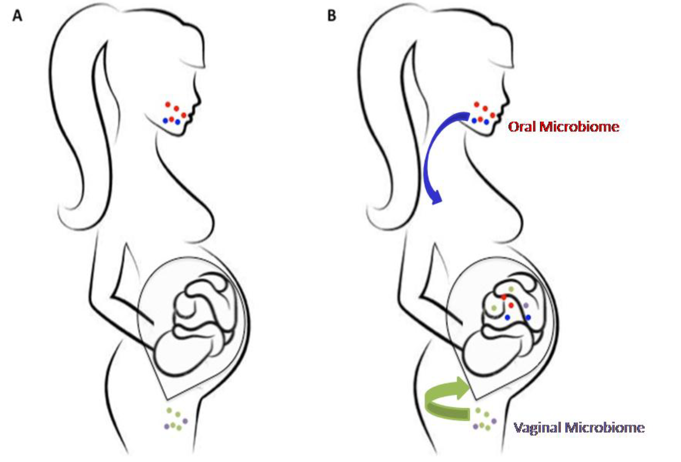
Figure 1. Prenatal microbiome colonization. (A) For years the uterus has been considered as a sterile womb in which the fetus is protected by the placenta from microbial colonization. (B) Recent studies have questioned this hypothesis showing that microbial colonization may already begin in utero; these microbes may reach the fetus from the maternal oral cavity/gut through the bloodstream (hematogenous route) and from the vagina (ascendant route).
High throughput 07 00038 g001